Quantitative analysis of TALE–DNA interactions suggests polarity
11
Quantitative analysis of TALE–DNA interactions suggests polarity effects Joshua F. Meckler 1 , Mital S. Bhakta 1 , Moon-Soo Kim 1 , Robert Ovadia 2 , Chris H. Habrian 2 , Artem Zykovich 1 , Abigail Yu 1 , Sarah H. Lockwood 1 , Robert Morbitzer 3 , Janett Elsa ¨ esser 3 , Thomas Lahaye 3 , David J. Segal 1, * and Enoch P. Baldwin 2, * 1 Genome Center and Department of Biochemistry and Molecular Medicine, University of California, Davis, CA 95616, USA, 2 Department of Molecular and Cellular Biology, University of California, Davis, CA 95616, USA and 3 Department of Biology, Institute of Genetics, Ludwig-Maximilians-University Munich, 82152 Martinsried, Germany Received November 26, 2012; Revised January 18, 2013; Accepted January 23, 2013 ABSTRACT Transcription activator-like effectors (TALEs) have revolutionized the field of genome engineering. We present here a systematic assessment of TALE DNA recognition, using quantitative electrophoretic mo- bility shift assays and reporter gene activation assays. Within TALE proteins, tandem 34-amino acid repeats recognize one base pair each and direct sequence-specific DNA binding through repeat variable di-residues (RVDs). We found that RVD choice can affect affinity by four orders of magni- tude, with the relative RVD contribution in the order NG > HD NN NI > NK. The NN repeat preferred the base G over A, whereas the NK repeat bound G with 10 3 -fold lower affinity. We compared AvrBs3, a naturally occurring TALE that recognizes its target using some atypical RVD-base combinations, with a designed TALE that precisely matches ‘standard’ RVDs with the target bases. This comparison revealed unexpected differences in sensitivity to substitutions of the invariant 5 0 -T. Another surpri- sing observation was that base mismatches at the 5 0 end of the target site had more disruptive effects on affinity than those at the 3 0 end, particularly in designed TALEs. These results provide evidence that TALE–DNA recognition exhibits a hitherto un- described polarity effect, in which the N-terminal repeats contribute more to affinity than C-terminal ones. INTRODUCTION Transcription activator-like effectors (TALEs) are sequence-specific DNA-binding proteins that the bacterial pathogen Xanthomonas injects into plant cells. Inside the plant cell, they bind to and activate specific host pro- moters (1). Their promoter specificity is conferred by a series of tandem protein repeats, typically 34 amino acids in length. Unlike any previously described DNA- binding domain, each repeat recognizes a single DNA base pair. Amino acids at positions 12 and 13, known as repeat variable di-residues (RVDs), determine the base preferences of a repeat. Deciphering the correspondence between RVD composition and target DNA bases created the ‘TALE DNA binding code’, making TALEs the first DNA-binding protein class for which robust and comprehensive rules of DNA recognition are known (2,3). Sequence-specific DNA binding is achieved by simple assembly of individual repeats with desired base specificities. Recent crystallographic work revealed the structural basis for TALE–DNA recognition (4,5). Each repeat consists of two alpha helices connected by a three-residue loop that contains the RVDs (the ‘RVD loop’). Sequential repeats interact to form a solenoid that binds to one DNA strand, with the TALE N-terminal to C-terminal direction aligned with the DNA 5 0 to 3 0 direction. Position 13 contacts the target base in the major groove through hydrogen bonds or van der Waals interactions, while position 12 stabilizes the RVD loop structure. Thus, repeat sequence preferences are essentially determined by a single amino acid–base interaction. *To whom correspondence should be addressed. Tel: +1 530 754 9134; Fax: +1 530 754 9658; Email: [email protected] Correspondence may also be addressed to Enoch P. Baldwin. Tel: +1 530 752 1108; Fax:+1 530 752 3085; Email: [email protected] The authors wish it to be known that, in their opinion, the first two authors should be regarded as joint First Authors. Present address: Moon-Soo Kim, Department of Chemistry, 1906 College Heights Boulevard, Western Kentucky University, Bowling Green, KY 42101, USA. 4118–4128 Nucleic Acids Research, 2013, Vol. 41, No. 7 Published online 13 February 2013 doi:10.1093/nar/gkt085 ß The Author(s) 2013. Published by Oxford University Press. This is an Open Access article distributed under the terms of the Creative Commons Attribution Non-Commercial License (http://creativecommons.org/licenses/ by-nc/3.0/), which permits unrestricted non-commercial use, distribution, and reproduction in any medium, provided the original work is properly cited. Downloaded from https://academic.oup.com/nar/article/41/7/4118/1069959 by guest on 22 December 2021
Transcript of Quantitative analysis of TALE–DNA interactions suggests polarity
Quantitative analysis of TALE–DNA interactionssuggests polarity effectsJoshua F. Meckler1, Mital S. Bhakta1, Moon-Soo Kim1, Robert Ovadia2,
Chris H. Habrian2, Artem Zykovich1, Abigail Yu1, Sarah H. Lockwood1,
Robert Morbitzer3, Janett Elsaesser3, Thomas Lahaye3, David J. Segal1,* and
Enoch P. Baldwin2,*
1Genome Center and Department of Biochemistry and Molecular Medicine, University of California, Davis,CA 95616, USA, 2Department of Molecular and Cellular Biology, University of California, Davis, CA 95616, USAand 3Department of Biology, Institute of Genetics, Ludwig-Maximilians-University Munich, 82152 Martinsried,Germany
Received November 26, 2012; Revised January 18, 2013; Accepted January 23, 2013
ABSTRACT
Transcription activator-like effectors (TALEs) haverevolutionized the field of genome engineering. Wepresent here a systematic assessment of TALE DNArecognition, using quantitative electrophoretic mo-bility shift assays and reporter gene activationassays. Within TALE proteins, tandem 34-aminoacid repeats recognize one base pair each and directsequence-specific DNA binding through repeatvariable di-residues (RVDs). We found that RVDchoice can affect affinity by four orders of magni-tude, with the relative RVD contribution in the orderNG>HD�NN � NI>NK. The NN repeat preferredthe base G over A, whereas the NK repeat bound Gwith 103-fold lower affinity. We compared AvrBs3, anaturally occurring TALE that recognizes its targetusing some atypical RVD-base combinations, with adesigned TALE that precisely matches ‘standard’RVDs with the target bases. This comparisonrevealed unexpected differences in sensitivity tosubstitutions of the invariant 50-T. Another surpri-sing observation was that base mismatches at the50 end of the target site had more disruptive effectson affinity than those at the 30 end, particularly indesigned TALEs. These results provide evidencethat TALE–DNA recognition exhibits a hitherto un-described polarity effect, in which the N-terminalrepeats contribute more to affinity than C-terminalones.
INTRODUCTION
Transcription activator-like effectors (TALEs) aresequence-specific DNA-binding proteins that the bacterialpathogen Xanthomonas injects into plant cells. Inside theplant cell, they bind to and activate specific host pro-moters (1). Their promoter specificity is conferred by aseries of tandem protein repeats, typically 34 aminoacids in length. Unlike any previously described DNA-binding domain, each repeat recognizes a single DNAbase pair. Amino acids at positions 12 and 13, known asrepeat variable di-residues (RVDs), determine the basepreferences of a repeat. Deciphering the correspondencebetween RVD composition and target DNA bases createdthe ‘TALE DNA binding code’, making TALEs thefirst DNA-binding protein class for which robust andcomprehensive rules of DNA recognition are known(2,3). Sequence-specific DNA binding is achieved bysimple assembly of individual repeats with desired basespecificities.
Recent crystallographic work revealed the structuralbasis for TALE–DNA recognition (4,5). Each repeatconsists of two alpha helices connected by a three-residueloop that contains the RVDs (the ‘RVD loop’). Sequentialrepeats interact to form a solenoid that binds to one DNAstrand, with the TALE N-terminal to C-terminal directionaligned with the DNA 50 to 30 direction. Position 13contacts the target base in the major groove throughhydrogen bonds or van der Waals interactions, whileposition 12 stabilizes the RVD loop structure. Thus,repeat sequence preferences are essentially determined bya single amino acid–base interaction.
*To whom correspondence should be addressed. Tel: +1 530 754 9134; Fax: +1 530 754 9658; Email: [email protected] may also be addressed to Enoch P. Baldwin. Tel: +1 530 752 1108; Fax: +1 530 752 3085; Email: [email protected]
The authors wish it to be known that, in their opinion, the first two authors should be regarded as joint First Authors.
Present address:Moon-Soo Kim, Department of Chemistry, 1906 College Heights Boulevard, Western Kentucky University, Bowling Green, KY 42101, USA.
4118–4128 Nucleic Acids Research, 2013, Vol. 41, No. 7 Published online 13 February 2013doi:10.1093/nar/gkt085
� The Author(s) 2013. Published by Oxford University Press.This is an Open Access article distributed under the terms of the Creative Commons Attribution Non-Commercial License (http://creativecommons.org/licenses/by-nc/3.0/), which permits unrestricted non-commercial use, distribution, and reproduction in any medium, provided the original work is properly cited.
Dow
nloaded from https://academ
ic.oup.com/nar/article/41/7/4118/1069959 by guest on 22 D
ecember 2021
Because of their modularity, easy programmability andreliability, TALE proteins have become the preferredDNA-binding domain to create artificial transcriptionfactors (ATFs) and nucleases (TALENs), and haverapidly transformed the field of genome engineering(6–10). These properties also provide a special opportunityfor synthetic biology, in which quantitative and predict-able interactions of modular transcription factor andpromoter parts are required to engineer gene regulatorycircuits (11). However, few direct quantitative assessmentsof TALE–DNA affinities and specificities have beenreported and, as of yet, there is no predictive frameworkin place. Extant data largely consist of cell-based tran-scription factor reporter assays in which the readout canbe complicated by numerous factors in addition to TALEDNA-binding affinity. To understand and predict TALErepeat affinities, specificities and functionalities, quantita-tive binding data are required for members of this import-ant new class of DNA-binding domain.
We present here the first systematic quantitativeassessment of TALE DNA affinity and recognition. Wecombined quantitative gel shift and transcription reporterassays to explore the relative affinities of individual RVDsin vitro and their relation to activity in vivo. We alsoexamined the specificity of two G-recognition RVDs,NN and NK, and the distribution of binding affinityover the length of the repeat region. Our data providephysical explanations and quantification of previouslyreported trends and suggest some complexities in thisseemingly simple mode of DNA recognition. In particular,we demonstrate that the N-terminal TALE repeatsinteract more strongly with DNA than the C-terminalrepeats, suggesting a polarity effect of TALE binding.
MATERIALS AND METHODS
Designed TALE construction
Designed TALE (dTALE) repeat arrays were modularlyassembled using the Golden Gate cloning reagentsdescribed in (12), with slight modifications to the proced-ures. The 17.5-repeat arrays were assembled in two stepsof cut-ligation reactions. The first reaction assembled twofive-repeat arrays and one seven-repeat array. Eachcut-ligation reaction used 75 ng of appropriate plasmidswith BsaI-HF (New England Biolabs) and T4 ligase(New England Biolabs) that were incubated at 37�C for5 h. On sequence verification, the three segments wereassembled in a second cut-ligation reaction using avector containing the last half-repeat to form a complete17.5-repeat array (5+5+7+0.5=17.5). Final 17.5-repeatarrays were cloned by StuI/AatII digestion intopPreTALE111-42 and pPreTALE94-42, which containedtruncated N- and C-termini of the naturally occurringTALE PthXo1 in pAH103 (5), generated by polymerasechain reaction (PCR) using primers listed in Supple-mentary Table S1. XhoI and AgeI sites incorporated atthe termini allowed subcloning of entire dTALEs into ex-pression vectors.
In this work, the RVD-containing repeat region is takenas starting at the beginning of the ‘0 repeat’ (residue 255
for AvrBs3, LTDGQ . . . ) and ending at the end of thecomplete ‘0.5 repeat’ (residue 897, . . . SRPDP). Thus, the111-42 truncation refers to a variant that retains 111N-terminal and 42 C-terminal residues from the full-length TALE, appended to the RVD repeat region(Supplementary Figure S1A).
Protein preparations
AvrBs3254-180 and dTALEs were cloned using BamHI/AgeIand XhoI/AgeI, respectively, into pMAL-TEV, a prokary-otic expression plasmid derived from pMAL-c5x (NewEngland Biolabs) that contained a site for the TobaccoEtch Virus (TEV) protease. TALE reading frames werebounded by an N-terminal maltose-binding protein (MBP)tag, a TEV protease cleavage site and a His6 C-terminalHis-tag (Supplementary Figure S1B). Tandem affinity puri-fication allowed isolation of homogeneous full-lengthMBP-TALE–His6 fusion proteins (Supplementary Figure S2B).BL21 cells (Novagen) were transformed and grown over-night on Luria Broth agar containing 100mg/ml carbenicil-lin. Single colonies were inoculated into 25ml of Luria Brothcontaining 100mg/ml carbenicillin, and grownwith vigorousshaking at 37�C. At an OD600 of 0.4, incubation wascontinued at 30�C to an OD600 of 0.6–0.8. Isopropyl-b-D-thiogalactopyranoside (IPTG) (0.1mM final) wasadded and the cultures were shaken at 30�C for 3–4h.Cells were pelleted (10min, 2000g) and stored at �80�C.Purificationwas carried out at 4�C, and all buffers contained2mM sodium azide. Cells were resuspended in 40ml lysis/wash buffer (500mM NaCl, 5mM imidazole, 20mMTris-Cl, pH 7.9) and lysed using a microfluidizer(Microfluidics Corp., Model M100-Y). The resultinglysate, including washes (100ml total), was clarified by cen-trifugation (40min, 15 000g), and the supernatant waspassed through a 2-ml column bed of Ni-IDA resin (2–3ml/min, Novagen). The column was washed with 100mlof lysis/wash buffer, 100ml of high-salt wash buffer (2MNaCl, 5mM imidazole, 20mM Tris-Cl, pH 7.9) to com-pletely remove bound nucleic acids and another 100ml oflysis/wash buffer. The MBP–TALE fusion proteins wereeluted in five 2-ml fractions using His elution buffer(500mM NaCl, 500mM imidazole, 20mM Tris-Cl, pH7.9). Fractions containing more than 0.1 OD280 werepassed through a 1-ml Luer lock syringe column containing0.75ml of amylose resin (New England Biolabs) (�0.3ml/min). The columns were then washed with 20ml of TALEstorage buffer (480mM KCl, 1.6mM ethylenediaminete-traacetic acid (EDTA), 2mM dithiothreitol (DTT), 12mMTris-Cl, pH 7.5). The highly purified fusion protein waseluted in 0.5-ml aliquots with TALE storage buffer contain-ing 10mMmaltose. The most concentrated fractions (1 to 4OD280) were dialyzed against 2� 300ml of TALE storagebuffer, quantified by ultraviolet absorbance and flash-frozenat �80�C in 50 -mL aliquots. The zinc finger DNA-bindingdomain of Zif268 (13) was subcloned into pMAL-TEV andpurified as described for dTALEs, except buffers contained100mM ZnCl2. The molar extinction coefficients at 280nmwere 81820, 81820, 92820 and 69330 for the MBP-TALE111-42, MBP-TALE94-42, MBP-AvrBs3254-180 andMBP-Zif268 proteins, respectively (EXPASY). Typical
Nucleic Acids Research, 2013, Vol. 41, No. 7 4119
Dow
nloaded from https://academ
ic.oup.com/nar/article/41/7/4118/1069959 by guest on 22 D
ecember 2021

final concentrations were 10–20mM, with an overall yield of1–5mg protein (4–20mg/l media). These proteins main-tained binding activity for at least 1 week at 4�C. Forcases in which the MBP tag was removed, quantitativecleavage was achieved by incubation of 10–20mM dTALEwith TEV proteinase (20mg/ml final, a gift from ChrisFraser, UC Davis) and 5mM DTT overnight at 4�C.
Electrophoretic mobility shift assay
Biotin-labeled DNA targets were generated by PCR amp-lification using a 50 biotinylated forward primer of 69-meroligonucleotides containing 19-base pair TALE targetsites or the Zif268 site (Supplementary Table S2). PCRreactions contained unlabeled reverse primer in a 4:1ratio over the biotinylated primer. Amplified targetswere column purified (Qiagen). Binding reactions weremixed on ice and placed in the dark for 1 h at roomtemperature (22�C) in 1� TALE electrophoretic mobil-ity shift assay (EMSA) buffer (12mM Tris-Cl, pH 7.5,60mM KCl, 2mM DTT, 0.05% NP-40, 50 ng/mLdouble-stranded poly (deoxyinosine-deoxycytosine)n(dIdC), 0.1mg/ml bovine serum albumin (BSA), 5%glycerol, 5mM MgCl2, 0.2mM EDTA). As indicated,zinc finger binding reactions were performed in 1 xTALE EMSA buffer supplemented with 100 mM ZnCl2,or in Zinc Buffer A (ZBA: 100mM Tris, 90mM KCl,1mM MgCl2, and 90 mM ZnCl2, pH 7.5) with 5%glycerol, 0.1mg/ml BSA, 0.05% NP-40. All binding reac-tions contained 25–55 pM target DNA, and purifiedproteins with a concentration of 0.1 – 2500 nM. Afterthe room-temperature binding reaction, samples wereplaced at 4�C for 30min. For all experiments, besidesthe ‘polarity’ assays, gel electrophoresis was performedon a 1.3% agarose gel using Amresco BiotechnologyGrade Agarose I in 0.5� tris-borate-EDTA (TBE)buffer (Bio-Rad). Gels were pre-run at 105V in 0.5�TBE buffer at 4�C for 30min before loading. Binding re-actions were loaded onto the gel while the current was on,and run for 20–30min. Using a wet-transfer apparatus(Bio-Rad), the DNA was blotted onto a Biodyne Bnylon membrane (Pierce) for 20min at 100V at 4�C.The DNA was cross-linked to the membrane with anultraviolet cross-linker (Stratagene) for 4min. Thebiotinylated DNA was visualized using the LightShiftChemiluminescent EMSA Kit (Pierce) according to themanufacturer’s protocol. Equilibrium binding constants(apparent KD) were calculated from protein titration ex-periments. Gel images on X-ray film (Denville Scientific)were scanned and then quantitated using ImageJ. Allreported EMSA measurements were averages of at leastthree experiments performed with independent protein di-lutions. For the ‘polarity’ gel shifts, the protocol was thesame, except that tris-borate (TB) buffer was substitutedfor TBE buffer (in the gel and running buffer) at identicalconcentrations. Representative EMSA data are shown inSupplementary Figure S3.
ATF Assay
dTALEs were cloned using XhoI and AgeI into thephosphoglycerate kinase (PGK) promoter-driven
mammalian expression vector pPGK-VP64 (14), whichappended an N-terminal HA epitope tag and nuclear lo-calization sequence, and a C-terminal VP64 transcrip-tional activation domain (15) (Supplementary FigureS1C). Target sites for the dTALEs were cloned betweenNotI and XhoI sites upstream of the SV40 promoter inpGL3-control plasmids (Promega), using primers listedin Supplementary Table S4. In 24-well plates, HEK293Tcells at 80% confluency in Dulbecco’s Modified EagleMedium (DMEM) supplemented with 10% fetal calfserum, 1 U/ml of penicillin and 1 mg/ml of streptomycinwere co-transfected with 100 ng of dTALE ATF expres-sion plasmid, 25 ng of modified pGL3-control fireflyluciferase reporter plasmid containing a dTALE targetsite and 25 ng of pRL-TK-Renilla Luciferase plasmid (asa transfection control, Promega), using Lipofectamine2000 (Invitrogen). Cells were harvested 48 h post-transfection by removing media, washing with 500 mL of1� Dulbecco’s Phosphate-Buffered Saline (DPBS) andthen followed by lysis in 100mL of 1� passive lysisbuffer (Promega) with 1� complete protease inhibitors(Roche). Clarified cell lysates (20mL) were used to deter-mine luciferase activity using DualGlo reagents (40mL,Promega) in a Veritas microplate luminometer (TurnerBiosystems). All experiments were performed in duplicateand repeated on two different days.
Binding site specificity assay using massively parallelsequencing (Bind-n-Seq)
Bind-n-Seq was performed essentially as described (13)using a full-length MBP–AvrBs3 fusion protein(AvrBs3254-267) as bait. AvrBs3254-267 was purified frominduced cells using amylose affinity resin (New EnglandBiolabs) according to manufacturer’s instructions. Bar-coded 93-mer double-stranded oligonucleotide targets con-taining Illumina primer binding sites and a 21-nt randomregion were incubated with 450nM AvrBs3254-267 in 1�TALE EMSA buffer. Bound complexes were precipitatedusing amylose resin and enriched by six wash steps in thecorresponding salt buffer. Eluted DNA was sequenced onan Illumina sequencer. Sequencing reads were filtered andsorted using custom Perl scripts found in the MERMADEpackage, an updated version of the Bind-n-Seq dataanalysis pipeline. MERMADE is freely available withuser documentation at http://korflab.ucdavis.edu/Datasets/BindNSeq. Briefly, high-quality reads (composedonly of A, C, T or G, with a valid constant region [‘AA’]and unique random region) were retained and split intoseparate files based on their unique 3-nt barcode(MERMADE scripts: sequence_converter.pl, debarcode.pl). For motif analysis, recovered sequences wereanalyzed relative to a file of unenriched background21-mer sequences using a sliding window of 6-12bp(MERMADE scripts: kmer_counter.pl, kmer_selector.pl).Sequences showing �2-fold enrichment relative to back-ground were then analyzed by MERMADE using an itera-tive motif searching approach (MERMADE scripts:mermade.pl, motif_expander.pl). The graphical representa-tion of the sequence motif was rendered using WebLogo.
4120 Nucleic Acids Research, 2013, Vol. 41, No. 7
Dow
nloaded from https://academ
ic.oup.com/nar/article/41/7/4118/1069959 by guest on 22 D
ecember 2021
RESULTS
Generating a scaffold
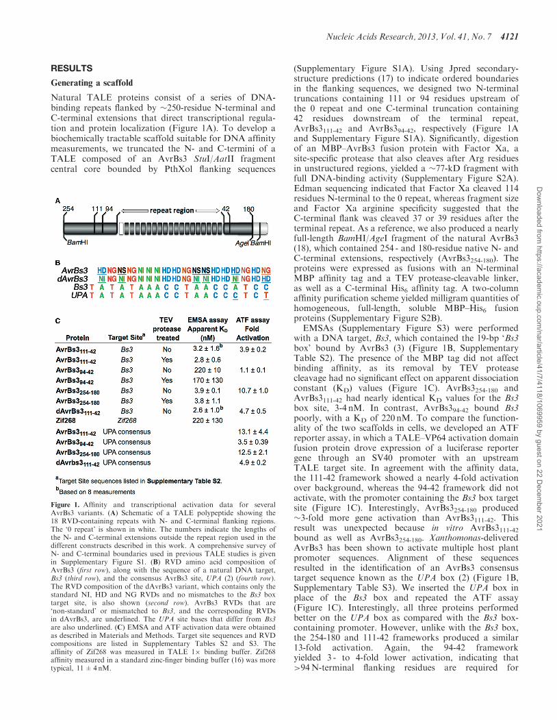
Natural TALE proteins consist of a series of DNA-binding repeats flanked by �250-residue N-terminal andC-terminal extensions that direct transcriptional regula-tion and protein localization (Figure 1A). To develop abiochemically tractable scaffold suitable for DNA affinitymeasurements, we truncated the N- and C-termini of aTALE composed of an AvrBs3 StuI/AatII fragmentcentral core bounded by PthXol flanking sequences
(Supplementary Figure S1A). Using Jpred secondary-structure predictions (17) to indicate ordered boundariesin the flanking sequences, we designed two N-terminaltruncations containing 111 or 94 residues upstream ofthe 0 repeat and one C-terminal truncation containing42 residues downstream of the terminal repeat,AvrBs3111-42 and AvrBs394-42, respectively (Figure 1Aand Supplementary Figure S1A). Significantly, digestionof an MBP–AvrBs3 fusion protein with Factor Xa, asite-specific protease that also cleaves after Arg residuesin unstructured regions, yielded a �77-kD fragment withfull DNA-binding activity (Supplementary Figure S2A).Edman sequencing indicated that Factor Xa cleaved 114residues N-terminal to the 0 repeat, whereas fragment sizeand Factor Xa arginine specificity suggested that theC-terminal flank was cleaved 37 or 39 residues after theterminal repeat. As a reference, we also produced a nearlyfull-length BamHI/AgeI fragment of the natural AvrBs3(18), which contained 254 - and 180-residue native N- andC-terminal extensions, respectively (AvrBs3254-180). Theproteins were expressed as fusions with an N-terminalMBP affinity tag and a TEV protease-cleavable linker,as well as a C-terminal His6 affinity tag. A two-columnaffinity purification scheme yielded milligram quantities ofhomogeneous, full-length, soluble MBP–His6 fusionproteins (Supplementary Figure S2B).EMSAs (Supplementary Figure S3) were performed
with a DNA target, Bs3, which contained the 19-bp ‘Bs3box’ bound by AvrBs3 (3) (Figure 1B, SupplementaryTable S2). The presence of the MBP tag did not affectbinding affinity, as its removal by TEV proteasecleavage had no significant effect on apparent dissociationconstant (KD) values (Figure 1C). AvrBs3254-180 andAvrBs3111-42 had nearly identical KD values for the Bs3box site, 3-4 nM. In contrast, AvrBs394-42 bound Bs3poorly, with a KD of 220 nM. To compare the function-ality of the two scaffolds in cells, we developed an ATFreporter assay, in which a TALE–VP64 activation domainfusion protein drove expression of a luciferase reportergene through an SV40 promoter with an upstreamTALE target site. In agreement with the affinity data,the 111-42 framework showed a nearly 4-fold activationover background, whereas the 94-42 framework did notactivate, with the promoter containing the Bs3 box targetsite (Figure 1C). Interestingly, AvrBs3254-180 produced�3-fold more gene activation than AvrBs3111-42. Thisresult was unexpected because in vitro AvrBs3111-42bound as well as AvrBs3254-180. Xanthomonas-deliveredAvrBs3 has been shown to activate multiple host plantpromoter sequences. Alignment of these sequencesresulted in the identification of an AvrBs3 consensustarget sequence known as the UPA box (2) (Figure 1B,Supplementary Table S3). We inserted the UPA box inplace of the Bs3 box and repeated the ATF assay(Figure 1C). Interestingly, all three proteins performedbetter on the UPA box as compared with the Bs3 box-containing promoter. However, unlike with the Bs3 box,the 254-180 and 111-42 frameworks produced a similar13-fold activation. Again, the 94-42 frameworkyielded 3 - to 4-fold lower activation, indicating that>94N-terminal flanking residues are required for
Figure 1. Affinity and transcriptional activation data for severalAvrBs3 variants. (A) Schematic of a TALE polypeptide showing the18 RVD-containing repeats with N- and C-terminal flanking regions.The ‘0 repeat’ is shown in white. The numbers indicate the lengths ofthe N- and C-terminal extensions outside the repeat region used in thedifferent constructs described in this work. A comprehensive survey ofN- and C-terminal boundaries used in previous TALE studies is givenin Supplementary Figure S1. (B) RVD amino acid composition ofAvrBs3 (first row), along with the sequence of a natural DNA target,Bs3 (third row), and the consensus AvrBs3 site, UPA (2) (fourth row).The RVD composition of the dAvrBs3 variant, which contains only thestandard NI, HD and NG RVDs and no mismatches to the Bs3 boxtarget site, is also shown (second row). AvrBs3 RVDs that are‘non-standard’ or mismatched to Bs3, and the corresponding RVDsin dAvrBs3, are underlined. The UPA site bases that differ from Bs3are also underlined. (C) EMSA and ATF activation data were obtainedas described in Materials and Methods. Target site sequences and RVDcompositions are listed in Supplementary Tables S2 and S3. Theaffinity of Zif268 was measured in TALE 1� binding buffer. Zif268affinity measured in a standard zinc-finger binding buffer (16) was moretypical, 11±4nM.
Nucleic Acids Research, 2013, Vol. 41, No. 7 4121
Dow
nloaded from https://academ
ic.oup.com/nar/article/41/7/4118/1069959 by guest on 22 D
ecember 2021
high-affinity binding comparable with AvrBs3254-180. The111-42 framework was used for all subsequentexperiments.
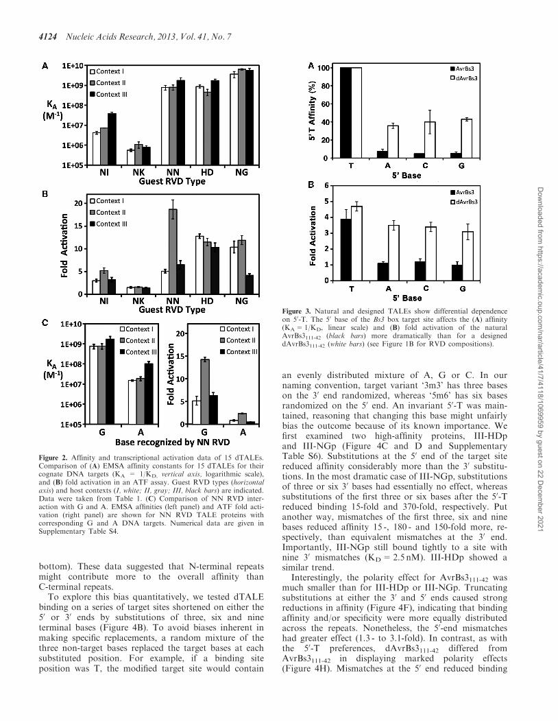
Using dTALEs to interrogate RVD relative affinities
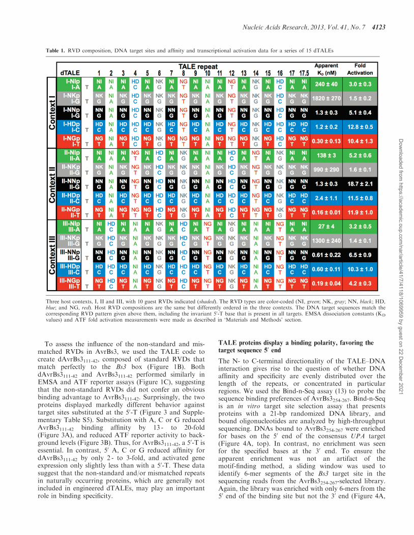
To compare the binding affinities of the five RVDs thathave been widely used to program dTALE specificities(HD, NI, NG, NN and NK), we used a ‘host-guest’design in which 10 ‘guest’ positions containing theRVDs to be tested were interspersed with eight constant‘host’ RVDs in a largely alternating pattern. The base 50 tothe target site was kept constant as T. Host contexts I – III(Table 1) sampled all base-step and base-triple combin-ations to examine potential context effects. Importantly,this setup avoided the structural peculiarities ofhomopolymeric runs in the target DNA sequences.Because the host repeats remained constant in each ofthe three contexts, we reasoned that their contributionto binding could be accounted for, allowing us to make dir-ect comparisons based on only the guest repeat identity.Fifteen dTALEs were constructed and matched with
corresponding DNA targets (Table 1). dTALE I-NIprefers to a protein with host context I and NI RVD-containing repeats at the guest positions, whereas thecognate DNA target is referred to as I-A. dTALEs con-taining G-recognizing NN and NK guests were comparedagainst identical G-containing target sites (e.g. I-G, II-Gand III-G). The proteins were expressed in E. coli andpurified to homogeneity (Supplementary Figure S2B).EMSA revealed that their apparent KDs spannedfour orders of magnitude, from 160 pM to 1.8 mM(Figure 2A, Table 1). Several trends became immediatelyapparent. The repeat type was the largest factor in affinitydifferences. The dTALEs with NG, HD and NN guestRVDs bound their targets with high affinity (160 pM –2.4 nM). The strongest affinities were for the three NGguest proteins, whereas III-NNp and III-HDp also hadpicomolar affinity. The NK guest dTALEs bound leastwell in all three contexts. Although consistently betterthan NK, the NI guest dTALEs also bound poorly butwith more variation. I-NIp had a KD of 240 nM, butIII-NIp bound with a KD of 27 nM. This 9-fold differencein affinity, despite the two proteins sharing the sameoverall distribution of repeat types, clearly demonstratesthe potential for significant contextual effects. Excludingthe NK-containing proteins, context III was the most fa-vorable setting for NI, NN and HD proteins, with a 3 - to9-fold advantage over contexts I and II. Taken in whole,the gel shift data suggested that the relative affinities ofindividual repeats can be ordered as NG>HD�NN �NI>NK.In the ATF assay, reporter gene activation by the
dTALE series ranged from 1.4 - to 19-fold (Figure 2B,Table 1). All three HD guest proteins were strong activa-tors, with levels at least 10-fold over background. As pre-dicted from the binding data, the three NK guest proteinswere the poorest activators. The correlation of affinity toactivation was not linear. A simple log-log modelproduced a reasonable fit of the data (R2=0.68,P=0.0002, Supplementary Figure S4), but several
dTALEs displayed considerable deviation. For example,the tight-binding III-NGp produced relatively low activa-tion (4-fold) compared with the two other NG guestproteins (>10-fold activation levels). Conversely,moderate-binding II-NNp showed the highest activationof the entire set (19-fold), higher than the other NNguests. There was an apparent demarcation in activationbetween 2.4 nM and 27 nM affinities (P=2� 10�12, basedon a comparison of 60 individual non-averaged fold acti-vation values in the two categories using a two-tailedheteroscedastic Student t-test). The six dTALEs withapparent KD� 27 nM had an average fold activation of2.7 fold, whereas those with KD� 2.4 nM had an averagefold activation of 10.2. Considering fold activation alone,the effectiveness of the five repeat types studied here wouldbe HD�NG�NN � NI>NK.
The NN RVD prefers G over A
According to correlations of natural TALE RVDs andtheir target sites (2,3), the NN repeat has a similar prefer-ence for G and A, and there are cases where this has beenshown in dTALEs (2,19). However, other reports (8,20)have shown instances where NN displays an apparentpreference for G. We compared NN repeat binding to Gand A by measuring I-NNp, II-NNp and III-NNpaffinities to the corresponding hosts containing G or Aguests (Figure 2C and Supplementary Table S4). Whenthe NN guest repeats were paired with A rather than Gin contexts I and II, binding was reduced 49 - and 41-fold,respectively. In context III, the reduction was less severe,but was still >17-fold. The same trend was apparent in theATF assay, with an 8-fold reduction in activation incontext II, and reduction to background levels incontexts I and III. These data indicate that NN RVDsprefer binding G over A, although on a per-repeat basis,the binding energy differences are relatively small (seeDiscussion).
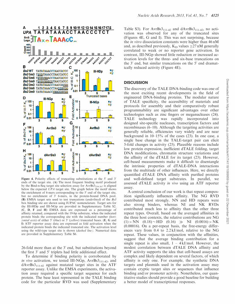
Natural and designed versions of AvrBs3 differ in theirrequirement for a 50T
Most naturally occurring TALEs bind to DNA sequencesbeginning with a T (2,3), but dTALEs have been reportedto recognize targets that have bases other than T in the 50
position (8,21) and in one case, the 50-T requirement wasdependent on the N-terminal extension length (21). Onemajor difference between natural TALEs and dTALEsproduced by available assembly kits is that the artificialproteins (22,23) are predominantly constructed using justthe HD, NG, NI, NN and NK RVDs. In contrast, mostnatural TALEs contain one or more ‘non-standard’RVDs, such as NS, N* (residue 13 deleted), HG andothers (1,2). In addition, ‘mismatches’ between RVDsand their consensus bases are rather typical. In fact,there has not been a single documented case of a naturallyoccurring TALE and a plant target promoter with aperfect code-predicted match. For example, AvrBs3,when bound to its Bs3 box target, has two HD-Amismatches and an NG-C mismatch. Additionally,AvrBs3 contains three ‘non-standard’ NS RVDs.
4122 Nucleic Acids Research, 2013, Vol. 41, No. 7
Dow
nloaded from https://academ
ic.oup.com/nar/article/41/7/4118/1069959 by guest on 22 D
ecember 2021
To assess the influence of the non-standard and mis-matched RVDs in AvrBs3, we used the TALE code tocreate dAvrBs3111-42, composed of standard RVDs thatmatch perfectly to the Bs3 box (Figure 1B). BothdAvrBs3111-42 and AvrBs3111-42 performed similarly inEMSA and ATF reporter assays (Figure 1C), suggestingthat the non-standard RVDs did not confer an obviousbinding advantage to AvrBs3111-42. Surprisingly, the twoproteins displayed markedly different behavior againsttarget sites substituted at the 50-T (Figure 3 and Supple-mentary Table S5). Substitution with A, C or G reducedAvrBs3111-42 binding affinity by 13 - to 20-fold(Figure 3A), and reduced ATF reporter activity to back-ground levels (Figure 3B). Thus, for AvrBs3111-42, a 50-T isessential. In contrast, 50 A, C or G reduced affinity fordAvrBs3111-42 by only 2 - to 3-fold, and activated geneexpression only slightly less than with a 50-T. These datasuggest that the non-standard and/or mismatched repeatsin naturally occurring proteins, which are generally notincluded in engineered dTALEs, may play an importantrole in binding specificity.
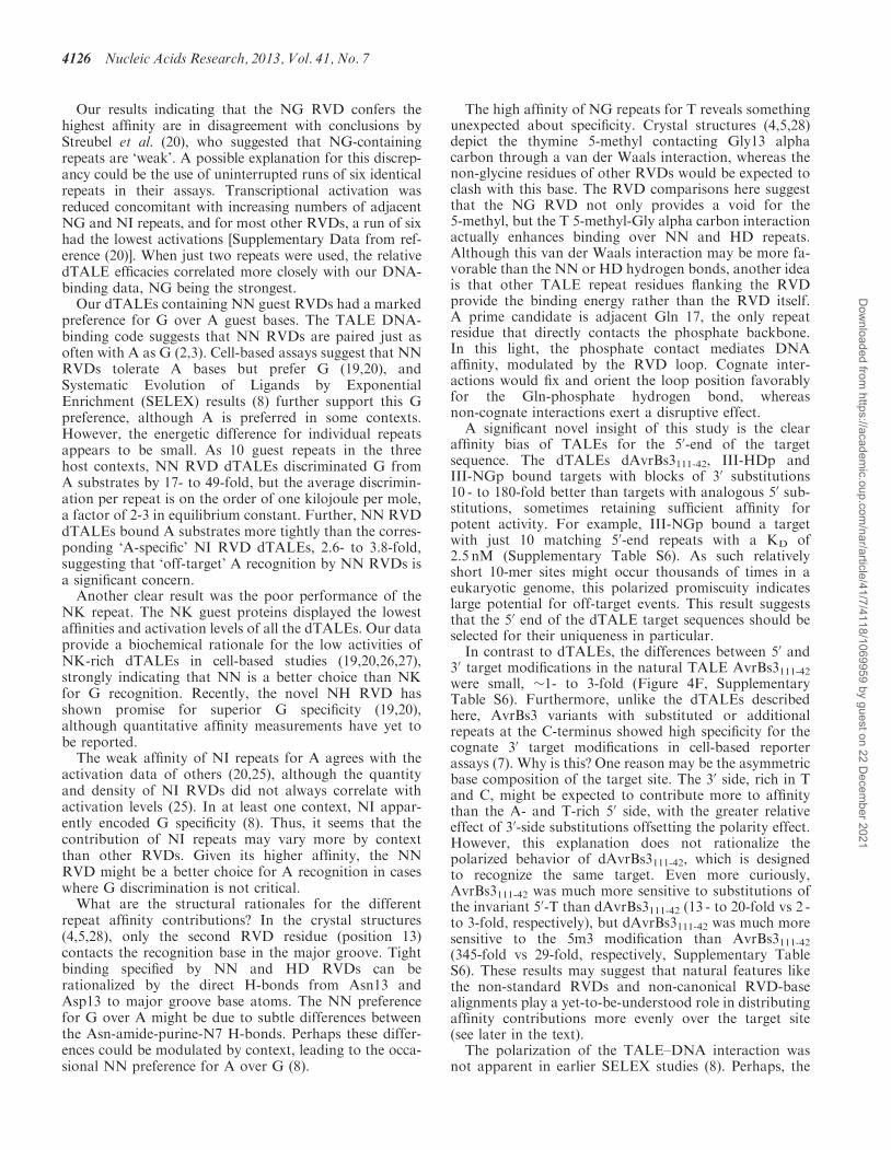
TALE proteins display a binding polarity, favoring thetarget sequence 50 end
The N- to C-terminal directionality of the TALE–DNAinteraction gives rise to the question of whether DNAaffinity and specificity are evenly distributed over thelength of the repeats, or concentrated in particularregions. We used the Bind-n-Seq assay (13) to probe thesequence binding preferences of AvrBs3254-267. Bind-n-Seqis an in vitro target site selection assay that presentsproteins with a 21-bp randomized DNA library, andbound oligonucleotides are analyzed by high-throughputsequencing. DNAs bound to AvrBs3254-267 were enrichedfor bases on the 50 end of the consensus UPA target(Figure 4A, top). In contrast, no enrichment was seenfor the specified bases at the 30 end. To ensure theapparent enrichment was not an artifact of themotif-finding method, a sliding window was used toidentify 6-mer segments of the Bs3 target site in thesequencing reads from the AvrBs3254-267-selected library.Again, the library was enriched with only 6-mers from the50 end of the binding site but not the 30 end (Figure 4A,
Table 1. RVD composition, DNA target sites and affinity and transcriptional activation data for a series of 15 dTALEs
Three host contexts, I, II and III, with 10 guest RVDs indicated (shaded). The RVD types are color-coded (NI, green; NK, gray; NN, black; HD,blue; and NG, red). Host RVD compositions are the same but differently ordered in the three contexts. The DNA target sequences match thecorresponding RVD pattern given above them, including the invariant 50-T base that is present in all targets. EMSA dissociation constants (KD
values) and ATF fold activation measurements were made as described in ‘Materials and Methods’ section.
Nucleic Acids Research, 2013, Vol. 41, No. 7 4123
Dow
nloaded from https://academ
ic.oup.com/nar/article/41/7/4118/1069959 by guest on 22 D
ecember 2021
bottom). These data suggested that N-terminal repeatsmight contribute more to the overall affinity thanC-terminal repeats.To explore this bias quantitatively, we tested dTALE
binding on a series of target sites shortened on either the50 or 30 ends by substitutions of three, six and nineterminal bases (Figure 4B). To avoid biases inherent inmaking specific replacements, a random mixture of thethree non-target bases replaced the target bases at eachsubstituted position. For example, if a binding siteposition was T, the modified target site would contain
an evenly distributed mixture of A, G or C. In ournaming convention, target variant ‘3m3’ has three baseson the 30 end randomized, whereas ‘5m6’ has six basesrandomized on the 50 end. An invariant 50-T was main-tained, reasoning that changing this base might unfairlybias the outcome because of its known importance. Wefirst examined two high-affinity proteins, III-HDpand III-NGp (Figure 4C and D and SupplementaryTable S6). Substitutions at the 50 end of the target sitereduced affinity considerably more than the 30 substitu-tions. In the most dramatic case of III-NGp, substitutionsof three or six 30 bases had essentially no effect, whereassubstitutions of the first three or six bases after the 50-Treduced binding 15-fold and 370-fold, respectively. Putanother way, mismatches of the first three, six and ninebases reduced affinity 15 -, 180 - and 150-fold more, re-spectively, than equivalent mismatches at the 30 end.Importantly, III-NGp still bound tightly to a site withnine 30 mismatches (KD=2.5 nM). III-HDp showed asimilar trend.
Interestingly, the polarity effect for AvrBs3111-42 wasmuch smaller than for III-HDp or III-NGp. Truncatingsubstitutions at either the 30 and 50 ends caused strongreductions in affinity (Figure 4F), indicating that bindingaffinity and/or specificity were more equally distributedacross the repeats. Nonetheless, the 50-end mismatcheshad greater effect (1.3 - to 3.1-fold). In contrast, as withthe 50-T preferences, dAvrBs3111-42 differed fromAvrBs3111-42 in displaying marked polarity effects(Figure 4H). Mismatches at the 50 end reduced binding
Figure 2. Affinity and transcriptional activation data of 15 dTALEs.Comparison of (A) EMSA affinity constants for 15 dTALEs for theircognate DNA targets (KA = 1/KD, vertical axis, logarithmic scale),and (B) fold activation in an ATF assay. Guest RVD types (horizontalaxis) and host contexts (I, white; II, gray; III, black bars) are indicated.Data were taken from Table 1. (C) Comparison of NN RVD inter-action with G and A. EMSA affinities (left panel) and ATF fold acti-vation (right panel) are shown for NN RVD TALE proteins withcorresponding G and A DNA targets. Numerical data are given inSupplementary Table S4.
Figure 3. Natural and designed TALEs show differential dependenceon 50-T. The 50 base of the Bs3 box target site affects the (A) affinity(KA=1/KD, linear scale) and (B) fold activation of the naturalAvrBs3111-42 (black bars) more dramatically than for a designeddAvrBs3111-42 (white bars) (see Figure 1B for RVD compositions).
4124 Nucleic Acids Research, 2013, Vol. 41, No. 7
Dow
nloaded from https://academ
ic.oup.com/nar/article/41/7/4118/1069959 by guest on 22 D
ecember 2021
26-fold more than at the 30 end, but substitutions beyondthe first 30 and 50 triplets had little additional effect.
To determine if binding polarity is corroborated byin vivo activation, we tested III-NGp, AvrBs3111-42, anddAvrBs3111-42 against mutated target sites in the ATFreporter assay. Unlike the EMSA experiments, the activa-tion assay required a specific target sequence for eachprotein. The base least represented in the TALE bindingcode for the particular RVD was used (Supplementary
Table S3). For AvrBs3111-42 and dAvrBs3111-42, no acti-vation was observed for any of the truncated sites(Figures 4E, G and I). This was not surprising, becausethe in vitro dissociation constants were higher than 46 nMand, as described previously, KD values �27 nM generallycorrelated to weak or no reporter gene activation. Incontrast, III-NGp showed little reduction or increased ac-tivation levels for the three- and six-base truncations onthe 30 end, but similar truncations on the 50 end dramat-ically reduced activity (Figure 4E).
DISCUSSION
The discovery of the TALE DNA-binding code was one ofthe most exciting recent developments in the field ofengineered DNA-binding proteins. The modular natureof TALE specificity, the accessibility of materials andprotocols for assembly and their comparatively robustprogrammability are significant advantages over othertechnologies such as zinc fingers or meganucleases (24).TALE technology was rapidly incorporated intodesigned site-specific nucleases, transcription factors andrecombinases (6–10). Although the targeting activities aregenerally reliable, efficiencies vary widely and are nearbackground in 10–15% of the cases (23). In one case, asingle base change in the TALE-target pair can elicit5-fold changes in activity (25). Plausible reasons includelow protein expression, inefficient dTALE folding, targetDNA modifications, chromatin structure variations andthe affinity of the dTALE for its target (23). However,cell-based measurements make it difficult to disentanglethe intrinsic properties of dTALE-DNA interactionsfrom the multitude of other influences. Here, we directlyquantified dTALE DNA affinity with purified proteinsand well-defined target substrates. In parallel, weassessed dTALE activity in vivo using an ATF reporterassay.A central conclusion of our work is that repeat compos-
ition significantly influences affinity. The NG RVDcontributed most strongly. NN and HD repeats werealso strong binders, whereas NI and NK RVDscontributed much less to affinity than the other threerepeat types. Overall, based on the averaged affinities inthe three host contexts, the relative contributions are NG(1)>NN (0.18)�HD (0.16) � NI (0.0016)>NK(0.00016). On a per-repeat basis, the free-energy differ-ences vary from 0.4 to 2.2 kJ/mol, relative to the NGrepeat. These values, in conjunction with the affinities,suggest that the average binding contribution for asingle repeat is also small, 1 – 4 kJ/mol. However, themodest correlation between dTALE DNA affinity andATF activity supports the idea that cell-based assays arecomplex and likely dependent on several factors, of whichaffinity is only one. For example, the synthetic DNAtargets and plasmids used in the activation assay maycontain cryptic target sites or sequences that influencebinding and/or promoter activity. Nonetheless, our quan-titative studies should provide a good baseline for buildinga better model of transcriptional responses.
Figure 4. Polarity effects of truncating substitutions at the 50 and 30
ends of the target site. (A) The most frequent binding motif producedby the Bind-n-Seq target site selection assay for AvrBs3254-267 is alignedbelow the expected UPA target site. The graph below the motif showsthe enrichment of 6-mers corresponding to the 50 end of the target site,but no enrichment of 30 6-mers, in the protein-bound DNA pool.(B) EMSA target sets used to test truncations (underlined) of the Bs3box binding site are shown using IUPAC nomenclature. Target sets forthe III-HDp and III-NGp are provided in Supplementary Table S2.(C, D, F and H) EMSA data are expressed as a percentage ofaffinity retained, compared with the 19-bp substrate, when the indicatedprotein binds the corresponding site with the indicated number (hori-zontal axis) of either 50 (blue) or 30 (yellow) truncated bases. (E, G andI) ATF reporter assay data are expressed as fold activation when theindicated protein binds the indicated truncated site. The activation levelusing the wild-type target site is shown (dashed line). Numerical dataare provided in Supplementary Table S6.
Nucleic Acids Research, 2013, Vol. 41, No. 7 4125
Dow
nloaded from https://academ
ic.oup.com/nar/article/41/7/4118/1069959 by guest on 22 D
ecember 2021
Our results indicating that the NG RVD confers thehighest affinity are in disagreement with conclusions byStreubel et al. (20), who suggested that NG-containingrepeats are ‘weak’. A possible explanation for this discrep-ancy could be the use of uninterrupted runs of six identicalrepeats in their assays. Transcriptional activation wasreduced concomitant with increasing numbers of adjacentNG and NI repeats, and for most other RVDs, a run of sixhad the lowest activations [Supplementary Data from ref-erence (20)]. When just two repeats were used, the relativedTALE efficacies correlated more closely with our DNA-binding data, NG being the strongest.Our dTALEs containing NN guest RVDs had a marked
preference for G over A guest bases. The TALE DNA-binding code suggests that NN RVDs are paired just asoften with A as G (2,3). Cell-based assays suggest that NNRVDs tolerate A bases but prefer G (19,20), andSystematic Evolution of Ligands by ExponentialEnrichment (SELEX) results (8) further support this Gpreference, although A is preferred in some contexts.However, the energetic difference for individual repeatsappears to be small. As 10 guest repeats in the threehost contexts, NN RVD dTALEs discriminated G fromA substrates by 17- to 49-fold, but the average discrimin-ation per repeat is on the order of one kilojoule per mole,a factor of 2-3 in equilibrium constant. Further, NN RVDdTALEs bound A substrates more tightly than the corres-ponding ‘A-specific’ NI RVD dTALEs, 2.6- to 3.8-fold,suggesting that ‘off-target’ A recognition by NN RVDs isa significant concern.Another clear result was the poor performance of the
NK repeat. The NK guest proteins displayed the lowestaffinities and activation levels of all the dTALEs. Our dataprovide a biochemical rationale for the low activities ofNK-rich dTALEs in cell-based studies (19,20,26,27),strongly indicating that NN is a better choice than NKfor G recognition. Recently, the novel NH RVD hasshown promise for superior G specificity (19,20),although quantitative affinity measurements have yet tobe reported.The weak affinity of NI repeats for A agrees with the
activation data of others (20,25), although the quantityand density of NI RVDs did not always correlate withactivation levels (25). In at least one context, NI appar-ently encoded G specificity (8). Thus, it seems that thecontribution of NI repeats may vary more by contextthan other RVDs. Given its higher affinity, the NNRVD might be a better choice for A recognition in caseswhere G discrimination is not critical.What are the structural rationales for the different
repeat affinity contributions? In the crystal structures(4,5,28), only the second RVD residue (position 13)contacts the recognition base in the major groove. Tightbinding specified by NN and HD RVDs can berationalized by the direct H-bonds from Asn13 andAsp13 to major groove base atoms. The NN preferencefor G over A might be due to subtle differences betweenthe Asn-amide-purine-N7 H-bonds. Perhaps these differ-ences could be modulated by context, leading to the occa-sional NN preference for A over G (8).
The high affinity of NG repeats for T reveals somethingunexpected about specificity. Crystal structures (4,5,28)depict the thymine 5-methyl contacting Gly13 alphacarbon through a van der Waals interaction, whereas thenon-glycine residues of other RVDs would be expected toclash with this base. The RVD comparisons here suggestthat the NG RVD not only provides a void for the5-methyl, but the T 5-methyl-Gly alpha carbon interactionactually enhances binding over NN and HD repeats.Although this van der Waals interaction may be more fa-vorable than the NN or HD hydrogen bonds, another ideais that other TALE repeat residues flanking the RVDprovide the binding energy rather than the RVD itself.A prime candidate is adjacent Gln 17, the only repeatresidue that directly contacts the phosphate backbone.In this light, the phosphate contact mediates DNAaffinity, modulated by the RVD loop. Cognate inter-actions would fix and orient the loop position favorablyfor the Gln-phosphate hydrogen bond, whereasnon-cognate interactions exert a disruptive effect.
A significant novel insight of this study is the clearaffinity bias of TALEs for the 50-end of the targetsequence. The dTALEs dAvrBs3111-42, III-HDp andIII-NGp bound targets with blocks of 30 substitutions10 - to 180-fold better than targets with analogous 50 sub-stitutions, sometimes retaining sufficient affinity forpotent activity. For example, III-NGp bound a targetwith just 10 matching 50-end repeats with a KD of2.5 nM (Supplementary Table S6). As such relativelyshort 10-mer sites might occur thousands of times in aeukaryotic genome, this polarized promiscuity indicateslarge potential for off-target events. This result suggeststhat the 50 end of the dTALE target sequences should beselected for their uniqueness in particular.
In contrast to dTALEs, the differences between 50 and30 target modifications in the natural TALE AvrBs3111-42were small, �1- to 3-fold (Figure 4F, SupplementaryTable S6). Furthermore, unlike the dTALEs describedhere, AvrBs3 variants with substituted or additionalrepeats at the C-terminus showed high specificity for thecognate 30 target modifications in cell-based reporterassays (7). Why is this? One reason may be the asymmetricbase composition of the target site. The 30 side, rich in Tand C, might be expected to contribute more to affinitythan the A- and T-rich 50 side, with the greater relativeeffect of 30-side substitutions offsetting the polarity effect.However, this explanation does not rationalize thepolarized behavior of dAvrBs3111-42, which is designedto recognize the same target. Even more curiously,AvrBs3111-42 was much more sensitive to substitutions ofthe invariant 50-T than dAvrBs3111-42 (13 - to 20-fold vs 2 -to 3-fold, respectively), but dAvrBs3111-42 was much moresensitive to the 5m3 modification than AvrBs3111-42(345-fold vs 29-fold, respectively, Supplementary TableS6). These results may suggest that natural features likethe non-standard RVDs and non-canonical RVD-basealignments play a yet-to-be-understood role in distributingaffinity contributions more evenly over the target site(see later in the text).
The polarization of the TALE–DNA interaction wasnot apparent in earlier SELEX studies (8). Perhaps, the
4126 Nucleic Acids Research, 2013, Vol. 41, No. 7
Dow
nloaded from https://academ
ic.oup.com/nar/article/41/7/4118/1069959 by guest on 22 D
ecember 2021
SELEX procedure was optimized to probe specificityrather than relative affinities of different positions,which would require collecting sequence data at everySELEX cycle (29). In contrast, the single enrichmentcycle used in Bind-n-Seq is ideal for revealing relativeaffinity contributions of different RVD positions.
DNA binding by dTALEs was sensitive to the N-terminaland C-terminal scaffold boundaries. The 111-42 frameworkis stable enough to allow production of soluble dTALEs inmilligram amounts and provided binding and activationbehavior comparable with AvrBs3254-180. Using thedHAX3/DNA co-crystal structure (4) as a guide, TALE–DNA contacts involve at least 27C-terminal residues.However, another AvrBs3 deletion variant retaining only20C-terminal residues exhibited full DNA-binding activity(data not shown), and a TALEN construct containing justtwo C-terminal residues was fully functional (30). Thus, theC-terminus seems tolerant to deletions.
The N-terminal extension seems more critical. ThePthXol-DNA (3UGM) crystal structure depicts one add-itional repeat-like module, the ‘-1 repeat’, contacting theconserved 50-T, but no well-ordered structures or DNAcontacts are observed N-terminal to that (5). Nonetheless,a 111-residue N-terminal extension was required for fullDNA-binding and ATF activity, whereas 94 residuesreduced DNA binding by 50-fold. Miller et al. (8) used‘optimized’ TALENs with a 102-residue extension, suggest-ing that just eight extra residues confer optimal DNAbinding. What is the function of the N-terminal extension?Secondary structure predictions and proteolysis by FactorXa, which cleaves arginine residues in unstructuredpeptides, suggest that this region is a boundary betweenordered and disordered segments. Comprising potentiallyone or two additional 34-residue repeats, the N-terminusmay be an organizing center for DNA binding. One idea isthat this region, including the -1 repeat, forms a folding unitthat contacts the 50-T (5). This productive DNA-bindinginteraction then initiates nucleation of the repeat super-helical filament that wraps around the target. Recentwork by Gao et al. (31) provides support for this idea bydemonstrating that the TALE N-terminal region 148-288autonomously binds DNA non-specifically.
Our finding that the 50-repeat interactions contributemore to DNA affinity is consistent with the model of anN-terminal organizing center. If the N-terminus serves asan anchor, then the diminishing contribution of the moredistant repeats could result from registration mismatchbetween the repeat and DNA helices. Variations inhelical pitch and geometries of adjacent-repeat andDNA base-step transitions would compound withincreasing distance from the 50-T, de-phasing theprotein–DNA interaction and degrading the quality ofthe contacts. Indeed, in the PthXol-DNA structure, thelast three RVD repeats do not contact the DNA, eventhough the DNA sites are available for binding.
Extending this idea further, the local geometry of par-ticular DNA sequences might be more or less compatiblewith the TALE superhelix geometry, leading to an add-itional level of TALE DNA discrimination throughindirect readout. Runs of the same nucleotides, in particu-lar polydA/polydT (32,33), have characteristic helical
parameters and deformability that differ from those for‘average’ B-DNA. Registration mismatches could explainthe dramatic reductions in activation with increasing runlengths described previously (20). We attempted tominimize homopolymeric runs in our host-guest systemout of concern about this possibility. The insensitivity ofIII-NGp binding to the 3m3 target truncation may be amanifestation of this phenomenon, as the last four targetresidues are T. This idea finds structural support in thedHAX3/DNA co-crystal structure (3VPT), whichcontains a T3 run. While, in the first NG-T interaction,the T 5-Methyl and Gly13 C-alpha contact each otherclosely, 3.7 A, either of the subsequent NG repeats takeon unusual RVD loop conformations that increase the5-methyl-C-alpha separation by 2 A, out of van derWaals contact distance. Perhaps these RVD conform-ational variations serve to re-phase the downstreamcontacts but sacrifice affinity to do so. Utilization ofnon-standard repeats like NS and N*, and non-canonical‘mismatched’ RVD-base combinations may carry out thesame function, by loosening local structural constraints torealign the TAL and DNA helices. Taken in this light, it isperhaps not surprising that dAvrBs3111-42 andAvrBs3111-42 differ in their sensitivity to substitutions atthe 50 and 30 ends. It may be that a number of apparentdTALE specificity anomalies may be rooted in the regis-tration differences between TALEs and DNA.The overall implication is that for some proportion of
dTALEs, we do not yet have rules that reliably predicttheir interaction with their targets. However, from surveysof TALE efficacy, it should be possible to deduce rules forthe more complex behavior. The discrepancies betweenDNA-binding affinity and transcriptional activationsuggest that affinity is a necessary, but not sufficient,property for highly active synthetic dTALEs. High specifi-city for the target is another desirable trait. It may be that, aswith zinc fingers (34), some dTALEs with very high affinitiesmay prove to be problematic. Consider that III-NGp, whichbinds with low nanomolar affinity to targets containing onlythe first 10 of 19 bases, could occupy thousands of perfect10-base-pair matches expected in a mammalian genome.Such an example underscores the potential for significantoff-target activity by some dTALEs.Overall, these first systematic biochemical measure-
ments of TALE DNA affinities reveal the potential forboth high affinity and high variability, as well ascomplexities underlying the TALE DNA-binding code.These measurements will also provide calibration for bio-engineering of regulatory networks that are constructedusing the TALE–DNA platform.
SUPPLEMENTARY DATA
Supplementary Data are available at NAR online: Supple-mentary Tables 1–6 and Supplementary Figures 1–4.
ACKNOWLEDGEMENTS
We kindly thank Adam Bogdanove, Iowa StateUniversity, for the PthXo1 clone in pAH103, and the
Nucleic Acids Research, 2013, Vol. 41, No. 7 4127
Dow
nloaded from https://academ
ic.oup.com/nar/article/41/7/4118/1069959 by guest on 22 D
ecember 2021
Chris Fraser Lab, MCB, UC Davis, for providing purifiedTEV protease. We thank Jose Zarasoga for assistance inprotein production.
FUNDING
National Institutes of Health (NIH) [GM097073 to D.J.S.and E.P.B.]. Funding for open access charge: NIH[GM097073].
Conflict of interest statement. None declared.
REFERENCES
1. Boch,J. and Bonas,U. (2010) Xanthomonas AvrBs3 Family-TypeIII Effectors: Discovery and Function. Annu. Rev. Phytopathol.,48, 419–436.
2. Boch,J., Scholze,H., Schornack,S., Landgraf,A., Hahn,S., Kay,S.,Lahaye,T., Nickstadt,A. and Bonas,U. (2009) Breaking the codeof DNA binding specificity of TAL-type III effectors. Science,326, 1509–1512.
3. Moscou,M.J. and Bogdanove,A.J. (2009) A simple cipher governsDNA recognition by TAL effectors. Science, 326, 1501.
4. Deng,D., Yan,C., Pan,X., Mahfouz,M., Wang,J., Zhu,J.K., Shi,Y.and Yan,N. (2012) Structural basis for sequence-specificrecognition of DNA by TAL effectors. Science, 335, 720–723.
5. Mak,A.N., Bradley,P., Cernadas,R.A., Bogdanove,A.J. andStoddard,B.L. (2012) The crystal structure of TAL effectorPthXo1 bound to its DNA target. Science, 335, 716–719.
6. Christian,M., Cermak,T., Doyle,E.L., Schmidt,C., Zhang,F.,Hummel,A., Bogdanove,A.J. and Voytas,D.F. (2010) TargetingDNA double-strand breaks with TAL effector nucleases. Genetics,186, 757–761.
7. Morbitzer,R., Romer,P., Boch,J. and Lahaye,T. (2010) Regulationof selected genome loci using de novo-engineered transcriptionactivator-like effector (TALE)-type transcription factors. Proc.Natl Acad. Sci. USA, 107, 21617–21622.
8. Miller,J.C., Tan,S., Qiao,G., Barlow,K.A., Wang,J., Xia,D.F.,Meng,X., Paschon,D.E., Leung,E., Hinkley,S.J. et al. (2011) ATALE nuclease architecture for efficient genome editing. Nat.Biotechnol., 29, 143–148.
9. Mercer,A.C., Gaj,T., Fuller,R.P. and Barbas,C.F. 3rd (2012)Chimeric TALE recombinases with programmable DNA sequencespecificity. Nucleic Acids Res., 40, 11163–11172.
10. Perez-Pinera,P., Ousterout,D.G. and Gersbach,C.A. (2012)Advances in targeted genome editing. Curr. Opin. Chem. Biol., 16,268–277.
11. Garg,A., Lohmueller,J.J., Silver,P.A. and Armel,T.Z. (2012)Engineering synthetic TAL effectors with orthogonal target sites.Nucleic Acids Res., 40, 7584–7595.
12. Morbitzer,R., Elsaesser,J., Hausner,J. and Lahaye,T. (2011)Assembly of custom TALE-type DNA binding domains bymodular cloning. Nucleic Acids Res., 39, 5790–5799.
13. Zykovich,A., Korf,I. and Segal,D.J. (2009) Bind-n-Seq:high-throughput analysis of in vitro protein-DNA interactionsusing massively parallel sequencing. Nucleic Acids Res., 37, e151.
14. Szczepek,M., Brondani,V., Buchel,J., Serrano,L., Segal,D.J. andCathomen,T. (2007) Structure-based redesign of the dimerizationinterface reduces the toxicity of zinc-finger nucleases. Nat.Biotechnol., 25, 786–793.
15. Beerli,R.R., Segal,D.J., Dreier,B. and Barbas,C.F. 3rd (1998)Toward controlling gene expression at will: specific regulation ofthe erbB-2/HER-2 promoter by using polydactyl zinc fingerproteins constructed from modular building blocks. Proc. NatlAcad. Sci. USA, 95, 14628–14633.
16. Kim,M.S., Stybayeva,G., Lee,J.Y., Revzin,A. and Segal,D.J.(2011) A zinc finger protein array for the visual detection ofspecific DNA sequences for diagnostic applications. Nucleic acidsresearch, 39, e29.
17. Cuff,J.A., Clamp,M.E., Siddiqui,A.S., Finlay,M. and Barton,G.J.(1998) JPred: a consensus secondary structure prediction server.Bioinformatics, 14, 892–893.
18. Schornack,S., Meyer,A., Romer,P., Jordan,T. and Lahaye,T.(2006) Gene-for-gene-mediated recognition of nuclear-targetedAvrBs3-like bacterial effector proteins. J. Plant Physiol., 163,256–272.
19. Cong,L., Zhou,R., Kuo,Y.C., Cunniff,M. and Zhang,F. (2012)Comprehensive interrogation of natural TALE DNA-bindingmodules and transcriptional repressor domains. Nat. Commun., 3,968.
20. Streubel,J., Blucher,C., Landgraf,A. and Boch,J. (2012) TALeffector RVD specificities and efficiencies. Nat. Biotechnol., 30,593–595.
21. Sun,N., Liang,J., Abil,Z. and Zhao,H. (2012) Optimized TALeffector nucleases (TALENs) for use in treatment of sickle celldisease. Mol. Biosyst., 8, 1255–1263.
22. Cermak,T., Doyle,E.L., Christian,M., Wang,L., Zhang,Y.,Schmidt,C., Baller,J.A., Somia,N.V., Bogdanove,A.J. andVoytas,D.F. (2011) Efficient design and assembly of customTALEN and other TAL effector-based constructs for DNAtargeting. Nucleic Acids Res., 39, e82.
23. Reyon,D., Tsai,S.Q., Khayter,C., Foden,J.A., Sander,J.D. andJoung,J.K. (2012) FLASH assembly of TALENs forhigh-throughput genome editing. Nat. Biotechnol., 30,460–465.
24. Silva,G., Poirot,L., Galetto,R., Smith,J., Montoya,G.,Duchateau,P. and Paques,F. (2011) Meganucleases and othertools for targeted genome engineering: perspectives and challengesfor gene therapy. Curr. Gene Ther., 11, 11–27.
25. Zhang,F., Cong,L., Lodato,S., Kosuri,S., Church,G.M. andArlotta,P. (2011) Efficient construction of sequence-specific TALeffectors for modulating mammalian transcription. Nat.Biotechnol., 29, 149–153.
26. Huang,P., Xiao,A., Zhou,M., Zhu,Z., Lin,S. and Zhang,B. (2011)Heritable gene targeting in zebrafish using customized TALENs.Nat. Biotechnol., 29, 699–700.
27. Christian,M.L., Demorest,Z.L., Starker,C.G., Osborn,M.J.,Nyquist,M.D., Zhang,Y., Carlson,D.F., Bradley,P.,Bogdanove,A.J. and Voytas,D.F. (2012) Targeting G with TALEffectors: A Comparison of Activities of TALENs Constructedwith NN and NK Repeat Variable Di-Residues. PloS One, 7,e45383.
28. Deng,D., Yin,P., Yan,C., Pan,X., Gong,X., Qi,S., Xie,T.,Mahfouz,M., Zhu,J.K., Yan,N. et al. (2012) Recognition ofmethylated DNA by TAL effectors. Cell Res., 22, 1502–1504.
29. Roulet,E., Busso,S., Camargo,A.A., Simpson,A.J., Mermod,N.and Bucher,P. (2002) High-throughput SELEX SAGE method forquantitative modeling of transcription-factor binding sites. Nat.Biotechnol., 20, 831–835.
30. Mussolino,C., Morbitzer,R., Lutge,F., Dannemann,N., Lahaye,T.and Cathomen,T. (2011) A novel TALE nuclease scaffold enableshigh genome editing activity in combination with low toxicity.Nucleic Acids Res., 39, 9283–9293.
31. Gao,H., Wu,X., Chai,J. and Han,Z. (2012) Crystal structure of aTALE protein reveals an extended N-terminal DNA bindingregion. Cell Res., 22, 1716–1720.
32. Nelson,H.C., Finch,J.T., Luisi,B.F. and Klug,A. (1987) Thestructure of an oligo(dA).oligo(dT) tract and its biologicalimplications. Nature, 330, 221–226.
33. Peck,L.J. and Wang,J.C. (1981) Sequence dependence of thehelical repeat of DNA in solution. Nature, 292, 375–378.
34. Pattanayak,V., Ramirez,C.L., Joung,J.K. and Liu,D.R. (2011)Revealing off-target cleavage specificities of zinc-finger nucleasesby in vitro selection. Nat. Methods, 8, 765–770.
4128 Nucleic Acids Research, 2013, Vol. 41, No. 7
Dow
nloaded from https://academ
ic.oup.com/nar/article/41/7/4118/1069959 by guest on 22 D
ecember 2021