Prise en charge de la transposition des gros vaisseaux · Le point de vue du cardiopédiatre . ......
40
Dr Daniela Laux UE3C-Paris et M3C-CCML Prise en charge de la transposition des gros vaisseaux Le point de vue du cardiopédiatre
-
Upload
trinhkhanh -
Category
Documents
-
view
214 -
download
0
Transcript of Prise en charge de la transposition des gros vaisseaux · Le point de vue du cardiopédiatre . ......
Dr Daniela Laux UE3C-Paris et M3C-CCML
Prise en charge de la transposition des gros vaisseaux
Le point de vue du cardiopédiatre
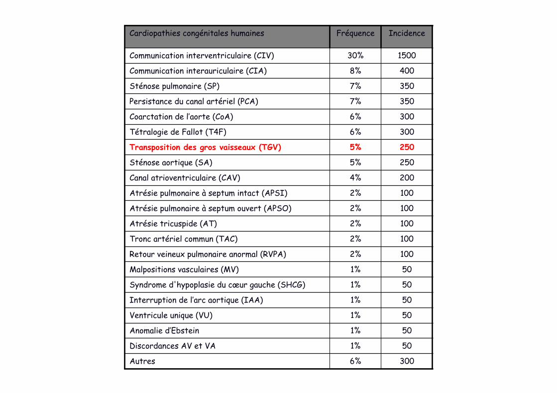
Cardiopathies congénitales humaines Fréquence Incidence Communication interventriculaire (CIV) 30% 1500 Communication interauriculaire (CIA) 8% 400 Sténose pulmonaire (SP) 7% 350 Persistance du canal artériel (PCA) 7% 350 Coarctation de l’aorte (CoA) 6% 300 Tétralogie de Fallot (T4F) 6% 300 Transposition des gros vaisseaux (TGV) 5% 250 Sténose aortique (SA) 5% 250 Canal atrioventriculaire (CAV) 4% 200 Atrésie pulmonaire à septum intact (APSI) 2% 100 Atrésie pulmonaire à septum ouvert (APSO) 2% 100 Atrésie tricuspide (AT) 2% 100 Tronc artériel commun (TAC) 2% 100 Retour veineux pulmonaire anormal (RVPA) 2% 100 Malpositions vasculaires (MV) 1% 50 Syndrome d'hypoplasie du cœur gauche (SHCG) 1% 50 Interruption de l’arc aortique (IAA) 1% 50 Ventricule unique (VU) 1% 50 Anomalie d’Ebstein 1% 50 Discordances AV et VA 1% 50 Autres 6% 300
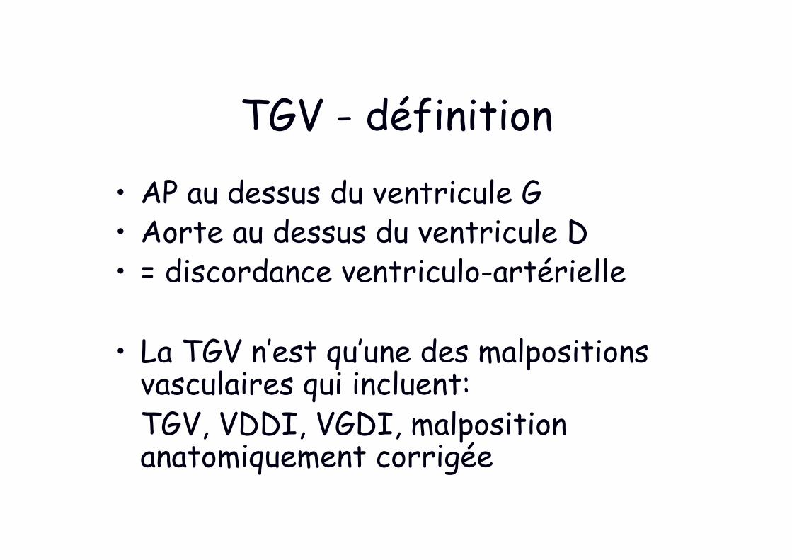
TGV - définition
• AP au dessus du ventricule G • Aorte au dessus du ventricule D • = discordance ventriculo-artérielle
• La TGV n’est qu’une des malpositions vasculaires qui incluent: TGV, VDDI, VGDI, malposition anatomiquement corrigée
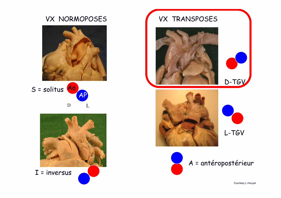
VX NORMOPOSES VX TRANSPOSES
Ao AP
D-TGV
L-TGV
S = solitus
I = inversus A = antéropostérieur
Courtesy L.Houyel
L D
Rappel embryologique
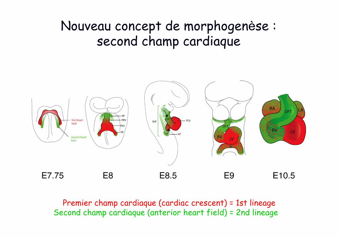
Nouveau concept de morphogenèse : second champ cardiaque
Premier champ cardiaque (cardiac crescent) = 1st lineage Second champ cardiaque (anterior heart field) = 2nd lineage
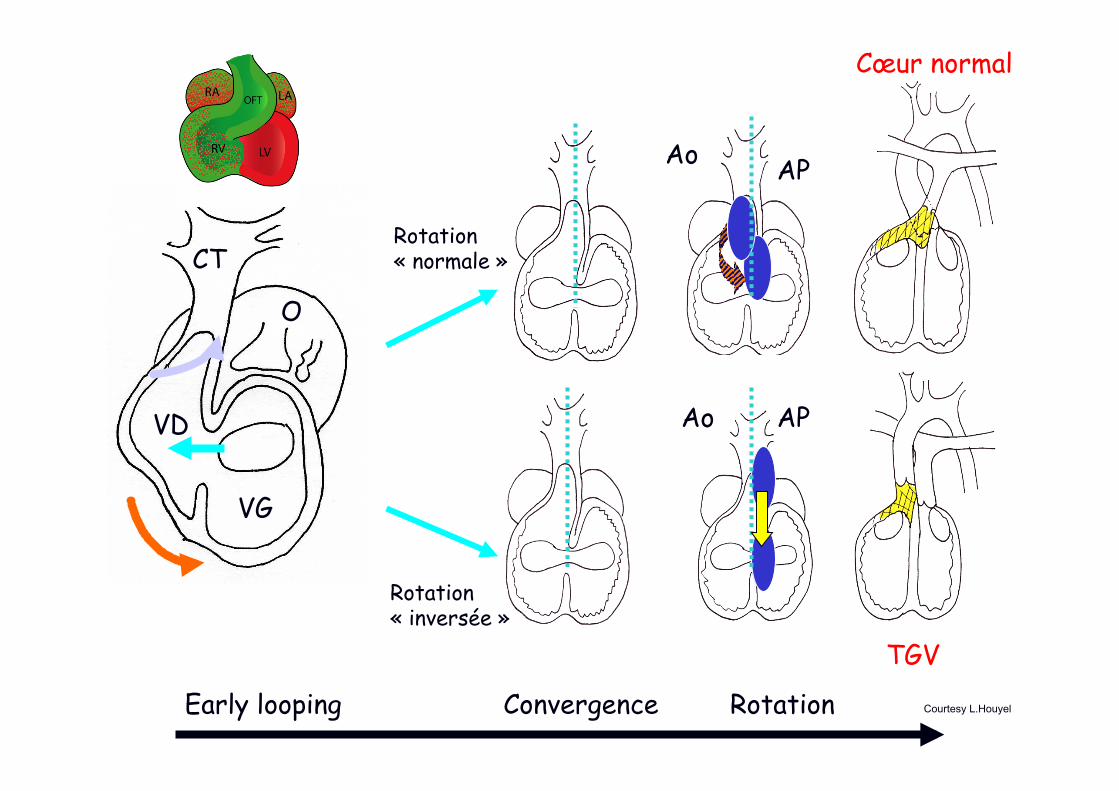
Early looping Convergence Rotation
Ao
Ao
AP
AP
Rotation « normale »
Rotation « inversée »
TGV
Cœur normal
VD
VG
O
CT
Courtesy L.Houyel
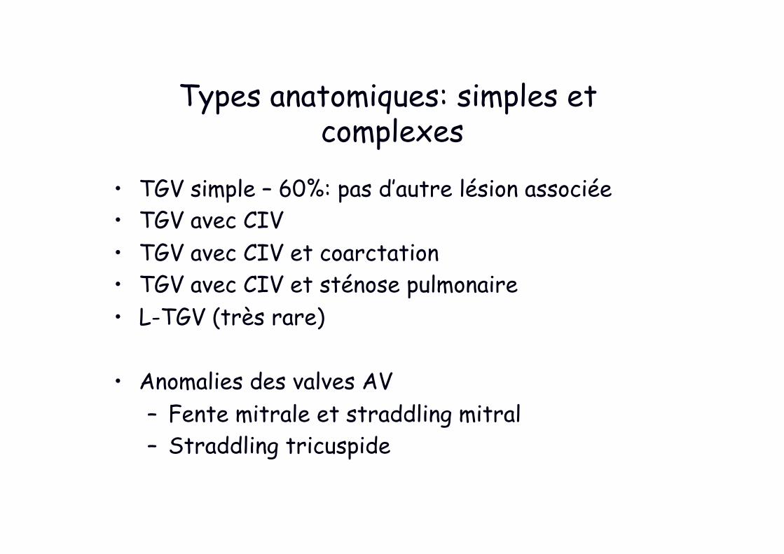
Types anatomiques: simples et complexes
• TGV simple – 60%: pas d’autre lésion associée • TGV avec CIV • TGV avec CIV et coarctation • TGV avec CIV et sténose pulmonaire • L-TGV (très rare)
• Anomalies des valves AV – Fente mitrale et straddling mitral – Straddling tricuspide
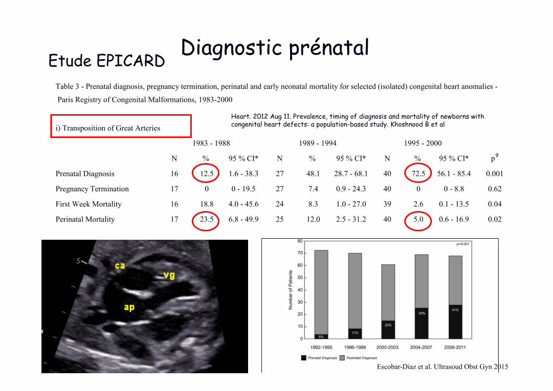
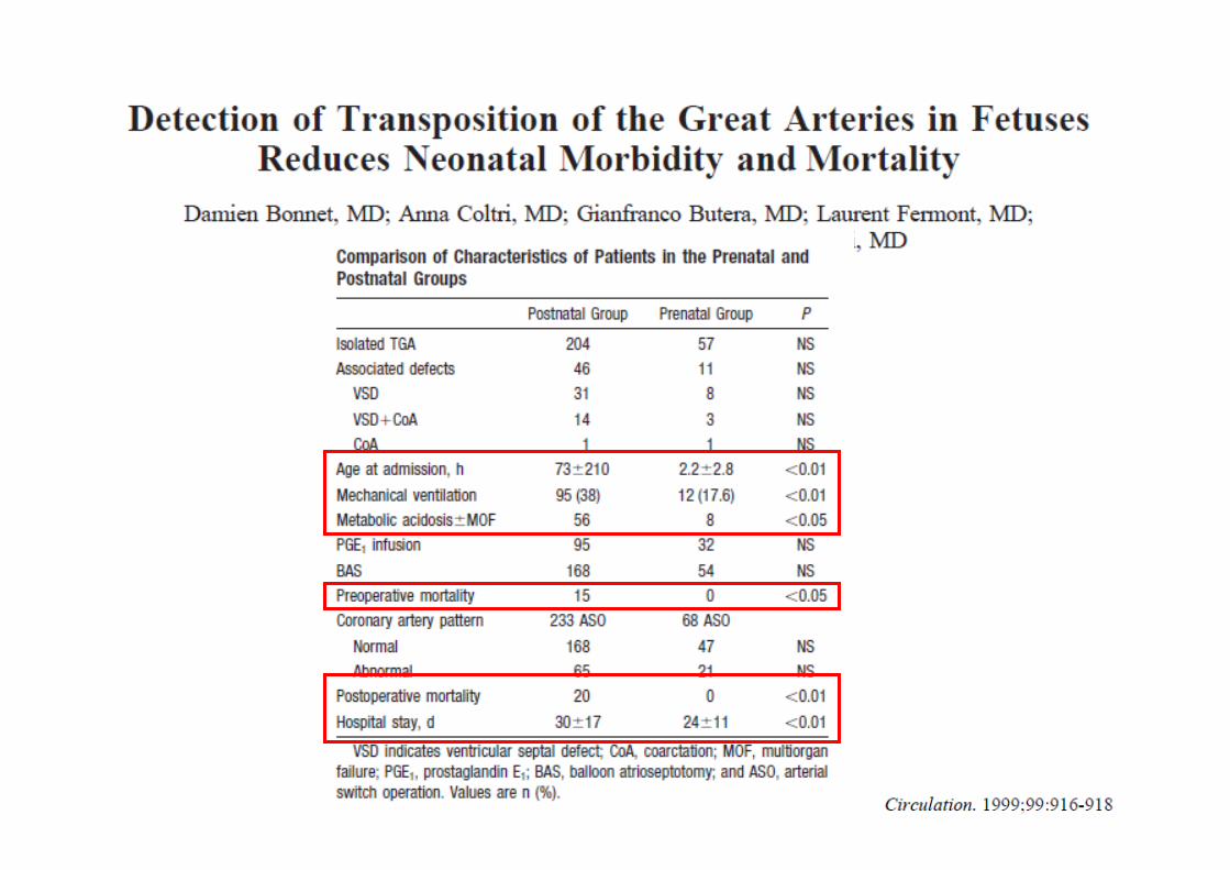
Diagnostic prénatal Table 3 - Prenatal diagnosis, pregnancy termination, perinatal and early neonatal mortality for selected (isolated) congenital heart anomalies -
Paris Registry of Congenital Malformations, 1983-2000
i) Transposition of Great Arteries
N % 95 % CI* N % 95 % CI* N % 95 % CI* pV
Prenatal Diagnosis 16 12.5 1.6 - 38.3 27 48.1 28.7 - 68.1 40 72.5 56.1 - 85.4 0.001
Pregnancy Termination 17 0 0 - 19.5 27 7.4 0.9 - 24.3 40 0 0 - 8.8 0.62
First Week Mortality 16 18.8 4.0 - 45.6 24 8.3 1.0 - 27.0 39 2.6 0.1 - 13.5 0.04
Perinatal Mortality 17 23.5 6.8 - 49.9 25 12.0 2.5 - 31.2 40 5.0 0.6 - 16.9 0.02
ii) Hypoplastic Left Heart Syndrome
N % 95 % CI* N % 95 % CI* N % 95 % CI* pV
Prenatal Diagnosis 22 31.8 13.9 - 54.9 29 82.8 64.2 - 94.2 27 88.9 70.8 - 97.6 < 0.001
Pregnancy Termination 22 13.6 2.9 - 34.9 29 72.4 52.8 - 87.3 27 63.0 42.4 - 80.6 < 0.001
First Week Mortality 18 83.3 58.6 - 96.4 8 75.0 34.9 - 96.8 10 50.0 18.7 - 81.3 0.12
Perinatal Mortality 19 84.2 60.4 - 96.6 8 75.0 34.9 - 96.8 10 50.0 18.7 - 81.3 0.10
1983 - 1988 1989 - 1994 1995 - 2000
1983 - 1988 1989 - 1994 1995 - 2000
Etude EPICARD
Heart. 2012 Aug 11. Prevalence, timing of diagnosis and mortality of newborns with congenital heart defects: a population-based study. Khoshnood B et al
Escobar-Diaz et al. Ultrasoud Obst Gyn 2015
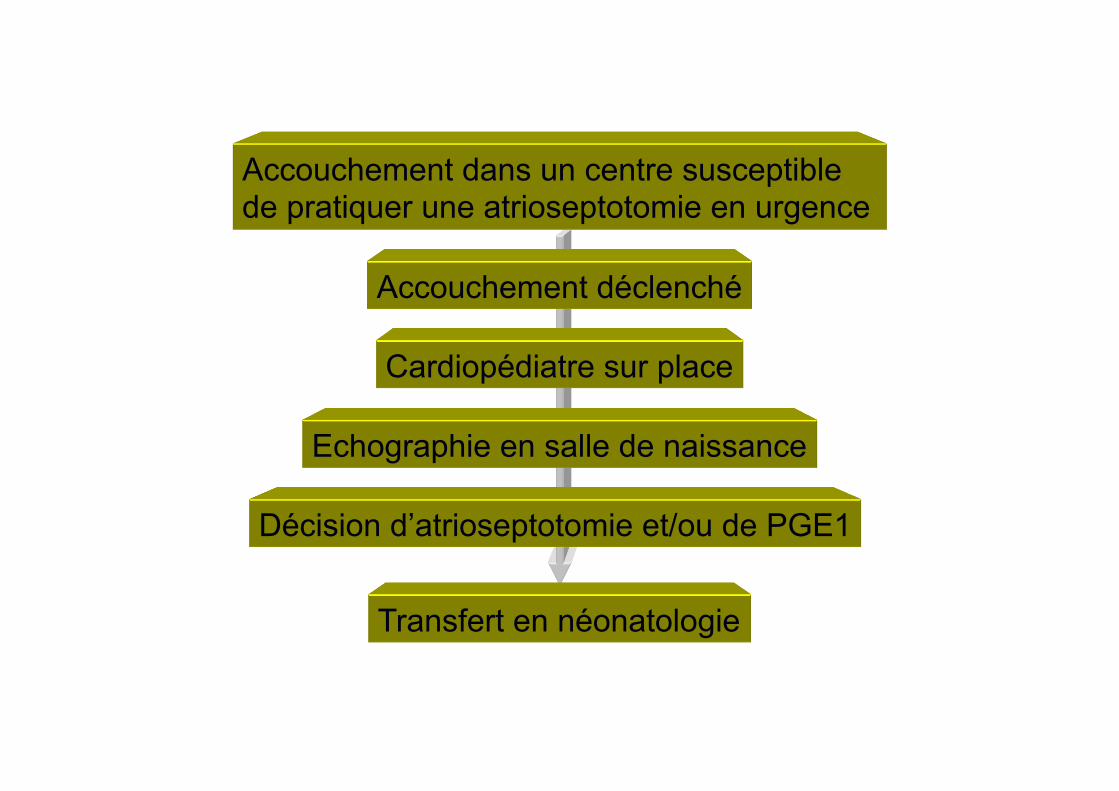
Accouchement dans un centre susceptible de pratiquer une atrioseptotomie en urgence
Accouchement déclenché
Cardiopédiatre sur place
Echographie en salle de naissance
Décision d’atrioseptotomie et/ou de PGE1
Transfert en néonatologie
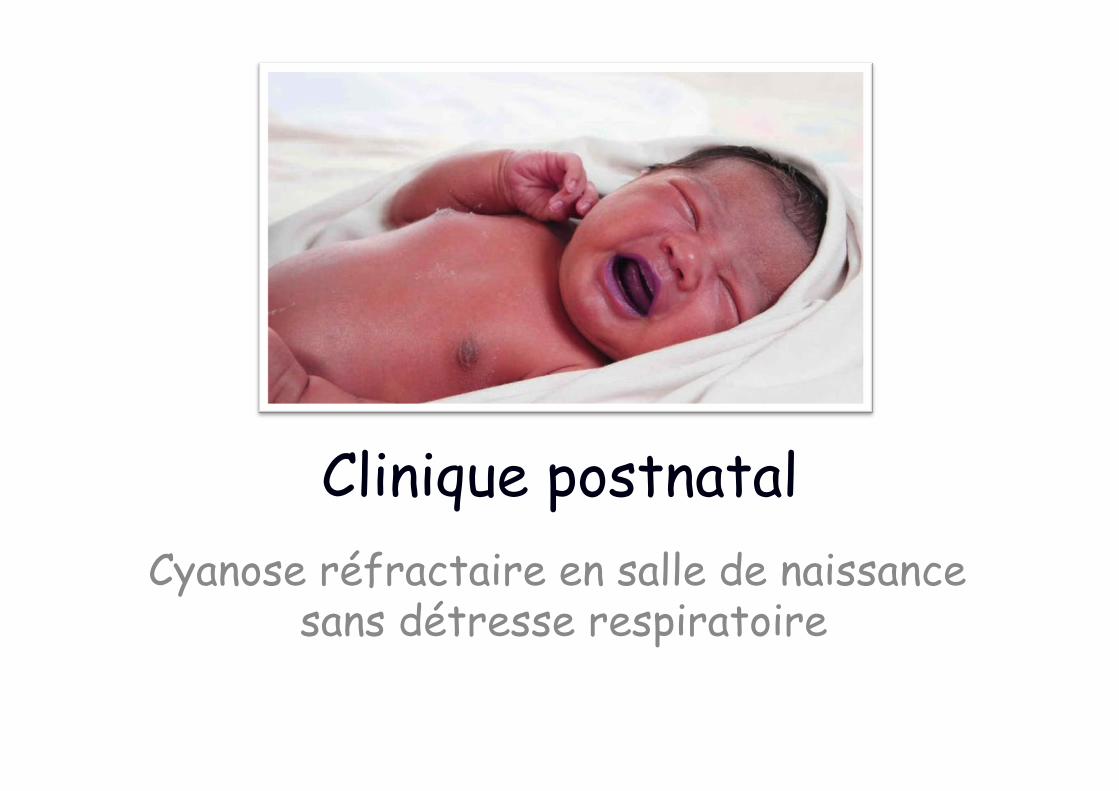
Clinique postnatal Cyanose réfractaire en salle de naissance
sans détresse respiratoire
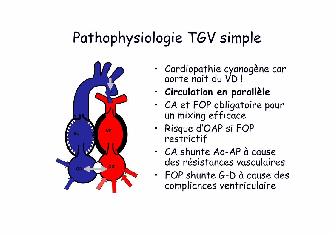
Pathophysiologie TGV simple
• Cardiopathie cyanogène car aorte nait du VD !
• Circulation en parallèle • CA et FOP obligatoire pour
un mixing efficace • Risque d’OAP si FOP
restrictif • CA shunte Ao-AP à cause
des résistances vasculaires • FOP shunte G-D à cause des
compliances ventriculaire
OD OG
VG VD
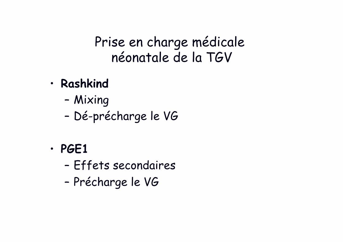
Prise en charge médicale néonatale de la TGV
• Rashkind – Mixing – Dé-précharge le VG
• PGE1 – Effets secondaires – Précharge le VG
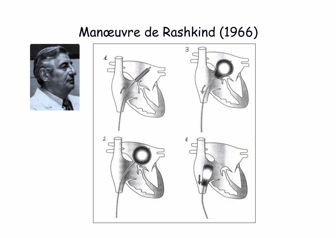
Manœuvre de Rashkind (1966)
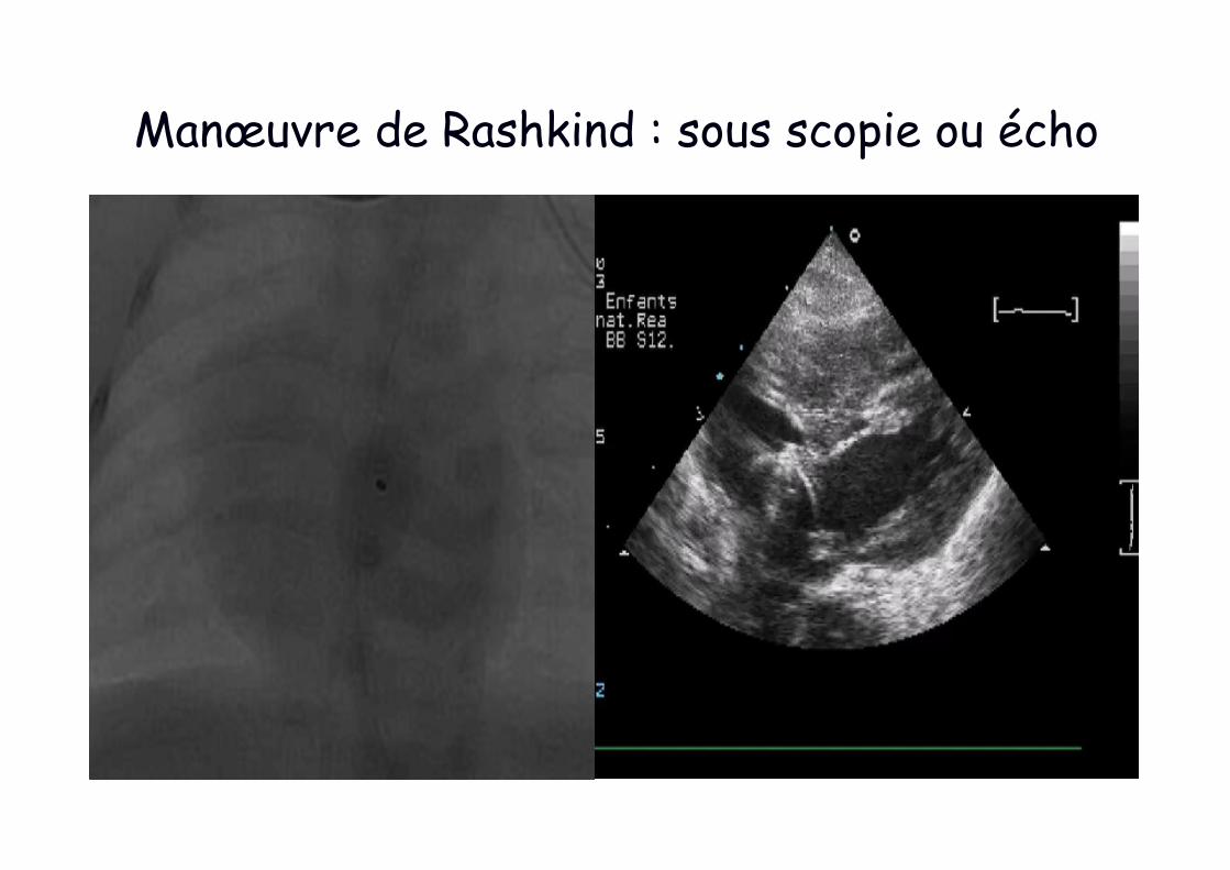
Manœuvre de Rashkind : sous scopie ou écho
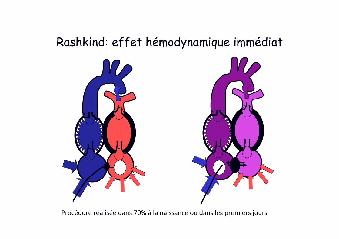
Rashkind: effet hémodynamique immédiat Ballon gonflé dans l’OG
Procédure réalisée dans 70% à la naissance ou dans les premiers jours
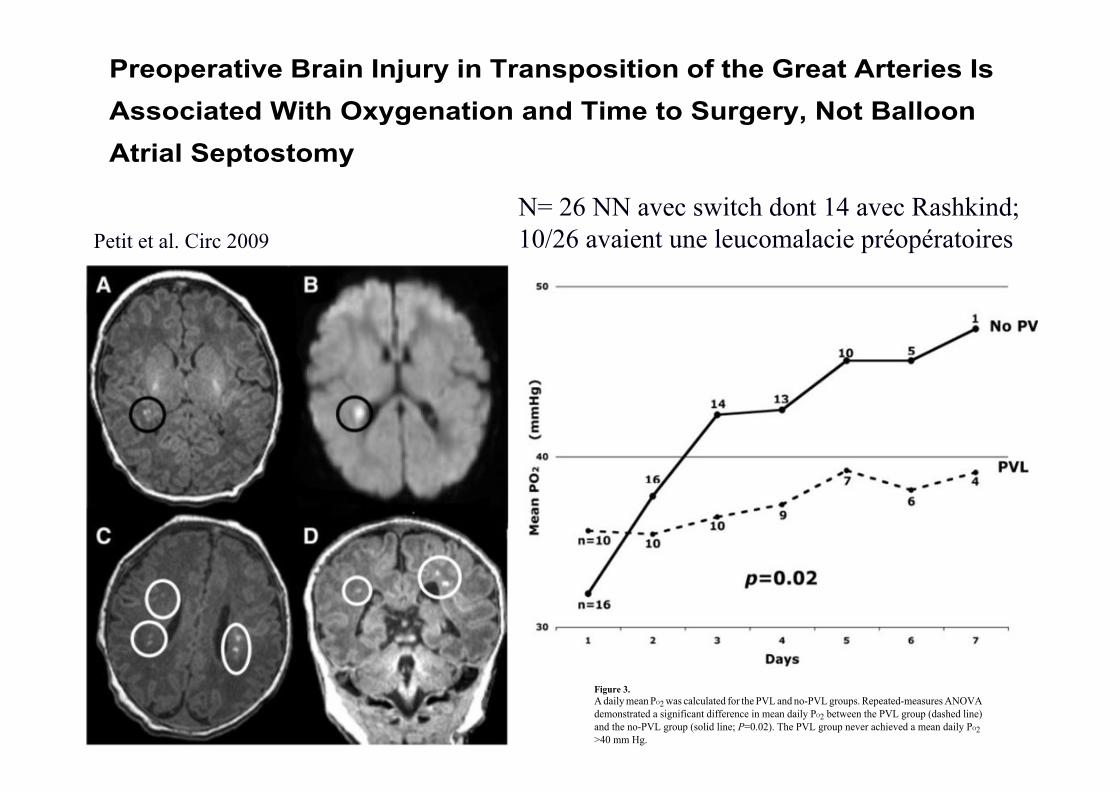
Preoperative Brain Injury in Transposition of the Great Arteries IsAssociated With Oxygenation and Time to Surgery, Not BalloonAtrial Septostomy
Christopher J. Petit, MD, Jonathan J. Rome, MD, Gil Wernovsky, MD, Stefanie E. Mason,BS, David M. Shera, ScD, Susan C. Nicolson, MD, Lisa M. Montenegro, MD, Sarah Tabbutt,MD, PhD, Robert A. Zimmerman, MD, and Daniel J. Licht, MDDivision of Cardiology, Department of Pediatrics (C.J.P., J.J.R., G.W., S.T.); Division of Critical CareMedicine, Department of Anesthesia and Critical Care Medicine (G.W., S.T.); Division of Neurology,Department of Pediatrics (S.E.M., D.J.L.); Division of Biostatistics and Epidemiology (D.M.S.);Division of Cardiothoracic Anesthesia, Department of Anesthesia and Critical Care Medicine(S.C.N., L.M.M.); and Department of Radiology (R.A.Z.), The Children’s Hospital of Philadelphiaand the University of Pennsylvania School of Medicine, Philadelphia, Pa
AbstractBackground—Preoperative brain injury is an increasingly recognized phenomenon in neonateswith complex congenital heart disease. Recently, reports have been published that associatepreoperative brain injury in neonates with transposition of the great arteries with the performance ofballoon atrial septostomy (BAS), a procedure that improves systemic oxygenation preoperatively. Itis unclear whether BAS is the cause of brain injury or is a confounder, because neonates who requireBAS are typically more hypoxemic. We sought to determine the relationship between preoperativebrain injury in neonates with transposition of the great arteries and the performance of BAS. Wehypothesized that brain injury results from hypoxic injury, not from the BAS itself.
Methods and Results—Infants with transposition of the great arteries (n=26) were retrospectivelyincluded from a larger cohort of infants with congenital heart disease who underwent preoperativebrain MRI as part of 2 separate prospective studies. Data collected included all preoperative pulseoximetry recordings, all values from preoperative arterial blood gas measurements, and BASprocedure data. MRI scans were performed on the day of surgery, before the surgical repair. Of the26 neonates, 14 underwent BAS. No stroke was seen in the entire cohort, whereas 10 (38%) of 26patients were found to have hypoxic brain injury in the form of periventricular leukomalacia.Periventricular leukomalacia was not associated with BAS; however, neonates with periventricularleukomalacia had lower preoperative oxygenation (P=0.026) and a longer time to surgery (P=0.028)than those without periventricular leukomalacia.
Conclusions—Preoperative brain injury in neonates with transposition of the great arteries isassociated with hypoxemia and longer time to surgery. We found no association between BAS andbrain injury.
© 2009 American Heart Association, Inc.Correspondence to Christopher J. Petit, MD, Texas Children’s Hospital, 6621 Fannin St, MC-19345 C, Houston, TX 77030. [email protected].
NIH Public AccessAuthor ManuscriptCirculation. Author manuscript; available in PMC 2009 September 2.
Published in final edited form as:Circulation. 2009 February 10; 119(5): 709–716. doi:10.1161/CIRCULATIONAHA.107.760819.
NIH-PA Author Manuscript
NIH-PA Author Manuscript
NIH-PA Author Manuscript
Figure 2.Brain MRI of preoperative infants with TGA: T1 imaging (A) and diffusion-weighted imaging(B) in a patient with mild PVL, which is a unifocal, small (<3 mm) white matter lesion. Thelower MRI images demonstrate axial (C) and coronal (D) T1 imaging in a patient with bilateral,multifocal (moderate PVL) white matter disease.
Petit et al. Page 11
Circulation. Author manuscript; available in PMC 2009 September 2.
NIH
-PA
Author M
anuscriptN
IH-P
A A
uthor Manuscript
NIH
-PA
Author M
anuscript
Figure 3.A daily mean PO2 was calculated for the PVL and no-PVL groups. Repeated-measures ANOVAdemonstrated a significant difference in mean daily PO2 between the PVL group (dashed line)and the no-PVL group (solid line; P=0.02). The PVL group never achieved a mean daily PO2>40 mm Hg.
Petit et al. Page 12
Circulation. Author manuscript; available in PMC 2009 September 2.
NIH
-PA
Author M
anuscriptN
IH-P
A A
uthor Manuscript
NIH
-PA
Author M
anuscript
Petit et al. Circ 2009 N= 26 NN avec switch dont 14 avec Rashkind; 10/26 avaient une leucomalacie préopératoires
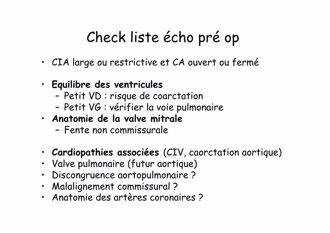
Check liste écho pré op • CIA large ou restrictive et CA ouvert ou fermé
• Equilibre des ventricules – Petit VD : risque de coarctation – Petit VG : vérifier la voie pulmonaire
• Anatomie de la valve mitrale – Fente non commissurale
• Cardiopathies associées (CIV, caorctation aortique) • Valve pulmonaire (futur aortique) • Discongruence aortopulmonaire ? • Malalignement commissural ? • Anatomie des artères coronaires ?
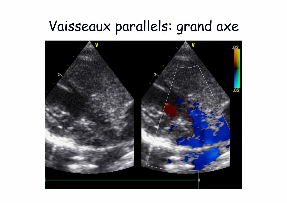
Vaisseaux parallels: grand axe
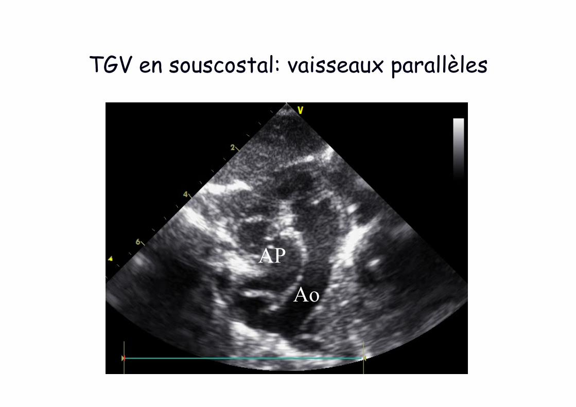
TGV en souscostal: vaisseaux parallèles
Ao AP
Ao
AP
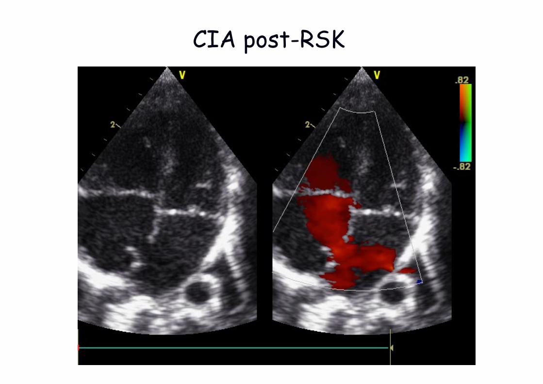
CIA post-RSK
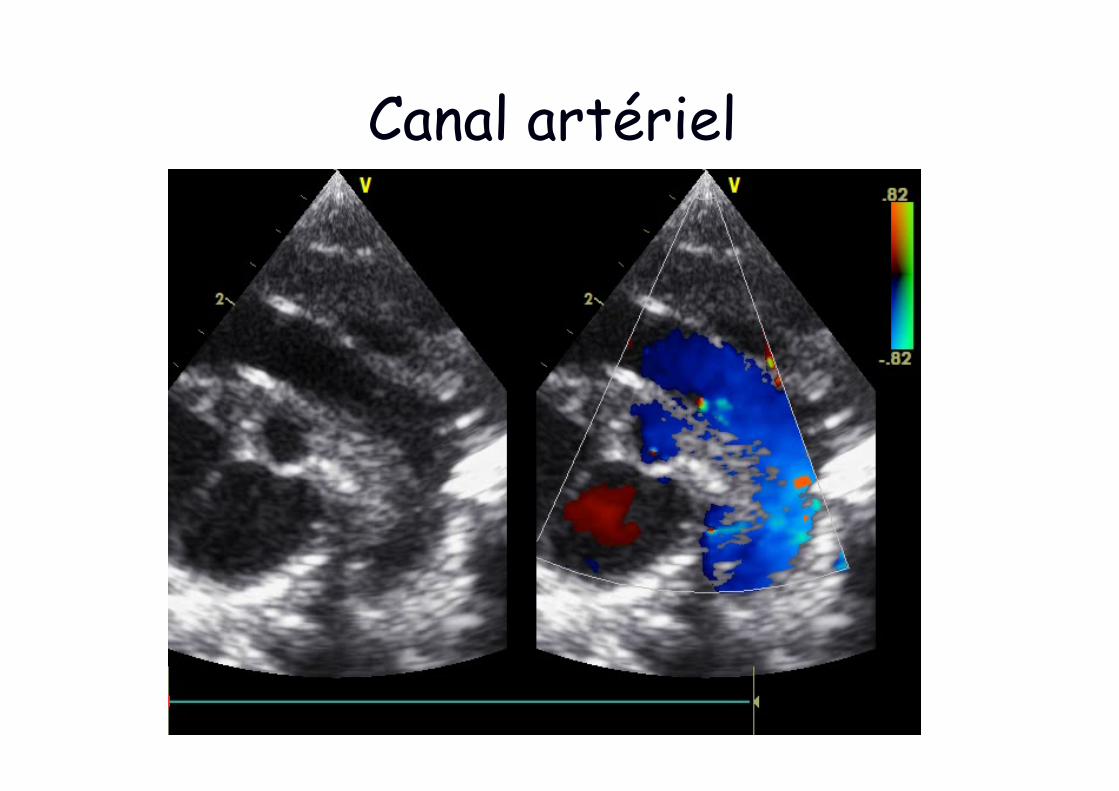
Canal artériel
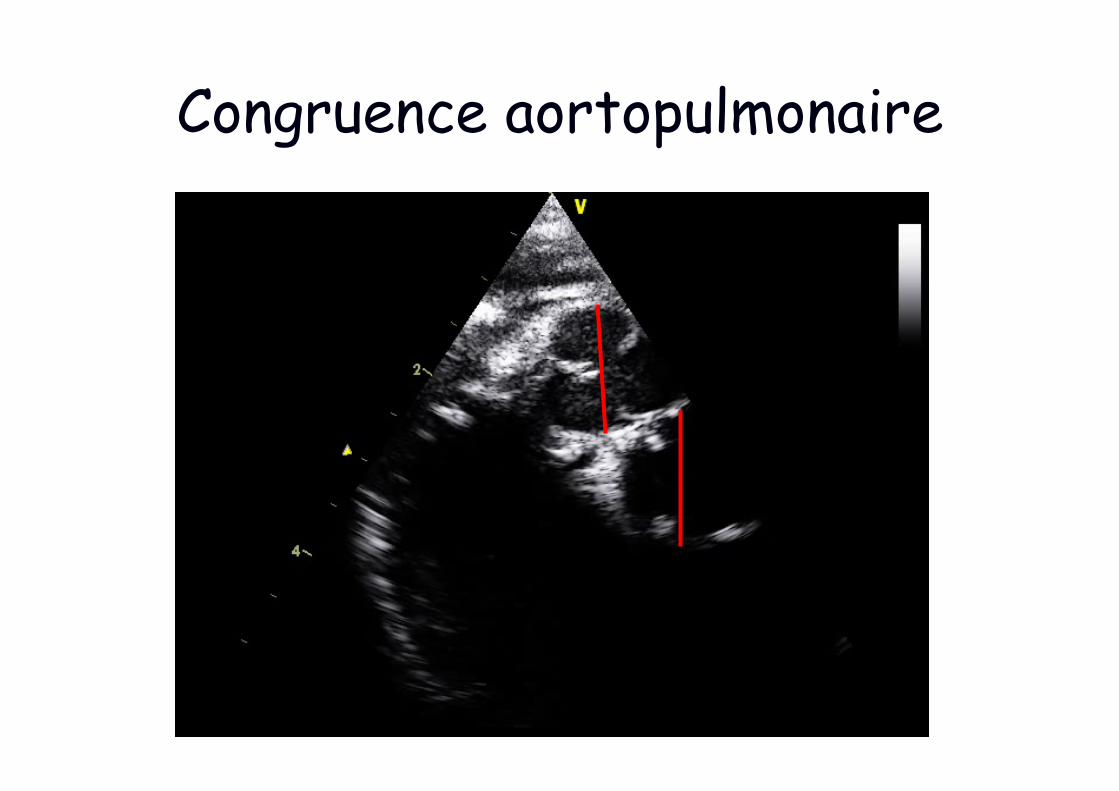
Congruence aortopulmonaire
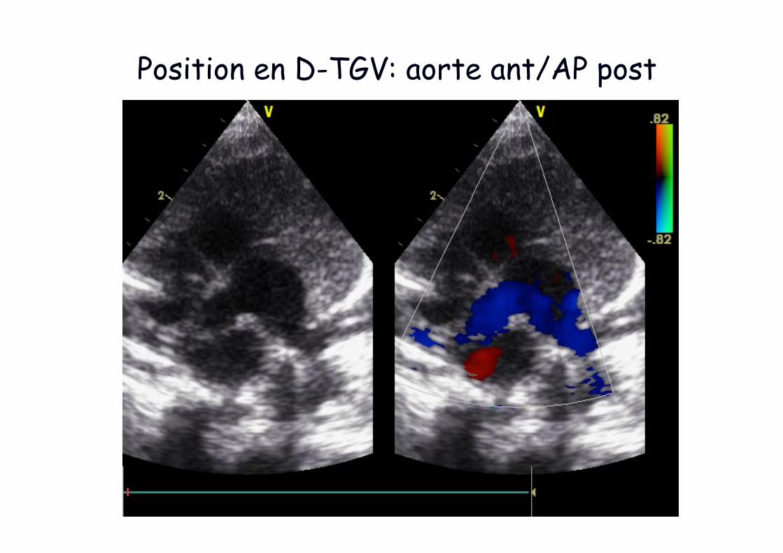
Position en D-TGV: aorte ant/AP post
AP
Ao
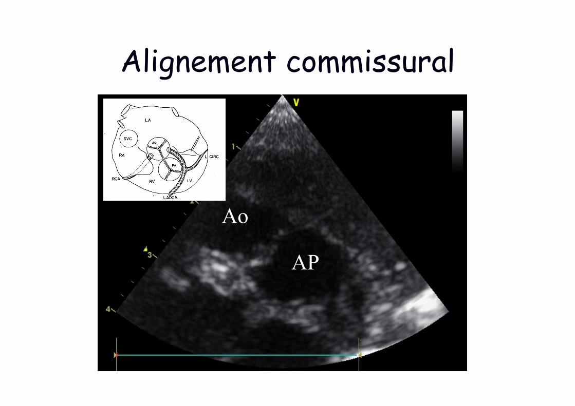
Alignement commissural
AO
AP
AP
Ao
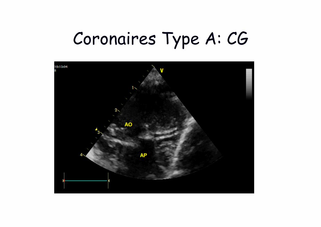
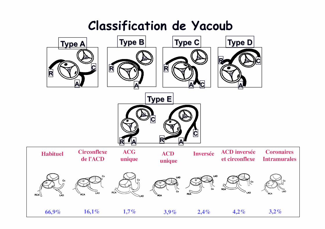
Coronaires Type A: CG
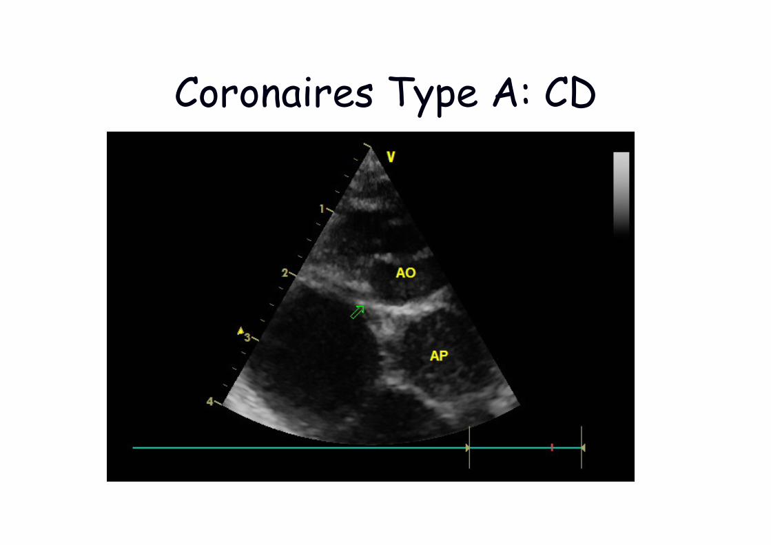
Coronaires Type A: CD
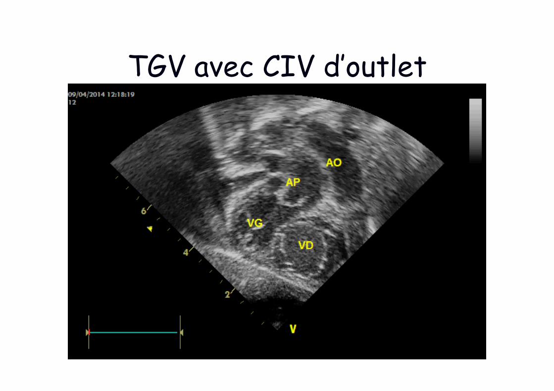
TGV avec CIV d’outlet
R
A
A
PA
R A
A
PA
C R
A
A
PA
C
R
A
A
PA C
R A
A
PA
C R A
A
PA
C
Type A Type B Type C Type D
Type E
Classification de Yacoub
• « normale » : 60% • boucle antérieure et/ou postérieure : 35% • entre gros vaisseaux (intramurale) : 5%
Anatomie coronaire
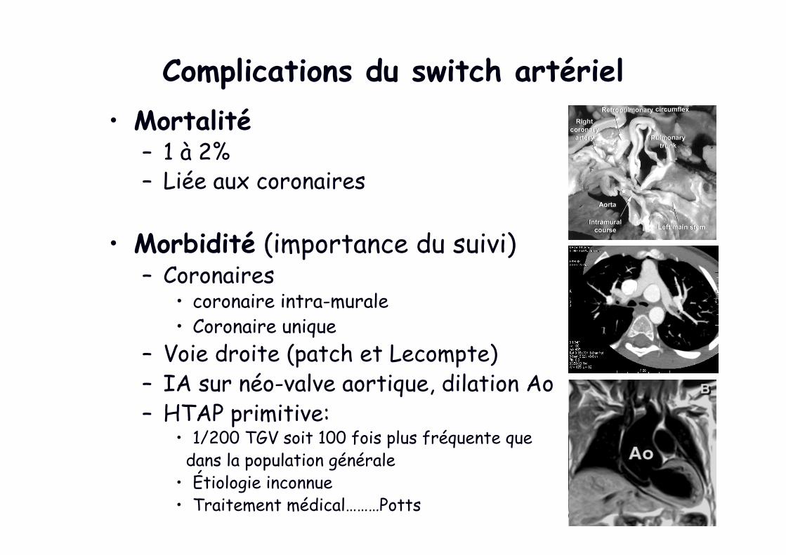
Complications du switch artériel • Mortalité
– 1 à 2% – Liée aux coronaires
• Morbidité (importance du suivi) – Coronaires
• coronaire intra-murale • Coronaire unique
– Voie droite (patch et Lecompte) – IA sur néo-valve aortique, dilation Ao – HTAP primitive:
• 1/200 TGV soit 100 fois plus fréquente que dans la population générale • Étiologie inconnue • Traitement médical………Potts
Les lésions coronaires après switch artériel
Comment les détecter?
ECG et échographie (IM!!!) Coroscanner si signe d’ischémie
Coronarographie si doute Test d’ischémie (scintigraphie)
Coroscanner systématique à 5 ans
Que faire ?
Rappeler votre chirurgien…

Devenir
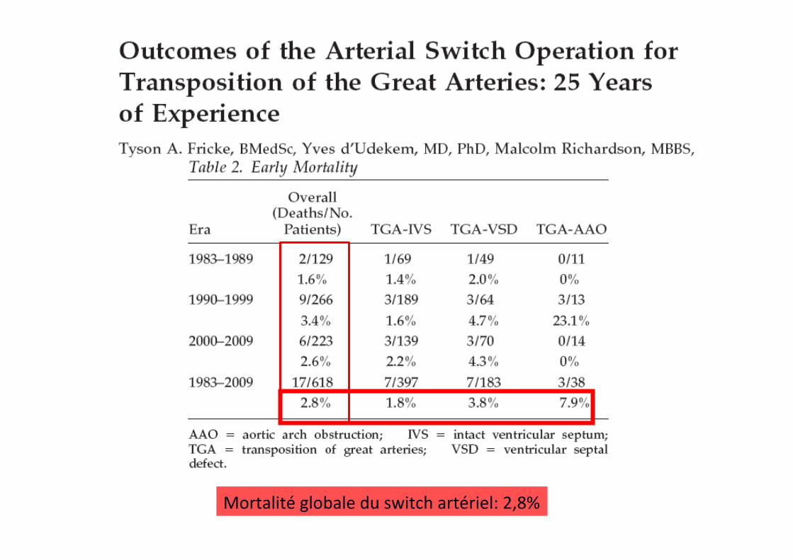
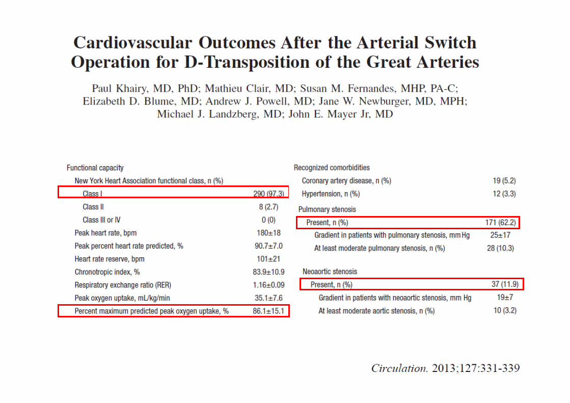
Mortalité globale du switch artériel: 2,8%
NIH-PA Author ManuscriptNIH-PA Author ManuscriptNIH-PA Author Manuscript
Villafañe et al.
Page 29
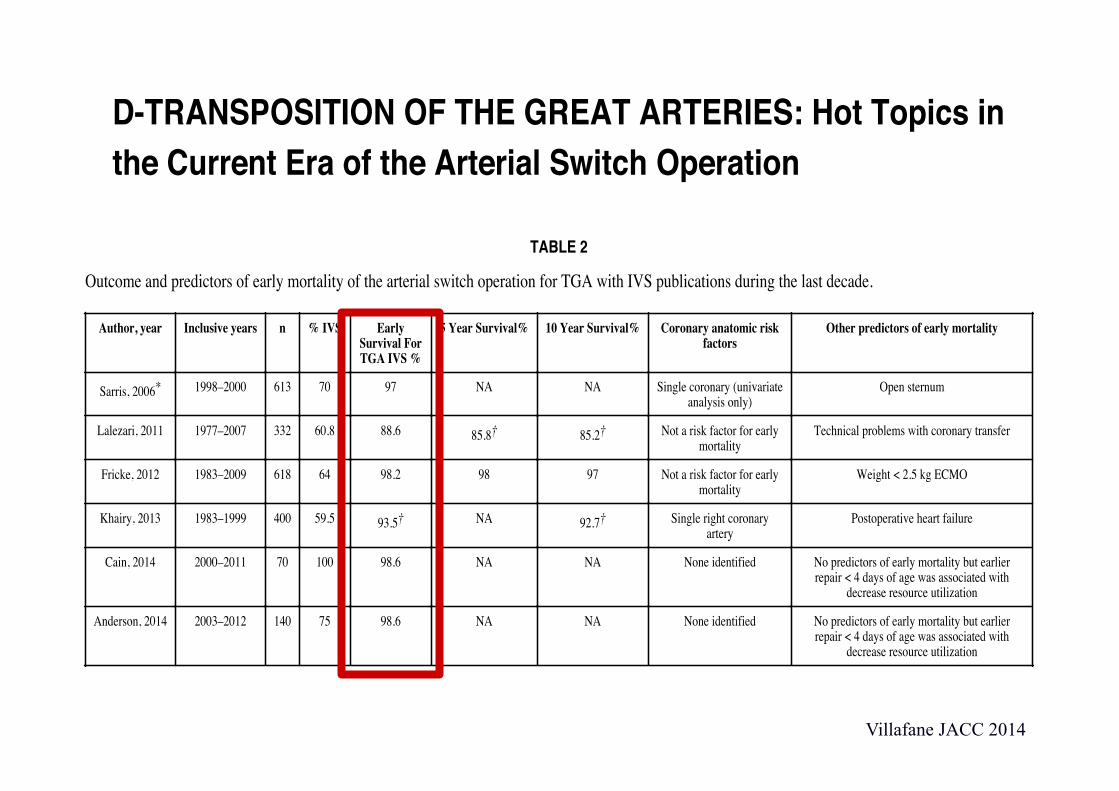
TABLE 2
Outcome and predictors of early mortality of the arterial switch operation for TGA with IVS publications during the last decade.
Author, year Inclusive years n % IVS Early Survival For TGA IVS %
5 Year Survival% 10 Year Survival% Coronary anatomic risk factors
Other predictors of early mortality
Sarris, 2006* 1998–2000 613 70 97 NA NA Single coronary (univariate analysis only)
Open sternum
Lalezari, 2011 1977–2007 332 60.8 88.6 85.8† 85.2† Not a risk factor for early mortality
Technical problems with coronary transfer
Fricke, 2012 1983–2009 618 64 98.2 98 97 Not a risk factor for early mortality
Weight < 2.5 kg ECMO
Khairy, 2013 1983–1999 400 59.5 93.5† NA 92.7† Single right coronary artery
Postoperative heart failure
Cain, 2014 2000–2011 70 100 98.6 NA NA None identified No predictors of early mortality but earlier repair < 4 days of age was associated with
decrease resource utilization
Anderson, 2014 2003–2012 140 75 98.6 NA NA None identified No predictors of early mortality but earlier repair < 4 days of age was associated with
decrease resource utilization
*Multicenter study of early results only from the European Congenital Heart Surgeons Association.
†Results include outcome of all patients undergoing the arterial switch operation and are not confined to those with intact ventricular septum.
ECMO = extracorporeal membrane oxygenation; IVS = intact ventricular septum; TGA = transposition of the great arteries with intact septum.
J Am
Coll C
ardiol. Author m
anuscript; available in PM
C 2015 A
ugust 05.
D-TRANSPOSITION OF THE GREAT ARTERIES: Hot Topics in the Current Era of the Arterial Switch Operation
Juan Villafañe, MD*, M. Regina Lantin-Hermoso, MD†, Ami B. Bhatt, MD‡, James S. Tweddell, MD§, Tal Geva, MD‖, Meena Nathan, MD¶, Martin J. Elliott, MD#, Victoria L. Vetter, MD, MPH**, Stephen M. Paridon, MD††, Lazaros Kochilas, MD, MSCR‡‡, Kathy J. Jenkins, MD, MPH§§, Robert H. Beekman III, MD‖‖, Gil Wernovsky, MD¶¶, and Jeffrey A. Towbin, MD##
On behalf of the American College of Cardiology Adult Congenital and Pediatric Cardiology Council*Department of Pediatrics (Cardiology), University of Kentucky, Lexington, Kentucky†Pediatric Cardiology, Texas Children’s Hospital, Baylor College of Medicine, Houston, Texas‡Adult Congenital Heart Disease Program, Massachusetts General Hospital, Harvard Medical School, Boston, Massachusetts§Cardiothoracic Surgery, Children’s Hospital of Wisconsin, Medical College of Wisconsin, Milwaukee, Wisconsin‖Department of Cardiology, Boston Children’s Hospital, Harvard Medical School, Boston, Massachusetts¶Department of Cardiac Surgery, Boston Children’s Hospital, Harvard Medical School, Boston, Massachusetts#Department of Pediatric Cardiothoracic Surgery, The Great Ormond Street Hospital for Children, NHS Foundation Trust, LONDON, UK**Children’s Hospital of Philadelphia, Perelman School of Medicine, University of Pennsylvania, Philadelphia, Pennsylvania††Department of Exercise Physiology, Perlman School of Medicine, University of Pennsylvania School of Medicine, Philadelphia, Pennsylvania‡‡University of Minnesota Children’s Hospital, Minneapolis, Minnesota§§Boston Children’s Hospital, Harvard Medical School, Boston, Massachusetts‖‖Division of Cardiology, Cincinnati Children’s Hospital Medical Center, University of Cincinnati College of Medicine, Cincinnati, Ohio
Address for correspondence: Juan Villafañe, MD, FACC, 743 East Broadway, #300, Louisville, Kentucky 40202, Ph: (502) 584-3200, Fax: (502) 458-8875, [email protected]. Juan Villafane has disclosed his relationship with Biomedical. James Tweddell has disclosed his relationship with CorMatrix. Tal Geva has disclosed his relationship with Medtronic. Meena Nathan has disclosed her grant support through the Thoracic Surgery Foundation for Research and Education. Lazaros Kochilas has disclosed his support by NIH/HNLBI (1R01 HL122392-01). Kathy Jenkins has disclosed her grant support through Johns Hopkins Medical Center, Medtronic and NuMed. Robert H. Beekman, III has disclosed his relationship with St. Jude Medical. The remaining authors have no relationships to disclose.
NIH Public AccessAuthor ManuscriptJ Am Coll Cardiol. Author manuscript; available in PMC 2015 August 05.
Published in final edited form as:J Am Coll Cardiol. 2014 August 5; 64(5): 498–511. doi:10.1016/j.jacc.2014.06.1150.
NIH
-PA
Author M
anuscriptN
IH-P
A A
uthor Manuscript
NIH
-PA
Author M
anuscript
Villafane JACC 2014
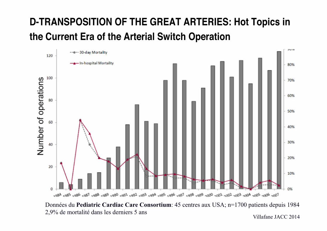
Figure 5. Neonatal ASO and postoperative mortalityData from the PCCC registry.ASO=arterial switch operation; D-TGA=d-transposition of the great arteries; PCCC=pediatric cardiac care consortium.
Villafañe et al. Page 27
J Am Coll Cardiol. Author manuscript; available in PMC 2015 August 05.
NIH
-PA Author Manuscript
NIH
-PA Author Manuscript
NIH
-PA Author Manuscript
D-TRANSPOSITION OF THE GREAT ARTERIES: Hot Topics in the Current Era of the Arterial Switch Operation
Juan Villafañe, MD*, M. Regina Lantin-Hermoso, MD†, Ami B. Bhatt, MD‡, James S. Tweddell, MD§, Tal Geva, MD‖, Meena Nathan, MD¶, Martin J. Elliott, MD#, Victoria L. Vetter, MD, MPH**, Stephen M. Paridon, MD††, Lazaros Kochilas, MD, MSCR‡‡, Kathy J. Jenkins, MD, MPH§§, Robert H. Beekman III, MD‖‖, Gil Wernovsky, MD¶¶, and Jeffrey A. Towbin, MD##
On behalf of the American College of Cardiology Adult Congenital and Pediatric Cardiology Council*Department of Pediatrics (Cardiology), University of Kentucky, Lexington, Kentucky†Pediatric Cardiology, Texas Children’s Hospital, Baylor College of Medicine, Houston, Texas‡Adult Congenital Heart Disease Program, Massachusetts General Hospital, Harvard Medical School, Boston, Massachusetts§Cardiothoracic Surgery, Children’s Hospital of Wisconsin, Medical College of Wisconsin, Milwaukee, Wisconsin‖Department of Cardiology, Boston Children’s Hospital, Harvard Medical School, Boston, Massachusetts¶Department of Cardiac Surgery, Boston Children’s Hospital, Harvard Medical School, Boston, Massachusetts#Department of Pediatric Cardiothoracic Surgery, The Great Ormond Street Hospital for Children, NHS Foundation Trust, LONDON, UK**Children’s Hospital of Philadelphia, Perelman School of Medicine, University of Pennsylvania, Philadelphia, Pennsylvania††Department of Exercise Physiology, Perlman School of Medicine, University of Pennsylvania School of Medicine, Philadelphia, Pennsylvania‡‡University of Minnesota Children’s Hospital, Minneapolis, Minnesota§§Boston Children’s Hospital, Harvard Medical School, Boston, Massachusetts‖‖Division of Cardiology, Cincinnati Children’s Hospital Medical Center, University of Cincinnati College of Medicine, Cincinnati, Ohio
Address for correspondence: Juan Villafañe, MD, FACC, 743 East Broadway, #300, Louisville, Kentucky 40202, Ph: (502) 584-3200, Fax: (502) 458-8875, [email protected]. Juan Villafane has disclosed his relationship with Biomedical. James Tweddell has disclosed his relationship with CorMatrix. Tal Geva has disclosed his relationship with Medtronic. Meena Nathan has disclosed her grant support through the Thoracic Surgery Foundation for Research and Education. Lazaros Kochilas has disclosed his support by NIH/HNLBI (1R01 HL122392-01). Kathy Jenkins has disclosed her grant support through Johns Hopkins Medical Center, Medtronic and NuMed. Robert H. Beekman, III has disclosed his relationship with St. Jude Medical. The remaining authors have no relationships to disclose.
NIH Public AccessAuthor ManuscriptJ Am Coll Cardiol. Author manuscript; available in PMC 2015 August 05.
Published in final edited form as:J Am Coll Cardiol. 2014 August 5; 64(5): 498–511. doi:10.1016/j.jacc.2014.06.1150.
NIH
-PA
Author M
anuscriptN
IH-P
A A
uthor Manuscript
NIH
-PA
Author M
anuscript
Données du Pediatric Cardiac Care Consortium: 45 centres aux USA; n=1700 patients depuis 1984 2,9% de mortalité dans les derniers 5 ans
Villafane JACC 2014
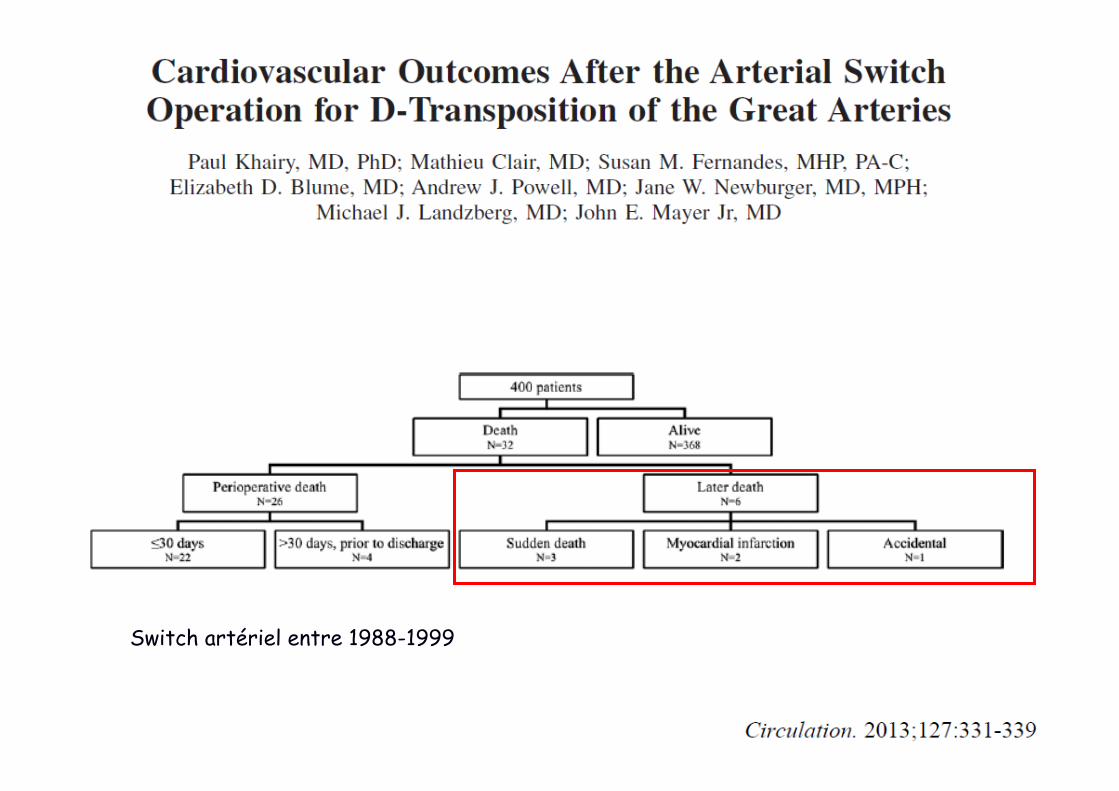
Switch artériel entre 1988-1999
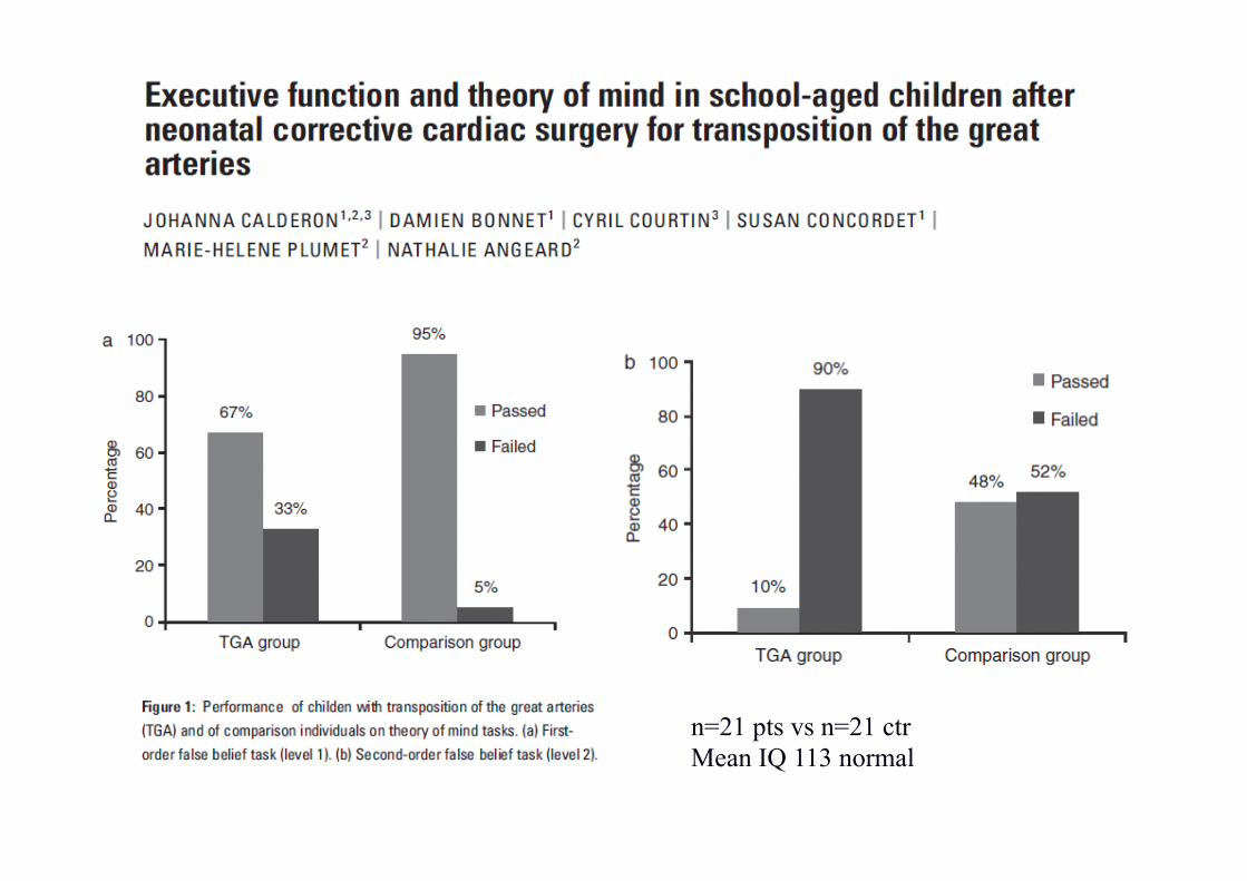
n=21 pts vs n=21 ctr Mean IQ 113 normal