par Programmes de Biologie Moléculaire Thèse présentée à ... · Université de Montréal...
178
Université de Montréal Identification et caractérisation de candidats régulateurs du cycle cellulaire chez le dînoflagellé L ïngulodïnïum polyedrum par Thierry Bertomeu Programmes de Biologie Moléculaire’ Faculté des Études Supérieures Thèse présentée à la Faculté des Études Supérieures en vue de l’obtention du grade de doctorat en biologie moléculaire Juillet, 2007 © Thierry Bertomeu, 2007
-
Upload
duonghuong -
Category
Documents
-
view
219 -
download
0
Transcript of par Programmes de Biologie Moléculaire Thèse présentée à ... · Université de Montréal...
Université de Montréal
Identification et caractérisation de candidats
régulateurs du cycle cellulaire chez le dînoflagellé
Lïngulodïnïum polyedrum
par
Thierry Bertomeu
Programmes de Biologie Moléculaire’
Faculté des Études Supérieures
Thèse présentée à la Faculté des Études Supérieures
en vue de l’obtention du grade de doctorat
en biologie moléculaire
Juillet, 2007
© Thierry Bertomeu, 2007

Q
3
o
o
Université flde Montréal
Direction des bibliothèques
AVIS
L’auteur a autorisé l’Université de Montréal à reproduite et diffuser, en totalité
ou en partie, par quelque moyen que ce soit et sur quelque support que ce
soit, et exclusivement à des fins non lucratives d’enseignement et de
recherche, des copies de ce mémoire ou de cette thèse.
L’auteur et les coauteurs le cas échéant conservent la propriété du droit
d’auteur et des droits moraux qui protègent ce document. Ni la thèse ou le
mémoire, ni des extraits substantiels de ce document, ne doivent être
imprimés ou autrement reproduits sans l’autorisation de l’auteur.
Afin de se conformer à la Loi canadienne sur la protection des
renseignements personnels, quelques formulaires secondaires, coordonnées
ou signatures intégrées au texte ont pu être enlevés de ce document. Bien
que cela ait pu affecter la pagination, il n’y a aucun contenu manquant.
NOTICE
The author of this thesis or dissertation has granted a nonexclusive license
allowing Université de Montrèal to reproduce and publish the document, inpart or in whole, and in any format, solely for noncommercial educational and
research purposes.
The author and co-authors if applicable retain copyright ownership and moral
rights in this document. Neïther the whole thesis or dissertation, flotsubstantial extracts from it, may be printed or otherwise reproduced without
the author’s permission.
In compliance with the Canadian Privacy Act some supporting forms, contact
information or signatures may have been removed from the document. While
this may affect the document page count, it does flot represent any loss of
content from the document.
Université de Montréal
Faculté des Études Supérieures
Cette thèse intitulée
Identification et caractérisation de candidats régulateurs
du cycle cellulaire chez le dinoflagellé Lingulodiniumpolyedrum
présentée par
Thierry Bertomeu
a été évaluée par un jury composé des personnes suivantes:
Martine Raymond, président-rapporteur
David Morse, directeur de recherche
Daniel Matton, membre du jury
Hervé Moreau, examinateur externe
Trang Hoang, représentant du doyen de la FES

III
Résumé
Les dinoflagellés sont des eucaryotes unicellulaires qui composent une
majeure partie du phytoplancton. Leur biologie et leur organisation nucléaire sont
uniques. Leur chromosomes sont condensés en permanence et l’organisation de
leur ADN génomique est hors du commun car ils n’ont pas d’histone ni de
nucléosome. Leur noyau ne se désagrège pas à la mitose et la ségrégation du
matériel génétique se fait via un fuseau mitotique cytoplasmique passant au
travers du noyau par des invaginations nucléaires avec un contact indirect aux
chromosomes attachés à la surface interne du noyau.
Lingulodinium polyedrum est un dinoflagellé modèle pour l’étude des
mécanismes de contrôle des rythmes circadiens car plusieurs de ses aspects
physiologiques (bioluminescence, photosynthèse, migration verticale et mitose)
sont circadiens. Les intervenants biochimiques de ces rythmes circadiens doivent
être ciblés par l’horloge circadienne pour effectivement contrôler l’occurrence de
ces rythmes. L’isolement et la caractérisation de candidats régulateurs du cycle
cellulaire de ce dinoflagellé pourraient alors nous faire découvrir d’autres
particularités du cycle cellulaire des dinoflagellés tout en nous permettant de
découvrir quel contrôle biochimique amène une division cellulaire circadienne.
Nous avons isolé deux gènes d’une banque d’ADNc de Lingulodinium
insérée dans un vecteur d’expression chez la levure capables de complémenter de
manière fonctionnelle un mutant conditionnel létal de Saccharomyces cerevisiae
(pour ses cyclines CIni, Ctn2 et Cln3).
Le premier gène encode une cycline authentique que l’on a appelé LpCycl.
Cette cycline possède un domaine repliement-cycline ainsi qu’une boîte de
iv
destruction cycline typique des cyclines mitotiques. Un anticorps, dirigé contre sa
partie N-terminale excluant ses domaines cyclines, reconnaît une protéine de 68
kDa par transfert de type Western. Ce signal est plus abondant sur des extraits
protéiques de cultures aux cellules enrichies en G2 plutôt que sur des extraits
protéiques de cultures aux cellules enrichies en Gi.
Le second gène encode une protéine AAA (ATPases Associées à
différentes Activités cellulaires) appelée LpAAA. Elle est similaire à des protéines
bactériennes impliquées dans la sporulation et à des AAA de plantes supérieures
aux fonctions inconnues. Une augmentation des niveaux de transcrits de la cycline
CLB5 a été détectée chez la levure exprimant LpAAA et nous proposons que
LpAAA pourrait remodeler et inactiver l’inhibiteur Whi5 du facteur de transcription
SBF responsable de la transcription de CLB5. Une activité similaire de LpAAA
chez les dinoflagellés est prometteuse.
Nous avons purifié et microséquencé une protéine, LpCDK5, reconnue par
un anticorps anti-PSTAIRE, un motif caractéristique de plusieurs CDKs.
Malheureusement, cette CDK ressemble à un type non-impliqué dans les étapes
de transition du cycle cellulaire. L’absence de changement de phosphorylation de
cette CDK dans le temps, détecté par transfert Western de gels SDS-PAGE 2D,
est en accord avec cette proposition.
La découverte de LpCycl et LpCDK5 laisse présager la présence de
plusieurs familles de cyclines et CDKs chez les dinoflagellés. Il se pourrait ainsi
que le cycle cellulaire des dinoflagellés soit contrôlé en suivant les principes
généraux déjà connus d’autres organismes, tout en ayant des mécanismes qui
restent à découvrir qui leurs sont propres.
Mots-clés : Cycline, CDK, AAA, cycle cellulaire, dinoflagellé, rythme circadien
V
Abstract
Dinoflagellates are unicellular eukaryotes that comprise a large part of
phytoplankton. Their biology and nuclear organisation are uniques. Their
chromosomes are permanently condensed and organisation of their genomic DNA
differs from that of other eukaryotes because they lack both classic histone and
nucleosome. Their nuclei do not disassemble at mitosis and segregation of their
genetic material is done by a cytoplasmic mitotic spindle passing through the nuclei
by several nuclear invaginations and chromosomes being attached indirectly to
them by the internai surface of the nuclei.
Lingulodinium polyedrum is a dinoflagellate used for studying the control
mechanisms of circadian rhythms because many aspects of its physiology
(bioluminescence, photosynthesis, vertical migration and mitosis) are circadian.
The biochemical components of these circadian rhythms must be targets of the
circadian dock in order to effectively control the occurrence of these rhythms. The
isolation and caracterization of candidate regulators of the celI cycle from this
dinoflageliate could not only uncover new peculiarities of the celI cycle of
dinoflagellates but also uncover the targets for the circadian control of ce!! division.
We isolated two genes from a cDNA library from Lingulodinium in a yeast
expression vector able to functionally complement a conditional lethal mutant of
Saccharomyces cerevisiae (for cyclins Cmi, C1n2 and C1n3).
The first gene encodes an authentic cyclin we called LpCycl. This cyclin
has a cyclin-fold domain and a cyclin destruction-box motif, typical of mitotic
cyclins. We raised an antibody directed against the N-terminal part of the protein,
excluding the cyclin domains, and it recognizes a 68 kDa band on Western blot.

vi
The signal is more abundant in extracts from cultures enriched in G2 phase cells
compared to cultures enriched in Gi phase celis.
The second gene encodes an AAA protein (TPases Associated with
different cellular Activities) we called LpAAA. This protein is similar to bacterial
proteins implicated in sporulation and to AAA 0f unknown function from higher
plants. Augmentation 0f the cyclin CLB5transcript levels was detected in the yeast
expressing LpAAA and we propose that LpAAA might remodel and inactivate
Whi5, an inhibitor of the transcription factor SBF responsible for the transcription of
CLB5. A similar activity cf LpAAA in the dinoflagellate is possible.
We purified and microsequenced a protein, LpCDK5, reacting to an anti
PSTAIRE antibody, a motif common to many CDKs. Unfortunately, this CDK looks
like a type of CDK not implicated in the different transitions of the celI cycle. The
lack of changes in the phosphorylation state of LpCDK5 over time, as detected by
Western blotting of 2D SDS-PAGE gels, is in accordance with no such role.
The discovery 0f LpCycl and LpCDK5 suggests there are many different
families of CDKs and cyclins in dinoflagellates. It is therefore possible that the cell
cycle cf dinoflagellates is controlled with the same general rules already discovered
in other organisms, while still having their own peculiarities that are still to be
discovered.
Keywords Cyclin, CDK, AAA, cell cycle, dinoflagellate, circadian rhythm
vii
Table des matières
Résumé iii
Abstract y
Table des matières vii
Liste des tableaux x
Liste des figures xi
Liste des abréviations Xiii
Remerciements xviii
Avant-propos xix
1 Introduction 1
1 .1 Les dinoflagellés 1
1 .1 .1 Biologie générale 1
1.1.2 Matériel génétique et noyau 4
1.1.3 Biochimie particulière 7
1.1.4 Lingulodinium polyedrum et rythmes biologiques 9
1.2 Contrôle du cycle cellulaire 14
1.2.1 Cycle cellulaire : principes de base 14
1 .2.2 Cycle cellulaire : régulation 15
1.2.2.1 Isolement biochimique de MPF : une kinase et une cycline 15
1.2.2.2 Les mutants cdc : rôle central de Cdc2 16
1 .2.2.3 Cyclines et kinases cyclines-dépendantes (anglais : CDK5) 18
1 .2.3 Regard sur S. cerevisiae 21
1 .3 Études du cycle cellulaire chez les dinoflagellés 27
1 .4 Approches et objectifs du projet 33
2 Publication #1 Isolation of a dinoflagellate mitotic cyclin by functional
complementation in yeast 37
viii
2.1 Abstract .38
2.2 Introduction 38
2.3 Experimental Procedures 42
2.4 Resu Its 45
2.4.1 Functional cloning of a mitotic cyclin 45
2.4.2 The cyclin sequences are derived from dinoflagellates 53
2.4.3 Cyclin expression is controlled post translationally 60
2.5 Discussion 62
2.6 Acknowledgements 68
3 Publication #2: A Dinoflagellate AAA Family Member Rescues a Conditional
Yeast GuS Phase Cyclin Mutant through Increased CLB5 Accumulation 69
3.1 Abstract 70
3.2 Introduction 70
3.3 Resu Its 74
3.3.1 A Gonyaulax AAA protein rescues the yeast cInlcIn2cln3 mutant
phenotype 74
3.3.2 The level of Siclp necessary to inhibit celI growth is not altered by
GpAAA 82
3.3.3 GpAAA interacts only weakly with Cdc28p in a two-hybrid assay 86
3.3.4 GpAAA rescues the cInlcIn2cIn3 phenotype by increasing CLB5
transcript levels 89
3.4 Discussion 90
3.5 Methods 94
3.6 Acknowledgements 97
4 Publication #3 A dinoflagellate CDK5-like cyclin-dependent kinase 98
4.1 Abstract 99
4.2 Introduction 99
ix
4.3 Results.103
4.4 Discussion 118
4.5 Methods 120
4.6 Acknowledgements 126
5 Discussion générale et perspectives 129
6 Bibliographie 146

X
Liste des tableaux
Table 1 Codon usage of Cycll5 compared to other Gonyaulaxgenes 55
Table 2 Sequence identities in cyclin domains 56
Tableau 3 Nombre de séquences nucléotidiques disponibles sur GenBank par
espèces de dinoflagellés 140
Tableau 4 Projets de séquençage d’ESTs de dinoflagellés publiés ou en cours de
réalisation 141
xi
Liste des figures
Figure 1.1 Marée rouge 3
Figure 1 .2 Les chromosomes des dinoflagellés sont condensés à l’interphase 5
Figure 1.3 Temps d’occurrence de 4 rythmes circadiens de Lingulodinium
po/yedrum (bioluminescence, photosynthèse, migration et division cellulaire).
11
Figure 1 .4 Sommaire des intervenants moléculaires principaux du cycle cellulaire
de S. cerevisiae (voir le texte pour plus de détails) 22
Figure 2.1 Rescue of the C1n123 mutant yeast phenotype by GonyaulaxcDNAs. 46
Figure 2.2 Gonyaulax cyclin contains two conserved cyclin domains and a
degradation box 49
Figure 2.3 Two different Gonyaulaxcyclin genes are expressed 52
Figure 2.4 Gonyaulaxcontains multiple cyclin genes 57
Figure 2.5 Phylogeny of Gonyaulax cyclin sequence is consistent with known
relationships 59
Figure 2.6 Gonyaulaxcyclin is more abundant in G2 cells 61
Figure 3.1 Partial rescue of cInlcIn2cIn3 mutant phenotype by a dinoflagellate
cDNA 77
Figure 3.2 Cycl7 encodes an AAA protein 81
Figure 3.3 Schematic representation of possible mechanisms allowing GpAAA to
pass the START checkpoint in the absence of CLN3 84
Figure 3.4 GpAAA does flot compete with the CKI Sici p 85
Figure 3.5 GpAAA activates CLB5 transcription 88
Figure 4.1 OeIl cycle analysis of L. polyedrumover time 105
Figure 4.2 Chromatography of LpCycl and an anti-PSTAIRE reactive protein. .. 108
xii
Figure 4.3 Microsequence analysis of proteins bound to p13-Sepharose beads.
111
Figure 4.4 Identification of an anti-PSTAIRF reactive protein following two
dimensional SDS/PAGE 112
Figure 4.5 Anti-PSTAIRE reactive protein corresponds to a CDK5 isoform 114
Figure 4.6 No variation upon the expression of LpCDK5-Iike is detected over time.
116
Figure 4.7 Isoelectric point of LpCDK5-like is invariant over time 117
Supplementary Figure 4.8 LpCDK5 partial sequence alignment 128
Figure 5.1 Hypothèse de la fenêtre d’opportunité du cycle circadien donnée au
cycle cellulaire pour le contrôle du moment d’entrée en mitose via l’enlèvement
du phosphate inhibiteur de la CDK1 132
xl”
Liste des abréviations
2D bidimensionnel
5-FOA (anglais : 5-EluoroQrotic Acid)
AAA ATPases associées à différentes activités cellulaires (anglais: ATPases
Associated with different cellular Activities
AD domaine d’activation (anglais : activation domain)
ADN acide désoxyribonucléique
APC complexe favorisant l’anaphase (anaphase promoting complex)
ARN acide ribonucléique
ARNm acide ribonucléique messager
ARNr acide ribonucléique ribosomal
ARS séquence autonome de réplication (anglais : autonomous replication
sequence)
ATP adénosine tri-phosphatée
BD domaine de liaison (anglais : binding domain)
BLAST (anglais : basic local alignment search tool)
bp paire de base (anglais : base pair)
BSA albumine sérique de boeuf (anglais : bovine serum albumin)
C Celsius
CAK kinase activatrice de CDK (anglais : CDK-activating kinase)
CCD (anglais : charged-couple device)
CCMP (anglais : national culture collection of marine phytoplankton)
CDC cycle de division cellulaire (anglais : celI division cycle)
CDK kinase cycline-dépendante (anglais s cyclin-dependent kinase)
xiv
cDNA (anglais : complementary deoxyribonucleic acid)
CHAPS (anglais cholamidopropyl-dimethylammonio propanesulfonate)
CIP phosphatase intestine de veau (anglais : calf intestinal phosphatase)
CKJ inhibiteur de CDK (anglais CDK inhibitor)
C02 dioxyde de carbone
CI temps circadien (anglais : circadian time)
Cyc cycline
DAPI (anglais 4’, 6-diamidino-2-phenylindole)
DEAE (anglais : diethylamino ethanol)
DEPC diéthylène pyrocarbonate
DIT (anglais : dithiothreitol)
EDTA acide éthylènediamine tétraacétique
EGIA acide éthylène glycol-bis (2-aminoéthyle éther)-N, N, N’, N’-tétraacétique
EST (anglais exp ressed sequence tag)
FPLC (anglais : fast protein liquid chromatography)
g gravité
Gi (anglais : gap 1)
G2 (anglais : gap 2)
Gp Gonyaulax polyedra
GPD (anglais : glyceraldehyde 3-phosphate dehydrogenase)
GSI (anglais : glutathione s-tranferase)
h heure
HABs population explosive d’algues nocives (anglais : harmful algal blooms)
HCI chlorure d’hydrogène
hv lumière
HEPES (anglais : 4-(2-hydroxyethyl)-1 -piperazineethanesulfonic acid)
HSP protéine de choc thermique (anglais : heat-shock protein)

xv
IEF (anglais : isoelectric focusing)
IgG immunoglobuline G
IPG (anglais : immobilized pH gradient)
IPTG isopropyl thio-f3-galactoside
kb kilobase
kDa kiloDalton
L litre
LBP protéine liant la luciférine (anglais : Luciferin-Binding Protein)
LD jour/nuit (light/dark)
LiOAc acétate de lithium
Lp Lingulodinium polyedrum
m mètre
M molaire
MAPs protéines associées aux microtubules (anglais : Microtubule-Associated
Proteins)
MBF (anglais : Miul Oeil Cycle BOX inding Eactor)
tg microgramme
mg milligramme
Mg CI2 chlorure de magnésium
mL millilitre
im micromètre
mM millimolaire
imoI micromolaire
MPF facteur initiant l’entrée en mitose I la maturation (anglais: mitosislmaturation
promoting factor)
mRNA (anglais : messenger ribonucleic acid)
MS spectroscopie de masse (anglais : mass spectroscopy)
xvi
MTOC centre organisateur des microtubules (anglais : microtubules organisation
center)
NaOAc acétate de sodium
NaOH hydroxide de sodium
Ni-NTA (anglais : nickel nitrilo-tri-acetic acid)
NL non-linéair
NLS signal de localisation nucléaire (anglais : nuclear localization signal)
00 densité optique (anglais : optical density)
ORC (anglais : origin recognition complex)
ORF cadre de lecture ouvert (anglais open reading frame)
PBS (anglais : phosphate-buffered saline)
PCP protéine-a péridinine-chlorophylle (anglais : peridinin-chlorophyll a-protein)
PCR réaction en chaîne de la polymérase (anglais : polymerase chain reaction)
pg picogramme
phase Gi (anglais : gap 1)
phase G2 (anglais : gap 2)
phase M phase mitotique
phase S phase de synthèse d’ADN
PI iodure de propidium (anglais : propidium iodide)
PMSF (anglais phenylmethylsulfonyl fluoride)
PoIy(A) polyadénylé
PVDF (anglais : polyvinylidene fluoride)
RB rétinoblastôme (anglais : retinoblastoma)
RNA (anglais ribonucleic acid)
RNAse ribonucléase
rRNA (anglais ribosomal RNA)
rpm tours par minute (anglais : rounds per minute)

xvii
RT-PCR réaction en chaîne de la polymérase amorcée par une transcriptase
inverse (anglais : reverse transcriptase)
RuBisCO (anglais : ribulose-J, 5-bisphosphate carboxylase-oxygenase)
s seconde
SEF (anglais wi4/6 OeIl Cycle BOX Sinding Eactor)
SCB (anglais Swi4/6 OeIl Cycle BOX)
SCF (anglais : Skpl/Cullin/F-box protein)
SDS sulfate dodécyle de sodium (anglais s sodium dodecyl sulfate)
SDS-PAGE (anglais s SDS polyacrylamide gel electrophoresis)
SL (anglais s spliced leader)
SPB (anglais s spindle pole body)
SPF facteur initiant l’entrée en phase S (anglais : S phase promoting factor)
SRH (anglais : second region of homology)
ssDNA acide désoxyribonucléique simple brin (anglais single stranded
deoxyribonucleic acid)
TBP protéine liant la boîte TATA (anglais : TATA-box binding protein)
UTR région non-traduite (anglais : untranslated region)
UV ultra violet
xviii
Remercïements
Je tiens premièrement à remercier mon directeur de recherche David Morse.
Son esprit scientifique, son humour et ses qualités humaines m’avaient attiré au
baccalauréat et m’ont convaincu d’aller étudier ce petit être bizarre qu’est
Gonyaulax. Un grand merci pour sa patience, ses bonnes idées, son support
financier et moral et ses approches scientifiques qui me serviront tout au long de
ma carrière.
Je tiens aussi à remercier mes partenaires et camarades de laboratoire
Nasha Nassoury, Yunling Wang, Mathieu Lapointe et Tyler Mackenzie. J’ai partagé
avec vous les hauts et les bas de travailler avec Gony et c’est une expérience
unique. Votre amitié, votre humour et un votre aide à ma formation m’ont fait me
sentir au labo comme un deuxième chez moi.
Je remercie tous les autres professeurs, post-docs et élèves de l’IRBV qui
ont participé chacun à leur manière à créer la magnifique ambiance que l’on y
retrouve. Un grand merci à Mario Cappadocia et aux membres présents et passés
de son laboratoire pour tous les gâteaux d’anniversaire, sorties à La Stanza et
« vols » de matériel. Un merci particulier à Martin O’Brien pour sa précieuse amitié
en dedans et en dehors du cadre de travail.
Je tiens à remercier mon père Philippe Bertomeu pour l’amour de la science
que j’ai trouvé initialement chez lui et ma soeur Christine Bertomeu pour toutes les
belles choses que nous avons vécues ensemble.
Finalement, un immense merci à mon épouse Marie-Claude Joly pour les
encouragements et la patience d’avoir bien voulu attendre la fin de mes études.
xix
Avant-propos
Au cours de mes études, l’organisme modèle sur lequel je travaillais,
Gonyaulax polyedra, a changé de nom pour Lingulodinium polyedrum. Afin de
respecter la nouvelle nomenclature, j’y réfèrerai par son nouveau nom. Toutefois,
les articles déjà soumis ou publiés sous l’ancienne nomenclature n’ont pas été
modifiés et le lecteur de cette thèse doit voir les deux termes comme
interchangeables, se référant à la même espèce. Les mêmes règles s’appliquent à
leurs abréviations respectives (Gp et Lp).

1
1 Introduction
1.1 Les dinoflagellés
1.1.1 Biologie générale
Les dinoflagellés (division Pyrrhophytes, classe Dinophycea) sont des
protistes regroupant près de 4000 espèces. Ils vivent en eau douce et dans les
océans où ils composent une partie importante du phytoplancton et sont donc ainsi
à la base de la chaîne alimentaire. Environ 50% des espèces sont phototrophes et
nagent librement. Les autres espèces démontrent une grande diversité dans leur
mode de vie et peuvent être hétérotrophes, mixotrophes (phototrophes et
hétérotrophes), être en symbiose au sein d’autres protistes ou invertébrés ou en
être les parasites. Parmi les exemples de symbiose, certains dinoflagellés se
retrouvent au sein de coraux et pourraient leur procurer jusqu’à 50% de leur apport
en carbone. La survie de certains dinoflagellés étant très sensible à des hausses
de température, on assiste parfois à un phénomène de blanchiment des coraux
(anglais coral bleaching) qui perdent leur symbiote dinoflagellé s’il fait trop chaud.
L’étude des dinoflagellés est donc à l’avant-plan des effets du réchauffement
global de la planète qui pourrait justement compromettre l’apport de 002 fixé par
les dinoflagellés.
Tous les dinoflagellés possèdent à un moment de leur cycle de vie un stade
motile. Deux flagelles, un transversal et l’autre longitudinal, leur procurent le
mouvement caractéristique des dinoflagellés (grec dinos: tourbillonnant). Tous
contiennent aussi des sacs (alvéoles), en dessous de leur membrane plasmique.
Certaines sont emplies de thèques de cellulose (ou autre) aux formes variées qui
sont utilisées comme critère principal de classification taxonomique. Plusieurs

2
espèces marines sont bioluminescentes et il est proposé que ce phénomène leur
soit bénéfique en réduisant leur prédation par les copépodes (Mesinger et Case
1992).
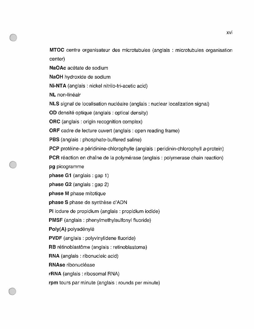
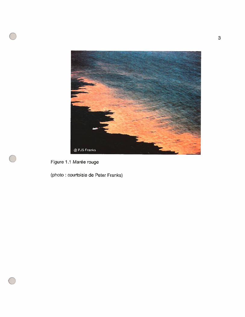
À diverses occasions de nature mal comprise, on assiste à l’apparition très
rapide d’une forte concentration de dinoflagellés, surtout en période chaude et en
eaux peu profondes. Les dinoflagellés étant généralement de couleur rouge-
brunâtre, on parle alors de tels phénomènes en tant que « marées rouges » (voir
Figure 1.1, p.3). Mais, il est plus exact de se référer à ce phénomène de
surpopulation explosive d’algues nocives pour l’humain en tant que HABs (anglais
harmful algal blooms) car ce ne sont pas toutes les espèces qui produisent des
toxines et plusieurs dinoflagellés ne sont pas colorés. Les intoxications humaines
par des dinoflagellés arrivent la plupart du temps par l’ingestion d’organismes
marins filtrants qui auront concentré leurs toxines. Les toxines de la famille des
saxitoxines sont les plus répandues et causent une paralysie musculaire via une
liaison très forte aux canaux sodiques. La biologie générale des dinoflagellés est
substantiellement décrite par Hackett, Anderson, Erdner et Bhattacharya (Hackett,
Anderson et al. 2004), Taylor et Pollingher (Taylor et Pollingher 1987) et Spector
(Spector 1984).
3
Figure 1.1 Marée rouge
@PJSFranks
(photo : courtoisie de Peter Franks)
4
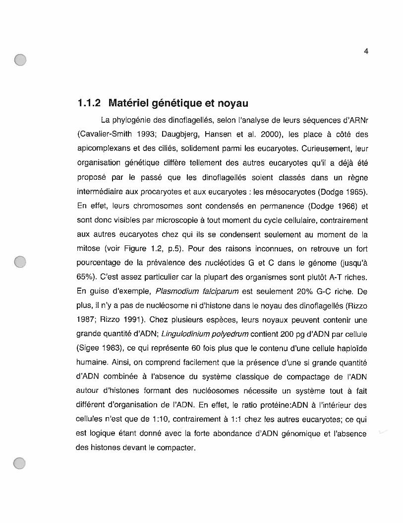
1.1.2 Matériel génétique et noyau
La phylogénie des dinoflagellés, selon l’analyse de leurs séquences d’ARNr
(Cavalier-Smith 1993; Daugbjerg, Hansen et al. 2000), les place à côté des
apicomplexans et des ciliés, solidement parmi les eucaryotes. Curieusement, leur
organisation génétique diffère tellement des autres eucaryotes qu’il a déjà été
proposé par le passé que les dinoflagellés soient classés dans un règne
intermédiaire aux procaryotes et aux eucaryotes : les mésocaryotes (Dodge 1965).
En effet, leurs chromosomes sont condensés en permanence (Dodge 1966) et
sont donc visibles par microscopie à tout moment du cycle cellulaire, contrairement
aux autres eucaryotes chez qui ils se condensent seulement au moment de la
mitose (voir Figure 1 .2, p.5). Pour des raisons inconnues, on retrouve un fort
pourcentage de la prévalence des nucléotides G et C dans le génome (jusqu’à
65%). C’est assez particulier car la plupart des organismes sont plutôt A-T riches.
En guise d’exemple, Plasmodium fa/ciarum est seulement 20% G-C riche. De
plus, il n’y a pas de nucléosome ni d’histone dans le noyau des dinoflagellés (Rizzo
1987; Rizzo 1991). Chez plusieurs espèces, leurs noyaux peuvent contenir une
grande quantité d’ADN; Lingulodinium polyedrum contient 200 pg d’ADN par cellule
(Sigee 1983), ce qui représente 60 fois plus que le contenu d’une cellule haploïde
humaine. Ainsi, on comprend facilement que la présence d’une si grande quantité
d’ADN combinée à l’absence du système classique de compactage de l’ADN
autour d’histones formant des nucléosomes nécessite un système tout à fait
différent d’organisation de l’ADN. En effet, le ratio protéine:ADN à l’intérieur des
cellules n’est que de 1:10, contrairement à 1:1 chez les autres eucaryotes; ce qui
est logique étant donné avec la forte abondance d’ADN génomique et l’absence
des histones devant le compacter.
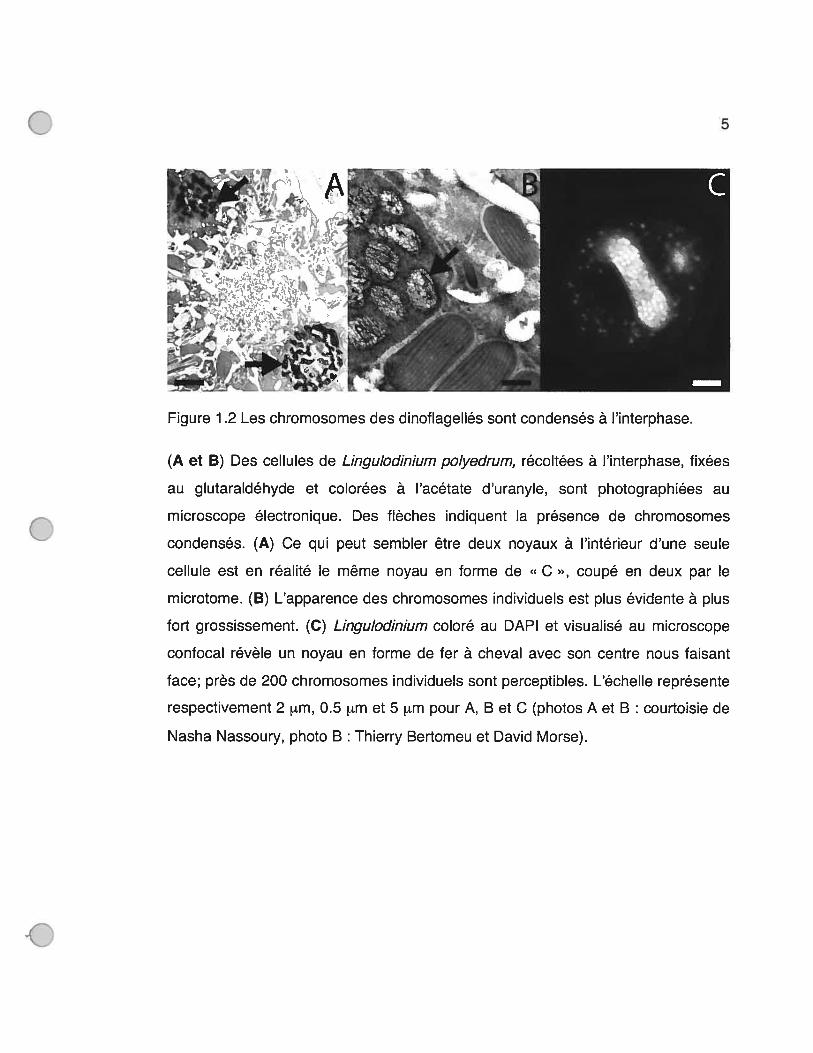
5
(A et B) Des cellules de Lingulodinium polyedrum, récoltées à l’interphase, fixées
au glutaraldéhyde et colorées à l’acétate d’uranyle, sont photographiées au
microscope électronique. Des flèches indiquent la présence de chromosomes
condensés. (A) Ce qui peut sembler être deux noyaux à l’intérieur d’une seule
cellule est en réalité le même noyau en forme de « C », coupé en deux par le
microtome. (B) L’apparence des chromosomes individuels est plus évidente à plus
fort grossissement. (C) Lingulodinium coloré au DAPI et visualisé au microscope
confocal révèle un noyau en forme de fer à cheval avec son centre nous faisant
face; près de 200 chromosomes individuels sont perceptibles. L’échelle représente
respectivement 2 im, 0.5 tm et 5 im pour A, B et C (photos A et B : courtoisie de
Nasha Nassoury, photo B : Thierry Bertomeu et David Morse).
ø’1kf I
Figure 1 .2 Les chromosomes des dinoflagellés sont condensés à l’interphase.
6
Il a été démontré que la partie interne des chromosomes des dinoflagellés
contenait de l’ADN sous la forme Z, enroulé sur la gauche (Soyer-Gobillard,
Geraud et al. 1990). À l’inverse, de longues branches d’ADN sous la forme
classique B, enroulé vers la droite, ont été vues au pourtour des chromosomes. La
transcription de gènes semble improbable dans le centre ultra-condensé des
chromosomes ayant de l’ADN sous forme de cristal liquide cholestérique (Costas
et Goyanes 2005), une forme chirale de la matière. Il est alors tentant de penser
que les branches décondensées des chromosomes sont garantes de la
transcription de gènes à l’interphase malgré une condensation permanente des
chromosomes.
De nombreuses hypothèses ont été avancées pour expliquer une
condensation de l’ADN en absence d’histones. 12 à 68 % des nucléotides
thymidines (selon les espèces de dinoflagellés) sont remplacés dans leur génome
par le nucléotide analogue hydroxyméthyluracil (Colette 1984). Seuls les
dinoflagellés ont ce nucléotide particulier et il se pourrait qu’il ait la propriété de
favoriser la condensation des chromosomes. Un autre mécanisme de
condensation pourrait aussi faire intervenir la grande quantité de cations divalents
présents dans le noyau qui pourraient contribuer à neutraliser les charges
négatives de l’ADN. Enfin, de nombreuses protéines aux propriétés basiques ainsi
que d’autres protéines ressemblant à des histones eucaryotiques et à des
protéines bactériennes liant l’ADN ont été isolées (Sala-Rovira, Geraud et al. 1991;
Taroncher-Oldenburg et Anderson 2000; Wong, New et al. 2003). Mais, l’affinité
testée de l’une d’entre elles est spécifique seulement à certaines séquences
d’ADN (Chudnovsky, Li et al. 2002) et n’a donc pas la propriété de lier tout ADN
qu’il soit tel que chez les histones classiques.

7
La mitose particulière des dinoflagellés est aussi un phénomène unique à
cette classe, tellement que l’on parle d’elle en tant que dinomitose (Chatton 1920).
En effet, le noyau des dinoflagellés, qui peut se retrouver sous plusieurs formes
(sphérique chez Crypthecodinium cohni pyramidale chez Gymnodinium dodgel et
en forme de fer à cheval chez Lingulodinium polyedrum), ne se dissout pas au
moment de la mitose (Rae 1970). Cette persistance de l’enveloppe nucléaire
pendant la mitose a déjà été rapportée chez d’autres organismes (levures,
diatomées et euglènes). Toutefois, dans leur cas, un fuseau mitotique
intranucléaire sert à la ségrégation des chromosomes, tandis que chez les
dinoflagellés, un fuseau mitotique s’établit à partir du cytoplasme et traverse le
noyau par une ou plusieurs invaginations nuléaires ou canaux (Spector 1984). Les
chromosomes sont attachés à la surface interne de la membrane nucléaire tandis
que les microtubules s’y attachent du côté cytoplasmique, adjacents au point
d’ancrage des chromosomes (Bhaud, Guillebault et al. 2000).
1.t3 Biochimie particulière
L’étude des dinoflagellés continue de produire d’étonnantes découvertes
concernant plusieurs aspects de leur biochimie qui leur est singulière et unique. En
guise d’exemple, aucune séquence promotrice n’a encore été caractérisée chez un
dinoflagellé. Cela est dû en partie à l’absence de techniques pouvant leur
introduire un transgène. Toutefois, lorsque l’on examine les séquences d’ADN
génomique en amont de différents gènes devant normalement contenir un
promoteur, on ne retrouve pas de boîte TATA (Le, Markovic et al. 1997; Li et
Hastings 1998). Chez les autres eucaryotes, le promoteur est lié par la protéine
TBP (anglais: TATA-box binding protein) qui sert à l’initiation de la transcription.
Une protéine analogue à TBP a été séquencée du dinoflagellé Crypthecodinium

8
cohnhi (Guillebault, Sasorith et al. 2002) et il a été montré que celle-ci a plus
d’affinité envers une séquence HU que TATA. Également, dans les séquences 3’
UTR des ARNm, on ne retrouve aucune séquence ressemblant de près ou de loin
au signal de polyadénylation (AAUAAA) fréquemment retrouvé 30 nucléotides en
amont du début de la queue de poly A. Cette séquence chez les autres eucaryotes
permet le positionnement de la coupure du transcrit suivie de l’ajout d’une queue
de poly A. Les mécanismes de base de la transcription et de la traduction chez les
dinoflagellés sont donc différents de ceux classiquement connus chez les
eucaryotes supérieurs. Finalement, une séquence de 22 nucléotides de long,
appelée SL (anglais : spliced leader), vient tout juste d’être rapportée à l’extrémité
5’ de tous les transcrits nucléaires des dinoflagellés (Zhang, Hou et al. 2007). La
séquence SL s’agit d’un ajout post-transcriptionnel que l’on retrouve chez plusieurs
autres classes d’organismes tels les nématodes, planaires, trypanosomes et
euglènes.
En plus du noyau, les chloroplastes des dinoflagellés démontrent aussi des
caractéristiques peu communes. Parmi ces caractères, on retrouve la présence de
trois membranes autour des chloroplastes qui nécessitent un mécanisme spécial
pour le ciblage des protéines (Nassoury, Cappadocia et al. 2003; Patron, Waller et
al. 2005). Il y a aussi la présence de la protéine soluble de l’antenne, PCP
(anglais peridinin-chlorophyll a-protein), qui a une structure unique (Hofmann,
Wrench et al. 1996) et qui ne se retrouve dans aucun autre organisme. Il y a
également l’utilisation d’une RuBisCO de forme Il pour fixer le carbone (Morse,
Salois et al. 1995; Whitney, Shaw et al. 1995). Les dinoflagellés sont ainsi les seuls
eucaryotes ayant cette forme inusitée de cette enzyme auparavant retrouvée
seulement chez des espèces procaryotiques anaérobiques. Quant aux gènes
chloroplastiques, plusieurs gènes typiquement transcrits dans les chloroplastes ont
9
été transférés au noyau des dinoflagellés (Zhang, Green et al. 1999). Aussi, le peu
de gènes encore présents dans les chloroplastes des dinoflagellés sont portés sur
des structures appelées « minicercles ». Finalement, une queue de poly U a été
trouvée à l’extrémité 3’ des transcrits des gènes chloroplastiques (Wang et Morse
2006), en place de la queue de poly A plus classique. La fonction d’une telle
modification demeure inconnue, mais reste définitivement un mécanisme hors du
commun lors de l’expression génique des organites.
1.1.4 Lingulodïnïum polyedrum et rythmes biologiques
Lingulodinium polyedrum est une espèce phototrophe d’un diamètre
d’environ 40 im qui nage librement en milieu salin. Elle est le sujet de très
nombreuses études chronobiologiques depuis plus de 50 ans (Hastings 2001) car
elle possède toute une panoplie de rythmes circadiens.
Les rythmes circadiens peuvent être définis comme étant des propriétés
physiologiques se reproduisant une fois par jour en avec une rythmicité proche de
24 heures. Ils sont entraînés par des signaux journaliers, tels que des
changements lumineux ou de température, qui amènent leur rythmicité à
exactement 24 heures. En condition constante, leur rythmicité persiste et il doit
aussi exister un mécanisme de compensation envers la température qui empêche
la période du rythme de changer en fonction des changements de température.
Enfin, les rythmes circadiens sont engendrés par un contrôle à partir d’horloges
moléculaires, mais ne sont pas en soi des horloges.
Or, Lingulodinium possède plusieurs rythmes circadiens classiques biens
étudiés : bioluminescence, photosynthèse (comprenant l’évolution d’02 et la
fixation du 002), migration verticale et division cellulaire (Figure 1.3, p.11). Ainsi,
ce dinoflagellé produit une lumière bleutée seulement en phase de nuit, est

10
capable de photosynthèse surtout en phase de jour, migre vers la surface de
l’océan le jour et descent à une profondeur de -10 m la nuit et ne se divise qu’une
heure après le lever du soleil (McMurry et Hastings 1972).

11
NUIT JOUR18:00 0:00 6:00 12:00 18:00
LD12 LD1S [DO LD6 CD12
02 02
I hu
h’i hu hu hu hu huhu hu hu
I t t î î î CO CO CO?
-10m]CO2CO2CO2CO,
Figure 1 .3 Temps d’occurrence de 4 rythmes circadiens de Lingulodinium
polyedrum (bioluminescence, photosynthèse, migration et division cellulaire).
Les moments d’occurence de 4 rythmes circadiens classiques de Lingulodinium
sont présentés sur une période de 24 heures avec 12 heures d’obscurité et 12
heures de lumière (LD12 :12). La concordance des temps chronobiologiques (LD,
de l’anglais Light-Dark) et journaliers est montrée; LDO représente le moment où le
soleil se lève. La bioluminescence, illustrée par des cellules bleues émettant de la
lumière (hu), a lieu la nuit. La photosynthèse, où il y a simultanément émission d’02
et absorption de C02, a lieu le jour. Lingulodinium, un peu avant la tombée de la
nuit, migre pour atteindre une profondeur de -10 m et, peu avant le lever du jour,
remonte vers la surface. La division cellulaire n’a lieu qu’une heure après l’aube
(Image : Thierry Bertomeu).
12
Puisque les dinoflagellés sont unicellulaires, il faut donc que la nature même
de ces rythmes soit biochimique. Or, dans une chaîne classique de réactions
enzymatiques, il est souvent possible de moduler toute la cascade en modulant
l’enzyme présente en quantité limitante. On parle alors de l’étape à vitesse
limitante (anglais: rate-limiting step). Il est donc possible et même probable que le
contrôle circadien des rythmes de bioluminescence, de fixation du carbone et de
division cellulaire se fasse sur la protéine limitante de ces réactions.
Les protéines responsables de la bioluminescence chez Lingulodinium ont
déjà été caractérisées. La luciférase (nom générique donné aux enzymes variées
d’organismes différents catalysant la bioluminescence) de Lingulodinium est
l’enzyme qui oxyde le substrat luciférine (Bae et Hastings 1994) et LBP (anglais
luciferin-binding protein) (Lee, Mittag et al. 1993) est la protéine qui séquestre ce
substrat et le relâche à la luciférine au bon moment. Il a déjà été montré qu’il y a un
rythme journalier d’abondance de ces deux protéines avec un maximum atteint la
nuit, coïncidant avec le rythme circadien de la bioluminescence (Johnson, Roeber
et al. 1984; Morse, Milos et al. 1989). Un contrôle circadien de la synthèse ou de la
dégradation de ces protéines peut par conséquent parfaitement expliquer le
mécanisme de contrôle du rythme circadien de la bioluminescence.
Des mesures du flux d’électrons à travers le photosystème II faites in vitro
montrent une plus grande activité le jour que la nuit et il a été proposé qu’une
modulation de ce flux puisse expliquer le rythme circadien d’évolution d’oxygène
(Samuelsson, Sweeney et al. 1983). La RuBisCO de forme II est la seule enzyme
chez Lingulodinium qui fixe le 002 (Morse, Salois et al. 1995) et est considérée
comme l’étape limitante du cycle de Calvin. Il a été montré que l’abondance de
cette enzyme est constante dans le temps mais que sa distribution dans le
13
chloroplaste de Lingulodinium change avec une localisation dans les pyrénoïdes
(la partie évasée dirigée vers l’intérieur des chloroplastes auréolés) correspondant
avec le maximum d’activité de fixation du carbone (Nassoury, Fritz et al. 2001).
Ainsi, un contrôle de localisation protéique à l’intérieur des plastides eux-mêmes
pourrait expliquer le rythme circadien de fixation du carbone chez les dinoflagellés.
La mitose de Lingulodinium ne se passe qu’à une heure précise du jour et
est donc sous contrôle circadien. Toutefois, puisque le temps de génération d’une
cellule est typiquement plus long qu’une journée, ce contrôle circadien est plutôt
perçu comme donnant une opportunité au cycle cellulaire de se poursuivre
seulement à certains moments du jour. C’est cette hypothèse de fenêtres
d’opportunités (anglais gating) momentanées, données au cycle cellulaire par le
cycle circadien, que nous proposons afin de concilier la concordance des cycles
circadiens et cellulaires sur le moment d’entrée en mitose. Le mécanisme de
contrôle du rythme circadien de la division cellulaire n’est pas encore connu.
Toutefois, par analogie avec les rythmes de bioluminescence et de photosynthèse,
on peut penser qu’une caractérisation biochimique des protéines responsables de
l’entrée en phase M de Lingulodinium pourrait nous indiquer quel mécanisme de
contrôle l’horloge circadienne utilise pour moduler le rythme circadien de la division
cellulaire. Ainsi, un autre mécanisme de contrôle d’un rythme circadien chez L.
polyedrum pourrait être mis à jour, au côté de ceux déjà connus pour les rythmes
circadiens de la bioluminescence et de la photosynthèse.
En relation avec les singularités de la dinomitose et l’organisation de l’ADN
des dinoflagellés, le fonctionnement et la régulation des composantes du contrôle
du cycle cellulaire pourraient exposer un fonctionnement biochimique nouveau,
exclusif aux dinoflagellés vis-à-vis leur cycle cellulaire. C’est pour cette raison, tout
en gardant l’objectif à long terme d’une découverte du mécanisme de contrôle du

14
rythme circadien de la division cellulaire, que le sujet de mon doctorat consiste en
la recherche de composantes de contrôle du cycle cellulaire du dinoflagellé
Lingulodinium polyedrum.
1.2 Contrôle du cycle cellulaire
1.2.1 Cycle cellulaire: principes de base
Le cycle cellulaire, ou cycle de division cellulaire (cdc), peut être défini
comme la série d’étapes ordonnées entre deux mitoses d’une cellule, amenant le
dédoublement fidèle du matériel génétique et sa ségrégation précise entre les deux
nouvelles cellules filles. C’est depuis Howard & PeIc en 1951 (Howard et Pelc
1951) que l’on sait que la duplication de l’ADN génomique des eucaryotes n’arrive
qu’à un moment précis et pas tout au long du cycle cellulaire. On appelle ainsi
phase S, le moment de la synthèse d’ADN et phase M, le moment de la mitose.
Les temps intermédiaires à ces deux phases sont Gi et G2 (anglais : Gap), de
sorte qu’un cycle complet est composé des phases Gi, S, G2 et M. L’ensemble
des phases G1, S et G2 est appelé interphase et c’est surtout durant cette période
que la cellule croît, de manière à fournir le gain de volume et de matériel
nécessaire à la future division mitotique.
La transition entre ces quatre étapes n’est pas simplement liée au passage
du temps. Elle est plutôt liée à la satisfaction de « points de contrôle » (anglais
checkpoints), disposés à des moments critiques du cdc, où n’est autorisée une
progression du cycle que lorsque certaines conditions sont atteintes (Nurse 1994).
Ce concept (Hartwell et Weinert 1 989) vient du fait que certaines cellules arrêtent
la progression de leur cycle cellulaire à des moments précis du cycle suivant des
stimuli particuliers et que cet arrêt peut souvent être brisé par la mutation d’un seul
gène. Le point de contrôle le plus connu est celui appelé « START » chez les
15
levures ou point de restriction chez les mammifères. Il s’agit d’un moment en Gi
d’engagement à procéder plus tard à la phase S en réaction à un milieu favorable
(Cross 1995). Parmi les autres points de contrôle, on retrouve entre autres celui
d’arrêt du cycle cellulaire en tout temps suivant une coupure d’ADN double-brin, un
contrôle en G2 s’assurant que tout l’ADN est répliqué avant l’entrée en phase M et
un contrôle en phase M s’assurant que tous les chromosomes sont attachés aux
microtubules avant leur ségrégation (Stem, Baserga et al. 1998).
Les premières propriétés de base du contrôle du cdc furent élucidées par la
fusion de cellules (hétérocaryons) à différentes phases et de l’observation des
changements associés à leurs noyaux (Johnson et Rao 1970). Une cellule en Gi
fusionnée à une cellule en phase S entre en phase S. En parallèle, une cellule en
G2 fusionnée à une cellule en phase M entre en phase M. Il devait donc exister
des facteurs initiant l’entrée en phase S et d’autres facteurs initiant l’entrée en
phase M. Toutefois, il a été trouvé que ces facteurs agissaient de manière
passagère puisque qu’une cellule en G2 ne fait pas entrer une cellule en G1, en
phase S et vice-versa pour une cellule en G1 qui ne fait pas entrer une cellule en
G2, en phase M. Finalement, une cellule en G2, fusionnée à une cellule en phase
S, n’entreprend pas un deuxième cycle de duplication de son ADN, ce qui montre
qu’il doit y avoir des mécanismes inhibiteurs de retour à une phase précédente si
elle vient de se terminer.
Y 22 Cycle cellulaïre : régulation
t2.2.1 Isolement biochimique de MPF: une kinase et une cycline
Notre compréhension actuelle du cycle cellulaire combine des approches
expérimentales variées sur des organismes très différents. L’expérience historique
qui a le plus réussi à unifier les efforts de plusieurs approches fut probablement la

16
purification du facteur entraînant l’entrée en mitose. La stimulation d’oocytes de
grenouilles arrêtés en G2 par des hormones provoque leur maturation en les
taisant entrer en méiose I. L’injection d’un peu de cytoplasme provenant d’un oeuf
mature dans le cytoplasme d’un oeuf immature, même avec l’utilisation d’inhibiteurs
de synthèse protéique, est suffisante pour les faire entamer la maturation (Masui et
Markert 1971). Cette expérience offrait donc un système de détection du facteur
mitant la maturation. La purification biochimique de MPF (anglais: maturation
promoting factor) par une série de fractionnements chromatographiques (Lohka,
Hayes et al. 1988) révéla deux composantes majeures: une kinase (Gautier,
Norbury et al. 1988) et une cycline (Gautier, Minshull et al. 1990). MPF se révéla
capable d’induire la mitose, composée d’une série d’évènements similaires à la
méïose si ce n’est l’alignement des chromosomes, chez des organismes
hétérologues (Tachibana, Yanagishima et al. 1987). Des cyclines et des kinases
similaires sont maintenant reconnues comme des protéines universelles aux
eucaryotes. On se réfère désormais à MPF en tant que facteur initiant la mitose
plutôt que la maturation.
1.2.2.2 Les mutants cdc : rôle central de Cdc2
La kinase de MPF se révéla être l’homologue de la kinase Cdc2 (Gautier,
Norbury et al. 1988) auparavant isolée d’un mutant thermoconditionnel létal de
Schizosaccharomyces pombe. La génétique des levures S. pombe et S.
cerevisiae, comme système d’isolement de gènes responsables du contrôle du
cycle cellulaire, s’est montrée avec le temps très efficace (Lee et Nurse 1988). En
effet, ce sont des eucaryotes qui peuvent être maintenus à l’état haploïde, ce qui
facilite les études génétiques. Aussi, ces deux levures possèdent des caractères
morphologiques servant à diagnostiquer à quelle étape du cdc elles sont rendues.
S. pombe, aussi connue sous le nom de levure fissipare, s’allonge tout au long du
17
cycle cellulaire et une cellule ne se divisant plus devient anormalement grosse. S.
cerevisiae, aussi connue sous le nom de levure bourgeonnante, produit un
bourgeon à partir de la transition GuS qui grossit à mesure que la cellule approche
de la phase M. Ainsi, une mutation dans un gène initiant l’entrée en phase S chez
S. cerevisiae produira des levures ayant toutes un très petit bourgeon, arrêtées à
ce point de contrôle. La génération de collections de mutants et leur
complémentation génétique pour rétablit leur phénotype est un outil fort puissant
chez les levures pout déterminer les gènes responsables d’une fonction
particulière. Puisque des gènes contrôlant le cycle cellulaire doivent justement
empêcher la progression du cycle cellulaire s’ils sont mutés, des levures
thermoconditionnelles létales furent sélectionnées avec une température
permissive de 25 oc et une température conditionnelle de 36 °c. Des collections de
mutants du cdc chez S. cerevisiae et S. pombe (nommés cdcl, cdc2...) furent
établies par les chercheurs Leland Hartwell et Paul Nurse respectivement. Toutes
les cellules d’un mutant particulier mis en condition restrictive présentent les même
caractères morphologiques et donc un arrêt au même stade du cdc. Pour diverses
raisons, les mécanismes de contrôle d’entrée en phase S sont plus connus chez S.
cerevisiae, tandis que pour S. pombe, ce sont ceux d’entrée en mitose.
Or, il a été montré que Cdc2 de S. pombe (Lee et Nurse 1988) est une
kinase dont l’activité est maximale à la transition G2/M et qui, lorsque inactivée par
la chaleur, arrête la levure à la transition G2/M. De plus, avec d’autres mutants de
S. pombe arrêtant aussi le cdc à la transition G2/M, il a été montré génétiquement
que l’action de plusieurs gènes intervenait via CDC2, ce qui démontre que ce gène
est un acteur clé de l’entrée en phase M. Parmi ces mutants, cdc25 est une
protéine tyrosine phosphatase qui déphosphoryle et active cdc2 (Millar, McGowan
et al. 1991). D’ailleurs, le phénotype de cdc25 en condition restrictive est une

18
cellule très allongée qui tarde à entreprendre une mitose. À l’inverse, la protéine
Weel (anglais : petit) procure à la levure mutée pour ce gène une morphologie
petite. Il s’agit d’une tyrosine et sérine/thréonine kinase qui phosphoryle Cdc2 et
l’inactive (Parker, Atherton-Fessier et al. 1992). La morphologie petite s’explique
alors parfaitement par une entrée précoce en phase M par Cdc2 activée trop tôt.
Finalement, la protéine Cdcl3 est requise à l’activation de Cdc2 (Moreno, Hayles
et al. 1989). Cdcl3 est une cycline et se retrouve associée à Cdc2 à la mitose.
Cette convergence de mécanismes d’action de contrôle d’entrée en mitose via
Cdc2 plaçait donc cette kinase à l’avant-plan du contrôle de l’entrée en mitose.
Aussi, les intervenants génétiques kinase cdc2 et cycline cdcl3 chez S. pombe
ayant été trouvés et l’isolement biochimique faisant entrer les oeufs de grenouilles
en phase M d’un facteur composé d’une kinase homologue à Cdc2 et d’une cycline
démontrait l’existence universelle d’un contrôle d’entrée en mitose chez différentes
espèces (Nurse 1990).
1.2.2.3 Cyclines et kinases cyclines-dépendantes (anglais:CDKs)
Des homologues de Cdc2 ont été trouvés chez tous les eucaryotes
séquencés. Aujourd’hui on se réfère à ces kinases en tant que CDKs car elles
nécessitent une liaison avec une cycline pour être actives (Pines 1995). Ce sont
des sérine/thréonine kinases qui possèdent la plupart des autres domaines
conservés retrouvés chez les autres kinases, en plus d’avoir dans leur domaine III
une séquence identique ou similaire à la séquence PSTAIRE retrouvée chez Cdc2.
Les CDK5 possèdent surtout deux sites de phosphorylation. Le premier, sur la
tyrosine 15 de Cdc2, se trouve dans la boucle-P (anglais: P-loop) en N-terminal
qui est un site où une phosphorylation limite l’accès à l’ATP et est donc inhibitrice
pour Cdc2 (Smits et Medema 2001). Le deuxième site, sur la thréonine 161 de
19
Cdc2, est un site activateur qui réoriente la boucle-T (anglais : T-loop), change la
conformation de la CDK et la rend active (Smits et Medema 2001). C’est la kinase
Weel qui est responsable de la phosphorylation inactivatrice, la phosphatase
Cdc25 qui enlève ce phosphate inhibiteur et la CAK (anglais: CDK-activating
kinase) qui est responsable de la phosphorylation activatrice.
Chez l’humain, on retrouve plus de 10 CDK5 avec la CDK1 comme
homologue de la kinase Cdc2 de levure (Stem, Baserga et al. 1998). La structure
cristallographique de la CDK2 humaine en association avec la cyclmne A humaine
révèle que le domaine III contenant la séquence PSTAIRE et la boucle-T contenant
le site de phosphorylation activatrice de la CDK sont en étroite association avec la
cycline (Jeffrey, Ruso et al. 1995). Cette association change la conformation de la
CDK qui voit son site catalytique s’ouvrir et devient alors potentiellement active.
Les cyclines ont originellement été découvertes comme des protéines
cycliquement détruites après chaque division dans des oeufs d’oursins de mer en
mitoses répétées (Evans, Rosenthal et al. 1983). Toutefois, la définition d’une
cycline a changé pour maintenant être attribuée aux protéines ayant la séquence
consensus « repliement-cycline » (anglais : cyclmn-fold), contenant deux domaines
boîte-cycline (anglais: cyclin-box) d’environ 150 acides aminés chacun, et étant
capable de lier et d’activer une CDK (Kobayashi, Stewart et al. 1992). Chez
l’humain, près de 25 cyclmnes différentes ont été rapportées dont seulement la
moitié ont un rôle à jouer dans le cycle cellulaire (Stem, Baserga et al. 1998).
Toutes les cyclines sont classées en famille chez les organismes selon leur
similarité de séquence mais aussi selon leur patron d’expression et leur similarité
structurale. En effet, contrairement aux CDKs dont l’expression est généralement
constante, les cyclmnes aux fonctions reliées au cycle cellulaire ont des pics
d’expression associés au moment du cycle cellulaire où leur activité est nécessaire

20
à la progression du cdc (Murray 2004). Chez les mammifères, ce sont surtout les
cyclines des familles A, B, D et E qui sont impliquées dans le cycle cellulaire
(Stem, Baserga et al. 1998). L’hypothèse selon laquelle les cyclines, en plus
d’activer leurs partenaires CDKs, déterminent la spécificité du substrat de leurs
kinases, est assez répandue (Loog et Morgan 2005).
MPF phosphoryle les substrats aux sites consensus S/T*PXK/R (Lewin
2004) où X représente n’importe quel acide aminé. Chez S. pombe, on retrouve
plus de 200 substrats de MPF (Ubersax, Woodbury et al. 2003) dont des histones,
des condensines et des protéines associées aux microtubules (MAP5, anglais
microtubule-asocciated proteins). Il est proposé que la phosphorylation d’histones
pourrait favoriser la condensation de l’ADN mais cette relation n’est pas encore
prouvée. Les condensines phosphorylées s’associent en complexes qui lient l’ADN
et participent à sa condensation (Belmont 2006). Les multiples phosphorylations
de protéines associées aux microtubules (Wittmann, Hyman et al. 2001) participent
à l’élaboration du fuseau mitotique qui s’associe aux chromosomes. Aussi, chez
les organismes supérieurs ayant un noyau qui se désassemble à la mitose
contrairement aux levures, la phosphorylation des lamines nucléaires, les protéines
formant un réseau soutenant la structure du noyau à sa surface interne, provoque
leur dépolymérisation (Pines 1995). Le rôle physiologique de la phosphorylation de
la grande majorité des substrats de MPF demeure encore inconnu (Ubersax,
Woodbury et al. 2003).
Puisque les CDKs ont absolument besoin de se lier à une cycline pour être
actives, la destruction des cyclines est donc un mécanisme efficace d’inactivation
de leur activité kinase. D’ailleurs, il existe deux mécanismes de protéolyse de
cyclines bien documentés. Le premier fait intervenir des séquences PEST dans les
cyclines impliquées à la transition GuS. Il s’agit de régions riches en acides
21
aminés proline, acide glutamique, sérine et thréonine qui rendent instables les
protéines avec de tels motifs qui seront alors dégradées par le protéasome suite à
une poly-ubiquitination (Alberts, Johnson et al. 2002). Le second fait intervenir une
séquence appelée « boîte de destruction » (anglais destruction-box) présente en
N-terminal chez toutes les cyclines impliquées à la transition G2/M (Pines 1995).
On se réfère souvent à ce deuxième type de cycline en tant que cyclines de type A
ou B, en se basant sur la classification des cyclines de mammifères. La séquence
consensus des boîtes de destruction est R-X-X-L-X-X-(L/l)-X-N, où X représente
n’importe quel acide aminé, et la dégradation des cyclines mitotiques via leur boîte
de destruction se fait aussi via l’ubiquitination (Glotzer, Murray et al. 1 991).
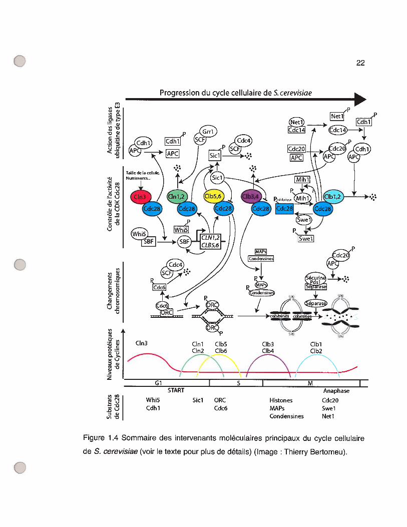
1.2.3 Regard sur S. cerevïsiaePour donner un aperçu de l’implication moléculaire des CDKs et cyclines à
tous les stades du cycle cellulaire, de leur contrôle et de leur effet, une partie de ce
qui est connu du contrôle du cdc chez S. cerevisiae est présenté plus en détail.
Ainsi, même si les autres organismes possèdent des intervenants différents,
certains principes généraux sont appliqués à tous les eucaryotes. Le lecteur de
cette thèse pourra se référer à la Figure 1.4 (p.22) pour mieux visualiser les
intervenants du cycle cellulaire qui seront décrits.
22
Figure 1 .4 Sommaire des intervenants moléculaires principaux du cycle cellulaire
de S. cerevisiae (voir le texte pour plus de détails) (Image : Thierry Bertomeu).
Progression du cycle cellulaire de S.cerevisiae(nL.1J
CJciJ,
—c-J
-
c-i
L’) CY<.3
‘w
-ç œ
— ul-çJC)
C)çJ‘o
—Q)g-Du
1 -1O o
- ou
L,,w
o: i
o =L-u
o->’xL)Dj
>
z
l—1L.. u
DO)La-i 0
C1n3 Cmi C1b5 C1b3 CIblCmn2 C1b6 C1b4 C1b2
/ NI
Whi5Cdli 1
Gi I S I M I‘I
START Anaphase
Sici ORC Histones Cdc2OCdc6 MAPs Swel
Condensines Neti
23
La levure bourgeonnante possède 5 CDKs dont seulement Cdc28,
l’homologue de Cdc2 de S. pombe, est impliquée dans le cdc et ce à toutes les
étapes (Mendenhall et Hodge 1998). S. cerevisiae possède aussi 22 cyclines dont
seulement 9 s’associent à Cdc28 et donc servent au contrôle du cdc (Measday et
Andrews 1998). Cmi, Cln2 et C1n3 sont les cyclines de type Gi tandis que les
cyclines Cibi, Clb2, C1b3, Clb4, Clb5 et Clb6 sont de type mitotique.
C1n3 est présente à travers tout le cycle cellulaire avec une transcription un
peu plus élevée en début Gi. Elle agit en amont des cyclines Cmi et Cln2 et
influence directement le passage du point de contrôle START (Tyers, Tokiwa et al.
1993) en étant un des senseurs de la taille de S. cerevisiae (Cross 1995). Un des
substrats de Cln3/Cdc2S est la protéine Whi5 qui, à l’image de la protéine
analogue Rb (Rétinoblastôme) des mammifères (Kaelin 1999), agit comme
inhibiteur du point de contrôle START. Whi5 lie et inhibe le facteur de transcription
SBF (anglais wi4/6 Celi Cycle Box (SCB) inding Eactor) et sa phosphorylation
par Cln3/Cdc28 enlève cette inhibition (Costanzo, Nishikawa et al. 2004; de Bruin,
McDonald et al. 2004). SBF, qui est capable d’induire la transcription de près de
200 gènes (lyer, Horak et al. 2001), va alors induire la transcription de CLN7
(Partridge, Mikeseil et al. 1997), CLN2 (lyer, Horak et al. 2001), CLB5 (Bean,
Siggia et al. 2005) et CLB6(Iyer, Horak et al. 2001).
Il existe des protéines capables de lier des CDKs, en association ou non
avec une cycline, et qui inhibent leur activité. On appelle CKI (anglais : CDK
inhibitors) de telles protéines et la protéine Sici de levure est justement une CKI
capable d’inhiber Cdc28 en complexe avec une cycline de type mitotique,
Clb/Cdc28 (Schwob, Bohm et al. 1994). Cette inhibition est enlevée par la
phosphorylation de Sici par Cdc28 en association avec CIni ou C1n2 (Verma,
Annan et al. 1997). La forme phosphorylée de Sici est reconnue par la protéine
24
Cdc4. Cdc4, en association avec le complexe SCF (anglais: Skpl/Cullin/F-box
protein), est une ligase ubiquitine de type E3 (Nash, Tang et al. 2001) qui
engendre donc la dégradation de Sici phosphorylée. Ce mécanisme d’inhibition
par la CKI et sa dégradation après l’activation de Clnl/Cdc28 et Cln2/Cdc28
s’assure donc que le peu de Cdc28/Clb présent en Gi ne puisse initier START.
D’ailleurs, une mutation dans le gène SIC7 rend viable le phénotype auparavant
non-viable d’une levure ayant ses trois 3 cyclines GuS mutées (cIni, cln2 et cln3)
(Tyers 1996).
Les mécanismes par lesquels l’activité kinase des CDK5 arrive à induire
l’initiation de la réplication ne sont pas encore très bien compris. On sait toutefois
que la phosphorylation par des CDKs des protéines Sld2/Drcl nécessaires à la
réplication est essentielle à l’initiation de cette réplication (Masumoto, Muramatsu
et al. 2002). Les protéines de l’ORC (anglais: origin recognition complex) forment
un complexe autour des sites ARS (anglais : autonomous replication sequence),
sites d’origine de la réplication chez la levure. Les complexes C1b5/Cdc28 et
C1b6/Cdc28 phosphorylent les protéines de l’ORC (Weinreich, Liang et al. 2001)
ainsi que la protéine Cdc6. Cdc6 lie l’ORC et engendre le recrutement de protéines
nécessaires à la formation du complexe de préinitiation. Sa phosphorylation par
Clb/Cdc28 la fait relâcher l’ORC, ce qui initie la réplication. Cdc6 phosphorylée est
ensuite dégradée et la persistance de l’activité Clb/Cdc28 en G2 empêche ainsi la
formation de nouveaux complexes de préinitiation en maintenant les niveaux de
Cdc6 bas (Donaldson et Blow 1999). Les cyclines Cml, C1n2 et Clb6 sont ciblées à
la dégradation proche de la transition GuS par l’ubiquitine ligase SCF couplée à la
protéine Grrl activée (Bloom et Cross 2007).
Les cyclines mitotiques sont exprimées par paires : C1b5 et C1b6 exprimées
à la transition G lIS, Clb3 et Clb4 à la fin de la phase S et Clbl et C1b2 10 minutes
25
avant l’anaphase (Epstein et Cross 1992). Clb3ICdc28 et Clb4/Cdc28 ont pour
effet d’initier la formation du fuseau mitotique (Richardson, Lew et al. 1992) alors
que Clbl/Cdc28 et Clb2ICdc28 initient la mitose (Surana, Robitsch et al. 1991). La
kinase Swel, un homologue de la protéine Weel de S. pombe, ajoute un
phosphate inhibiteur à la CDK Cdc28 couplée aux cyclines mitotiques (Booher,
Deshaies et al. 1993). La phosphatase Mihi, un homologue de la protéine Cdc25
de S. pombe, enlève ce même phosphate inhibiteur de Cdc28 (Mendenhall et
Hodge 1998). Or, la protéine Swel est justement un substrat de Clbl/Cdc28 et
C1b2/Cdc28, ce qui favorise sa dégradation via le complexe ubiquitine ligase APC
(anglais : Anaphase-promoting complex) (Asano, Park et al. 2005). Chez S.
pombe, la phosphorylation de Cdc25 par Cdc2/Cdcl3 rend celle-ci plus active, ce
qui active davantage le complexe CDK/cycline (Smits et Medema 2001). Si l’on
admettait que ce même mécanisme de rétroaction positive sur Cdc25 fonctionne
de la même manière chez S. cerevisiae, on aurait ainsi deux systèmes de
rétroaction amenant une activation explosive des complexes Clbl/Cdc28 et
Clb2/Cdc28 via la dégradation de l’inhibiteur Swel et l’activation de l’activateur
Mihl. C’est cette réaction en chaîne, élucidée chez S. pombe, qui empêche un
retour en arrière une fois la phase M commencée.
L’établissement du fuseau mitotique composé de microtubules nécessaires
à la future division mitotique se fait à la phase M. Chez les mammifères, deux
centrosomes cytoplasmiques se forment d’où partent des filaments de
microtubules terminés à chacune des extrémités par des complexes protéiques
appelés centres organisateurs des microtubules. Le noyau de la levure demeurant
intact, le réseau de microtubules s’installe alors à l’intérieur du noyau à partit
d’homologues de centtosomes, des SPBs (anglais : spindle pole body)
périnucléaires.

26
La dégradation des cyclines mitotiques à la fin de la mitose est capitale car
la surexpression de celles-ci, ou l’utilisation de formes non-dégradables, bloque les
cellules en métaphase (Murray 2004). L’APC est un complexe ubiquitine ligase de
type E3 qui induit l’anaphase suite, entre autres, à la dégradation de cyclines.
Chez S. cerevisiae, l’APC peut être associé aux protéines Cdc2O ou Cdhl et
celles-ci spécifient au complexe quelles protéines seront ubiquitinées (Peters
1999). La phosphorylation de Cdc2O par Clbl,2/Cdc28 permet l’association et
l’activation du complexe APC 20 (Pines 2006). APCC 20 est responsable de la
dégradation de Pdsl, une sécurine, et de cyclines mitotiques. La sécurine est une
protéine qui lie et maintient inactif une séparase, une protéase capable de couper
les protéines cohésines qui lient et retiennent ensemble les chromatides soeurs. La
dégradation des cohésines initie donc l’anaphase et la séparation des
chromosomes tirés par l’instabilité dynamique des microtubules. Il existe un point
de contrôle s’assurant que l’APC2° ne soit actif que lorsque tous les kinétochores
des chromosomes sont attachés au fuseau mitotique pour ne pas provoquer
d’accidents mitotiques mais l’identité de ce mécanisme n’est pas connu. Vers la fin
de la mitose, la phosphorylation de la protéine du nucléole Netl par CIbi ,2/Cdc2S
libère la phosphatase Cdcl4. Elle déphosphoryle les substrats de Clb/Cdc28,
enlève le phosphate inhibiteur de la CKI Sici et déphosphoryle Cdhl (Bloom et
Cross 2007). Cdhl déphosphorylée forme APC actif qui participe aussi à la
dégradation de cyclines mitotiques (Bloom et Cross 2007). La cytocynèse suit par
la fermeture d’un cercle contractile d’actine associée à la membrane de la cellule et
coupant aussi le noyau et la mitose est alors terminée. Le complexe APChl est de
nouveau inactivé par la phosphorylation de Cdhl par Cln3/Cdc28. Les deux
cellules filles sont de nouveau en G1.
27
113 Études du cycle cellulaire chez lesdinoflagellés
Aucun des génomes de dinoflagellés n’a encore été séquencé et le fait qu’ils
soient si gros y est probablement pour quelque chose. Aussi, jusqu’en 2002,
aucune collection d’ESTs dépassant 1000 séquences provenant d’un dinoflagellé
n’avait encore été réalisée. Il était alors peu étonnant que l’on ne retrouve encore
aucun régulateur du cycle cellulaire parmi le peu de séquences publiées.
Malheureusement, seulement deux publications rapportent des tentatives
d’isolement de régulateurs du cycle cellulaire chez un dinoflagellé (Salois et Morse
1996; Salois et Morse 1997). L’approche PCR et le criblage de banques d’ADNc
avec un anticorps dirigé contre l’homologue de Cdc2 ont ainsi toutes deux été
infructueuses. Dans ces conditions, l’étude du fonctionnement du cycle cellulaire
des dinoflagellés s’est limitée qu’à certains types d’expérience. La cytométrie en
flux permet un diagnostic fiable de l’état (G0/G1, S et G2IM) du cycle cellulaire où
se trouve une cellule. Les études microscopiques permettent d’observer les
changements d’ordre morphologique à l’intérieur de la cellule en rapport avec le
cycle cellulaire. L’utilisation d’anticorps contre des protéines régulatrices du cycle
cellulaire établies chez d’autres organismes peut nous indiquer le comportement
de protéines chez les dinoflagellés. La purification par affinité envers la protéinep13sucl de levure fissipare est une technique qui fonctionne chez plusieurs
organismes hétérologues et permet l’enrichissement du complexe protéique MPF.
Les tests d’activités kinase avec de l’ATP marqué radioactivement sur le substrat
histone Hi peut nous indiquer le niveau d’activité des CDKs mais il faut se rappeler
que presque toutes les kinases ont cette activité in vitro. Il faut donc ainsi s’assurer
que les protéines responsables des activités mesurées sont bel et bien des CDKs.
Finalement, des drogues arrêtant le cycle cellulaire à des points de contrôle précis

28
peuvent être utilisées et, si elles ont le même effet chez les dinoflagellés, la
présence de ce point de contrôle peut être confirmée.
Crypthecodinium cohnii est un dinoflagellé hétérotrophe avec un cycle
cellulaire partiellement synchronisable grâce à la technique de relâchement d’un
stade motile en Gi suite à une culture sur milieu solide (Bhaud, Salmon et al.
1991). C’est probablement cette raison pratique qui a fait de cette espèce celle sur
laquelle le plus d’études biochimiques des régulateurs du cycle cellulaire ont été
réalisées. Un anticorps dirigé contre la séquence peptidique de 7 acides aminés
PSTAIRE, la séquence conservée de la CDK1 qui sert à son association avec les
cyclines (Jeffrey, Ruso et al. 1995), reconnaît chez cette espèce une protéine de
34 kDa (Rodriguez, Cho et al. 1993), une taille similaire à toutes les autres CDKs.
L’utilisation de pi31 couplée à des billes de sépharose sur un extrait de
Crypthecodinium cohnhi arrive à retenir la protéine réagissant à l’anticorps anti
PSTAIRE tel que testé par transfert de type Western. Une protéine similaire à
CDK1 doit donc être présente chez les dinoflagellés. Toutefois, dans cette
expérience (Rodriguez, Cho et al. 1993), le degré d’enrichissement du signal de
l’anticorps obtenu par l’affinité à p13 n’a pas été testé.
Une activité kinase est associée aux protéines purifiées par affinité à pi3
couplée à des billes de sépharose, tel que testé par essai-kinase in vitro envers
l’histone Hi (Rodriguez, Cho et al. 1993). Ce même test d’activité kinase retrouve
60 à 70% plus d’activité dans des extraits provenant de cellules en phase M
comparées à des cellules en interphase (Bhaud, Barbier et al. 1994; Barbier, Albert
et al. 1995). Lorsque ce test d’activité est étalé sur un cycle cellulaire complet avec
des extraits pris aux heures, on voit un pic d’activité s’étalant sur toute la phase G2
avec un sommet (2.5 fois plus d’activité que le minimum d’activité détectée) atteint
2 heures avant la cytocynèse (Leveson, Wong et al. 1997). Ce comportement est
29
très surprenant puisque chez les autres organismes modèles du cycle cellulaire,
l’activité de MPF augmente rapidement seulement à la transition G2/M pour
ensuite aussi rapidement décliner (Smits et Medema 2001). Aussi, la différence
d’activité kinase de MPF entre l’interphase et la phase M des organismes modèles
est de beaucoup supérieure à un facteur de seulement 2.5. Il est dommage que les
protéines actuellement retenues par affinité envers p131 n’est pas été analysées.
Toujours chez Crypthecodinium cohni la détection de cyclines a été tentée
par l’utilisation d’anticorps envers des cyclines d’autres organismes. En utilisant un
anticorps contre la cycline Cdci3 de levure fissipare, une protéine exclusivement
cytoplasmique de 56 kDa est détectée dont l’abondance ne varie pas entre les
phases Gi et M (Barbier, Albert et aI. 1995). Ce résultat est plutôt surprenant si la
protéine détectée est effectivement une cycline mitotique. Aussi, un extrait
protéique immunoprécipité avec le même anticorps possède une activité kinase.
En utilisant cette fois un anticorps dirigé contre les boîtes-cyclines d’oursins de
mer, 4 bandes sont reconnues 50, 65, 75 et 90 kDa (Leveson, Wong et al. 1997).
Étonnament, la bande de 56 kDa n’est pas reconnue. Seule la bande de 50 kDa
est retenue par affinité envers p13 et les bandes de 50 et 65 kDa ont toutes
deux un pic d’expression correspondant à la phase S tandis que l’expression des
bandes de 75 et 90 kDa est constante.
Grâce à l’utilisation du nocodazole, une drogue dépolymérisatrice des
microtubules, il est possible de prolonger la phase M de Crypthecodinium cohnhi
avec un arrêt souvent à la métaphase (Yeung, New et al. 2000). Aussi, la cycline
Bi humaine est dégradée plus rapidement lorsque mélangée à un extrait de C.
cohnli en Gi plutôt qu’à un autre provenant de cellules en G2IM. Grâce à ces
expériences, la présence d’un point de contrôle inhibant l’activation de l’APC suite
30
au mésappariement des chromosomes au fuseau mitotique était fortement
démontrée.
La morphologie des chromosomes de Crypthecodinium cohnli a été
observée sur un cycle cellulaire grâce à des populations synchronisées en G1,
G2/M et en phase S grâce à l’aphidicoline, une drogue qui bloque la polymérase
d’ADN (Bhaud, Guillebault et al. 2000). Ainsi, en Gi, les chromosomes paraissent
lâchement condensés pour l’être un peu plus en fin Gi. À la phase S, ils se
déroulent pour se compacter plus intensément en G2. À la prophase, les
chromatides soeurs auparavant condensées ensemble en une seule entité en G2
se séparent partiellement pour former un V attaché via leur kinétochore à une
invagination nucléaire traversée par le fuseau mitotique extra-nucléaire.
Finalement, lors de la métaphase, le noyau s’aplatit dans le sens attendu d’un
alignement classique des chromosomes à la métaphase mais les kinétochores
attachés aux invaginations nucléaires ne semblent pas s’aligner sur un seul plan
médian.
Chez le dinoflagellé Gambierdiscus toxicus un anticorps anti-PSTAIRE
reconnaît aussi une protéine de 34 kDa (Van Dolah, Leighfield et al. 1995). En
utilisant ce même anticorps pour immunoprécipiter des extraits du dinoflagellé à
différentes périodes du cycle cellulaire, une activité kinase envers l’histone Hi est
détectée avec un maximum atteint avec des cellules en phase M. Toutefois, il est
plutôt curieux que les auteurs aient utilisés cet anticorps pour immunoprécipiter
l’activité de MPF. En effet, un anticorps anti-PSTAIRE ne reconnaît que la forme
monomérique d’une CDK non-dénaturée (Fines et Hunter 1990), donc inactive
sans cycline.
31
Chez Karenia brevis, un anticorps anti-Cdcl3 et un anticorps anti-boîtes
cyclines d’oursins de mer reconnaissent tous deux sur transfert de type Western
une bande de 56 kDa (Barbier, Leighfield et al. 2003). Ainsi, seule une bande est
rapportée suite à l’utilisation de l’anticorps anti boîtes-cyclines chez Karenia bre vis,
contrairement aux 4 bandes chez Crypthecodinium cohnli (Leveson, Wong et al.
1997). Ce signal de 56 kDa se retrouve dans un extrait protéique immunoprécipité
avec un anticorps anti-PSTAIRE. La présence de cette protéine est détectée tout
au long du cycle cellulaire et seulement dans le cytoplasme et le nucléole (Barbier,
Leighfield et al. 2003).
Chez Lingulodinium po/yedrum, il a été montré par cytométrie en flux que la
phase S, tout comme la mitose, est circadienne et commence 6 heures après la
transition lumière/noirceur lorsque des cellules sont cultivées sous un cycle
d’éclairage 12 heures lumière/12 heures noirceur (Homma et Hastings 1989). Les
auteurs proposaient alors qu’un contrôle circadien d’entrée en phase S et une
durée de phase G2 constante pouvaient accommoder une mitose synchronisée à
un cycle journalier. Ainsi, l’horloge circadienne n’ayant qu’un contrôle sur l’entrée
en phase S suffirait à produire le rythme circadien de la mitose. Ce modèle
demeure toutefois hypothétique.
L’utilisation d’un anticorps anti- PSTAI R E chez Lingulodinium polyedrum
révèle une bande de 32 kDa par transfert de type Western (Salois et Morse 1996).
Ce résultat est en contradiction avec les mêmes expériences faites sur
Crypthecodinium cohnfi et Gambierdiscus toxicus qui reconnaissent une bande de
34 kDa, une taille plus typique des CDK5. La protéine liée par l’anticorps fut clonée
par le criblage d’une banque d’ADNc avec cet anticorps et la protéine reconnue
n’est pas une kinase. Aussi, plusieurs approches PCR pour isoler une séquence
32
codant pour une CDK de Lingulodinium ont été infructueuses (Salois et Morse
1996; Salois et Morse 1997).
33
1.4 Approches et objectifs du projetIl est étonnant que si peu d’études d’ordre biochimique aient été réalisées
sur le cycle de division cellulaire des dinoflagellés. Leur propension à former très
rapidement des HABs donne pourtant à la recherche sur leur cdc une certaine
importance d’ordre économique. Aussi, leur organisation nucléaire si particulière
laisse présager plusieurs découvertes de mécanismes nouveaux de contrôle du
cdc, liées au domaine de la recherche fondamentale.
En 1999, au début de mes études graduées, à peine près de 1000
séquences nucléotidiques de dinoflagellés étaient disponibles dans les banques de
données publiques et, évidemment, aucune d’entre elles n’étaient liées au contrôle
du cycle cellulaire. Dans un contexte où les données de séquences étaient si
rares, une utilisation systématique d’anticorps hétérologues pour l’étude du cdc
des dinoflagellés semblait logique et était donc l’alternative la plus utilisée. En
relation avec cet énoncé, lorsque l’on sait qu’un organisme tel que S. cerevisiae
possède à lui seul 22 cyclines, on peut toutefois se questionner sur l’identité réelle
des protéines reconnues avec un anticorps anti-(boîte-cycline). Il y avait donc un
manque flagrant de composantes du cycle cellulaire déjà isolées ainsi qu’un
manque de données de séquence s’y rattachant nous permettant de pouvoir les
étudier plus en détail.
Puisque les approches classiques d’amplification par PCR de régions
conservées de CDKs (Salois et Morse 1996; Salois et Morse 1997) et de cyclines
(Salois et Morse, non-publié; Bertomeu et Morse, non-publié) demeurraient
infructueuses, il nous fallait adopter une autre technique pour arriver à les isoler.
34
à les isoler. Un séquençage systématique d’un très grand nombre d’ESTs
(anglais : Expressed Sequence Tag) de notre organisme était une possibilité.
Toutefois, un manque de ressources humaines et financières ainsi que l’existence
d’une méthode alternative aux possibilités nouvelles, la complémentation
fonctionnelle, nous a fait plutôt privilégier celle-ci. La complémentation
fonctionnelle est définie comme étant le remplacement en tout ou en partie d’une
fonction biologique perdue par un organisme par un gène provenant d’un autre
organisme. Ainsi, il était possible grâce à cette technique de faire des criblages
permettant d’isoler des gènes de L. polyedrum selon leur capacité à complémenter
une perte de fonction spécifique. Puisque la fonction et l’identité du ou des gènes
inactivés chez l’organisme à complémenter sont connues, il est fort probable que
les gènes isolés par cette technique en soient les homologues fonctionnels et aient
donc une fonction similaire.
La levure bourgeonnante est un organisme modèle unicellulaire
eucaryotique pour lequel il existe des milliers de mutants conditionnels pour toute
une panoplie de fonctions biologiques, y compris le contrôle du cdc. Elle est donc
un outil idéal pour effectuer des criblages de complémentation fonctionnelle d’une
banque d’ADNc de L. polyedrum insérée dans un vecteur d’expression chez la
levure. Ainsi, pour autant qu’il existe un mutant conditionnel d’une fonction
spécifique de la biologie de cette levure et que l’on sache qu’il y ait cette même
fonction chez les dinoflagellés, un criblage fonctionnel aurait de très bonnes
chances de réussir à isoler le gène responsable de cette fonction chez les
dinoflagellés. Évidemment, tous les gènes des dinoflagellés n’ont pas forcément
leur équivalent chez la levure et vice-versa, Il est aussi possible qu’un gène à la
séquence très rapprochée et ayant sensiblement la même fonction chez les deux
organismes ne puisse pas complémenter l’absence de son homologue dans l’autre

35
organisme. Par exemple, le choix du promoteur utilisé pourrait influer sur le succès
de la complémentation. Toutefois, on est en droit de penser que, pour un
mécanisme aussi fondamental que le contrôle du cycle cellulaire, il puisse y avoir
conservation de fonction des intervenants clé, tel que les cyclines et CDKs.
D’ailleurs, les premières cyclines humaines furent isolées par complémentation
fonctionnelle de S. cerevïsiae mutante conditionnelle pour ses cyclines GuS (Lew,
V. et al. 1991).
Avec tous les projets de créations de lignées mutantes conditionelles
nouvelles de S. cerevisïae, dont en autre un projet de délétions systématiques de
chacun de ses gène (Winzeler, Shoemaker et al. 1999), et pour autant que les
mutants résultants provoquent un phénotype distinct du type sauvage, les
possibilités de criblages fonctionnels sont immenses. Aussi, puisqu’à ce jour il
n’existe pas encore de méthode pour transformer un dinoflagellé, le report de la
génétique de ce protiste dans un organisme aussi facilement manipulable
génétiquement que la levure ouvre la voie aux expériences de type «gains de
fonction » aux dinoflagellés. Ainsi, le nombre d’expériences possibles avec un
système de complémentation de la levure par des gènes de dinoflagellés ne
seraient limitées que par l’imagination de l’investigateur. Comme exemple de
criblages innovateurs, on pourrait penser mettre dans la région 5’ non-traduite d’un
gène létal pour la levure une séquence d’ARN que l’on sait liée par un inhibiteur de
la traduction chez les dinoflagellés, et effectivement réussir à isoler cet inactivateur
avec un tel criblage. Ainsi, l’établissement d’une banque d’ADNc de L. polyedrum
dans un vecteur d’expression chez le levure pour fin d’isolement de composantes
du cycle cellulaire de notre dinoflagellé avait aussi pour objectif secondaire d’établir
un nouveau type d’approche de l’étude de la génétique des dinoflagellés.

36
Pour arriver à contrôler le moment de l’entrée en mitose chez Lingulodinium,
on peut penser à plusieurs mécanismes biochimiques différents, Il pourrait y avoir
un contrôle transcriptionnel ou traductionnel par l’horloge circadienne ne
permettant aux intervenants de l’entrée en mitose leur expression qu’à un moment
précis du jour. Aussi, une modification post-traductionnelle, telle qu’une
phosphorylation d’un site activateur ou la déphosphorylation d’un site inactivateur,
pourrait aussi être une possibilité. L’objectif à long terme de cette thèse est donc
d’établir les prémisses de la découverte du mécanisme de contrôle du rythme
circadien d’entrée en mitose des dinoflagellés, à savoir l’isolement de candidats
régulateurs du cycle cellulaire du dinoflagellé Lingulodinium polyedrum.
Hypothèses:
- Le contrôle du cycle de division cellulaire des dinoflagellés utilise les mêmes
sortes de régulateurs que les autres eucaryotes.
- Le contrôle du cdc par l’horloge circadienne se fait sur l’un de ces régulateurs.
Objectifs
- Isoler des gènes de Lingulodinium po/yedrum potentiellement impliqués dans le
contrôle de son cycle cellulaire.
- Caractériser le rôle de ces gènes chez Lingulodinium.
Approche:
- Établir un système de complémentation fonctionnelle de la levure avec des gènes
de Lingulodinium pour cribler des mutants conditionnels du cycle cellulaire.
2 Publication #1 : Isolation of a dinoflagellatemitotîc cyclîn by functîonalcomplementatîon in yeast
Thierry Bertomeu and David Morse
Publié dans: Biochemical and Biophysical Research Communications 323 (2004)
1172-1183
Toutes les procédures expérimentales furent effectuées par moi et cet article a été
écrit conjointement avec D.M.
38
2.1 AbstractDinoflagellates are protists with permanently condensed chromosomes that
lack histones and whose nuclear membrane remains intact during mitosis. These
unusual nuclear characters have suggested that the typical celI cycle regulators
might be slightly different than those in more typical eukaryotes. To test this, a
cyclin has been isolated from the dinoflagellate Gonyaulax polyedra by functional
complementation in clnl23 mutant yeast. This GpCycl sequence contains two
cyclin domains in its C-terminal region and a degradation box typical of mitotic
cyclins. Similar to other dinoflagellate genes, GpCycl has a high copy number,
with 5000 copies found in the Gonyau/axgenome. An antibody raised against the
N-terminal region of the GpCYC1 reacts with a 68 kDa protein on Western blots
that is more abundant in celI cultures enriched for G2-phase cells than in those
containing primarily Gi-phase cells, indicating its cellular level follows a pattern
expected for a mitotic cyclin. This is the first report of a celI cycle regulator cloned
and sequenced from a dinoflagellate, and our results suggest control of the
dinoflagellate celI cycle will be very similar to that of other organisms.
2.2 Introduction
The cell cycle, and in particular entry into M and S phases, is tightly
regulated in eukaryotic cells. Regulation relies on activation and subsequent
inactivation of cyclin-dependent kinases (CDKs), a family of serine/threonine
protein kinases whose activity depends on protein partners termed cyclins (Morgan
1 997; McGowan 2003). Cyclins regulate the total activity of the kinase subunit, and
individual members of the cyclin family are thought to allow the cell to control the
timing and location of the activated kinase (Murray 2004). Eukaryotes in general
39
contain several types of cyclins, each defined by many factors including the stage
of the ceil cycle at which they activate a CDK. Budding yeast for example, use
members of the GuS cyclin family (CLN1,2,3) to permit entry into S phase while
members 0f the mitotic cyclin family (CLB1 ,2,3,4) promote entry into mitosis.
Cyclins derive their name from their periodic synthesis and degradation, and
in principle, these changes in cyclin levels are necessary and sufficient to regulate
CDK activity. However, in practice, additional post-translational controls are
overlaid on top of the oscillating cyclin levels. For example, several additional
proteins can act as CDK inhibitors. In addition, both activatory and inhibitory
phosphorylation events contribute to the final levels of CDK activity. A CDK
activating kinase is required for activity, while the Weel kinase and the Cdc25
phosphatase act to add or remove an inhibitory phosphate, respectively.
To date, no molecular players in the dinoflageliate cell cycle have been
cloned and sequenced. Dinoflagellates are eukaryotic protists with a number of
nuclear features that, taken together, suggest their celi cycle regulation may have
some unusual modifications. First, their DNA is organized inside the nucleus
without histones (Rizzo 1991), so that unlike the beads-on-a-string arrangement
characterized by the presence of nucleosomes, dinoflagellate chromatin is
observed as smooth threads. This suggests that the histone code for defining
different functional regions in the genome is not used in dinoflagellates (Wargo and
Rizzo 2001) and that the histone kinase activity associated with CDKs is without
functional significance in these organisms. Secondly, dinoflagellate chromosomes
remain condensed at ail phases of the celI cycle, suggesting that condensins, if
present, will not be CDK substrates. It is possible that the chromosomes may
experience transient unwinding in order to allow replication to occur (Spector,
Vasconcelos et al. 1981), and filaments protruding from the core have been
40
proposed to account for transcription (Soyer-Gobillard, Gillet et al. 1999). Indeed,
general transcription rates may be 10w, as two dinoflagellate genes occur as
multiple copy f> 1000) tandem repeats (Le, Markovic et al. 1997; Li and Hastings
1998). lnterestingly, the tandem repeat structure has been exploited to isolate
potential upstream transcriptional regulatory sequences. These intergenic
sequences contain no known regulatory sequence motifs, including the TATA box
sequence otten found in the promoters 0f other eukaryotic genes. This later may be
explained by the recent observation that dinoflagellates express a form 0f the
TATA box-binding protein (normally part 0f the TFIID complex) that displays an
unusually high affinity for the sequence T1TF (Guillebault, Sasorith et al. 2002).
Lastly, the mechanics of mitosis itself in dinoflagellates is different from what is
normally seen in other cells. The nuclear membrane remains intact during mitosis,
implying that components 0f the nuclear lamina are also unlikely to be CDK
substrates. The mechanism of chromosome separation involves an extranuclear
spindle that traverses the nucleus inside cytoplasmic channels (Fritz and Treimer
1983). The chromosomes bind to the inside 0f the nuclear envelope, while the
spindle microtubules bind to kinetochores assembled on the cytoplasmic surface 0f
the envelope. This is reminiscent of the segregation 0f prokaryotic genomes, which
occurs concurrently with elongation of the nucleus.
Despite these differences, there is considerable evidence to suggest the celi
cycle in dinoflagellates will be found to be similar to that in other organisms. For
example, the celI cycle is known to involve discrete S and M phases (Homma and
Hastings 1 989; Bhaud, Barbier et al. 1 994), and entry into these different phases
must thus be tightly controlled. It has been shown that passage through GuS
boundary in many species is inhibited by olomoucine, a competitive inhibitor of
ATP binding to the p342 kinase (Abraham, Acquarone et al. 1995; Van Dolah and

41
Leighfield 1999). Furthermore, inhibition 0f the microtubule polymerization by
nocodazole prolongs metaphase and suggests that dinoflagellates have a spindle
checkpoint during M phase (Yeung, New et al. 2000). There s also evidence
supporting the presence 0f a cdc2-like kinase. First, histone kinase activity can be
purified from dinoflagellate extracts by p13 chromatography. The p131 clearly
binds strongly to the CDK in yeast, although its role is not yet clear (Vogel and
Baratte 1996), and phosphorylation of histones is considered a hallmark of the
CDK activity. In C. cohnhi, a peak of histone kinase activity has been reported in S
phase (Leveson, Wong et al. 1997) and in M phase (Bhaud, Barbier et al. 1994).
Furthermore, several studies have identified a protein of 34 kDa in dinoflagellate
extracts that cross-reacts with an anti-PSTAIR antibody (Rodriguez, Cho et al.
1993; Bhaud, Barbier et al. 1994; Van Dolah, Leighfield et al. 1995). The PSTAIR
motif, implicated in cyclin binding, is a highly conserved feature 0f the kinase
subunit (Morgan 1997), although a caveat to these observations is that a 34 kDa
protein cross-reacting with the anti-PSTAIR antibody previously cloned from
Gonyaulax had no conserved kinase motifs (Salois and Morse 1996). Lastly,
protein extracts from C. cohnli have been shown to cross react with antibodies
raised against the 56 kDa cyclin B from clam (Barbier, Albert et aI. 1 995; Leveson,
Wong et al. 1997; Barbier, Geraud et al. 1998). A caveat here is that experimental
results appear to depend on the antibody used. In some studies, only a single band
0f 56 kDa is seen on Westerns (Barbier, Albert et aI. 1995) while in other studies
bands at 50, 65, 75 and 90 kDa have been observed (Leveson, Wong et al. 1997).
Furthermore, the levels of immunoreactive protein have been reported to either be
10w in M phase (Leveson, Wong et al. 1 997) or constant throughout the celI cycle
(Barbier, Albert et aI. 1995; Barbier, Leighfield et al. 2003).

42
As a first step to understanding the cell cycle regulation in dinoflagellates,
we report here the first cyclin sequence isolated from this group of organisms. Dur
sequence, called GpCycl, is similar to other mitotic cyclins and can complement
Cln123 mutant yeast, underscoring the conservation in regulation cell cycle in
eukaryotes. Identification of an authentic dinoflagellate cyclin thus opens the way to
extensive immunological characterization and two-hybrid screening for interacting
partners, including the CDK itself. These studies are important from a basic
science perspective, as it is possible that the basic ceil cycle machinery has been
modified to accommodate the unusual features of dinoflagellate chromosome
architecture and mitosis. Second, from a practical point of view, the control of celI
proliferation will ultimately rely on understanding how the basic machinery
operates. This is especially important for the “red tide” species, which can also
pose substantial health risks because of their ability to proliferate to extremely high
levels in ocean waters and produce a variety of potent toxins (Van Dolah 2000).
2.3 Experimental Procedures
Ce!! culture. Cultures of Gonyaulax polyedra (now Lingulodinium polyedrum,
CCMP 1936) were obtained from the Provasoli-Guillard Culture Center and grown
in f/2 medium under 12 h light (40 tmol/m2s cool white fluorescent lights) and 12 h
dark at a temperature 0f 18 ± 1 OC to a cell density of 12-14,000 cells/mL. The
timing of cells samples taken under the light regime are termed LD, with LD O
corresponding to lights on and LD 12 to lights off. For filtration selection of different
cell cycle phases, cells were filtered through a 41 m pore size Nitex nylon
membrane as previously described (Homma and Hastings 1988). Mitosis in
Gonyau!ax occurs at LD1, just after dawn, at which time the celi volume is halved
(Homma and Hastings 1989). These studies demonstrated that large ceils retained

43
at LD 22 could be considered as enriched in G2-phase celis, while small ceils
passing through the filter at LD 4 could be considered as enriched in Gi-phase
ceils. b verify the enrichment protocol, oeils were examined using a Nikon Eclipse
TE2000-U equipped with a 000lSnap CCD. Oeil areas, quantitated using Image-
Pro plus from randomly selected microscope fields, were used to calculate oeil
volumes.
Yeast transformation. Transformation of yeast used the standard prot000l
developed by the Geitz laboratory (Agatep, Kirkpatrick et al. 1998). The yeast
strain mutant in CIbi, 2, 3 & 4 (strain SBY175, a gift from Dr. S. Reed, Scripps
Research lnstitute) contains mutations in four Clb genes (CIbi 234) but is able to
express a Olbi from the Gali promoter (Richardson, Lew et al. 1992). The yeast
strain mutant in Cmi, 2 & 3 (strain BF305-15d, a gift from Dr. B. Futcher, Cold
Spring Harbor Laboratories) is mutated in Cml, C1n2 and Cln3, and expresses a
C1n3 from a Gail promoter (Xiong, Connolly et al. 1991).
Antibody production. The cyclin cDNA was digested with PvuII, a blunt
ended fragment corresponding to 150 amino acids from the N-terminal end of the
proteins (lacking the cyclin domains) was cloned into the SmaI site of pQE31
(Qiagen) and the clone was verified by sequencing. This N-terminal region of
GpCYC1 has no homology to any known sequence in BLAST searches. The His
tag fusion protein expressed by the bacteria was purified by Ni-NTA affinity
chromatography. Rabbit polyclonal antibodies were prepared by National Biological
Laboratory (Dugald, Man, Canada) using their standard protocol and purified by
protein A chromatography (Millipore) prior to use. Western blots were performed
with a 1/300 dilution of this antibody. Affinity purified antibody was prepared from
the IgG fraction using nitrocellulose membranes to which had been transferred the

44
purified fusion protein after electrophoresis on SDS gels (Plaxton 1989). This
antibody was also used at a 1/300 dilution.
Preparation of Iibrary. RNA, extracted from cells at LD 23, was purified using
standard methods (Jones, Dunsmuir et al. 1985) and enriched for polyadenylated
ANA by oligo(dT) chromatography. A directional cDNA synthesis kit (Stratagene)
was used to synthesize cDNA from 5 ig mRNA. The cDNAs were cloned into a
yeast expression vector derived from the pYES2 (Invitrogen). The inducible Gail
promoter was excised from this vector by Spel digestion, the ends blunted with
Klenow fragment, and the remaining vector ligated to a blunt-ended GPD promoter
prepared from the p426GPD (Vernet, Dignard et al. 1987) vector by SacI-XbaI
double digest followed by treatment with Klenow. The GPD promoter is strong and
constitutive. Sequencing confirmed that the promoter initiates transcription toward
a multiple cloning site containing first EcoRi and then XhoI. The titer of the bank
prior to amplification was 4 x 10⁶ independent clones of which 90 ¾ contained an
insert.
Sequence analysis. AIl sequences were analyzed using MacVector
(Accelrys). The sequences used for phylogenetic analysis were : Anopheles
(EAAOO1 83), Arabidopsis (D96505), Asterina (BAA1 4010), 50$ (CAA48398),
Brassica (AAA51 660), Carassius (AAF82778), Danio (AAK1 5021), Dictyoste/ium
(AAC46498), Drosophila (P14785), Gallus (P43449), Giardia (EAA41 683),
Gonyaulax (this study; deposited under accession number AY61 8995), H. sapiens
(AAH36346), Hydra (P51986), Leishmania (CAD2O131), Mus (P51943),
Neurospora (EAA3461 5), Nicotiana (T02968), Oryza (AAR8721 2), Paramecium
(AAD08957), Patella (P24861), Pisum (CAB77269), Plasmodium (PF13_0022),
Pneumocystis (AAC78639), Saccharomyces (CAA49202), Schizosaccharomyces
pombe (A34948), Strongylocentrotus (AAF67075), Trypanosoma (CAD43046),
45
Xenopus (P47827). AIl sequences were truncated to roughly 280 amino acids
(containing the two cyclin boxes) and aligned using Clustal W before preparation of
a neighbor-joining tree (Poisson corrected with gap sites ignored). The nodes
where bootstrap support in 10,000 replicates exceeded 90% were marked.
2.4 Results
2.4.1 Functïonal clonîng of a mitotîc cyclin
We transformed yeasts carrying conditional mutations in cyclin genes with a
Gonyaulax cDNA Iibrary inserted into a yeast expression vector. Thus, growth of
transformed ceils under restrictive conditions indicated that the Gonyaulax library
had supplied a functional replacement for the yeast mutant gene. Yeast celis
carrying mutations in C1b1234 and C1n123 were transformed and tested for growth
under selective conditions. These screens were successful for the C1n123 mutant,
where 136 independent clones were recovered from 6,000,000 colonies
screened (1.5 fold coverage of the Iibrary). No functional rescue of the C1b1234
mutant yeast was observed, even when clones capable of functional rescue in the
C1n123 mutant yeast were used (Figure 2.1, p.46). This phenotype initially
suggested that we had isolated S phase cyclins.
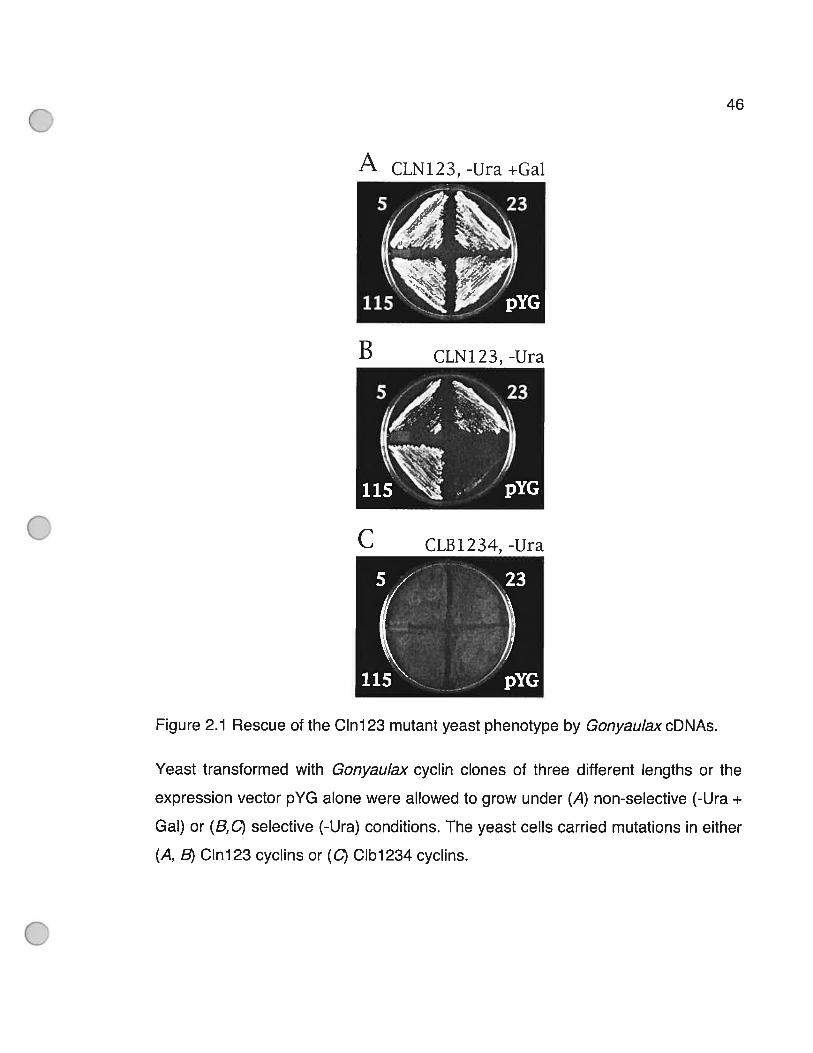
46
A CLN123,-Ura+Gal
YG
Figure 2.1 Rescue of the C1n123 mutant yeast phenotype by GonyaulaxcDNAs.
Yeast transformed with Gonyaulax cyclin clones of three different lengths or the
expression vector pYG aTone were allowed to grow under (A) non-selective (-Ura +
Gal) or (8,0) selective (-Ura) conditions. The yeast celis carried mutations in either
(A, 8) C1n123 cyclins or (C) C1b1234 cyclins.
B CLN123,-Ura
C CLB 1234, -Ura
47
A detailed analysis of the 136 clones (numbered from Cyci through Cyc136)
was carried out by first placing the clones into groups with identical restriction
maps based on digestion with both EcoRi and XhoI. One member 0f each group
was then completely sequenced (19 clones sequenced in total). 0f the 136
recovered sequences, 131 sequences feu into a single group that had different
lengths at the 5’ end but were otherwise identical. We have called this common
sequence GpCycl. AIl GpCycl members had two conserved cyclin domains that
shared 30 ¾ amino acid identity with corresponding domains from various
organisms, and aIl but two clones (designated Cyc3 and Cyc23) also possessed
the degradation box motif (RxxLxxlxN) typical of mitotic cyclins (Figure 2.2, p.49).
These characteristics indicated that we had cloned a mitotic cyclin instead of an S
phase cyclin. The presence of mitotic cyclins in a screen for GuS cyclins might be
related to the finding that mitotic cyclins have been observed to promote S phase
as well as mitosis in budding yeast (Haase and Reed 1999; Moore, Kirk et al.
2003).
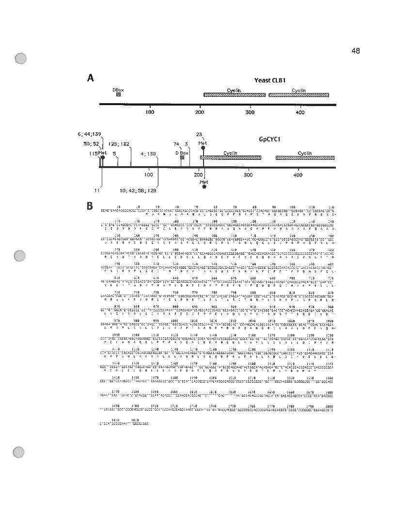
48
A YeastCLBl
OBox Cyc lin Cvo linQ r.W/41’7½¼<z4/oz4-:.z½1 V”?ZQZ’//.½Z’v2Z-WM
I I I
100 200 300 400
644139 23N N GpCYCI
5052 125132 74 3 MetN N’
1 isMet 5 4’ 139 D Box Cyclin Cyclinr ‘//////////Z%¼Y//////1 x½Z’////zVWW//4
I I I
100 200 300 400
Met
11 10;42;58;128
BG
L LtL
G
0: GO
ÛG
L0
Il O oLr E ‘:11k L’ G L P k (‘L r t’ vs.’’ o o’ 0911v P: S k G k L G’
31G 860 87E 090 09E 900 91E 910 93k 940 91k 960G1’GGa’G’GGGGGG’GG’ ‘. CGGIGGOvOO’C1OG0003’11601:OGIGG:11’GGGAGO:1 ‘GG’Cv’G’GvGGGG’GvGG’vGvGGv:.GGOGG.oGv’GG’GG.OGOG
L o G :‘ G o o L : vo E E O : E r r o o o O L ‘: ‘6 y k 3 ‘‘Gr o k -‘316 960 99E 090E 10GO :016 :030 106k 1610 106k 1010 108E
G:vGv’Gor:. GO’G’GGCGG ‘G’.GC’CG ‘1G’ ‘GGGGGGCG’G:GG:GG:lvvG G6’GGG°G GG%OGIOGOGGGCGIO ‘CC’G 1GGG1G ‘1044’’GGGG’GICOGGvL soL’; r o ‘,:,vr’y.: ii F “ko P Gv kG krn’. Lv L ir k S GO’
G630 lIGE 1110 11G 1G:E 11GO .10k 116E 11’G IGOG 110E 1:00IICGOG:oG:1cvGGcv:G41:::’G:1G::G1:G11:GGGGOGrG1GvGOOG:4:G:GGGG4GGGG1CGG ‘vl’GGGIOGGG:GGGG’GG:.:v’:GGGGllG
F G L:, k’ GO L F:’ P vo G vI 9030G G Lv o’L L 0Ev IL 0G k ‘:0’
1110 11,0 110 1110 11’Q L50 11’0 11GO 119E 130G 310 10.9G:G::c’:GGv:r:G:vlGIGGGG:G:’’GG ‘‘G1::v:1v4:1-1v11:G.G::GG::GG: ‘‘lGIlvGGcCGG ‘:1:1:114—c-11:11 ‘:1’lCvlv.161111’Œv
Ev Poli L’ vo’ L IL ‘9ko S G 0k (0 r vs L vO L’ G ‘6E tEL C’
033G 13.19 031G 136E .310 :EE 1±9E 010E 17.0 1.110 0.130 14.1011 G’’lCcv’I116GCGGG,I:v11’C1 ‘lLvv1llC1l1vGvGl ‘lG’cvGL ‘v’l.lGlvlCvv VvCllGlvvcGvCvGCl’vlv1G1v1ClvllCG:GG CclCv
o 0 4 F L G Q E L 0 0 y k o 6 E o G o r o s F ‘ F ii ‘ E “ L “ k II F y
dO 1160 11E 14GO 1199 100k 111E 11.0 lirE rIO db 116k:11 “Gdl ‘14101G
111G 118E 019E 1’IGE :510 1610 1690 1540 1510 156E 1570 150GG’.’’ d’vClOGGlOvV’G ‘‘‘‘ 4’ ‘16180GGGG’I’G2’1CG’.dG116111CG10’1811G1
159G rIE 1110 1’.O dIE :140 1110 P09 1’”O 110G 119k 1800‘:cv,1:1GOG:10011’11:y’’::’Gv’lvc:010GG ‘GIIIGGIIOGIGGG’11601v1G1Gd1:1’1:111G:’1111G1:0’1
141G 1010 -
49
Figure 2.2 Gonyaulax cyclin contains two conserved cyclin domains and a
degradation box.
(A) Schematic comparison of the longest Gonyau/ax cyciin sequence with that of
yeast CLB1 shows a similar orientation 0f the degradation box (D box) and the two
cyclin domains. The positions corresponding to the 5’ end of the 1 9 sequenced
clones are shown by clone number (unes) as well as the position of the first three
ATG codons (circles, marked as Met since ail three are in trame) for the Gonyau/ax
cyclin. Shaded boxes mark the position cf the degradation box and the twa cyclin
domains. (B) The nucleotide and derived amino acid sequence of GpCycll5.
Translation has been assumed to start at the first ATG.

50
it is important to note that the cloning vector used lacks an initiation codon,
so that any transcript produced must contain its own in-frame AUG in order to be
transiated. Indeed, this may be the reason why an S phase cyclin was not picked
up by our screen. There are three AUGs in the N-terminal region of the deduced
protein sequence of the longest clone (Figure 2.2A, p.49). The first is immediately
downstream from the 5’end of clone Cycll5, while the second and third are
located close together at the beginning of the first cyclin domain. The length of
clones Cyci 15, Cyc5 and Cyc23 is such that they could initiate translation from the
first, second and third AUG, respectively. There is no detectable phenotypic
difference between the three clones. Interestingiy, no shorter sequences were
recovered in our screen. It is possible that no shorter sequences were synthesized
during the cDNA synthesis reaction. However, it seems more likely that a GpCYC1
produced by translation initiation from the next methionine (inside the first cyclin
domain) is too short to function properly. In any event, it seems clear that the
degradation box is not required for phenotypic rescue as Cyc23 lacks this
sequence element.
To test the size cf the transcript from which the clones were derived, the
entire cDNA sequence was radiolabeled and hybridized to poly(A) RNA on
Northern blots. Interestingly, two bands were observed instead cf the one expected
(Figure 2.3, p.52). The upper band, at 2.2 kb, is large enough to encode the 1.83
kb Cycl 15 clone isolated from the genetic screen while the smaller band, at 1 .6 kb
appears too short. We also observed that the smaller of the two hybridization
signaIs was not obtained when a probe derived from the 3’ UTR was used,
suggesting that this band lacks much of the roughly 600 nucleotide 3’ UTR found in
ail the sequenced clones. Although it is theoretically possible that the smaller
51
mRNA may represent a post-transcriptionally modified form 0f GpCycl, this seems
unhikely given that it was not recovered in our genetic screen. Instead, t seems
more probable that the sequence represents a different cyclin box-containing
sequence that was flot identified in our screen because of the requirement for an
in-frame methionine upstream of the cyclin domains. The size of the larger RNA in
this analysis also suggests that the GpCycl sequence is still missing roughly 370
bp from the 5’ end.
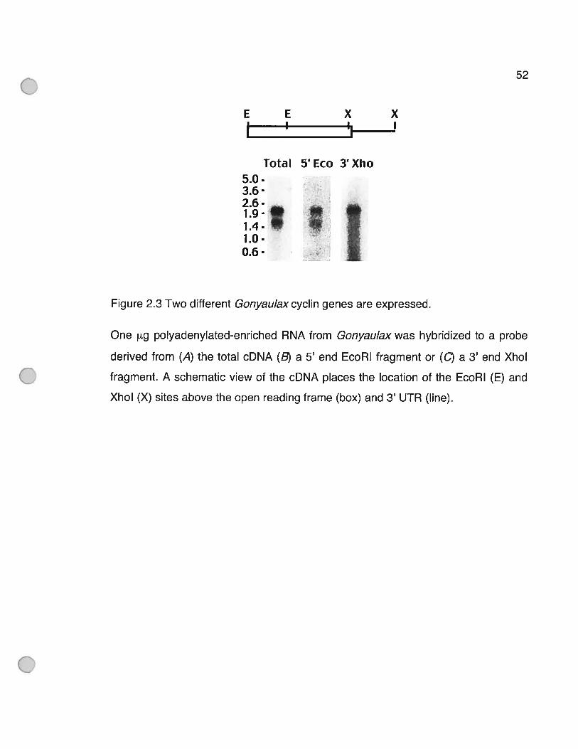
52
E E X X
Total 5’ Eco 3’ Xho5.0-3.6-
1g1410-06-
Figure 2.3 Two different Gonyaulaxcyclin genes are expressed.
One g polyadenylated-enriched RNA from Gonyaulax was hybridized to a probe
derived from (A) the total cDNA (B) a 5’ end EcoRi fragment or (C) a 3’ end XhoI
fragment. A schematic view of the cDNA places the location of the EcoRi (E) and
XhoI (X) sites above the open reading frame (box) and 3’ UTR (une).

53
Slight differences were also observed in the length of the 3’end of the
clones. Ail sequences recovered had minor differences in the position cf the
poly(A) tract, with 20 nucleotides separating the shortest from the longest clone.
However, as the sequence was identical up to the the poly(A) tract, either site
recognition for poiyadenylation is somewhat leaky or we have cloned transcripts
expressed from different genes, each with a slightly different location for their
putative polyadenylation signal. This behavior has not been previously noted in
other analyses of dinoflagellate gene expression, such as for the sequences POP
(Le, Markovic et ai. 1997) or LBP (Machabee, Wall et ai. 1994), and to date the
polyadenylation signal has not been defined for any dinofiagellate transcript.
The remaining 5 clones isolated from the screen falI into a second group of
identical sequences with different 5’ end lengths. Unlike the first group, these
sequences share no homology with known cyclins, despite being able to
complement the yeast Cln123 mutation. Curiously, the only similar sequences
currently available in GenBank contain an uncharacterized Sp0IIIAA (ATPase)
domain conserved in prokaryotes and a putative protein-encoding gene from
Arabidopsis. The identity cf this clone and the mechanism whereby it is able to
rescue the mutation in the cln 123 yeast strain are not yet known (see discussion).
2A.2 The cyclîn sequences are derïved fromdînoflagellates
Five lines of evidence support the contention that the clones isolated did
indeed correspond to authentic dinoflagellate cyclins. First, the sequences are 66.5
% GC-rich, as are other nuclear encoded Gonyaulax genes (Machabee, Wall et al.
1994; Le, Markovic et al. 1997). Second, the codon usage in the cyclin cDNA is
54
consistent with that found in a compendium of ail other known Gonyaulax coding
sequences currently available in GenBank (Table 1, p.55; R2 = 0.528). Third, a
search of dinoflagellate EST sequences recovered one cyclin sequence with 77 %
amino acid sequence identity to the C-terminal end of the Gonyaulax sequence
from 6000 non-redundant Alexandrium ESTs (these ESTs were sequenced from
the 3’end (Hackett, Yoon et al. 2004)). Although only a short fragment of the
sequence is available, this is the highest level of sequence identity found with any
organism and is roughly equivaient to the degree of identity found between cyclins
found in plants, in fungi or in animais (boxed areas in Table 2, p.56). In contrast,
the level of sequence identity between GpCycl and any other cyciin is uniformly
low (30 ¾), as is the similarity between cyclins from different groups. Fourth,
measurement of the number of gene copies per dinoflagellate nucleus suggest that
5000 gene copies are present (Figure 2.4, p.57). This is consistent with the
generally high copy number of dinoflagellate genes, including the 1000 copies of
the luciferin binding protein (LBP) gene (Lee, Mittag et al. 1993) and the 5000
copies of the peridinin-chlorophyll a-binding protein (PCP) gene (Le, Markovic et al.
1997).
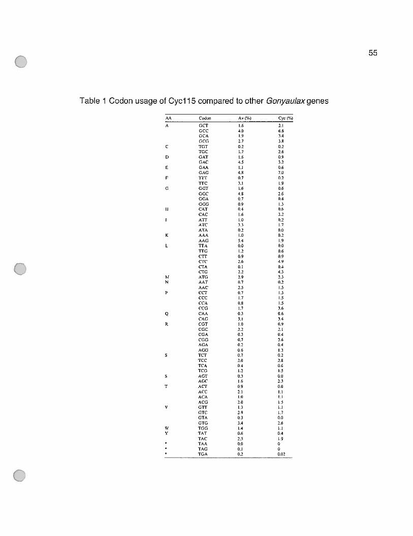
55
Table 1 Codon usage of Cycl 15 compared 10 other Gonyaulax genes
Cc,don Av (%) C’yc (%)
A GCT 1.6 2.1GCC 3.0 6.6GCA 1.9 3.4GCC1 2.7 3.8
C RiT 0.2 0.2TGC 1.7 2.6
D GAT 1.6 0.9GAC 4.5 3.2
E GAA 1.1 0.6GAG 4.8 7.0
F iTT 0.7 0.2TTC 3.1 1.9
G GGT 1.6 0.6GGC 4.8 2.6GGA 0.7 0.4GGG 0.9 1.3
II CAT 0.4 0.6CAC 1.6 3.2ATT 1.0 0.2AtC 3.3 1.7
ATA 0.2 0.0K ÂAA 1.0 0.2
AAG 5.4 1.9L TTA 0.0 0.0
TTG 1.2 0.6cii’ 0.9 0.9
2.6 4.9tTA 0.! 0.4CTG 2.2 4.3
M ATG 2.9 2.3N AAT 0.7 0.2
AAC 2.5 1.5P CCT 0.7 1.3
CCC 1.7 1.5CCA 0.8 1.5CCG 1.7 3.6
Q CAA 0.3 0.6CAG 3.1 3.4
R CGT 1.0 0.9CGC 2.2 2.1CGA 0.3 0.4CGG 0.7 2.6AGA 0.2 0.4AGG 0.6 1.3
S TCr 0.7 0.2TCC 2.0 2.8TCA 0.4 0.6TCG 1.2 1.5
S AGT 0.3 0.0AGC 1.6 2.3
T AET 0.9 0.0ACC 2.1 1.1ACA 1.0 1.1ACG 2.0 1.5
V GTT 1.3 1.1GTC 2.9 1.7GTA 0.3 0.0GTG 3.4 2.6
W TGG 1.4 1.1Y TAT 0.6 0.4
TAC 2.5 1.9TAA 0.0 0TAG 0.1 0TGA 0.2 0.02
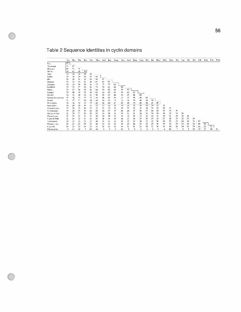
I us,,nnvn
‘‘Il,
t),,’
(ioIdI,hOan,,[.1n9%fl
l’aIda.tfl,’l,,!, ha,,,,.,f) ,,ba
(n,,ltgj,rh) I
•î•l•)’Il’.nl0
(rjnh,ua,:’,o,,
3oa’l (C (64)
r»”’1”
(‘7i4 14
:34 29 12 32(4 (112253127 2% 25 27 26 3’)7 .1)) 3)) 1)) 2,) 33 2f,12 32 11 2’) 31 29 76 36
1’) 27 2) 27 2% 25 25 2’) 3)3’ 33 1’) 10 3% 23 23 33 2% 4232 2’) 3f) 29 23 3’) 25 34 31 4’) T]34 93 3) 9] 36 36 23 2) 29’34 (3t 1 5 6 III 7 S S Il 2 12 Il) (I
Table 2 Sequence identities in cyclin domains
56
3’,,,, N,,, ((o, t),, NI,,, uiI t),, I f,,,,, Na,, 14,’) 14,,,, la,,, Pal SI, II,1 I)r,., (J,,,, 1 L_a, t),,, t’a, (lb Na,, t’,,a 16,1,,
60 11 91M13 1% 16 3411 36 34 3334 36 31, 3333 15 35 134 30 19 313% 36 37 3435 37 36 3433 3), 34 3234 31 31 3237 iI, I) 3531 (7 37 04
13 14 13 1339 3,(4 (1 ‘6
24 33 3% 222’) 27 2’)
15 3) il II2331,31 3124 21 34 3%
29 24 3’) 2’)23 30 2%
S II L)) 7
15 95 f
‘‘
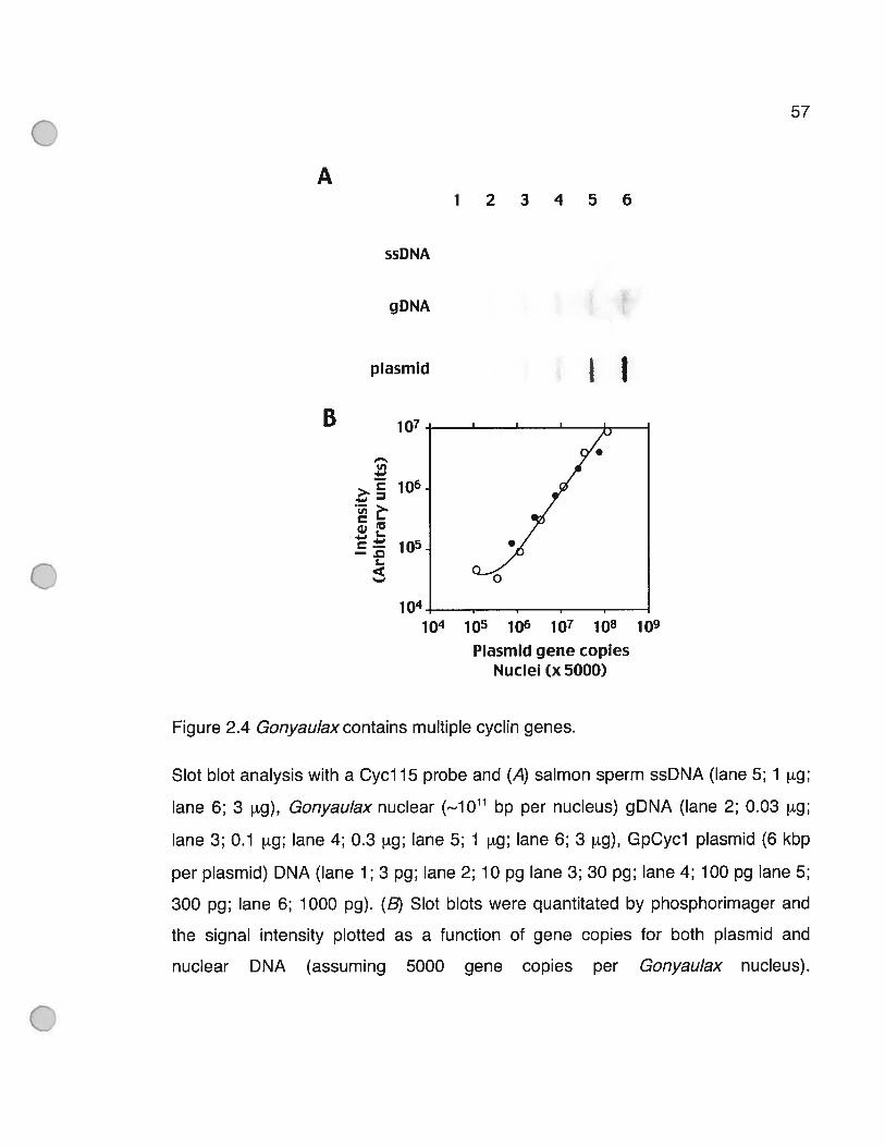
57
A
B
ssDNA
gDNA
plasmîd
u,
. >
—.
t
s—
123456
Figure 2.4 Gonyaulaxcontains multiple cyclin genes.
I I
Siot blot analysis with a Cycl 15 probe and (A) salmon sperm ssDNA (Pane 5; 1 pg;
lane 6; 3 pg), Gonyaulax nuclear (,.1Oh1 bp per nucleus) gDNA (Pane 2; 0.03 g;
Pane 3; 0.1 pig; Pane 4; 0.3 tg; Pane 5; 1 tg; Pane 6; 3 pig), GpCycl plasmid (6 kbp
per plasmid) DNA (Pane 1; 3 pg; Pane 2; 10 pg Pane 3; 30 pg; Pane 4; 100 pg Pane 5;
300 pg; Pane 6; 1000 pg). (B) SPot blots were quantitated by phosphorimager and
the signal intensity plotted as a function of gene copies for both plasmid and
nuclear DNA (assuming 5000 gene copies per Gonyaulax nucleus).
10
1041O 1O 106 10 108 70
Plasmïd gene copiesNucleï Cx 5000)

58
Lastly, we performed phylogenetic analysis with the dinofiagellate cyclin
sequence to see if the predicted phylogenetic relationships were compatible with
those determined using other sequences. Mitotic cyclin sequences were selected
to represent the widest range of available taxa, and the sequences truncated to
contain the cyclin fold (roughiy 250 amino acids). Our phylogeny recovers the
major groups of fungi, plants and animais with high bootstrap support (Figure 2.5,
p.59). The GpCycl sequence tails outside these three ciades as expected
(Baidauf, Roger et al. 2000; Van de Peer, Baldauf et ai. 2000). However, we also
expected the dinofiageilate sequence to group within a clade containing ciliate and
apicomplexan sequences. The ciliates are represented in our analyses by
Paramecium, and aithough ciliates do flot form a ciade with our Gonyaulax cyclin,
there is no significant bootstrap support for exciuding Gonyaulax from such a
relationship. Ciearly, the 10w degree in phylogenetic discrimination found in the
conserved cyclin domains preciudes making a definite statement as to the
reiationship between ciliates and dinoflagellates. The apicomplexan cyciin is
represented in our analysis by the cyclin4 sequence from Plasmodium, the only
apicompiexan sequence with two cyciin domains. This sequence shares littIe
homology with the dinoflageilate sequence, and indeed, is used as an outgroup in
this analysis. This is presumably due to the very 10w (7-12 %) sequence identity
between the apicomplexan sequence and other cyclins (Table 2, p.56), and it has
been suggested that the unusual ceil cycle of Plasmodium may have ied to highly
derived cyciin sequences (Merckx, Le Roch et al. 2003). Dur analysis is thus not
inconsistent with the estabiished phylogenetic position of the dinofiagellates. Taken
together, therefore, our data provide compelling evidence that GpCycl is a bone
flUe dinofiagellate cyciin.

59
Figure 2.5 Phylogeny 0f Gonyaulax cyclin sequence is consistent with known
relationships.
Protein sequences were aligned by ClustaIW and analyzed using neighbor joining.
The Gonyaulax cyclin is not part of the clusters representing fungi, animais and
higher plants.
Plasmodium
95
99 I NeurosporaI .Fungi
Pneumocystis
S.pombe
100 Brassica
100L Arabidopsis
Pea PIant
Oryza
Nicotiana
Gonyaulax
0.2
Leischmania
Paramecium
Dictyostelium

60
2.4.3 Cyclin expression is controlled post translationally
To address the issue of cyclin expression at the protein level, a rabbit
polyclonai antibody was raised against the N-terminal region 0f GpCYC1 for use on
Western biots. We anticipated that a mitotic cyclin synthesized from our clone
should have a molecular weight of roughly 64 kDa and should vary in amount
over the course of the celi cycle. However, ceils under our growth conditions have
a roughly five day doubling time (Figure 2.6A, p.61). Thus, even though mitosis
occurs only in a window of several hours just following dawn (Homma and Hastings
1989), the ceils are not synchronized with respect to the celi cycle. We therefore
prepared samples enriched for G2 or Gi ceils using a modification of a previously
described filtration protocol (Homma and Hastings 1988) (Figure 2.6B, p.61). The
average celi volume in samples enriched for G2 phase celis is roughly twofold
greater than in samples enriched for Gi phase celis as expected (Figure 2.6D,
p.61). Western blots analysis of Gi- and G2-enriched celi extracts using an
antibody raised against the N-terminal region of the cyclin showed that a band at
68 kDa increased in abundance trom G1-enriched to G2-enriched cell extracts
(Figure 2.60, p.61). Preincubation of the antibody with the bacterially expressed
GpCYC1 N-terminus specifically reduced the level of reaction with the 68 kDa
band, while affinity purification of the antibody using the bacterially expressed
protein transferred to nitrocellulose after SDS-PAGE reduced the level of reaction
with ail bands other than that at 68 kDa. We conciude from this experiment that
GpCycl encodes a protein of —P68 kDa that accumulates to higher leveis in celis
prior to mitosis compared to recently divided celis. Thus, our sequence has the
characteristic properties expected for a mitotic cyclin.

G
(D mitosis oFiltration Filtration
/
enrichedI ©
___
G?- 0Venriched © Gt
Figure 2.6 Gonyaulaxcyclin is more abundant in G2 ceils.
(A) Oeil concentration measured daily over a three-week intervai shows a roughly
five day doubling time. (B) Scheme of fiiter-enrichment for G2-phase ceils uses
ceils harvested prior to mitosis and retained by filtration, whiie fiiter-enrichment for
Gi-phase ceils uses ceils harvested foliowing mitosis that pass through the filter.
(C) Western blot of Gonyaulax extracts enriched for Gi or G2-phase ceiis probed
with an antibody raised against the N-terminai region of the Gonyaulax cyciin (a
CycN), the same antibody preincubated with bacteriaiiy-produced cyclin (a-OycN +
cyciin), the same antibody affinity purified on nitroceliulose membranes (aOycN*)
or a-POP as a control for protein loading. Ail ianes had simiiar amounts of protein
as judged by Ponceau red staining. (D) Ce11 volumes were caicuiated from
measurements of ceil area on over 100 ceiis in random microscope fieids using ceii
sampies taken from LD 23, either enriched for G2 celis or not, and from LD 4,
either enriched for G1 cells or not.
A B
61
LDT4
‘l.’.III.
Day
C 61 G2
6BkDa+ a-CycN
— —68 kfla + a—CycN + Cyclin
68kDa+ ci-CyCNr
32 kDa-
ct-PCP
Dqu,000
I Celi volumer ——-:
30000b—f---
2O,OOO —eio.000b
— zItL_
G2 61
LD23 LD4
62
2.5 DïscussïonThe purpose of the present study was to isolate a dinoflagellate cyclin and to
determine if it possessed any distinguishing features that might be related to the
unusual nuclear features cf dinoflagellates. To this end, we screened a
dinoflagellate cDNA library for cDNAs able to functionally complement yeast
harboring mutations in either Gi phase or M phase cyclins. Although screens for
mitotic cyclins were not successful, our screens for functional complementation cf
Gi cyclins produced a single cyclin sequence. Roughly one cyclin clone was
recovered for every 50,000 clones screened, but it is important to recognize that
this may not necessarily reflect cyclin clone abundance. Indeed, this number
probably underestimates the total number of clones since our expression vector
begins translation of the inserted sequence from its first AUG. Other cyclin genes
could be present and yet might not be selected by the screen if, for example, they
are not long enough to reach the authentic initiation codon and do not have an
AUG in-frame. Our Northern blots showing two distinct size classes of mRNA
hybridizing to the full length cyclin cDNA suggests that at least one additional cyclin
not recovered by our screen may indeed be present.
The second group of sequences able to rescue the mutant cyclin phenotype
was recovered from our screen with a much lower frequency (less than 1 clone per
million screened). This clone does not share any sequence similarity with known
cyclins BLAST searches. Instead, significant sequence identity was observed only
with proteins of unknown function in prokaryotes that contain a Sp0IIIAA (ATP
binding) domain (an E-value cf 4 x e20) and Arabidopsis protein with a putative
ATPase activity. It should be noted that other non-cyclin proteins such as RINGO

63
(Karaiskou, Perez et al. 2001) have been shown to activate CDKs, presumably
because of a three-dimensionai structure similar to the cyciin fold. We will attempt
to address the function of these clones using two-hybrid assays to look for
interactions with an authentic yeast CDK as well as by examining the phenotype of
homozygous Arabidposis mutants whose copy of the gene has been inactivated by
a T-element insertion. For the present work, we have concentrated our efforts on
the sequence clearly identified as a cyclin by sequence homology.
With respect to the sequence 0f the dinoflagellate cyclin, its N-terminal cyclin
domain shares 30 % sequence similarity with mitotic cyclins from other
organisms (Table 2, p.56). Clearly, both sequence analysis and the presence of a
degradation box place our sequence among the mitotic cyclins. Dur sequence
shares 54 % amino acid sequence identity in the N-terminal cyclin domain with the
sequence AAB92250, reported to be from the dinoflagellate Alexandrium.
However, the full sequence corresponding to this fragment was not reported, and
more recently, a cyclin gene fragment (CK432882) was found in an Alexandrium
EST project whose 79 C-terminal amino acids shared 70 % sequence identity at
the amino acid level with GpCycl. This level of sequence homoiogy is consistent
with the high levels of sequence identity found in members of the plants, animais,
and fungi (Table 2, p.56). It is not possible to determine the degree of similarity
between the two reported Alexandrium sequences as they correspond to different
regions of the mature protein.
The phylogenetic anaiysis of GpCYC1 was expected to place the
dinoflageliates next to the apicomplexans and ciliates, consistent with the
molecular phylogeny of multiple protein and rRNA sequences (Baldauf, Roger et al.
2000; Van de Peer, Baldauf et aI. 2000). However, the only available
Apicomplexan cyclin containing two cyclin domains shares very limited sequence
64
similarity with our Gonyau/ax sequence (Merckx, Le Rech et al. 2003) (10 %
sequence identity at the amine acid level). This 10w degree cf similarity presumabiy
refiects the unusual nature cf the apicomplexan sequences, which also share iittle
homoiogy with cyclins from other organisms (7-12 % sequence identity). It must be
noted that a reiationship with Paramecium is net exciuded by our data, since the
cyciin domains contain toc many conserved residues to have much discriminative
power. Paramecium, as a representative cf the dilates, wouid have been expected
te be the next ciosest relative to the dinoflageilates after the Apicomplexans. It is
clear, however, that there is strong bootstrap support for the clade representing
plant and animal cyciins, and that the GpCYC1 does net ferm a part cf this group.
Dur sequence 15 thus consistent with estabiished relatienships given the Iimited
number cf informative characters in the sequence used.
It is aise important te note that the unusuai nuclear architecture cf the
dinefiagellates has not resuited in highiy atypicai cyciin sequences, as found in the
wideiy divergent Plasmodium sequence for exampie. It is thus reasonabie to
expect the CDK sequences might possibiy aise dispiay simiiar phyiogeny. If so,
then our phyiegenetic analyses impart an important new direction in our application
cf PCR based strategies to isciate dinefiagellate CDKs. Dur previous work in this
regard, to date unsuccessful, has been based on simiiarity with yeast hemoiogs of
the ceii cycle machinery. This phyiogenetic anaiysis new suggests that either plant
or animai sequences shouid instead be empioyed as a basis for the seiection of
censerved regions. It is interesting, in this regard, that immunoiegicai studies using
antibedies directed against heterologous cyclins (see beiew) give resuits more
ciesely appreximating those described here when antibodies directed against clam
cyclins rather than yeast cyclins are used.
65
The screening protocols that were tested in the present study inciuded both
a screen for mitotic cyclins (the Clb1234) and the Gi cyclins (C1n123). The screen
for mitotic cyclins was unsuccessful, and furthermore, even when the mitotic
cyclins isolated from the C1n123 screen were directly tested in the CIbi 234 mutant,
no functional rescue was observed. The Clb mutations appear quite complex
phenotypically, and it has been previously shown that both Cibi and C1b3 are
required to compiement a mutation in C1b2 (Fitch, Dahmann et al. 1992). Perhaps
our Gonyaulax cyciin alone is unable to substitute for the lack 0f the four Clb
genes. In contrast, the Cln123 mutant appears more amenable to functional
complementation. Indeed, this mutation has been previously used to isolate various
cyclins (including mitotic cyclins) from human (Lew, Dulic et al. 1991) and
Arabidopsis libraries (Day, Reddy et ai. 1996).
The observation that our N-terminai antibody recognizes a protein whose
abundance changes over the course of the celI cycle is different from a series of
studies in C. cohnhi and K brevis using a polyclonal antibody raised against the
cdcl3 gene product of S. pombe. Here, the immunoreactive cyclins were observed
to have a molecular weight of 56 kDa and to be present at ail times over the cell
cycle (Barbier, Geraud et al. 1998; Barbier, Leighfield et al. 2003). This molecular
weight is lower that that seen in our analyses, but it must be noted that the fission
yeast cyclin domains share only 30 % sequence identity with our sequence over
the region encompassing the two cyclin domains. Another series of studies with the
same organism used monoclonal antibodies directed against a 36 amino acid
stretch in the cyclin domain of sea urchin cyclin B. In these experiments, the
antibody reacted with three bands at 97, 66 and 46 kDa (Yeung, New et al. 2000).
The bands at 97 and 66 were Iow in mitosis and high in G2, as we find with our
antibody, and the band at 66 kDa is similar in molecular weight to that seen with
66
our antibody. The peptide used ta generate this commercial antibody shares 58%
sequence identity with GpCYC1, althaugh the degree of similarity between
Gonyaulaxand C. cohniicyclins is not yet known. We raised our antibody to the N-
terminal damain of the pratein deliberately, reasaning that the lack of sequence
homoiogy ta any sequence in GenBank wouid tender it unlikely to crass-react with
any other proteins. Antibodies raised against a canserved cyclin domain, on the
other hand, might react with a number of different cyclins each having a different
distribution over the celI cycle. lndeed, if other cyclins were present in the
dinofiagellates, an antibody recognizing severai cyclins might make cell cycle
variations in a mitotic cyclin difficult ta detect.
The cyciin protein is not readily detectable by standard protein stains after
SDS-PAGE, suggesting that the cyciin is flot as abundant as the proteins LBP
(j—1% total pratein in night phase celis) or POP (up ta 5% total pratein). It is thus a
little surprising ta find that the number of cyciin gene copies is simiiar ta the —5000
copies found far POP, as a part af the ratianale previously invoked ta accaunt far
the large number 0f gene copies was protein abundance. This suggests instead
that numerous gene copies are a more fundamental feature af nuclear architecture
in Gonyaulax. This unusual gene structure may be related ta the unusuai
components of the transcriptional machinery identified in the dinoflagellates
(Guillebault, Sasorith et al. 2002) or the iack af recognizable motifs far transcription
factar binding in the putative pramaters af the LBP (Lee, Miffag et al. 1993), POP
(Le, Markavic et al. 1997) or luciferase (Li and Hastings 1998) genes. A definite
examination af the transcriptianal activity of these putative pramaters must await a
repraducible transformation system sa that their raies can be assessed in vivo.
However, it must be noted that our Narthern biat analyses suggest that twa distinct
67
size 0f transcript are present. Therefore, it is possible that the cyclin sequence
reported here may constitute only half the 5000 gene copies measured.
We currently have no data concerning the subcellular location of our cyclin.
However, our sequence has no recognizable nuclear localization sequence, and is
thus unhikely to enter the nucleus unless accompanied by another protein that is
able to promote nuclear entry. In agreement with this, cyclin B is not normally able
to enter the nucleus in animal ceNs, and will flot unless an NLS is added (Moore,
Kirk et al. 2003). The recent finding that the 56 kDa antigen reacting with the anti
cdcl3 is Iocated in the nucleolus 0f K brevis is thus puzzling (Barbier, Leighfield et
al. 2003). Possibly this antibody recognizes a different cyclin in the dinoflagellates.
The celI cycle in dinoflagellates 5 of considerable interest because 0f the potential
impact on human health of the massive summer blooms of toxic species (Van
Dolah 2000), but molecular studies have been hampered by the lack 0f authentic
celI cycle regulators.
The celI cycle is also 0f particular interest in Gonyau/ax because of the
delicate interplay between the cell cycle and the circadian (daily) dock. In this
organism, mitosis always occurs just after dawn even though the celI cycle can last
several days. Thus a window 0f opportunity opens at dawn for cells 0f sufficient
size to enter M phase, and mitosis is said to be “gated” by the circadian dock. The
mechanism for this is not known in dinoflagellates, although in regenerating liver
cells, activation 0f cdc2 kinase activity correlates with a decrease in expression 0f
weel RNA and protein (Matsuo, Yamaguchi et al. 2003). The transcription of the
weel kinase, whose activity results in inactivation of the kinase/cyclin complex, is
inhibited by the oscillating activity of the dock components CLOCK/BMAL.
Furthermore, the presence of the inhibitory phosphate on Tyrl5 of cdc2 can be
directly measured and is circadian in regenerating liver. Thus in liver cells, the
68
circadian window of opportunity opens during decreased levels cf the weel kinase,
an inhibitor of cdc2 kinase activity. Therefore, isolation of the regulatory elements
of dinoflagellate celI cycle regulation is a prerequisite to our long-term goal of
understanding the impact of the circadian dock on celI cycle control in
dinoflagellates.
2.6 AcknowledgementsWe thank Drs. P. Belhumeur and M. Raymond for technical advice and
yeast vectors, Drs. S. Dorion and J. Rivoal for assistance with the antibody
production and purification, and M. O’Brien for advice on cDNA synthesis and ANA
purification. We gratefully acknowledge the financial support of the National
Science and Engineering Research Council of Canada.
69
3 Publication #2 : A Dinoflagellate AAAFamïly Member Rescues a ConditionalYeast GuS Phase Cyclïn Mutant throughlncreased CLB5 Accumulation
Thierry Bertomeu and David Morse
Accepté chez Protist, sous presse en juillet 2007
Toutes les procédures expérimentales furent effectuées par moi et cet article a été
écrit conjointement avec D.M.
70
3.1 AbstractAn AAA protein from the dinoflagellate Gonyaulax polyedra (GpAAA) with
the unusual ability to rescue the phenotype 0f a yeast mutant lacking GuS phase
cyclins (cInlcIn2cIn3) has been isolated and the mechanism of rescue was
characterized. We find that GpAAA is flot a cyclin and has no similarity to any
known ceil cycle regulators. Instead, GpAAA forms a novel and strongly supported
clade with bacterial spoIIIAA proteins and an Arabidopsis gene of unknown
function. Since dinoflagellates cannot be transformed, we took advantage of the
powerful genetic tools available for yeast. We find that rescue of the cInlcIn2cIn3
phenotype does flot involve an effect on the CDK-inhibitor (CKI) Siclp, as GpAAA
does not alter the sensitivity to an inducible SICY. Instead, Northern blot analyses
show that GpAAA expression increases levels of CLB5, in agreement with the
observation that GpAAA is unable to rescue the quadruple mutant
c/nlcIn2cIn3cIb5. We propose that the increased transcription of CLB5 may be due
to a protein remodeling function of GpAAA alleviating inhibition of the transcription
factor SBF. Thus, although no known equivalents to the yeast SBF have been
documented in dinoflagellates, we conclude that dinoflagellates could indeed utilize
GpAAA as a celI cycle regulator.
3.2 IntroductionGiven the wealth of knowledge on control of cell cycle in mammalian celis
and model eukaryotes such as yeast, it is surprising that almost nothing is known
about control of the celi cycle in dinoflagellates. This is unfortunate, as
dinoflagellates are major contributors to the phytoplankton in the oceans (Taylor
and Pollingher 1987), and more importantly, rampant cell division can resuit in
71
large blooms, termed red tides, that can have a serious impact on the environment
and public health (Friedman and Levin 2005). Furthermore, the key mechanisms
regulating the ceIl cycle are also of interest in order to understand how they have
been modified to accommodate the characteristic permanently condensed
chromosomes of dinoflagellates and the fact that the nuclear membrane remains
intact through mitosis while having a cytoplasmic mitotic spindie (Moreno Diaz de
la Espina, Alverca et al. 2005). Unfortunately, the study of dinoflagellate
biochemistry is hampered by an inability to exploit forward and reverse genetic
tools, so we have instead chosen to analyze the function of dinoflagellate genes
that affect progression of the ceil cycle in yeast.
To facilitate interpretation of our analyses, a brief review of the yeast celI
cycle is required. As for other eukaryotes, the cell cycle of Saccharomyces
cerevisiae (budding yeast) is regulated by activation of the cyclin-dependent kinase
Cdc28p (also termed CDK1) (Morgan 1997). This CDK is involved in regulating
passage through both the GuS phase boundary and the G2/M phase boundary.
The passage between each of the boundaries requires CDK activation through
binding to the regulatory cyclins that are expressed at the appropriate times to
allow this passage. Once activated, CDKs phosphorylate a range of substrates
whose phosphorylation allows celis to initiate either DNA replication (S phase) or
mitosis (M phase), and the substrate specificity is dictated, at least in part, by the
cyclin subunit bound to the CDK (Loog and Morgan 2005). Three Gi cyclins, called
Clnl, C1n2 and C1n3 are involved in transitions across the G 1/5-phase boundary
(START) (Hadwiger, Wittenberg et al. 1989), with the CLN3gene acting upstream
from CLN7 and CLN2 (Tyers, Tokiwa et al. 1993). Budding yeast also contains six
B-type cyclins, 2 involved in DNA replication (CLB5and CLB6) (Toone, Aerne et al.
72
1997), 2 in spindie morphogenesis (CLB3and CLB4) (Richardson, Lew et al. 1992)
and 2 in mitosis (CLB7 and CLB2) (Surana, Robitsch et al. 1991).
There are many factors that influence CDK activity, in keeping with the
primordial role of this enzyme in ensuring an orderly progression through the celi
cycle (Mendenhall and Hodge 1998). In addition to the activatory interaction with
cyclins, there are also inhibitory interactions with CDK inhibitors (CKIs). One
example is Sici p, which binds and inactivates Clb/Cdc28 (Schwob, Bohm et al.
1994). Sici p is normally inactivated by Cln/Cdc28 phosphorylation allowing the celI
to initiate DNA replication. Furthermore, overlaid on the regulation conferred by
protein-protein interactions are post-translational modifications that affect the
conformation and activity of the kinase subunit. These modifications include an
activatory phosphorylation cf a conserved residue in the T-loop of the CDK (Lim,
Loy et al. 1 996), and the inhibitory phosphorylation cf conserved residues in the
ATP-binding pocket whose functional significance is clear in fission yeast (Enoch
and Nurse 1990) yet less so in budding yeast, despite changes through the ceIl
cycle (Amon, Surana et al. 1992).
Interestingly, recent studies have described non-cyclin proteins able to bind
and activate some CDKs. In neurons, for example, CDK5 can be activated by p35
(Lew, Huang et al. 1994; Tsai, Delalle et al. 1994), although the contribution cf p35
activation cf CDK to regulation cf the celI cycle progression is flot yet clear
(Dhavan and Tsai 2001). In addition, members cf the Ringc/Speedy family have
aise been shown to bind and activate CDKs (Ferby, Blazquez et al. 1999;
Lenormand, Dellinger et al. 1999). The Ringo/Speedy proteins appear to play an
important role in regulation cf the meiotic cycle in Xenopus oocytes (Ferby,
Blazquez et al. 1999). These proteins are only marginally similar to the cyclin
consensus, and the mechanism whereby they activate CDKs remains unclear.

73
The partial redundancy of the three yeast CLNgenes has been exploited for
the development of a yeast strain (cInlcIn2cIn3), which contains only one GuS
phase cyclin (CLN3), placed under control of an inducible galactose-regulated
promoter. Such a strain has been used to successfully isolate cyclin homologs from
a number of different organisms by functional complementation (Koff, Cross et al.
1991; Lahue, Smith et al. 1991; Leopold and OFarreli 1991; Lew, Dulic et al. 1991;
Soni, Carmichael et al. 1995). In this technique, libraries are screened for
heterologous genes able to overcome a requirement for an endogenous CLN in
cells grown in the presence of glucose. The isolation of non-cyclin sequences
through screening has been reported only once, and the Drosophila Cdc28p
homolog that was isolated was suggested to have a CLN-independent function in
yeast (Leopold and O’Farrell 1991). Interestingly, yeast lacking Siclp will grow
even when none of the three CLN genes is expressed, indicating that this CKI is
essential for imposing the requirement for Gi cyclins at START (Tyers 1996).
The molecular events occurring as yeast cells pass START involve
substantial changes in the pattern of gene transcription (Cho, Campbell et al. 1998;
Spellman, Sherlock et al. 1998). These changes result from activation of two
transcription factors, termed MBF and SBF, each of which recognizes different
binding sites in the promoters of the regulated genes (Iyer, Horak et al. 2001).
Curiously, these transcription factors are not directly substrates of Cln/Cdc28
(Wijnen, Landman et al. 2002). lnstead, the causal link between transcription factor
activation and CDK activation may involve phosphorylation and inactivation of a
repressor, such as the Whi5 repressor of SBF (Costanzo, Nishikawa et al. 2004).
The findings reported here are a part of an ongoing program to isolate
regulators of the dinoflagellate celI cycle by functional complementation (Bertomeu
and Morse 2004). In this study we have characterized a dinoflagellate AAA family

74
member that was able to complement a Gi cyclin mutant cInlcIn2cIn3 (Bertomeu
and Morse 2004). This protein, termed GpAAA, lacks any homology to known
cyclins. In general, members of the AAA family 0f ATPases are ubiquitous proteins
that exploit ATP hydrolysis for degrading or inducing conformational changes in a
wide range 0f protein substrates (Sauer, Bolon et al. 2004; Hanson and Whiteheart
2005). Our analysis 0f the mechanism of phenotypic rescue by GpAAA is
consistant with this, as our data point to a role in alleviating repression of SBF
activity thus allowing CLB5 transcription and progression through the yeast celI
cycle. We advance the intriguing possibility that a similar role might be played by
GpAAA within the dinoflagellates.
3.3 Results
3.3.1 A GonyaulaxAAA protein rescues the yeastcInlcIn2cIn3 mutant phenotype
The yeast triple mutant c/nlcIn2cIn3 carnes a mutation in ail three GuS
phase cyclins, and grows in the presence 0f galactose due to induction 0f an
additional CLN3 gene under control 0f an inducible GAL promoter. This strain was
transformed with a Gonyau/ax cDNA library in a yeast expression vector, and
roughly 6 x10⁶ clones (1.5-fold coverage of the library) were screened in the
presence 0f glucose to inhibit normal growth. The reversion frequency 0f this strain
on selective glucose plates is less than 10 (Xiong, Connolly et al. 1991). The
screen yielded 136 positive clones that were placed into one 0f two families of
identical sequences by restriction enzyme analysis. One group, comprising 131
clones, was found to encode an authentic dinoflagellate cyclin (LpCycl) by
sequence similarity and functional analysis (Bertomeu and Morse 2004). However,
the smaller second family (the remaining 5 clones) had no sequence similanity to
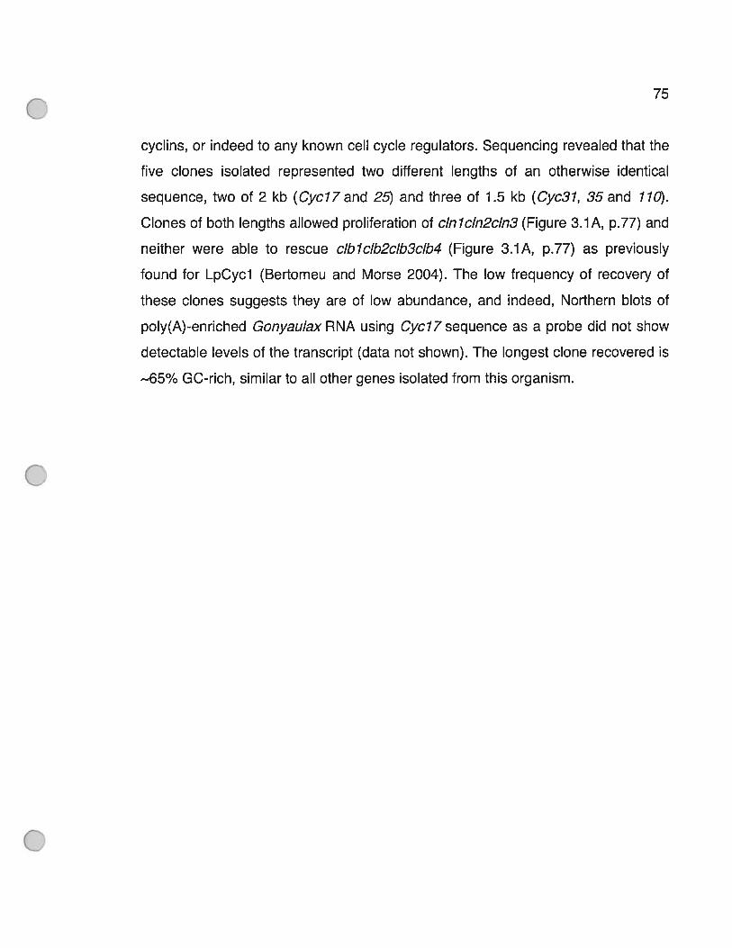
75
cyclins, or indeed to any known celi cycle regulators. Sequencing revealed that the
five clones isolated represented two different lengths of an otherwise identical
sequence, two of 2 kb (Cycl7and 25) and three of 1.5 kb (Cyc3l, 35and 710).
Clones of both lengths allowed proliferation 0f cInlcIn2cIn3 (Figure 3.YA, p.77) and
neither were able to rescue cIblcIb2cIb3cIb4 (Figure 3.1A, p.77) as previously
found for LpCycl (Bertomeu and Morse 2004). The 10w frequency of recovery of
these clones suggests they are of 10w abundance, and indeed, Northern blots 0f
poly(A)-enriched Gonyaulax RNA using Cycl7 sequence as a probe did not show
detectable levels 0f the transcript (data not shown). The longest clone recovered is
—65% GC-rich, similar to ail other genes isolated from this organism.
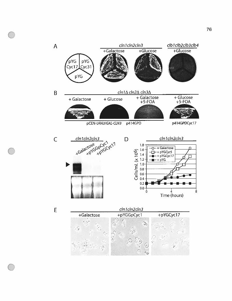
76
A cm 1 cIn2cIn3+Galactose +Glucose
(CYc[c3J)
db 1 cIb2cIb3cIb4+Gkjc ?
B+ Galactose + Glucose + Glucose
+5-FOA
___
IpcEN-URA3/GAL-CLN3 p414GPD p4l4GPDcycl 7
cm lcIn2cIn3e - \1
cm lcIn2cIn3- +pYGGpCycl
t’
cm lA cln2z\ cIn3A+ Galactose
+5-FOA
D cm JcIn2cIn31.8
C
E
X
-
L,
-J
EV)
U 0.4
+Ga lactose
)
—
F
+pYGCycl 7/
(
r/
t

77
Figure 3.1 Partial rescue of cInlcIn2cIn3 mutant phenotype by a dinoflagellate
cDNA.
(A) The yeast mutant cInl::HIS3 cIn2::TRP7 c/n3::URA3-GAL-CLN3 normally
requires galactose for growth (left panel), although in the presence 0f glucose,
dinoflagellate cDNAs Cycl7 and Cyc3l in yeast expression vector pYG can
substitute for the lack of CLN3 expression (middle panel). Growth of cIbl::URA3
cIb2::LEU2 cJb3::TRP7 cIb4::HIS2 GAL-CLBY-LEU2is flot rescued by either cDNA
(right panel). (B) The yeast mutant cinlA cIn2A c/n3A pCEN-URA3/GAL-CLN3
does not grow on Glucose medium or on Galactose medium supplemented with 5-
FOA. Transformation with p41 4GPDCyc1 7, followed by growth for 2 days in liquid
medium allowing for loss of pCEN-URA3IGAL-CLN3, result in yeast able to grow
on Glucose supplemented with 5-FOA, demonstrating rescue in the complete
absence of CLN3. (C) Northern blot using CLN3 as a probe on 5 tg total RNA
obtained from cInlcIn2cIn3 grown either with galactose (left lane), or with glucose
after transformation with the authentic dinoflagellate cyclin pYGLpCycl (middle
lane) or pYGCycl7 (right lane). (D) The c/nlcln2c/n3 mutant transformed with
pYGCyc17 (closed circles) grows more slowly in glucose than does cInlcIn2cIn3
cells transformed with pYGLpCycl (open squares), and more slowly than
cInlcIn2cIn3 cells grown in galactose (open circles). (E) cInlcIn2cIn3 cells grown in
glucose after transformation with pYGCycl7 (right panel) have an unusual
extended bud phenotype not observed when celI express authentic cyclins (left and
middle panels). AIl photographs are at the same scale.
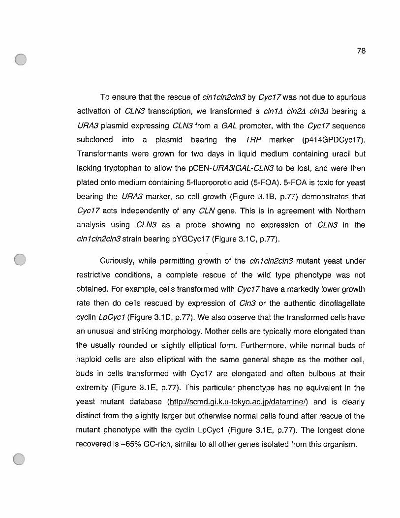
78
To ensure that the rescue of cInlcIn2cIn3 by Cycl7was flot due to spurious
activation of CLN3 transcription, we transformed a cinlA cIn2A cIn3A bearing a
URA3 plasmid expressing CLN3 from a GAL promoter, with the Cyc77 sequence
subcloned into a plasmid bearing the TRP marker (p414GPDCyc17).
Transformants were grown for two days in Iiquid medium containing uracil but
lacking tryptophan to aliow the pCEN-URA3/GAL-CLN3 to be lost, and were then
piated onto medium containing 5-fluoroorotic acid (5-FOA). 5-FOA is toxic for yeast
bearing the URA3 marker, so celi growth (Figure 3.1B, p.77) demonstrates that
Cyclf acts independentiy of any CLN gene. This is in agreement with Northern
analysis using CLN3 as a probe showing no expression of CLN3 in the
c/nlcln2cln3strain bearing pYGCycl7 (Figure 3.1C, p.77).
Curiousiy, while permitting growth of the cInlcIn2cIn3 mutant yeast under
restrictive conditions, a complete rescue of the wiid type phenotype was not
obtained. For exampie, ce! Is transformed with Cyc77 have a markediy iower growth
rate then do ceils rescued by expression of C1n3 or the authentic dinofiagellate
cyclin LpCycl (Figure 3.1 D, p.77). We also observe that the transformed ceils have
an unusual and striking morphoiogy. Mother ceils are typically more elongated than
the usually rounded or slightly elliptical form. Furthermore, while normal buds of
haploid ceils are also eliiptical with the same general shape as the mother ceil,
buds in celis transformed with Cycl7 are elongated and often buibous at their
extremity (Figure 3.1E, p.77). This particular phenotype has no equivalent in the
yeast mutant database (http://scmd.gi.k.u-tokyo.ac.jp/datamine/) and is cleariy
distinct from the slightly larger but otherwise normal celis found after rescue of the
mutant phenotype with the cyclin LpCycl (Figure 3.YE, p.77). The longest clone
recovered is —65% GC-rich, similar to ail other genes isolated from this organism.

79
The schematic view of the longest cDNA sequence presented here (Figure
3.2A, p.81) shows the 5’ end of the two lengths 0f clones isolated and the position
of the transiational start site (ATG) immediately downstream from the end of these
clones. Both of these ATG codons are in frame, and have an appropriate Kozak
context (Kozak 1 989). They presumably represent the start of translation since the
cloning vector itself does not contain an initiation codon after the promoter.
Database searches using BLASTP (Altschul, Madden et al. 1997) revealed thatthe
N-terminal end of the protein had no similarity to any known protein. Curiously, a
block of m-75 amino acids was found repeated three times within this region (Figure
3.2A-B, p.81). In contrast to the N-terminal end, a 230 residue region in the C
terminal end of the predicted protein had significant sequence identity with many
hypothetical and uncharacterized ATPases from bacteria (E value 5e25) and gene
At7g73170 from Arabidopsis thaliana (E value 4e20), a higher plant. This region
shares 32% amino acid sequence identity (52% sequence similarity) with the
Sp0IIIAA domain from bacteria. The function of this domain is still unknown but it is
found in the first gene of operon Spo III from many bacteria in which a defect stops
sporulation at stage 3. We thus tested if either of these two domains alone could
rescue cInlcIn2cIn3. A convenient Bcll restriction enzyme site (Figure 3.2A, p.81)
was exploited to clone the N and C-terminal regions separately into the expression
vector. Neither of the two regions alone was able to rescue the cInlcIn2cIn3
phenotype (Figure 3.20, p.81). We also note that, since the shorter form (Cyc37)
can also rescue, two of the three repeats in addition to the C terminal end are
sufficient for activity.

A EcoRl
Cycl7Cyc25
I Kepeat l Hepeat2 t Hepet3 ICyc3 ICyc35CycilO
D WalkerAG X X X X G K T X X X X X V
452GPTGSGKTTVTRDV46O
TG BcIlAAA
80
SIJ0IIIAA I
—> IçWalker A Walker B SRH
TGA]Xho 1
3’UTR
B it) 2t) 30
Repeatl 895 S S P C E A H LIR D A D P W H li S VC D P W E QIA H R G T Q S E G SRepeat2Repeat3 2385_LTPCQGHfI[RDPDPWHQSGi]DPWEQHHCRQçEGS
40 5)) 61) 70R K S K E E[JT R G A A E A E A S P W A T Y A P E W E S Q t) Q A W Q P[JE A E Ij163H K P R E E A T R GEAD A S P WA T YA P E S ES R H Q AWQ P T E A E H 237HESKEEVfflRGVAEAEA[AWVTYAPASQG. QARQPNEAEH310
C
f I Cycl7 \pYGCycl7’pYGCycI7
mCterm
E
Waiker BRXXGXXLXXXXXXD
513 R E H G A C L VAS AVG D 556
SRHVXLLXASNXXXXVDXAAXR
6O4VELLRADFIIRCIIVVLEADR922
MmPEX1
Sc PEX1 J Peroxisomal
BsFtsH
At FtsH Metalloproteases
SeFtsH JAt CDC48 HomotypicSc CDC48 J ER Fusion
ScRPT2Proteasome
Af26S JScSAP1
> MeiosisAtMEI JAtSEC18 t
‘ SecretionScSecl8 J
Cycl7 (GpAAA)
Ati g731 70
Pm AM SpoIIIAA
Ssp Spahi
Av Spahi
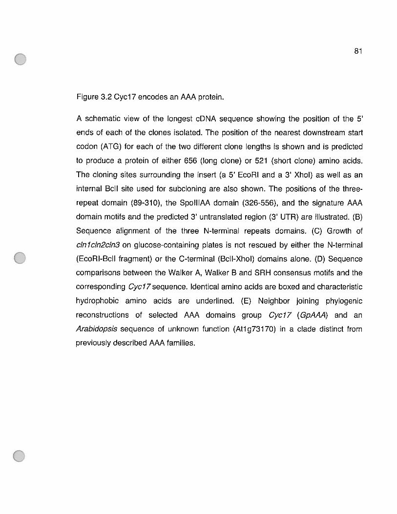
81
Figure 3.2 Cycl7 encodes an AAA protein.
A schematic view 0f the longest cDNA sequence showing the position of the 5’
ends of each 0f the clones isolated. The position of the nearest downstream start
codon (ATG) for each of the two different clone lengths is shown and is predicted
to produce a protein of either 656 (long clone) or 521 (short clone) amino acids.
The cloning sites surrounding the insert (a 5’ EcoRi and a 3’ XhoI) as well as an
internaI Bcll site used for subcloning are also shown. The positions of the three
repeat domain (89-310), the Sp0IIIAA domain (326-556), and the signature AAA
domain motifs and the predicted 3’ untranslated region (3’ UTR) are illustrated. (B)
Sequence alignment 0f the three N-terminal repeats domains. (C) Growth of
cInlcIn2cIn3 on glucose-containing plates is not rescued by either the N-terminal
(EcoRl-BclI fragment) or the C-terminal (Bcll-Xhol) domains alone. (D) Sequence
comparisons between the Walker A, Walker B and SRH consensus motifs and the
corresponding Cycl7sequence. Identical amino acids are boxed and characteristic
hydrophobic amino acids are underlined. (E) Neighbor joining phylogenic
reconstructions 0f selected AAA domains group Cycl7 (GpAAA) and an
Arabidopsis sequence of unknown function (At1g73170) in a clade distinct from
previously described AAA families.
82
The second half of the SpoIIIAA domain contains two conserved motifs
(Walker A and B) that form a ATP/GTP binding site fa P-loop) (Walker, Saraste et
al. 1982), as well as a SRH motif (Second region of homology) (Figure 3.2A-D,
p.81) (Patel and Latterich 1998). These three motifs are found in members of the
AAA (ATPases Associated with different cellular Activities) superfamily whose
members include metalloproteases, components of the 26S proteasome, and
proteins involved in membrane trafficking or organelle biogenesis. Interestingly,
these different functions are reflected in primary sequence homologies and can
thus be recovered using molecular phylogenetic analysis (Frohlich 2001). Using
only a small subset of sequences to represent the known different AAA family
members, as well as the Cycl7, At1g73170 and various Sp0IIIAA-containing
bacterial sequences, we recover the same general functional classes documented
previously (Frohlich 2001) with high bootstrap support (Figure 3.2E, p81). This
phylogeny clusters the Cycl7 sequence, together with the Sp0IIIAA domain
containing proteins, firmly outside the previously recognized AAA groups. This
suggests that both Cyci 7 and the Arabidopsis sequence may constitute members
of a new functional class of AAA proteins within the eukaryotic lineage. Taken
together, our analyses indicate that the Gonyaulax cDNA isolated here is a bone
flUe member of the AAA superfamily and it has therefore been named GpAAA.
33.2 Ihe level of Sici p necessary to inhibit celi growthïs not altered by GpAAA
We have considered three possible mechanisms by which GpAAA could
allow passage through START in yeast (Figure 3.3, p.84). The first mechanism is
suggested by the viability 0f the quadruple mutant clnlcln2cln3sicl (Tyers 1996),
as the lack of the CDK inhibitor Sici p allows activation of the C1b5/Cdc28 complex
83
and thus enables the mutant to overcome the normal requirement for Cmi, Cln2
and C1n3. To test if GpAAA might act to alleviate the inhibition of Cdc28/C1b5 by
Sici p (Figure 3.3 mechanism 1, p.84), either by inhibiting Sici p binding to Cdc28p
or by promoting Siclp degradation, we employed a cInlcIn2 GALY-SICY strain that
overexpresses this CKI when supplied with galactose. Since this strain contains a
CLN3 gene on a plasmid containing the URA3 selectable marker, we used plasmid
p414GPDGpAAA, which contains the compatible TRP selectable marker. GpAAA
from this vector was already shown to be active (Figure 3.1 B, p.77). We then
titrated galactose-induced expression of SIC1 with glucose. The growth 0f the
cInl2 GAL 7-SICY on galactose-containing plates is inhibited at 10w levels of
glucose, as expected (Figure 3.4, p.85). More importantly, expression of GpAAA
from the strong GPD promoter does not change the amount of glucose required to
inhibit the growth of these celis. We conclude that GpAAA does not rescue
cInlcIn2cIn3 by blocking the CKI activity of Sici p.

84
SCFSic1HP—* •:
?M GI S
Figure 3.3 Schematic representation cf possible mechanisms allowing GpAAA to
pass the START checkpoint in the absence of CLN3.
(1) GpAAA might bind and inactivate the CKI Siclp or promote Siclp degradation.
(2) GpAAA might substitute for CLN3 in activating and targeting Cdc2Bp to its
normal substrates. (3) GpAAA might activate CLB5 transcription, either by direct
activation of the CLB5 transcription factor SBF or by inactivation cf the SBF
repressor Whi5.
2
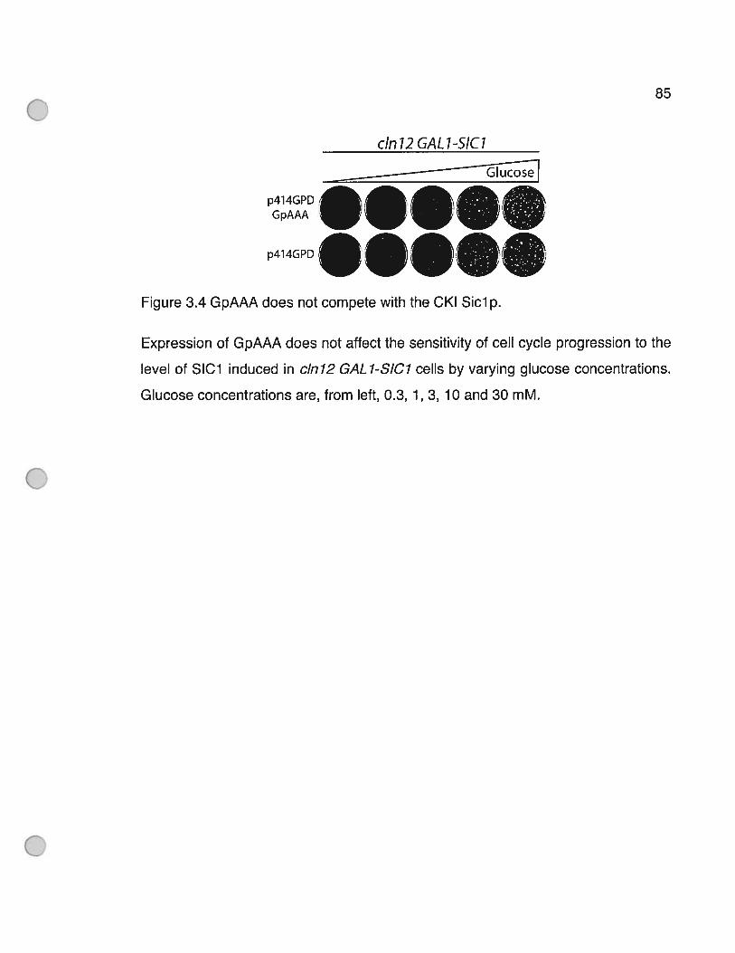
85
cm 12 GAL 1-SIC1
—TEose
p414GPD:
_____
GpAAA
____
p4J4GPD
Figure 3.4 GpAAA does flot compete with the CKI Sici p.
Expression of GpAAA does flot affect the sensitivity of celi cycle progression to the
level of SIC1 induced in cInl2 GAL 1-SIC7 cells by varying glucose concentrations.
Glucose concentrations are, from left, 0.3, 1, 3, 10 and 30 mM.
86
33.3 GpAAA ïnteracts only weakly wïth Cdc28p in a twohybrid assay
To test for a possible interaction between Cdc28p and GpAAA, as
demonstrated for other non-cyclin proteins such as RINGO/SPEEDY (Ferby,
Blazquez et aI. 1999; Lenormand, Dellinger et aI. 1999), we asked if GpAAA was
able to directly bind Cdc28p itself in a two hybrid assay. An interaction would be
required if GpAAA were to activate Cdc28p as if it were a cyclin (Figure 3.3
mechanism 2, p.84). We therefore constructed plasmids producing fusion proteins
between the Activation-Domain of Ga14 and GpAAA (pADGpAAA) and between the
Binding-Domain of Ga14 and Cdc28p (pBDCdc28) for use in the yeast strain PJ69-
4a. This strain carnes the HIS3 and ADE7 selectable markers under a GAL
promoter, allowing growth if there is interaction between the two fusion proteins.
We observe growth in the absence of histidine but not in the absence of adenine
(Figure 3.5A, p.88). Since adenine auxotrophy represents more stringent
conditions than histidine auxotrophy (James, Halladay et aI. 1996), these results
could indicate a weak interaction between Cdc28p and GpAAA. Growth on medium
lacking histidine but not on medium lacking adenine has been shown in a two
hybrid assay between C1b5 and Cdc2Sp (Cross and Jacobson 2000). Thus
mechanism 2 cannot be definitely ruled out, although it is important to stress that
there is no primary sequence similarity between GpAAA and any known cyclin.
Furthermore, the predicted secondary structure has nothing resembling a cyclin
fold (data not shown), in contrast to that proposed to explain the ability of p35 to
activate CDK5 (Lew, Huang et aI. 1994; Tsai, Delalle et al. 1994).
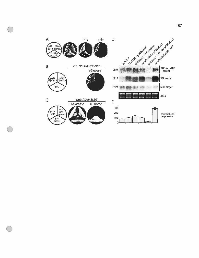
87
A -his
c1GA
p414p414 GPDGPD GpAAA
pYGGpCycl
D
(_ -, ‘?
x-i-
5Ç’ *\&\&
c4 4’ &‘ &ç’
PCLI I5t3roetYHPI .
s’ IMBF targete..— ..- “-
rRNA
300
200 relative CLB5expression
__
10Oflfl
cm 1 cIn2cIn3cIbScIb6
+Glucose
cm 1 cIn2cIn3cIbS+Galactose +Glucose
88
Figure 3.5 GpAAA activates CLB5 transcription.
(A) Yeast two-hybrid assay with pADGpMA and pBDCdc28 shows a weak
interaction, allowing growth on medium lacking histidine but not on medium lacking
adenine. pBDp53 and pADSV4O were used as a positive control, while pBDLaminC
and pADSV4O were used as a negative control. Neither pADGpAAA nor
pBDCdc28p alone grow on —His plates. (B) pYGGpAAA does not allow growth 0f
cinlA cIn2A cIn3A c/b5::ARG4 cIb6::ADE7 pCEN-URA3/GAL 1-CLN1 in restrictive
conditions. The authentic cyclin LpCycl was used as a positive control and the
empty vector as a negative control. (C) pYGGpAAA does flot allow growth of cinlA
cIn2A cIn3A cIb5::ARG4 pCEN-URA3IGAL7-CLN7 in restrictive conditions. (D)
Northern blot analysis of 4 ig of poly(A) enriched RNA of yeast strains grown in
SC-medium (supplemented with galactose instead of Glucose in the third lane)
probed with CLB5, PCL7 or YHPY. rRNA remaining in the samples was used as a
loading control. (E) Phosphorimager quantification of accumulated CLB5 RNA
levels in panel D.

89
3.3.4 GpAAA rescues the cInlcIn2cIn3 phenotype byincreasing CLB5transcript levels
A third possibility to explain phenotypic rescue of cInlcIn2cIn3 mutants by
GpAAA involves activation of CLB5 expression (Figure 3.3 mechanism 3, p.84).
This gene was originally isolated from a screen of S. cerevisiae genes able to
rescue cInlcIn2cIn3 (Epstein and Cross 1992). We therefore tested the ability of
GpAAA to rescue the quintuple cyclin mutant cInlcIn2cIn3cIb5cIb6 (Figure 3.5B,
p.88) and quadruple mutant cInlcIn2cIn3cIb5 (Figure 3.5C, p.88). These strains,
although rescued by overexpression of cyclin LpCycl, could not be rescued by
GpAAA. The inability of GpAAA to rescue the quadruple mutant indicates that
CLB6 alone is insufficient to effect rescue of the cInlcIn2cIn3 phenotype. This is in
agreement with the observation that CLB5 and not CLB6 was recovered from a
screen of S. cerevisiae genes able to overcome the c/nlcln2c/n3 phenotype
(Epstein and Cross 1992).
To confirm that the rescue of cInlcIn2cIn3 involved CLB5 transcription, we
next tested the effect of GpAAA on expression 0f different GuS activated genes.
Northern blot analyses (Figure 3.5D, p.88) showed a stronger expression 0f CLB5
in clnlcln2c/n3 cells overexpressing CLN3, as expected in the presence of
Galactose (de Bruin, McDonald et al. 2004), as well as in LpCycl and in celis
bearing a GpAAA construct. The size of the faint signal observed within the CLB5
probe in the clnlcln2cln3clb5c/b6 sample is indicative 0f hybridization with
ribosomal RNA. Since CLB5 transcription is activated by both SBF and MBF
transcription factors (Bean, Siggia et al. 2005), the expression of genes specifically
activated by the two factors separately were also tested. The expression pattern of
PCL7, a gene specifically transcribed at the GuS transition and activated only by
90
SBF (lyer, Horak et al. 2001) mirrors that of CLB5, while YHP1, another GuS
activated genes that is exclusively an MBF target does flot (Bean, Siggia et al.
2005). We conclude from this that GpAAA is likely to rescue the cInlcIn2cIn3
mutant phenotype by induction of CLB5 expression.
3.4 DiscussionWe report here the isolation of a dinoflagellate AAA encoding gene GpAAA,
which, when expressed in budding yeast, allows passage through the GuS
checkpoint in the absence of GuS phase cyclins. In a functional complementation
screen to rescue growth of the cInlcIn2cIn3 mutant yeast, GpAAA clones were
isolated five times from 6 x 10⁶ clones, although based on their sequence, only two
different clones were actually retrieved. By comparison, a dinoflagellate cyclin was
recovered 131 times during the same screen (Bertomeu and Morse 2004). This
shows that the GpAAA was poorly represented in the library, possibly due to weak
expression in the cells. In agreement with this later idea, Northern blots using the
GpAAA cDNA as a probe were unable to detect any expression of the gene. The
GpAAA thus appears to be a 10w abundance mRNA in our dinoflagellate.
The ability of GpAAA to rescue the cInlcIn2cIn3 mutant phenotype is
remarkable since GpAAA does not share any sequence similarity with any known
cyclins. Indeed, previous studies using libraries prepared from other organisms to
screen similar mutant yeast recovered only cyclins (Koff, Cross et al. 1991; Lahue,
Smith et al. 1991; Leopold and OFarrell 1991; Lew, Dulic et al. 1991; Soni,
Carmichael et al. 1995) with the sole exception of a drosophila Cdc28p homolog
that could apparently exhibit basal levels of activity without Gi cyclins (Leopold and
O’Farrell 1991). The sequence of GpAAA itself yields no due as to its function, as
the only feature that can be identified is a domain found in AAA proteins. The AAA
91
proteins have too large a spectrum of activities to provide any solid basis for
predicting the function of GpAAA (Hanson and Whiteheart 2005); the only feature
common to AAA family members is their ability to use ATP to induce
conformational changes in specific target proteins.
To address the mechanism underlying GpAAA action in yeast, a number cf
different possibilities were evaluated. First, it has been previously shown that Sici p
is an inhibitor cf Cdc28-Clb complexes, and that a cInlcIn2cIn3sicl yeast strain
grows because Siclp degradation is an essential function cf CLN genes
(Schneider, Yang et al. 1996; Tyers 1996). However, although members of AAA
superfamily are involved in protein degradation (Sauer, Bolon et al. 2004; Hanson
and Whiteheart 2005), it is unlikely that GpAAA acts to degrade Sici because the
inhibition cf celI growth during titration cf Siclp expression is unaffected by the
presence of GpAAA (Figure 3.4, p.85).
Another possibility was that GpAAA might directly activate the CDK in a
CLN-independent manner. lndeed, there are now several examples in the literature
where non-cyclin proteins have been observed to activate a CDK. One of these,
RINGO, activates both Cdc2 and CDK2, and plays a key role in allowing meiotic
G2/M progression in Xenopus oocytes, although the mechanism underlying this
activation is still unknown (Ferby, Blazquez et al. 1999). A second, SPEEDY,
appear to act similarly (Lenormand, Dellinger et al. 1999) and may in fact be
related to RINGO. A third, termed p35, has been found to activate Cdk5 in neurons
(Lew, Huang et al. 1994; Tsai, Delalle et al. 1994). While the physiological
importance of this activation is still unclear, the mechanism may result from a
similarity in the three dimensional structure cf the protein to a cyclin-fold, the
domain that binds and activates CDK5, an idea supported by the similarity in the
predicted alpha-helical nature of p35 and cyclins. The observation that GpAAA
92
interacts with Cdc28p in our two-hybrid assay could potentially support a CDK
activation by a non-cyciin protein. However, the predicted secondary structure 0f
GpAAA has no similarity at ail to known cyclins (data flot shown). Furthermore,
whiie activation of CDK kinase activity by a non-cyciin protein is possible, it is
difficuit to imagine how CDK activity could then be directed toward the substrates
whose phosphorylation is prerequisite for entry into S-phase entry. Lastiy, and
most importantiy, any direct activation cf Cdc28p by GpAAA would be expected to
be independent of the presence of C1b5, a prediction in direct contrast to the data
in Figure 3.5C. These considerations Iead us to conciude that a mechanism
involving direct activation of Cdc2Sp by GpAAA is extremeiy unhikeiy.
The iast possibility tested was that GpAAA might activate CLB5 transcription
in a CDK-independent manner. It was recentiy reported that activation of the
transcription factor SBF may involve Cin3-Cdc28 dependent phosphorylation of a
Whi5 repressor subunit, a mechanism conceptuaiiy similar to that cf the
retinobiastoma protein Rb (Costanzo, Nishikawa et aI. 2004; de Bruin, McDonald et
al. 2004). These new findings expiain activation of SBF by C1n3-Cdc28 despite the
iack of any evidence for direct phosphorylation of SBF or direct interaction between
C1n3-Cdc28 and SBF (Wijnen, Landman et al. 2002). We find that GpAAA
expression does indeed augment the level of CLB5 transcript accumulation to
levels above those found in ail the other strains tested. The cInlcIn2cIn3 rescue is
clearly due to CLB5 transcription activation as GpAAA cannot rescue
cInlcIn2cIn3cIb5 mutant and the CLB5 expression has already been shown to
rescue a CLN-deficient yeast. Furthermore, the expression of PCL7, a specific
target of SBF and not MBF, foliows the same pattern as CLB5, whiie the
abundance of a MBF-specific gene, YHP1, is flot significantly greater in yeast
expressing GpAAA. It thus seems likely that ceils expressing GpAAA have higher
93
SBF activity without affecting the activity of MBF. Based on the role of AAA
proteins in protein remodeling, we propose that this activation may be due to
GpAAA-induced remodeling or degradation of Whi5.
The group of AAA proteins to which GpAAA belongs includes bacterial
Sp0IIIAA proteins as wefl as an Arabidopsis gene. The bacterial proteins are
encoded by the first 0f eight genes in the spolliA operon, and mutants block spore
formation at a stage termed engulfment (Jedrzejas and Huang 2003). Mutants
blocked during this stage do flot activate the specific sigma factors required to
allow the continuing changes in gene expression required for completing the celi
differentiation program. Although the function of Sp0IIIAA is currently not known, it
is an intriguing possibility that Sp0IIIAA may help to modulate the changing sigma
factor activities in a manner similar to the prokaryotic transcription activator PspF, a
AAA protein believed to be involved in restructuring a a54-RNA polymerase
promoter complex (Rappas, Schumacher et aI. 2006). Thus, in bacterial systems
there is clear evidence for a role 0f AAA proteins in regulating gene expression.
Does the Arabidopsis thaliana At1g73170 have a similar action on transcription?
Homozygous Arabidopsis mutants have no apparent phenotype and the
At1g73770 cDNA does not rescue the yeast cInlcIn2cIn3 mutant phenotype (data
not shown). However, we note that At7g73 770 has significant similarity only with
the C-terminal AAA domain, and we further note that both N-terminal and C
terminal moieties of GpAAA are necessary for rescue (Figure 3.2C, p.81). Thus,
even if it were to act in regulating transcription, the At7g73770 may have an
alternate target flot detected in our functional complementation assay. It is tempting
to speculate that GpAAA may represent a new class of AAA functionalities involved
in regulating transcription in both prokaryotes and eukaryotes. It is important to
note in this regard that the yeast AAA protein Saplp associates with the

94
transcriptional repressor Sinlp (Liberzon, Shpungin et al. 1996), thus potentially
acting to alter gene expression.
To date, the only characterized ceil cycle regulator from a dinoflagellate s a
cyclin most similar to mitotic cyclins (Bertomeu and Morse 2004). However, a
search of the public EST databases revealed that there is at least one sequence
with homology to cyclin dependent kinases. Thus, it seems likely that
dinoflagellates will use ceil cycle regulators similar to those cf other eukaryotes.
This being the case, it is tempting to speculate that GpAAA might play a role in
regulation of the dinoflagellate celI cycle that is similar that reported here in yeast.
Unambiguously proving this contention will require tools not currently available,
however. For example, there are no other known regulators of the dinoflagellate
celi cycle, precluding tests of interactions with GpAAA. More importantly, the lack
of transformation systems for dinoflagellates precludes analysis of cell behavior
after introduction of a gene in sense or antisense orientations. Clearly, given these
limitations, the application of yeast genetics to study of the dinoflagellate ceil cycle
represents a particularly valuable experimental approach.
3.5 MethodsConstruction and screening of a yeast expression library: The construction
of a dinoflagellate cDNA library in the yeast expression vector pYG, containing the
2p. origin cf replication and the glyceraldehyde-3-phosphate dehydrogenase (GPD)
promoter, has been previously described, as has the c/nlcIn2cIn3 mutant
functional complementation protocol which yielded an authentic dinoflagellate
cyclin, LpCycl (Bertomeu and Morse 2004). The Cycl7/GpAAA sequence
described here has been deposited in GenBank with the accession number
DQ51 5200.
95
Yeast sfrains, cultures and transformations : Mutant yeast strains used here
include (j) strain SBY175 (obtained from Dr. S. Reed, Scripps Research Institute)
MATa his2 leu2 trp 1 ura3 db 1:: URA3 c/b2: :LEU2 c/b3:: TAPi clb4::HIS2 GAL
CLBJ-LEU2 (called clbYcIb2clb3clb4 in this study) (Richardson, Lew et aI. 1992),
(ii) strain BF305-15d (obtained from Dr. B. Futcher, CoId Spring Harbor
Laboratories) MATa his3 /eu2 trpi ura3 cIni::HIS3 cln2::TAP7 cln3::URA3-GAL-
CLN3 (called clnic/n2c/n3 in this study) (Xiong, Connolly et al. 1991), (iii) strain
1227-3A (obtained from Dr. F. Cross, Rockefeller University) MATa ciniA cln2A
cln3A pCEN-URA3/GAL-CLN3 (called ciniA cln2A cln3A in this study) (iv) strain
1851-1 (Dr. F. Cross) MATa cInlA cln2A cln3A GAL7-SIC1 pCEN-URA3/CLN3
(called clnlcln2GALi-SIC1 in this study) (Cross and Levine 1998), (y) strain 1581-
2C (Dr. F. Cross) MATa ciniA cln2A cln3A clb5::ARG4 clb6::ADE1 pCEN
URA3/GALY-CLN1 (called cInicln2cIn3clb5clb6 in this study) (Cross and Levine
1998), (vi) strain 1581-6A (Dr. F. Cross) MATa cinlA cln2A cIn3A cIb5::ARG4
pCEN-URA3/GALY-CLNY (called clnicln2cln3clb5 in this study); strains from the
Cross laboratory are based on the A364A background (Hartwell 1967). Strain
SEY621O (from Dr. P. Beihumeur, Université de Montréal), used as wild-type for
this study, MATa his3A200 Ieu3-3,772 lys2-80i trpiA9Oi suc2A9 ura3-52, (used
as wild-type for this study). Yeast were grown in Synthetic Complete medium (SC)
lacking the appropriate auxotrophic nutrients with different amounts of glucose
and/or galactose. Yeasts were transformed using a lithium acetate procedure
(Gietz and Woods 2002).
Phylogenetic analysis : AAA family sequences were obtained from GenBank
using a previously described tree (Frohlich 2001) to define the major families, and
aligned using CLUSTALW. Phylogenetic reconstruction was performed using
96
Neighbor joining with absolute number of differences using MacVector (Accelrys).
Bootstrap values for 10000 repetitions are reported for values of more than 70.
Plasmid construction . To produce pYGCycl 7 N-term, pYGCycl 7 (prepared
from New England Biolabs Dam negative E colistrain FR2925) was digested with
Bcll and Xhol to remove the second half of Cycl7then recircularized by ligation
with a dimerized pair of oligonucleotides (5’-GATCTGAHGGGGATCCATT-3’ and
5’-TCGAAATGGATCCCCAATCA-3’) bearing a new stop codon. To produce
pYGCycl7 C-term, pYGCycl7 was digested with EcoRi and Bcll to remove the
first half of Cycl7 then recircularized by ligation with a dimerized pair of
oligonucleotides (5’-AATTGCCATGGGACTGGATGAGTT -3’ and 5’-
GATCAACTCATCCAGTCCCATGGC -3’), bearing a new start codon and the
nucleotides necessary to encode the first five amino acids of the SpollIAA domain
that were cut with Bcll. For pADGpAAA construction, pYGCycl7 was digested with
EcoRi and Xhol to liberate the full length cDNA which was directionally cloned in
frame in pAD also digested with EcoRi and Xhol. For construction of pBDCdc28
CDC28 was PCR-amplified with Pwo polymerase (5’-
CATGAATTCATGAGCGGTGAATTAGCAAAUAC-3’ and 5’-
TAACTGCAGTTATGAHCHGGAAGTAGGGGTG-3’) from a plasmid bearing the
complete cDNA and after digestion with EcoRi and Pstl, Iigated into EcoRi and
Pstl digested pBDGaI4. The construct p414GPDGpAAA (also called
p414GPDCyc17) was prepared by directional cloning of an EcoRi and Xhol
digestion fragment of pYGCycl7 into p414GPD (Mumberg, Muller et al. 1995), a
centromeric vector containing a GPD promoter and a TAPi auxotrophic marker. Ail
constructs were confirmed by sequencing.
Two-HybridAssays :The yeast strain PJ69-4a (James, Halladay et aI. 1996)
specificaliy designed for two-hybrid assays was used for the Gal4-based protein
97
interaction experiments. It contains the markers HIS3 and ADE1 under a GAL
promoter. Plasmids pBD-Ga14 and pAD-Ga14 (cailed pBD and pAD in this study)
and pBDLaminC, pBDp53 and pADSV4O are constructs from the HybriZAP two
hybrid system (Stratagene).
Northern Blots : Yeast cultures (200 ml) were grown to an O.D. 600 of 1.0,
and the ceils pelleted and kept at 8O0C until use. Oeils were lysed in TRizol
reagent (Invitrogen) using zirconium beads and a BeaU Beater and treated
according to the manufacturer’s recommendations. Poly(A) RNA was enriched
using oligo-dT column chromatography, and either 4 ig cf poly(A)-enriched RNA or
5 tg total RNA was run on formamide/formaldehyde agarose gels and transferred
to positively charged nylon. The blots were hybridized against a radiolabeled probe
made against a DNA fragment obtained by POR on SEY621O genomic DNA using
the yeast Research Genetics flanking primers (YPR1200-FOR and YPR1200-REV
for CLB5, YALO400-FOR and YALO400-REV for CLN3, YNL2S9W-FOR and
YNL289W-REV for PCL1 and YDR451 O-FOR and YDR451O-REV for YHPJ). The
membranes were exposed for 1 to 5 days on a Phosphorimager screen and
scanned on a Typhoon 9200 (GE Healthcare).
3.6 AcknowledgementsWe thank S. Reed, B. Futcher, P. Beihumeur and specially F. Oross for
yeast strains and helpful advice. We gratefully acknowledge the financial support of
the National Science and Engineering Research Council cf Canada.
98
4 Publication #3 : A dinoflagellate CDK5-likecyclin-dependent kinase
Thierry Bertomeu, Jean Rivoal and David Morse
Accepté chez Biology of the Ceil, sous presse en juillet 2007
Toutes les procédures expérimentales furent effectuées par moi et cet article a été
écrit conjointement par les trois auteurs.

99
4.1 AbstractBackground information. Mitosis during the dinoflagellate celi cycle is
unusual in that the nuclear envelope remains intact and segregation of the
permanently condensed chromosomes uses a cytoplasmic mitotic spindle. To
examine regulation of the dinoflagellate celI cycle in the context of these unusual
nuclear features, it is necessary to isolate and characterize known cell cycle
regulators such as CDK (cyclin-dependent kinase).
Resuits. We report here the characterization of a CDK from the
dinoflagellate Lingulodinium polyedrum. This CDK reacts with an anti-PSTAIRE
antibody and was identified by protein microsequencing after partial purification.
The protein microsequence shows homology toward the Pho85/CDK5 clade of
CDK5. Neither the amount nor the phosphorylation state changed over the course
of the ceil cycle, in agreement with resuits reported for CDK5 family members in
other systems.
Conclusions. We conclude we likely isolated a CDK5-like protein. The data
reported here support identification of this protein as a CDK5 homolog, and
suggest that dinoflagellates may contain several CDK families.
4.2 Introduction
The celI cycle of dinoflagellates 5 of interest from both health and basic
science perspectives. First, many species of dinoflagellates are responsible for
causing harmful algal blooms (Wong and Kwok 2005) and a detailed knowledge of
the celI cycle of dinoflagellates could lead to better understanding, monitoring
and/or preventing this phenomenon. Second, aIl dinoflagellates show a number of
100
very unusual celi cycle-related characteristics: they are devoid of histones (Herzog
and Soyer 1981), their chromosomes are permanentiy condensed throughout the
celi cycle, their nuclear envelope remains intact during mitosis, and chromosome
segregation uses an extra-nuclear spindle that passes through nuclear
invaginations and makes indirect contact to the chromosomes attached to the
inside of the nuclear membrane (Bhaud, Guillebault et al. 2000). Finally, since
mitosis in dinoflagellates is under circadian control, identifying the cell cycle
regulators is a necessary prerequisite to uncovering the mechanism by which the
circadian dock controls this rhythm.
In eukaryotic cells, transition between the different phases of the ceil cycle is
tightly controlled by the activity of different CDKs (Sanchez and Dynlacht 2005).
The yeast Saccharomyces cerevisiae has 5 CDK5, of which only two (Cdc28 and
Pho85), are involved in celI cycle control (Toh-e and Nishizawa 2001). Cdc28
associates with different celi cycle phase-specific cyclins and is required for ail the
phase transitions throughout the ceil cycle (Mendenhall and Hodge 1998). Pho85
also appears to play a role in the GuS transition (Espinoza, Ogas et al. 1994), and
in particular, has been found to be involved in the degradation of the CDK inhibitor
(CKI) Sici which normally acts to inhibit entry into S phase (Wysocki, Javaheri et
al. 2006). In contrast, mammalian cells use many different CDKs at different steps
of the cell cycle, such as CDK4 and CDK6 for passage through Gi, CDK2 for entry
into and progression through S phase, and CDK1 for the G2/M transition (Obaya
and Sedivy 2002). CDK1 is a Cdc28 homologue, while CDK5 is homologous to
Pho85 (Liu and Kipreos 2000). CDK5 is unrelated to the control of the ceil cycle in
mammalian celis and appears instead to be involved in neuronal development
(Smith and Tsai 2002) and other diverse functions (Rosaies and Lee 2006).

101
Most members of the CDK group of serine/threonine protein kinases are
themselves regulated post-translationally by phosphorylation. An inhibitory
phosphorylation site is iocated within the P-loop at the N-terminal region cf CDKs,
while an activating phosphorylation site is Iocated in the T-Loop which otherwise
biocks the active site cleft. CDK5 in mammalian oeils is a notable exception to this
type cf regulation, as it does not require phosphorylation in the T-loop for activation
(Qi, Huang et al. 1995; Dhavan and Tsai 2001), and phosphorylation of Tyr’5 in the
P-loop increases rather than decreases activity cf the enzyme (Zukerberg, Patrick
et ai. 2000). This behavior is aiso observed for Pho85 in yeast, which is also
activated by phosphorylation in the P-loop and does flot require T-loop
phosphorylation for activity (Toh-e and Nishizawa 2001).
The activity cf CDKs are also reguiated by protein-protein interactions.
Cyclins, a group of proteins originally isolated because their abundance varied
throughout the oeil cycle, are absoiutely required for CDK aotivity. They bind CDKs
through interactions with their PSTAIRE motif (Glu-Gly-VaI-Pro-Ser-Thr-Ala-Ile-Arg
Giu-Iie-Ser-Leu-Leu-Lys-Giu motif) on the CDK. In addition, a group of proteins
called CDK inhibitors (CKIs) can bind CDKs, either bound with a cyclin or flot, and
block their activity.
The celi cycle of dinoflagellates has been the subject cf severai previcus
studies using as a model the rapidly dividing heterotroph Crypthecodinium cohnll.
This species contains a 34 kDa protein reacting to an anti-PSTAIRE antibody and
both this signal and histone Hi kinase activity can be enriched using pi3
immunoprecipitation (Rodriguez, Cho et al. 1993). A modest increase (less then 2-
foid) in histone Hi kinase activity cf proteins immunoprecipitated with p131 was
found in M phase compared with interphase oeil extracts (Bhaud, Barbier et al.
1994). Interestingly, an antibody raised against a Schizosaccharomyces pombe
102
cyclin (Cdcl 3) was found to cross react with a 56 kDa band in C. cohnii extracts
and this same antibody was able to immunoprecipitate a higher histone Hi kinase
activity (Barbier, Albert et aI. 1 995). Using an anti-(cyclin-box) antibody, four cross
reacting bands were found (Leveson, Wong et aI. 1997), of which two (at 50 and 65
kDa) accumulated in G2 and disappeared after M phase. OnIy the 50 kDa band
could be immunoprecipitated using p131 binding.
In Gambierdiscus toxicus, an anti-PSTAIRE antibody was used to
demonstrate the presence of a 34 kDa reacting band whose quantity did not vary
over time (Van Dolah, Leighfield et al. 1995). This same antibody used for
immunoprecipitation was able to enrich histone Hi kinase activity, and kinase
activity was at its maximum when extracts from ceils in M phase were used.
An anti-PSTAIRE reactive protein at around 32 kDa was also found in the
dinoflagellate Lingulodinium polyedrum, although cloning and analysis of the
sequence demonstrated clearly that it was not a CDK (Salois and Morse 1996).
More recently, a cyclin called GpCycl has been cloned by functional
complementation from this same species (Bertomeu and Morse 2004). Since
Gonyaulax polyedra has been renamed as Lingulodinium polyedrum, we shaH now
further refer to this cyclin as LpCycl. LpCycl rescues a mutant of S. cerevisiae
deficient in the GuS cyclins Cml, C1n2 and C1n3. The presence of a destruction-
box in its N-terminal sequence together with the more then 1 0-fold decrease in
amounts from G2-enriched to G1-enriched ceils, indicated this clone Iikely encodes
a mitotic cyclin. This protein was the first (and so far the only) celI cycle regulating
protein cloned and characterized from a dinoflagellate.
Interestingly, an EST (expressed sequence tag) project of Karenia brevis
(with 7001 independent ESTs) was found to contain the first potential dinoflagellate
103
CDK sequence (Accession number C0060059) (Lidie, Ryan et al. 2005). This
sequence is clearly a Serinerrhreonine protein kinase with similarity (E value: 2e10)
to the Drosophila melanogaster CDK1 -homologue. However, this sequence lacks
the first 100 amino acids normally containing the characteristic PSTAIRE motif and
its potential identity as a CDK is thus unconfirmed.
We report here the first biochemical isolation of a dinoflagellate CDK and its
identification by protein microsequencing. The microsequence data clearly shows
the conserved PSTAIRE motif and thus confirms the sequence as a CDK. The
sequence has, as expected, high similarity to a Toxoplasma gondli CDK based on
the phylogenetic relationships of dinoflagellates and apicomplexans. The sequence
also appears most similar to the CDK5/Pho85 group of CDK5 in phylogenetic
analyses. Furthermore, the abundance and phosphorylation state of this CDK is
constant over the cell cycle, similar to what is found with CDK5 from other species.
As this CDK does flot seem to associate with LpCycl, we conclude that
dinoflagellates are likely to express a number of different CDKs, as found for other
organisms.
4.3 ResultsThe L. polyedrum strain used here is different from that whose celI cycle
events were studied previously (Homma and Hastings 1989). We therefore used
flow cytometry to characterize the ceil cycle in our strain. For the present study,
cells were maintained in exponential phase growth by 2-fold dilution of the cells
every 3 days. Under these conditions, ceils have a shorter doubling time than
previously reported (Bertomeu and Morse 2004), with approx. 30% of the celis
entering S phase during the early night phase (Figure 4.1A-B, p.105). In agreement
with previous observations, S phase was found to occur within a 4 hour period
104
around LD (light/dark) 18 (Homma and Hastings 1989) and M phase occurs around
LD1, 1 hour after the start of the light phase (Sweeney and Hastings 1958). M
phase can be observed either as the disappearance of celis in G2/M (Figure 4.1B,
p.105) or as the appearance cf dividing cells, a population with the same FL2-A
(fluorescence amplitude) as G2 oeils but a FL2-W (fluorescence width)
intermediate between G2 cells and Gi doublets (Figure 4.1C, p.105).
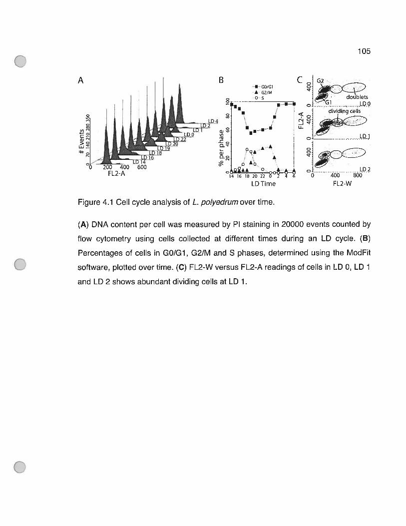
105
A
U DO
u. <dMdIflgceIIS
?LD2O
0 200 400 600tI A o
0 400 $00
LDTime FL2-W
Figure 4.1 OeIl cycle analysis of L. po/yedrum over time.
(A) DNA content per celI was measured by PI staining in 20000 events counted by
flow cytometry using ceils collected at different times during an LD cycle. (B)
Percentages of cells in G0/G1, 021M and S phases, determined using the ModFit
software, plotted over time. (C) FL2-W versus FL2-A readings of celis in LD 0, LD 1
and LD 2 shows abundant dividing cells at LD 1.
106
Interestingiy, although most of the G2 phase celis divided at LD 1, a small
percentage of G2 phase ceils can be found at ail time points measured between
LD 1 and LD 14. Indeed, depending on the particular experiment, up to 8% cf G2
phase celis could be found during the day phase (data flot shown). This
observation reinforces the idea that M phase is clock-controlled and does not
simply occur at a fixed time following S phase.
As an initial approach to isolation cf a dinoflagellate CDK, we thought to use
binding to the dinoflagellate cyclin LpCycl (Bertomeu and Morse 2004) as a
molecular marker that could be monitored following fractionation by column
chromatography. The presence of LpCycl was determined by an anti-LpCycl
antibody reaction. A crude protein extract from cells at LD 0.5 was first subjected to
ammonium sulfate precipitation, with proteins precipitating between 17 to 40%
containing ail the polypeptides immunoreacting with the anti-LpCycl serum (data
not shown). The precipitate was resuspended, desalted and subjected to anion
exchange chromatography. A single band at 68 kDa was recognized by the anti
LpCycl antibody and the LpCycl protein routinely eluted within a major peak on 12
fractions (30 to 41) (Figure 4.2A, p.108). A non-reproducible and minor LpCycl
reactive species is occasionally noted at a position prior (25 to 28) to the main
peak. Interestingly, challenging the same fractions with an anti-PSTAIRE antibody
identified a single peak of immunoreactive protein whose apparent molecular
weight on SDS-PAGE was 34 kDa, although this immunoreactive protein was only
found associated with the first several fractions of the major cyclin-containing peak.
The majority of anti-LpCycl reactive fractions (30 to 41) were then pooled and
fractionated by gel-filtration chromatography. The cyclin eluted within four fractions
(22 to 25) (Figure 4.2B, p.108) corresponding to an apparent molecular size of 160
107
± 20 kDa (data flot shown). However, the anti-PSTAIRE signai was no longer
associated with the cyclin, as the immunoreactive protein eluted at a position
corresponding to an apparent molecular size of 34 kDa.

108
A Anion-ExchangeFraction: 24 25 26 27 28 29 30 31 32 33 34 35 36 37 38 39 40
a-L pCyc i .r — 46$ kDa
a-PSTAIRE 134kDa
B Gel-Filtration C AffinityFraction: 21 22 23 24 252627 2829
U Ba-LpCycl H——— — —
468 kDaa-LpCyc1 168
a-PSTAIRE .H134kDa
Figure 4.2 Chromatography of LpCycl and an anti-PSTAIRE reactive protein.
(A) After fractionation by anion-exchange chromatography, LpCycl elutes in a
single broad peak (fractions 30 to 41) as detected by Western blotting, whereas a
protein reacting to an anti-PSTAIRE antibody co-elutes in the first five of these
fractions. (B) The gel-filtration chromatography of the partially purified LpCycl
shows separation of LpCycl and the anti-PSTAIRE reacting proteins. (C) LpCycl,
partially purified by gel-filtration does not bind to a p131 affinity chromatography
resin. U, unbound; B, bound.
109
These results suggested that our initial hypothesis of a CDK-LpCycl
interaction might have been incorrect. To verify if our experimental conditions had
disrupted the CDK-cyclin binding, we tested for LpCycl binding to p131-
Sepharose beads. Cyclin binding to this affinity column is thought to require CDK
cyclin interaction (Vogel L. 2002), but LpCycl failed to bind this resin as judged by
Western blot analysis (Figure 4.20, p.108). We also tested for co-elution of anti
cyclin and anti-PSTAIRE immunoreactivity on gel filtration on Sepharose CL4-B
using the crude extract directly, to ensure that the purification procedure itself did
not decrease association of the two proteins. The two were again found in different
fractions (data flot shown), consistent with a lack of interaction between LpCycl
and the anti-PSTAIRE reactive protein.
To test if a CDK flot identifiable by anti-PSTAIRE immunoreactivity was
present in the extracts, p13 chromatography was performed on a crude protein
extract (Figure 4.3, p.111). The identification of aIl protein bands visible by
Coomassie blue staining was then attempted by MS sequencing. Only 5 of the
eleven bands corresponding to Lingulodinium proteins could be identified by
significant sequence identity. Notably, neither cyclin (around 56 kDa) nor cdc2-like
(around 34 kDa) sequences were recovered in this analysis suggesting that
appreciable levels of CDK did flot bind to p13’ under these conditions.
At this point in our analysis, we reasoned that it might be possible to identify
the anti-PSTAIRE reactive protein enriched through the two columns in Figure 4.2
(p.108) by MS sequencing. lndeed, a Coomassie stained spot corresponding to the
cyclin, as judged by an anti-LpCycl Western done in parallel, was readily detected
(Figure 4.4A, p.1 12). Since a Coomassie stained spot was observed at the position

110
of the anti-PSTAIRE signal after two-dimensional SDS/PAGE (Figure 4.4B, p.112),
this protein was excised from the gel and microsequenced.

111
IL __—(l4OkDa)
101 kDa(120 kDa)
97kDa Luciferase(110kDa 51%)
%.,H5P90 (90 kDa 12%)
‘“‘LBP(75kDa,83%)54kDa
‘Rubisco (55 kDa, 25%)
Actin (45 kDa, 52%)
38 kDa GST-p131 (39 kDa)
——————(33 kDa)
29 kDa q—(28 kDa)
_—(23kDa)
(21 kDa)
Figure 4.3 Microsequence analysis 0f proteins bound to p131-Sepharose beads.
A ctude protein extract was bound to p132l and the specific eluate resolved by
SDS/PAGE. Shown are the 1 1 Lingulodinium bands that were excised and
microsequenced as weII as the GST-p13, which was also eluted from the beads.
Identity of the protein is written when MS sequencing yielded sufficient homology to
known proteins. The molecular weight cf the bands and the percentage cf
sequence coverage by MS are shown in parentheses.
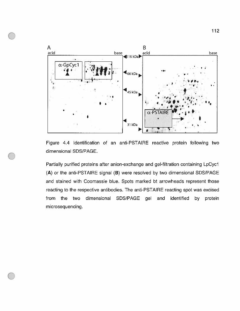
112
A BaclU base aclU base
4116 kDa
466kDa
45 kDa
3lkDa
Figure 4.4 Identification 0f an anti-PSTAIRE reactive protein following two
dimensional SDS/PAGE.
Partially purified proteins after anion-exchange and gel-filtration containing LpCycl
(A) or the anti-PSTAIRE signal (B) were resolved by two dimensional SDS/PAGE
and stained with Coomassie blue. Spots marked bt arrowheads represent those
reacting to the respective antibodies. The anti-PSTAIRE reacting spot was excised
from the two dimensional SDS/PAGE gel and identified by protein
m icroseq u e nci ng.
ŒGpCyc1
I
4I

113
The peptide sequences obtained were very similar to a variety cf different
CDKs, with the highest similarity (BLASTP E value: obtained with a
Toxoplasma gondil cdc2-like CDK (see Supplementary Figure Si at
http://www.biolcell.org/boc/O99/bocO99xxxxadd.htm or Supplementary Figure 4.8,
p.128 cf this thesis). This was encouraging, as the apicomplexans (te which T
gondil belong) are the closest relatives te dinoflagellates in phylogenetic analyses.
Curiously, however, when the protein sequence was compared with the thoroughly
sequenced genomes cf Caenorhabditis elegans, Danlo rerlo, D. melanogaster, S.
cerevisiae and Schizosaccharomyces pombe, a higher degree cf homology was
reproducibly found with the CDK5/Pho85 rather than the CDK1 family cf CDK5.
This relationship is best illustrated by a phylogenic analysis cf selected CDK1 and
CDK5 sequences. The inclusion cf our Lingulodinium sequence within a well
supported CDK5/Pho85 clade (Figure 4.5B, p.ii4) suggested that our protein
might correspond to a CDK5-like. Unfortunately, exhaustive attempts te clone a
DNA fragment cf LpCDK5-like using degenerate PCR primers on genomic and
complementary DNA, or using radio-labeled degenerate oligonucleotides te screen
a cDNA library were unsuccessful, thus precluding a more detailed analysis cf the
homclcgy. However, it is important te note that the peptide sequence reported here
covers around 35% cf the expected amine acid sequence, and includes the
characteristic PSTAIRE motif that unambiguously identifies the protein as a CDK.
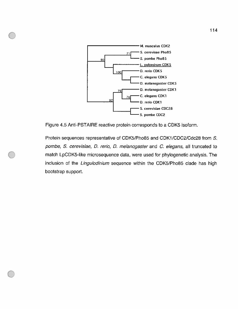
114
M. musculus CDKZ
S. cerevisae Pho8S
S. pombe Pho85
L. polyedrum CDK5
D. rerio CDK5
C. elegans CDK5
D. melanogaster CDK5
D. melanogaster COK1
C. elegans CDK1
D. reno CDK1
S. cerevisiae CDC28
S. pombe CDC2
Figure 4.5 Anti-PSTAIRE reactive protein corresponds to a CDK5 isoform.
Protein sequences representative of CDK5/Pho85 and CDKY/CDC2/Cdc28 from S.
pombe, S. cerevisiae, D. rerlo, D. melanogaster and C. elegans, ail truncated to
match LpCDK5-like microsequence data, were used for phylogenetic analysis. The
inclusion of the Lingulodinium sequence within the CDK5/Pho85 clade has high
bootstrap support.

115
The reaction 0f the anti-PSTAIRE with a protein identified as a bona fide
CDK by microsequence suggested that the antibody could be used to characterize
the behavior cf the enzyme. We first tested LpCDK5-Iike expression over a daily
period in Lingulodinium celis grown under an LD cycle, where mitosis is clearly
rhythmic. No major difference in the expression of LpCDK5-like was detected in
unsynchronized exponentially growing cells (Figure 4.6, p.116). Since CDKs can
be post-translationally modified by phosphorylation events, the isoelectric point of
LpCDK5-like through a celI cycle was also determined. However, no reproducible
changes in CDK phosphorylation could be observed. At low exposure times (Figure
4.7A, p.117), antibody reaction with the two dimensional SDS/PAGE gave rise to a
single specific band. Longer exposures of the films revealed several other spots
(Figure 4.7B, p.117) at molecular masses compatible with CDKs, mostly at LD O
and LD 12. However, the mobility of ail proteins detected in the over exposed films
does not change after incubation with CIP (calf intestinal phosphatase)
phosphatase, indicating they do not correspond to differentially phosphorylated
isoforms of LpCDK5-like (Figure 4.70, p.117). We had anticipated that an isoform
representing a major part of LpCDK5 in 30% of the cells entering mitosis at LD O
would have been readily detected with this technique if this kinase was differentially
phosphorylated at mitosis. This observation is in agreement with our assignment of
the Lingulodinium protein to the CDK5 family, which, unlike its CDK1 relative, does
not undergo differential phosphorylation during different phases cf the celi cycle.

116
LDIime4 6 8 10121416182022 0 2 4 6
Œ-PSTAIRE
a-Rubiscoîj_ p—
Ponceau rsta in in g
Figure 4.6 No variation upon the expression of LpCDK5-like is detected over time.
Protein samples of Lingulodinium collected at different LD times were resolved by
SDS/PAGE and the Western blot was probed with either anti-PSTAIRE or anti
RuBisCO antibodies. Arrow heads respectively represent either the 34 kDa or 55
kDa bands for the upper and middle panels respectively. The PVDF membrane
stained with Ponceau red is shown in the Iower panel.
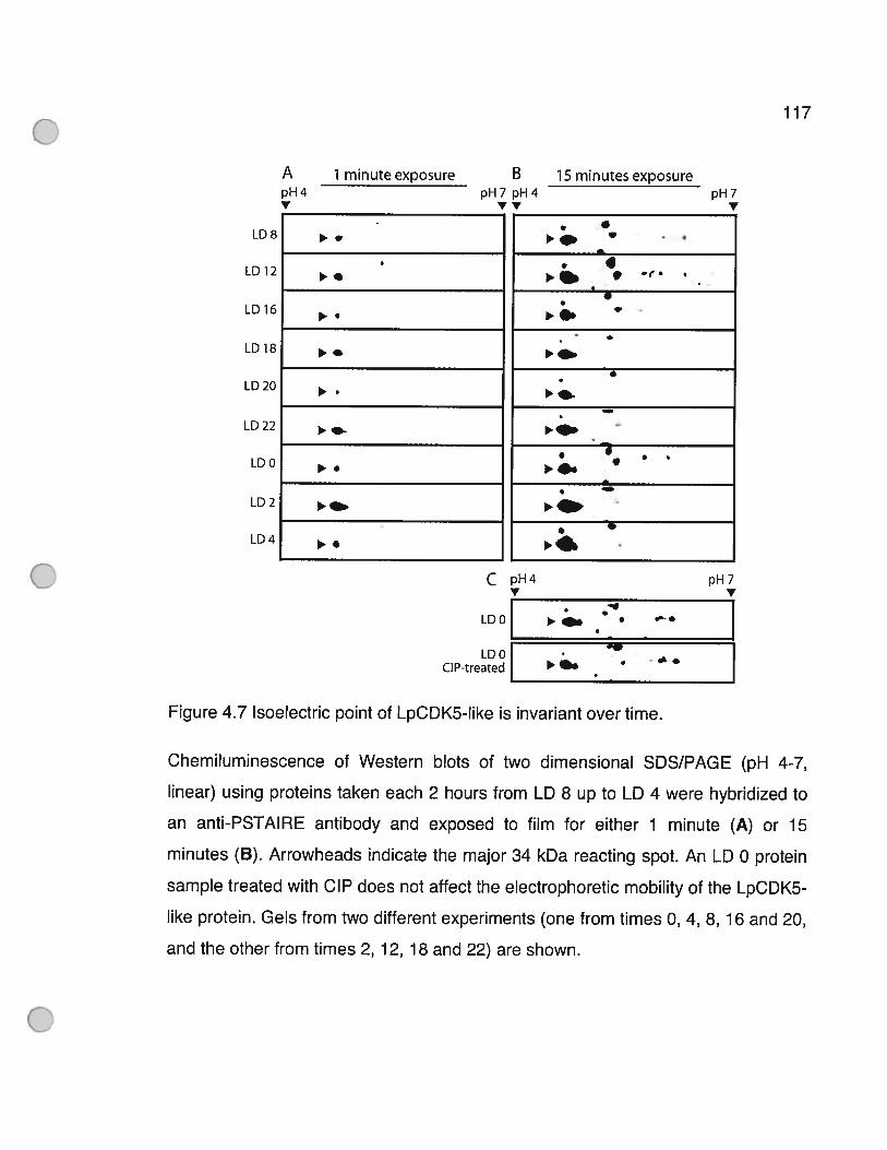
- e
a’..., e I
. .. , ...
.
.
. o
..
.
.
.i...
e
I
.p.. p..
Figure 4.7 Isoelectric point of LpCDK5-like is invariant over time.
Chemiluminescence of Western blots of two dimensional SDS/PAGE (pH 4-7,
linear) using proteins taken each 2 hours from LD 8 up to LD 4 were hybridized to
an anti-PSTAIRE antibody and exposed to film for either 1 minute (A) or 15
minutes (B). Arrowheads indicate the major 34 kDa reacting spot. An LD O protein
sample treated with CIP does not affect the electrophoretic mobility of the LpCDK5-
like protein. Gels from two different experiments (one from times 0, 4, 8, 16 and 20,
and the other from times 2, 12, 18 and 22) are shown.
ApH4V
1 minute exposure
117
BpH 7 pH 4
WV
15 minutes exposurepH 7
V
LD 8
LD 12
LD 16
LD 18
LD 20
LD 22
LD O
LD 2
LD 4
C
LD O
LDOCIP-treated
pH4 pH7V
e• . e..
OW
p... •
118
4.4 DiscussionIt has long been known that mitosis of L. polyedrum grown in a laboratory
setting is under circadian control. Under 12 h light/12 h dark (LD::12:12) conditions,
mitosis is restricted to 1 h after lights are turned on (LD 1). Since the generation
time of a single celI is typically greater then 24 h (Sweeney and Hastings 1958), the
cell cycle is said to be “gated” rather than controlled directly by the circadian dock.
AIl celis are thus given an opportunity to enter M phase each day, but only the cells
in an appropriate position in the ce!! cycle will do so. Flow cytometric
measurements of exponentially growing Lingulodinium confirm this, as although
most ceHs in G2 phase undergo mitosis at this time (Fig. 10), some G2 phase cells
do not divide at LD 1 and can be found later during the day phase. Interestingly,
these observations argue against the mode! of Homma and Hastings (Homma and
Hastings 1989), in which M phase was proposed to follow at a fixed time interval
from a circadian controlled S phase. G2 phase cells du ring day phase suggest they
were not ready to undergo mitosis at LD 1 and were thus obligated to wait for the
subsequent mitotic window.
With respect to S phase, our data showing onset of S phase approx. 9 h
prior to mitosis and lasting for approx, 4 h confirms previous observations (Homma
and Hastings 1989). Additional f!ow cytometry measurements of celis grown in
different lighting conditions wi!I be required to assess definitively if there is a
circadian control over both S and M phases.
To date, only one study has identified a dinoflageHate cDNA as a CDK
(Lidie, Ryan et al. 2005) and this identification is based on an incomplete EST

119
sequence. Curiously, if this sequence were completed using the N-terminal region
of the Drosophila CDK1 sequence, the predicted protein would have a molecular
mass of 43 kDa, much larger than the 34 kDa expected for CDKs. Moreover, this
kinase lacks the RD sequence in subdomain VIb present in ail CDK5 (Ferrari
2006). Thus it seems unlikely that this sequence corresponds to a true CDK. There
are, however, three other potential CDK sequences that can be found by screening
the dinoflagellate EST databanks. One, in Karenia bre vis (00061088), contains a
PLTAIRE motif with some similarity with CDK9. Two others, one in Oxyrrhis marina
(EG741876) and the other in Karlodinium micrum (KME00004803), contain a
PSTAIRE motif with some similarity with CDK1. These sequences thus support our
contention that dinoflagellates have many different CDKs as do other eukaryotes.
We report here protein microsequence data that confirms the identity of an
anti-PSTAIRE reactive protein as a CDK from a dinoflagellate. Our results do not
support an interaction of this CDK with the cyclin LpCycl previously reported from
this organism. LpCycl and LpCDK5-like do flot co-elute during gel-filtration, either
before or after partial purification on anion-exchange chromatography (Figure 4.2B,
p.108 and data not shown). The apparent molecular mass of the cyclin following
gel-filtration (around 160 kDa) suggests that LpCycl may be part of a protein
complex, possibly encompassing a CDK interacting with LpCycl, as the cyclin
monomer is expected to be 68 kDa. However, no other protein in the mixture reacts
to the anti-PSTAIRE antibody and LpCycl does flot bind to p13s affinity resin
(Figure 4.20, p.108).
The protein identified here by microsequence data appears most similar to
the Pho85/CDK5 family members. This conclusion is supported by phylogenetic
analysis with the sequence, albeit incomplete, that is currently available, as
LpCDK5-like is found in a CDK5/Pho85 clade with high bootstrap support.

120
Furthermore, the invariance cf phosphorylation state at important ceil cycle
transitions times is in agreement with a CDK flot implicated in ceIl cycle control,
such as CDK5, whereas CDK1 is the recognized key regulator of entry into M
phase. Since CDK1 and CDK5 types of cyclins are very closely related (Manning,
Whyte et al. 2002), a definite classification of dinoflagellates CDKs will require a
more complete catalog of dinoflagellate sequences.
The anti-PSTAIRE used in this study recognizes a single major protein of 34
kDa that elutes as a single peak after anion-exchange and gel-filtration. This
immunoreactive protein thus differs from that found in a previous study (Salois and
Morse 1996) in which an anti-PSTAIRE antibody (from another supplier) was found
to react with a 32 kDa band. This discrepancy may be due to differences in the
amino acids flanking the PSTAIRE antigen used for raising the different antibodies.
This suggests that care should be exercised in use of these antibodies to assess
the dinoflagellate celI cycle (Rodriguez, Cho et al. 1993; Van Dolah, Leighfield et al.
1995) unless independent confirmation of the identity cf the immunoreactive
protein can be obtained. It is unfortunate that we were unable to recover cDNA or
genomic sequence corresponding to our protein microsequence. It is possible that
the gene is cf 10w abundance in the RNA complement of the celI and thus may be
absent from our cDNA library. Furthermore, the genomic copy may contain a large
number of intervening sequences precluding amplification by PCR. We note
however that the lack cf a complete LpCDK5 sequence does not preclude use of
the antibody to assess the levels and phosphorylation state of the protein.
4.5 MethodsCe!! culture. Cultures of L. polyedrum (formerly G. polyedra) CCMP 1936
were obtained from the Provasoli-Guillard National Center for Culture cf Marine
121
Phytoplankton (Booth Bay Harbor, Maine, U.S.A.) and grown in F/2 medium under
12 h light (40 iimol/m2s cool white fluorescent light) and 12 h dark at a temperature
of 18 ± 1°C. The timing of ceil samples taken under this light regime is termed LD,
with LD O corresponding to lights on and LD 12 to lights off.
FIow Cytometry Analysis. 100 ml of celi cultures (approximately 8000
cells/ml) were centrifuged at 1000 g for 1 min and resuspended in 50 ml of 70%
(v/v) ethanol. This centrifugation and ethanol wash was repeated twice with the
cells in the last wash exposed to culture room lights for 30 min with gentle shaking
to bleach residual pigments. CelI samples were stored at 4°C until use, when they
were washed twice with water, tteated with 40 mg/ml RNase A for 30 min at 37°C
and stained for 1 h in the dark with 50 mg/ml Propidium lodide (Sigma). For each
time point, 20000 events were read for exponentially growing cells using a
FACScan (BD BioSciences) equipped with a 488 nm laser and a 586 nm filter for
the FL2 channel. Data was acquired using the CellQuest Pro software (BD
BioSciences). Cellular debris was excluded from our analysis using a gating
window in Forward and Side Scatter plots and cell doublets were excluded using a
gating window in FL2-area (FL2-A) versus FL2-width (FL2-W) plots. The
percentage of celis in each phase of the cell cycle was calculated using the ModFit
software (BD BioSciences).
Western analysis. Protein samples were boiled for 5 min in five packed ceT!
volumes of 2% (w/v) SDS, 0.7 mM 2-Mercaptoethanol, 62.5 mM Tris/HCI (pH 6.8)
and 10% (v/v) glycerol and resolved by SDS/PAGE at 150 V on 12% gels. The
proteins were then transferred electrophoretically to a PVDF membrane for 20 min
at 20 V using a semi-dry transfer ce!! (BioRad) and stained with Ponceau Red to
confirm transfer. Membranes were blocked for 10 min in TBS-T (Tris-buffered
saline containing Tween 20; 5 mM Tris/Hcl, pH 8.0, 137 mM NaCI, 2.5 mM KO! and

122
0.05% Tween 20) containing 5% (wlv) non-fat dry miik and incubated with
antibodies overnight at 4°C diluted in fresh buffer. The antibodies used in this study
were a previously described polyclonal antibody against the N-terminal region cf
LpCycl (Bertomeu and Morse 2004), a 1/4000 dilution of a commercially available
rabbit polyclonal antibody raised against a 15 amino acids epitope cf human CDK1
encompassing the PSTAIRE sequence (Santa Cruz Biotechnology; SC-53) and a
1/8000 dilution cf anti-(form II RuBisCO) (Nassoury, Fritz et al. 2001) were used as
primary antibodies. Membranes were washed 3 times for 10 min each in TBS-T,
incubated with 1/4000 goat anti-rabbit antibody coupled to peroxidase (Sigma),
washed three times and the position of antibody binding detected with the ECL®
Plus substrate (GE Healthcare). Films were exposed for between 20 s to 15 min
depending upon the intensity of the signal.
Biochemical purification of a CDK Lingulodinium ceils at LD 0.5 were
recovered from culture medium by filtration on Whatman 541 paper and were
frozen in iquid N2 for later use. Ail procedures were carried out at 4°C. The ceils
(10 g from approx. 50 1 of actively growing cultures), were broken in 35 mi cf
extraction buffer (20 mM Hepes pH 8.0, 20 mM [3-Glycerophosphate, 25 mM NaF,
2mM EGTA, 1.5 mM MgCI2, 300 mM PMSF, 3 mg/ml Leupeptin, 1 mM DTT
(dithiothreitol), 10 mg/ml cytochalasin B and 1 mM [y-S] ATP) by two runs through
a French Press (Thermo Electron Corporation) at 16000 psi in a 40 K celI. The
extract was clarified by centrifugation at 30000 g for 10 mm, and the supernatant
mixed with 0.2 vol. of 100% saturated (NH4)2S04 solution to yield a 17% solution.
Following a 10 min incubation on ice, the solution was centrifuged 10 min at 30000
g, and the supernatant mixed with 0.38 vol. of 100% saturated (NH4)2S04 to yield a
40% solution. Following a 10 min incubation on ice, the solution was centrifuged at
30000 g for 10 min and the pellet resuspended in 2 ml cf buffer A (20 mM HEPES
123
pH 8.0, 2 mM EGTA, 1.5 mM MgCI2, 300 1iM PMSF, 3 mg/ml Leupeptin, 1 mM
DTT). The sample was desalted in one ml aliquots through PD-10 desalting
columns (GE Healthcare) pre-equilibrated in buffer A. The desalted fraction was
Ioaded onto an 8 ml Fractogel EMD DEAE-650 (S) (VWR) column equilibrated with
buffer A. Chromatographic separation was performed with a FPLC (GE Healthcare)
running at 0.5 ml/min using a O-50% linear gradient of buffer B (buffer A containing
20 mM 3-Glycerophosphate, 25 mM NaF and 1 M KCI). A total of 77 1 ml fractions
were collected, with LpCycl detected by western analysis in a major peak of 12 ml
centered at around fraction 35. This chromatographic step resulted in a 8-fold
enrichment of the antibody reactive signal compared to the total protein content in
the sample. The fractions containing this peak was then concentrated to 500 d
using an Amicon Ultra-15 10000 (Millipore) and loaded onto a prepacked Superdex
200 high resolution 10/30 gel filtration column equilibrated with buffer A containing
100 mM KCI at a flow rate of 0.4 ml/min. Fractions of 0.5 ml were collected from
the start of loading and resu lted in a further 5-fold enrich ment of cyclin.
py3sucl affinity purification. Partially purified LpCycl was loaded onto a
Tricorn 5/20 column (GE Healthcare) containing 200 pi GST(glutathione S
transferase)-p13 coupled to Sepharose beads (Upstate) at 0.1 ml/min and
washed with 20 column volumes of solution A with 100 mM KCI. Fresh L.
polyedrum (2 g) collected at LD 0.5 were broken in 8 ml extraction buffer, cleared
and precipitated with (NH4)2S04 as determined above, then resuspended in 1 ml
PBS (pH 7.4) and loaded on a PD-1O column equilibrated with PBS (pH 7.4) to
remove residual (NH4)2S04. The resulting 1 .3 ml sample was rocked gently with
200 pi GST-p131 beads 3 hours at 4°C. The beads where washed four times for
10 min with PBS (pH 7.4). Protein was eluted by a 10 min incubation in Western
denaturing solution at 95°C.
124
Two-dimensional gel electrophoresis. Partially purified protein samples from
gel filtration chromatography (fractions 22 to 25, which contained the majority of the
LpCycl signal and fractions 28 to 31, which contained the anti-PSTAIRE signal)
were precipitated with 3 vol. of acetone and the pellets dried and resuspended in
265 !‘ cf 8M Urea, 4% Chaps, 20 mM DÎT and 0.5% pH 3-10 non-linear (NL)
immobilized pH gradient (IPG) buffer. Most (250 III) 0f this sample was used to
rehydrate a 13 cm IPG strip pH 3-10 NL and was electrophoresed using an
lPGphor isoelectric focusing system (GE Healthcare) for 20000 VoIt-hours. For the
second dimension, strips were incubated 20 min in SDS equilibration buffer (50
mM Tris/Hcl, pH 8.8, 6 M Urea, 30% (vlv) glycerol and 2% (w/v) SDS) and then
resolved by SDS/PAGE (12% gel). The gels were stained with 000massie Blue
and scanned. The rest of the protein sample (15 pi) was diluted to 250 cl using the
same buffer and treated similarly, except that the second dimension was
transferred on to a PVDF membrane for immunoblotting instead of 000massie Blue
staining. The single protein reacting with the anti-PSTAIRE was microsequenced
after identification of the corresponding spot on the Coomassie Blue-stained gel.
To determine the isoelectric points of the CDK as a function cf time, 200 ml of
Lingulodium cultures (around 8000 cells/ml) from each time point were centrifuged
and frozen in liquid N2. The cell pellets were broken in 1 ml buffer (80 mM [3-
Glycerophosphate, 25 mM NaF, 20 mM Hepes, pH 8.0, 5 mM EGTA, 300 mM
PMSF, 3 mg/ml Leupeptin, 1 mM DÎT and 1% phosphatase inhibitor cocktail 1 and
2 (Sigma)) with approx. 200 tl 0.5 mm zirconium beads in a Bead Beater for three
times 1 min at 4°C. After centrifugation at 20000 g for 10 min at 4 °C, the
supernatant was desalted by sequential addition cf 4 vol. methanol, 1 vol.
chloroform and 3 vol. H20, with vortexing between each addition. The samples
were then centrifuged for 2 min at 10000g and the supernatant, containing the
125
methanol, water and saits was discarded. An additional 4 Vol. of methanol was
added to the chloroform and denatured proteins, vortexed and centrifuged again.
The dried pellets were resuspended in 400 pi 8M Urea, 4% (w/v) Chaps, 20 mM
DTT and 0.5% pH 4-7 IPG buffer and 125 pi were resolved on 7 cm IPG strips pH
4-7. For the dephosphorylation experiment, 0.5 g of celis at LD O were broken
either in the same buffer described and kept on ice or into 1 ml of Promega’s iX
CIP buffer containing 300 mM PMSF and 3 mg/ml Leupeptin and centrifuged 5 min
at 20000 g. The supernatant was incubated 1 hour at 37°C with 100 units of CIP
(Promega) and then analyzed by two dimensional gel electrophoresis. The
SDS/PAGE gels were aIl run simultaneously using a Mini-PROTEAN 3 Dodeca CelI
(BioRad).
Protein microsequencing. Proteins were excised from the 000massie Blue
stained gel, extensively destained, subjected to trypsin digestion and the tryptic
fragments sequenced by MS. Sample preparation and analysis was performed by
the Harvard Microchemistry Facility (Cambridge, MA, U.S.A.) for LpCDK5-like and
the Institut de Recherche en Immunologie et en Cancérologie (Montreal, Qc,
Canada) for ail others.
Phylogenic tree. Sequences were analyzed using the MacVector software
(Acceirys). The sequences used for the phylogenic tree were: S. pombe (Pho85:
074456 and CDC2: P04551), S. cerevisiae (Pho85: NP_015294 and Cdc28:
NP_009718), D. rerlo (CDK5: AAH85381 and CDK1: NP_997729), C. elegans
(CDK5: AAD371 21 and CDK1: AAC60520), D. melanogaster (CDK5: AAA63754
and CDKY: NP_476797) and M. musculus (CDK2: N P_904326). Sequences were
aligned using Clustal W and amino acids for which we had no LpCDK5-iike
sequence data were removed before the neighbor-joining tree was done (Poisson
corrected).
126
4=6 Acknowledgements
The authors gratefully acknowledge the financial support of the National
Science and Engineering Research Council 0f Canada (171382-03, D.M.).

127
Se / MSGELANYKRLEKVGEGTYGVVYK,\ 25Tg 1 \fEKYQKLEK1GEGTYGVVYKA 21
Se 26 LDLRPGQGQRVVALKKIRLESEDEG 50Tg 22 QDHSG - - - - E I SALKK I R LEAEDEG 42Lp LEAEDEG
Se 5/ yjP S T A I R E I S L L K E LIK D DIN I V R L Y D 75Tg 43 [Tp S T A I R E 1 S L L K E L H H P N I V R L R D 67Lp II PSTA IRE I SLLKELHHPN I VR
Se 76 lOt)Tg ÔVIHTD-RRLTLVfEYLDQDLKLLD91Lp KLTLVFEYLDQDLKKI
Se 101 GI PKDQPLGADI VKKf!vIMQLCKGIi\ 125Tg 92 - -VCDGGLEPSTTKSfLfQLLCGIA 114Lp SFLYQLLR
Se 126 Y C H S H R I L H R D L K P Q N L L I N[D G N L 15(1Tg 115 YCHEHRVLHRDLKPQNLLINEGAL 139Lp DLKPQNLLINRI
5e 151 KLGDfGLARAfGVPLRAYTFIEIVTL 175Tg 140 KLADFGLARAfGIPVRSYTHEVVTL 164Lp L A D f G L A R
Se 176 W Y R A P E V L L G G K Q Y S T[V D T W S I G C 200Tg /65 WYRAPDVLMGSKTYSTPVDIWSVGC 189Lp A P DV LMG S R K YS T P I DMWS VGC
Se 201 IFAEMCNRKPIfSGDSEIDQIFKIf 225Tg 190 1 fAEMVNGRPLfPGTGNEDQLMKI f 214Lp 94 IfAEMVNGR
Se 226 RVLGTPNEAIWPDIVYLPDfKPSFP 250Tg2I5KVLGTPQVSEIJPQLAELPHWNRDFP219
Se 25/ QWRRKDLSQVVPS LDPRG I DLLDKL 275T 240 QF P P L PWDQVV P K LDP LGTD L L S RM 264
5e 276 LAYDP INR I SARRAA I HPYfQES 298Tg 265 LRFDSNQR I SARQAMQHPYfSDLAD 269
Tg 290 NIKRLATfRGE100
128
Supplementary Figure 4.8 LpCDK5 partial sequence alignment.
The LpCDK5 partial microsequence shares extensive homology to a Toxoplasma
gondil CDK (1g), and Cdc28 from Saccharomyces cerevisiae (Sc). Amino acids
identical to those of LpCDK5 are boxed.
129
5 Discussion générale et perspectivesLa stratégie d’utilisation de la génétique de la levure comme technique
d’isolement de gènes régulateurs du cycle cellulaire d’un dinoflagellé fut couronnée
de succès avec la découverte de la cycline LpCycl. La présence dans cette
protéine d’une boîte de destruction cycline et une plus grande abondance de celle-
ci dans des extraits protéiques de cellules à majorité en G2 comparées à des
cellules surtout en Gi prouve qu’il s’agit d’une cycline mitotique. Il s’agit, encore à
ce jour, de la seule composante de contrôle du cdc d’un dinoflagellé jamais
publiée. Sa découverte confirme un contrôle du cdc chez les dinoflagellés d’un
système utilisant CDKs et cyclines.
La CDK que la cycline LpCycl est censée activer n’a pas encore été
découverte. Il en va évidemment de même pour les substrats phosphorylés par ce
complexe CDK-cycline. Il serait intéressant de connaître l’identité de ces substrats
puisque nous pourrions ainsi expliquer les changements biochimiques requis pour
engendrer une mitose dans un contexte où le noyau reste intact et la ségrégation
des chromosomes se fait à partir d’un fuseau mitotique cytoplasmique. En relation
avec l’identification potentielle des substrats du complexe LpCycl/CDK, il serait
très intéressant de savoir à quels endroits dans la cellule LpCycl est localisée. En
effet, certaines cyclines mitotiques de mammifères sont transférées au noyau juste
avant l’entrée en phase M. Pour un organisme chez qui le fuseau mitotique est
cytoplasmique, il est tentant de penser que cette protéine est exclusivement
cytoplasmique. Notre anticorps (article #1) ne nous permettait pas de tester une
telle localisation cytologique. Toutefois, d’autres antisérums dirigés contre LpCycl
mériteraient définitivement d’être testés pour cette application.
130
Des analyses préliminaires sur le patron d’expression de LpCycl dans le
temps, en utilisant des extraits protéiques de cellules synchronisées par leur cycle
circadien mais pas dans leur cycle cellulaire, montrent des changements dans
l’abondance de LpCycl avec un large sommet proche du moment de la mitose.
Les niveaux d’expression les plus bas de LpCycl ne descendent jamais jusqu’à
zéro, même chez des cellules enrichies en G1 post-mitotiques. C’est à première
vue plutôt étrange pour une protéine devant être activement dégradée pour
effectivement permettre la sortie de l’étât mitotique. Plusieurs hypothèses peuvent
expliquer une telle observation. La première vient du fait que seule une partie des
cellules entre en phase M à chaque aube et que certaines cellules sont en phase
G2 durant le jour. Ainsi, il se pourrait que les quantités détectées de LpCycl le jour
proviennent des quelques cellules en G2 ne s’étant pas divisées durant l’aube et
que celles-ci aient déjà accumulé LpCycl en prévision de leur prochaine fenêtre
d’opportunité d’entrer en mitose à l’aube suivante. Alternativement, l’observation
d’un signal envers notre anticorps anti-LpCycl sur des extraits provenant de
cellules de jour vient peut-être d’une dégradation lente de LpCycl chez des
cellules venant tout juste de se diviser. Finalement, une expression basale
minimale de LpCycl à travers tout le cycle cellulaire, tel que pour C1n3 de S.
cerevisiae est aussi possible. L’établissement d’une méthode de synchronisation
efficace pour Lingulodinium facilitera la poursuite de l’étude de l’expression de
cette cycline.
Au cours de mes études, le mécanisme de contrôle circadien d’entrée en
phase M chez des cellules de foie de souris a été rapporté (Matsuo, Yamaguchi et
al. 2003). Chez cet animal, une composante de l’horloge circadienne induit
directement la transcription du gène WEEY, codant pour la kinase inhibitrice de
CDK1, qu’à certains moments du jour. Ainsi, même chez des cellules de foie ne

131
devant pas se diviser dans la journée, on retrouve une modulation circadienne de
la transcription de WEE7 par l’horloge. Ce mécanisme par lequel l’horloge
circadienne donne une occasion au cycle cellulaire d’entrer en phase M une fois
par jour est justement le reflet de notre hypothèse de « fenêtres d’opportunités ».
Si une kinase inhibitrice Weel était sous contrôle circadien chez Lingulodinium,
nous serions capables de vérifier son action en regardant le patron de
phosphorylation de son substrat CDK1. Une modification de son activité, même
dans des cellules ne devant pas se diviser dans la journée, confirmerait que ce
changement biochimique est la source du contrôle circadien de l’entrée en mitose.
Une schématisation d’un tel système de contrôle chez Lingulodinium est présenté
dans la Figure 5.1 (p.132). De plus, en utilisant la séquence d’ADN en amont d’un
gène WEEY, nous pourrions même espérer pouvoir isoler une composante de
l’horloge circadienne de dinoflagellé si celle-ci était, comme chez la souris, un
facteur de transcription se liant au promoteur de WEE1.

Cycle Circadien
132
T1i Jour l1 i Jour
:__
Gi I s IG2IMI
Cycle Cellulaire
Figure 5.1 Hypothèse de la fenêtre d’opportunité du cycle circadien donnée au
cycle cellulaire pour le contrôle du moment d’entrée en mitose via l’enlèvement du
phosphate inhibiteur de la CDK1.
Une fois par jour, l’horloge biologique de Lingulodinium donne l’opportunité
(encadrés en lignes pointillées) au cycle cellulaire de ce dinoflagellé d’effectuer la
transition G2/M. L’horloge biologique enlève tous les phosphates inactivateurs de
la CDK1 de ce dinoflagellé à l’aube. Toutefois, la CDK1 ne devient active
(représentée dans la figure par CDK entourée d’un cercle et non d’un rectangle)
que lorsque le phosphate inhibiteur est enlevé et qu’elle est aussi associée à une
cycline mitotique.
Nuit
I ‘irJour
II ne
CDKCycli9e
133
Il est décevant que le criblage de S. cerevisiae thermoconditionnelle létale
pour sa CDK Cdc28 avec les ADNc de Lingulodinium n’ait pas fonctionné.
Toutefois, les mutants conditionnels létaux de cette levure, au nombre
incessamment grandissant, deviennent autant d’outils disponibles à l’isolement
d’autres gènes de dinoflagellés avec la banque déjà construite. Par exemple, des
mutants conditionnels létaux des gènes homologues à WEEY (Bouquin, BarraI et
al. 2000) et CDC25 (McMillan, Longtine et al. 1999) de S. cerevisiae, un inhibiteur
et un activateur respectivement de CDK1, seraient très utiles. Aussi, il est possible
qu’une autre banque d’ADNc de Lingulodinium, construite différemment avec un
promoteur moins fort ou inductible ou encore avec un codon initiateur ATG fournis
en amont du site d’insertion des ADNc, serve à l’isolement de gènes pour lesquels
la banque actuelle ne fonctionnerait pas. En parallèle, le criblage de gènes de
dinoflagellés chez S. pombe, chez qui beaucoup de mutants sont aussi
disponibles, pourrait aussi devenir un système permettant l’isolement de gènes
spécifiques. D’ailleurs, les mutants arrêtant le cdc en phase M sont beaucoup
mieux caractérisés chez la levure fissipare que chez la levure bourgeonnante. Il
suffirait de fabriquer une autre banque d’ADNc dans un vecteur d’expression
compatible avec la levure fissipare.
Il est très probable que Lingulodinium exprime une CDK1. En effet, il est
logique de penser que la découverte du de la cycline mitotique LpCycl implique la
présence de la protéine régulée par celle-ci. De même, un EST récemment
disponible du dinoflagellé Karlodinium micrum encode justement une CDK1. Dans
ces conditions, on peut se questionner sur les raisons pouvant expliquer l’échec du
criblage fonctionnel pour la CDK1 de Lingulodinium chez S. cerevisiae mutante
pour CDC28. Il se pourrait que ce gène soit très faiblement exprimé, et donc

134
absent de la banque d’ADNc, ou encore qu’il représente un faible pourcentage de
celle-ci malgré son excellent titre primaire de 4 millions de clones indépendants. En
accord avec cette hypothèse, un criblage d’interaction deux-hybrides chez la levure
avec LpCycl comme appât n’a pas réussi à isoler d’interactants spécifiques, dont
évidemment la CDK.
Aussi, il se pourrait que l’expression des clones codants pour la CDK1 soit
compromise chez la levure. En effet, les ADNc utilisés pour faire la banque étaient
incomplets, n’ayant pas la totalité de leur extrémité 5’. Ce phénomène est dû à la
propriété inhérente du systême de synthèse d’ADNc utilisé qui utilise l’ARNase H
pour générer l’amorce nécessaire à la synthèse du second brin. En plus, la
présence de structures secondaires fortes dans un transcrit pourrait empêcher la
progression de la transcriptase inverse lors de la synthèse du premier brin et
produire un clone tronqué. Ainsi, l’absence du codon initiateur de clones de cette
banque empêche la traduction de la totalité de leur séquence codante et peut donc
empêcher une complémentation. Quant au criblage de LpCycl elle-même, la
présence dans ce gène d’un codon méthionine dans le bon cadre de lecture en
amont du domaine repliement-cycline a permis l’isolement de 131 clones sans
qu’aucun de ceux-ci n’aient la totalité de leur région 5’ non-traduite.
Mis-à-part les raisons liées aux propriétés de la banque d’ADNc, ii se
pourrait que la CDK1 de Lingulodinium n’arrive simplement pas à complémenter la
levure bourgeonnante sans Cdc28. En effet, il a été trouvé récemment que des
CDK5 peuvent présenter différentes spécificité de substrats à phosphoryler. Par
exemple, les CDK4 et CDK6 humaines, même si liées séparément à la même
cycline D, phosphorylent des substrats différents (Takaki, Fukasawa et al. 2005),
introduisant le concept que les CDKs, en plus des cyclines, déterminent aussi la
spécificité du substrat. On peut donc penser qu’il est possible que la spécificité de

135
substrat dictée par la CDK1 de Lingulodinium ne soit pas compatible avec la
phosphorylation des substrats nécessaires à la poursuite du cycle cellulaire de la
levure en absence de sa propre CDK1.
Lors du criblage de gènes de Lingulodinium complémentant S. cerevisiae
n’exprimant plus ses cyclines CIni, C1n2 et Cln3, nous avons isolé un ADNc qui ne
codait pas pour une cycline. Cette protéine fait partie de la famille des AAA et nous
l’avons donc appellée LpAAA. Parmi tous les criblages fonctionnels de S.
cerevisiae mutant pour ses cyclines avec des banques d’ADNc d’autres
organismes publiés, il n’est rapporté que l’isolement de cyclines, à l’exception
d’une CDK de Drosophile. La découverte d’une protéine non-cycline qu’est LpAAA
était étonnante et représentait potentiellement un nouvel outil pour l’étude du
mécanisme de contrôle du point de transition START de la levure bourgeonnante.
Nous avons donc décidé de découvrir comment LpAAA arrivait à faire continuer le
cdc de la levure au-delà du point START en condition d’absence de cyclines G 1/5.
Les mécanismes de contrôle de la transition GuS sont assez bien compris
chez S. cerevisiae. À partir des connaissances actuelles, nous avons émis trois
hypothèses selon lesquelles LpAAA pourrait faire surpasser le manque de cyclines
GuS et nous les avons mises à l’épreuve. Sachant qu’un quadruple mutant pour
les gènes CLN1, CLN2, CLN3 et SICY est viable, nous avons testé la capacité de
LpAAA à inhiber ou à compétionner avec la CKI Sicl dans le triple mutant cinlA,
cIn2A et cIn3A. L’expression ou non de LpAAA ne change en rien les niveaux
d’expression de Sicl nécessaires à l’inhibition de la croissance, tel que modulé par
un gradient de glucose inhibiteur du promoteur Gal chez un triple mutant cinlA,
cIn2A et cIn3A avec SIC1 sous promoteur Gal. LpAAA agit donc indépendemment
de l’activité de Sici.
136
La deuxième possibilité d’action de LpAAA aurait pu être que cette protéine
non-cycline arrive quand même à lier et activer la CDK Cdc28, tout comme le fait
une cycline. En effet, des protéines telles que p35 et Ringo/Speedy, n’étant pas
considérées comme des cyclines, arrivent à lier et activer des CDKs. Toutefois,
ces protéines possèdent une structure secondaire très similaire aux cyclines,
formant possiblement un domaine ressemblant à un repliement-cycline. LpAAA
arrive à lier faiblement Cdc28 dans un essai deux-hybrides, tout comme doivent le
faire les cyclines, mais est dépourvue d’une structure secondaire ressemblant à un
repliement-cycline. Aussi, LpAAA n’arrive pas à complémenter le quadruple mutant
cinlA, cIn2A, cIn3A et c/b5A, tandis que la cycline authentique LpCycl y arrive.
Étant donné que la surexpression d’une cycline arrive à complémenter ce
quadruple mutant mais que la surexpression de LpAAA n’y arrive pas, il est peu
probable que LpAAA active Cdc28 directement comme le fait une cycline.
Le fait que LpAAA puisse complémenter le triple mutant cinlA, cIn2A et
cIn3A mais pas le quadruple mutant cinlA, c/n2A, cIn3A et cIb5A nous donnait une
forte évidence génétique que la fonction de LpAAA était liée à CLB5. Il est à noter
que la cyciine C1b5 avait originellement été isolée d’un criblage de gènes de S.
cerevisiae complémentant l’absence des 3 cyclines G lIS. Ainsi, il était possible
que LpAAA affecte l’expression de CLB5 endogène au triple mutant cinlA, cIn2A et
cIn3A. En confirmation de cette hypothèse, une plus grande accumulation de CLB5
est détectée dans les lignées de levures exprimant LpAAA par analyses Northern.
Nous avons également pu montrer par des Northerns sur d’autres gènes que cette
activation de la transcription est probablement liée à l’activation du facteur de
transcription SBF et non MBF, les deux facteurs de transcription connus de CLB5.

137
Puisque les protéines AAA sont reconnues entre autres comme ayant un
rôle de remodelage de la structure tridimensionelle des protéines, nous proposons
que LpAAA puisse activer la transcription de CLB5 en remodelant et inactivant un
inhibiteur du facteur de transcription SBF. Or, Whi5 a récemment été identifiée
comme inhibiteur du facteur de transcription SBF. Cet inhibiteur semble avoir un
rôle analogue à la protéine de mammifères Rb qui lie et inactive le facteur de
transcription E2F (Cross et Roberts 2001), responsable de l’initiation de la
transcription des gènes en GuS permettant la poursuite du cdc. Il est donc tentant
de spéculer que LpAAA puisse inactiver Whi5. Nous ne pouvons cependant pas
nous prononcer sur la possibilité qu’un tel remodelage de Whi5 ait effectivement
lieu chez la levure de type sauvage. Il y a plusieurs protéines AAA propres à la
levure bourgeonnante, mais aucune d’entre elles ne présente d’homologie
frappante envers LpAAA. Nous ne pouvons pas non plus impliquer LpAAA dans le
contrôle du cdc chez les dinoflagellés. L’absence de techniques d’obtention
d’organismes transgéniques et le manque de données de séquences chez les
dinoflagellés (dont l’homologue de la protéine Rb) nous empêche de tester le rôle
proposé de LpAAA chez Lingulodinium. Étant donné l’état de nos connaissances
du cdc chez les dinoflagellés, on ne peut pas non plus exclure qu’il se pourrait que
LpAAA ait une activité similaire chez Lingulodinium à celle que nous avons
découverte chez la levure bourgeonnante.
Afin de contourner les difficultés rencontrées pour l’isolement de la CDK1 de
Lingulodinium par criblage génétique, nous avons entrepris une purification
chromatographique de cette kinase pensant qu’elle puisse être en complexe avec
la cycline. Après l’utilisation d’une colonne échangeuse d’anions et une colonne de
filtration sur gel, un enrichissement partiel de LpCycl d’un facteur d’environ 80 fois
a été atteint. L’analyse des protéines purifiées montrait un patron trop complexe
138
pour envisager de microséquencer toutes les protéines d’environ 34 kDa, la taille
attendue des CDKs. Nous avons donc décidé d’utiliser un anticorps anti-PSTAIRE
pour voir si nous pouvions diagnostiquer laquelle des protéines était celle que nous
recherchions. L’anticorps anti-PSTAIRE reconnaît une protéine de 34 kDa par
SDS-PAGE, taille compatible avec une CDK. Cette protéine reconnue semble
coéluer avec LpCycl seulement avec la colonne échangeuse d’anions et pas celle
de filtration sur gel. Aussi, en séparant les protéines totales de Lingulodinium
directement sur une colonne de filtration sur gel, la protéine reconnue par
l’anticorps anti-PSTAIRE élue à une taille d’environ 34 kDa, clairement séparée de
LpCycl qui fait partie d’un complexe éluant aux alentours de 1 60 kDa.
La dissociation des signaux immunoréactifs anti-PSTAIRE et anti-LpCycl
était surprenante mais la réaction spécifique envers l’anticorps anti-PSTAIRE et sa
taille compatible avec celle des CDKs nous ont convaincu d’entreprendre son
identification par microséquençage. La séquence obtenue couvre près de 30% de
la protéine attendue et nous confirme que nous avons bel et bien isolé une CDK.
Malheureusement, de nombreuses tentatives pour cloner un ADNc codant pour
cette protéine ont échoué.
En comparant la séquence disponible de cette CDK avec les autres CDK5
d’organismes exhaustivement séquencés, nous trouvons systématiquement plus
d’homologie entre notre CDK et la CDK5. Nous avons donc appelé cette protéine
LpCDK5-Iike. Les CDK1 et CDK5 sont très proches phylogénétiquement l’une de
l’autre mais seule la CDK1 est impliquée dans les étapes de transition du cycle
cellulaire. Les CDK5 chez les mammifères sont impliquées dans le métabolisme
des neurones et pas du tout dans le cycle cellulaire. L’homologue de levure de la
CDK5 est la protéine Pho85 qui est impliquée, entre autres, dans la régulation de
la transcription amenant un métabolisme du phosphate plus efficace. Finalement,
139
nous ne notons aucun changement des points isoélectriques de LpCDK5-like dans
le temps par analyses Western sur gel SDS-PAGE bidimensionnels. Les CDKs
impliquées à la transition mitotique subissent une phosphorylation activatrice ainsi
qu’une déphosphorylation activatrice. L’homologie de LpCDK5-like envers les
CDK5 de même qu’une absence de changements dans son état de
phosphorylation au moment de la mitose nous fait conclure que nous avons isolé
une CDK non-reliée au contrôle du cdc. La stratégie d’isolement de la CDK1 en
purifiant LpCycl reste quand même une stratégie valide. D’ailleurs, le poids
moléculaire de 160 kDa d’élution de LpCycl après filtration sur gel, alors que cette
protéine fait seulement 68 kDa, nous laisse croire qu’il est toujours possible de
purifier les protéines associées à cette cycline. Il ne faudrait pas cette fois-ci se fier
seulement à une liaison à un anticorps anti-PSTAIRE pour choisir quelle protéine
séquencer et probablement ajouter une ou deux autres étapes de purification
chromatographique. La récente possibilité d’effectuer le microséquençage de
protéines au sein de notre propre université, à des coûts 20 à 30 fois inférieurs
qu’auparavant, rend maintenant cette approche faisable.
En 2007, la révolution génomique que le monde de la biologie subit
commence à peine à rejoindre les dinoflagellés. On retrouve désormais plus de
50000 séquences d’ADN de dinoflagellés sur Genbank. Les espèces pour
lesquelles on retrouve le plus de séquences d’ADN disponibles publiquement sont
indiquées au Tableau 3 (p.140). Plus de 90% de ces séquences proviennent de
plusieurs projets de séquençage d’ESTs (Bachvaroff, Concepcion et al. 2004;
Hackett, Yoon et al. 2004; Tanikawa, Akimoto et al. 2004; Lidie, Ryan et al. 2005;
Patron, Waller et al. 2005) ainsi que deux projets toujours en cours du Dr. Patrick
Keeling et du Dr. David Yellowlees (voir Tableau 4, p.1 41)
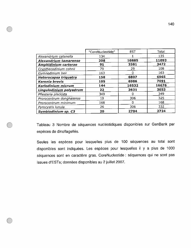
140
CoreN ucleotide’ EST Total
Alexandrium catenella 134 1 135
Alexandrium tamarense 208 10885 11093
Amphîdînium carterae 91 3381 3472
Crypthecodinium cohnif 79 29 108
Gymnodinium beii 163 0 163
Heterocapsa trîguetra 158 6807 6965
Kareniabrevis 105 6986 7091
Karlodiniummicrum 144 16532 16676
Lingulodiniumpolyedrum 22 3631 3653
Pfiesteria piscicida 349 0 349
Prorocentrum donghaiense 19 306 325
Prorocentrum minimum 168 0 168
Pyrocystis lunula 26 306 332
Symbiodinium sp. C3 20 2704 2724
Tableau 3 Nombre de séquences nucléotidiques disponibles sur GenBank par
espèces de dinoflagellés.
Seules les espèces pour lesquelles plus de 100 séquences au total sont
disponibles sont indiquées. Les espèces pour lesquelles il y a plus de 1000
séquences sont en caractère gras. CoreNucleotide : séquences qui ne sont pas
issues d’ESTs; données disponibles au 2 juillet 2007.
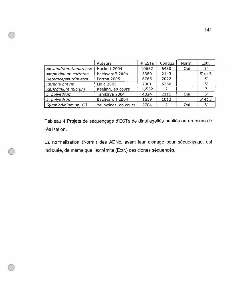
141
Auteurs # ESTs Contigs Norm. Extr
Tableau 4 Projets de séquençage d’ESTs de dinoflagellés publiés ou en cours de
réalisation.
La normalisation (Norm.) des ADNc, avant leur clonage pour séquençage, est
Alexandrium tamarense Hackett 2004 10632 6480 Oui 3’Amphidinium carterae Bachvaroff 2004 3380 2143 5’ et 3’Heterocapsa triguetra Patron 2005 6765 2022 5’Karenia brevis Lidie 2005 7001 5280 5’Karlodinium micrum Keeling, en cours 16532 ? ?L. polyedrum Tanikaya 2004 4324 2111 Oui 5’L. polyedrum Bachvaroff 2004 1519 1012 5’ et 3’
Symbiodinium sp. C3 Yellowlees, en cours 2704 ? Oui 3’
indiquée, de même que l’extrémité (Extr.) des clones séquencés.
142
Ayant maintenant accès à ces données, il est possible de voir si l’approche
par complémentation fonctionnelle que nous avons développée était plus
appropriée à la recherche de composantes du cdc des dinoflagellés qu’une
approche génomique plus empirique. Parmi les 58000 séquences disponibles de
dinoflagellés, on ne retrouve aucune séquence encodant un homologue des
régulateurs classiques Weel et Cdc25 de la levure fissipare. Aussi, outre la
séquence d’ADN de LpCycl que nous avons découverte, deux autres séquences
codent pour une cycline: CK432882 et KML0001122O. CK432882 d’Alexandrium
tamarense, à cause de sa très grande similarité de séquence avec LpCycl, est
probablement l’équivalent de LpCycl chez Alexandrium. Toutefois, si nous avions
eu seulement cet EST d’Alexandrium sans pouvoir comparer sa séquence avec
celle de LpCycl, l’identification de cette séquence en tant que cycline aurait pû être
manquée. En effet, un BLASTX de cette séquence donne une valeur E de
seulement 0.002 envers une cycline d’euglène. Quant à KML0001122O, elle
possède un domaine de repliement-cycline atypique qui n’a pas encore été associé
à une protéine ayant un rôle défini au cours de cycle cellulaire. Pour ce qui est de
CDK1, une séquence de Karlodinium micrum (KME00004803) encode sans
équivoque une kinase homologue. Ainsi, le séquençage d’ESTs commence à
mettre à jour des acteurs du cdc de dinoflagellés. Toutefois, il aura fallut plus de
50000 séquences pour trouver qu’une seule CDK et une seule cycline liée au cdc.
La complémentation fonctionnelle semble donc être une technique demandant
moins de ressources que les projets de génération d’ESTs. Aussi, sa propriété de
pouvoir faire découvrir des fonctionalités différentes et inattendues aux gènes
isolés autrement que simplement par comparaison de séquences, tel que ça a été
le cas pour LpAAA, rend cet outil précieux.
143
Il est indéniable que la disponibilité d’outils génomiques plus complets sur
les dinoflagellés faciliterait par beaucoup l’étude de leur cycle cellulaire. Par contre,
avec un organisme possédant 30 fois plus d’ADN que l’humain, seule une analyse
des séquences transcrites semble être envisageable à court terme. Dans un
contexte où il faut générer plus de 58000 ESTs pour trouver seulement 2
intervenants du cdc, il faudrait prévoir un nombre de séquences beaucoup plus
grand pour espérer tous les trouver. Une nouvelle méthode de séquençage, mise
au point par la compagnie 454 Lite Sequences, permet justement la génération de
près de 400000 séquences en moins d’une semaine, réduisant les coûts
traditionnels par paires de bases d’un facteur de près de 50. Ces séquences sont
longues de 300 nucléotides en moyenne, dans le cas d’organismes A-T riches ou
G-C riches tels que Lingulodinium, permettant une identification des clones par
BLASTX. Le grand nombre de séquences générées par cette technique, par
comparaison aux ESTs, augmenterait de beaucoup les chances de repérer une
séquence rare au sein d’une population d’ADNc, normalisée ou non. De plus,
puisque la génération des fragments d’ADN à séquencer se tait suite à un bris
mécanique par nébulisation, le séquençage plus fréquent de sections codantes et
non pas de régions non-traduites faciliterait d’autant plus l’identification de ces
fragments. Finalement, avec une telle technique, l’établissement d’un
transcriptome complet d’un dinoflagellé semble pour la première fois à notre
portée.
Dans le futur, il serait intéressant de savoir quels pourraient être les
substrats de MPF chez les dinoflagellés par une recherche des protéines
contenant la séquence consensus de phosphorylation par CDK1. En effet, il est
difficile de croire que la phosphorylation de certains substrats classiques, tels que
les lamines nucléaires, les histones et les condensines, ait lieu chez un organisme
144
à la mitose fermée et à l’ADN constamment condensé. On peut penser que la
phosphorylation des MAPs et de certains types de lamines nucléaires pourrait
expliquer le fonctionnement biochimique d’établissement d’invaginations à travers
du noyau à la mitose de même que la mise en place de l’attachement des
chromosomes à la paroi interne du noyau. Les dinoflagellés étant la seule classe
d’organisme à avoir une dinomitose, ce mécanisme doit forcément leur être propre.
Nous ne savons toujours pas quel est le mécanisme de contrôle du rythme
circadien de division cellulaire. Jusqu’à présent, parmis LpCycl, LpAAA et
LpCDK5-like, seule la cycline constitue un régulateur du cdc dont une
caractérisation chez Lingulodinium est en accord avec un contrôle du cdc.
Toutefois, son expression semble être plus en accord avec un contrôle par le cycle
cellulaire que par un cycle circadien.
Avec comme objectif à très long terme l’établissement d’une technique
permettant de détecter la potentialité dans un échantillon marin de l’apparition
d’une marée rouge, un anticorps anti-LpCycl ou encore un systême d’amplification
par RT-PCR du transcrit LpCycl pourrait peut-être servir d’outil pour mesurer la
vitesse de croissance de Lingulodinium. Avec la découverte d’un EST
d’Alexandrium à la séquence des plus similaires à LpCycl, on peut même penser
qu’un tel systême puisse être utilisé pour mesurer l’activité du cdc de tous les
dinoflagellés; quoique la présence universelle de LpCycl chez tous les
dinoflagellés n’ait pas encore été démontrée. Aussi, s’il s’avérait que le rôle
proposé de LpAAA de remodelage d’un inhibiteur d’entrée en phase GuS, tel que
Whi5 ou RB, soit effectivement vérifié chez les dinoflagellés, son potentiel d’outil
de détection de HAB5 en devenir serait très grand. En effet, on s’explique encore
mal pourquoi les dinoflagellés ont un taux de croissance très élevé dans certaines
conditions. Peut-être que LpAAA, surexprimée lorsque certaines conditions se

145
présentent, permet la surpopulation explosive des HABs, par analogie à
l’apparition d’un cancer chez les mammifères causé par un mauvais
fonctionnement de RB. Finalement, lorsque nous aurons une idée d’ensemble de
tous les mécanismes biochimiques du cycle cellulaire des dinoflagellés, peut-être
nous sera-t-il possible de produire un inhibiteur spécifique d’une réaction clé du
cycle cellulaire et d’effectivement utiliser cet inhibiteur pour contrôler l’apparition de
marées rouges indésirables.
146
6 BibliographïeAbraham, R. T., M. Acquarone, et al. (1995). “Cellular effects of olomoucine, an inhibitor
ofcyclin-dependent kinases.” Bio! CeIl 83(2-3): 105-20.Agatep, R., R. D. Kirkpatrick, et al. (1998). “Transformation of Saccharomyces cerevisiae
by the !ithium acetate/single-stranded carrier DNA!polyethylene glycol (LiAc/ssDNAJPEG) protocol.” Technical Tips Online (http://tto.trends.com).
Alberts, B., A. Johnson, et al. (2002). Molecular Biology of the Cell. New York, GarlandScience.
Altschul, S. F., T. L. Madden, et al. (1997). “Gapped BLAST and PSI-BLAST: a newgeneration of protein database search programs.” Nucleic Acids Res 25(17): 3389-402.
Amon, A., U. Surana, et al. (1992). “Regulation ofp34CDC28 tyrosine phosphorylation isnot required for entry into mitosis in S. cerevisiae.” Nature 355(6358): 368-71.
Asano, S., J. E. Park, et al. (2005). “Concerted mechanism of Swel/Weel regulation bymultiple kinases in budding yeast.” EMBO J. 24(12): 2194-2204.
Bachvaroff, T. R., G. T. Concepcion, et al. (2004). “Dinoflagellate Expressed SequenceTag Data Indicate Massive Transfer of Chloroplast Genes to the Nuclear Genome)’Protist 155(1): 65-78.
Bae, Y. M. and J. W. Hastings (1994). “Cloning, sequencing and expression ofdinoflagellate luciferase DNA from a marine alga, GonyauÏax poÏyedra.”Biochimica et Biophysica Acta 1219(2): 449-456.
Baldauf, S. L, A. J. Roger, et aI. (2000). “A kingdom-level phy!ogeny of eukaryotes basedon combined protein data.” Science 290(5493): 972-7.
Barbier, M., M. Albert, et al. (1995). “Cell cycle regulation of the primitive dinoflagel!ateCiypthecodiniunz cohnii biecheler - evidence for the presence of an homolog ofcyclin b.” Biology of the Cell 84(1-2): 35-42.
Barbier, M., M. L. Geraud, et al. (1998). “Colocalization of the cyclin b homologue p56and beta-tubulin during the celi cycle in a unicellular eucaryote dinoflage!tate.”Biology of the Ceil 90(1): 63-76.
Barbier, M., T. A. Leighfield, et al. (2003). “Permanent expression of a cyclin Bhomologue in the celi cycle of the dinoflagellate Karenia brevis.” Journal ofEukaryotic Microbiology 50(2): 123-131.
Bean, J. M., E. D. Siggia, et al. (2005). “High functional overlap between MluI celi-cyclebox binding factor and Swi4/6 cell-cycle box binding factor in the GuStranscriptional program in Saccharomyces cerevisiae.” Genetics 171(1): 49-61.
Belmont, A. S. (2006). “Mitotic chromosome structure and condensation.” Current Opinionin Ceil Biology 18(6): 632-63 8.
Bertomeu, T. and D. Morse (2004). “Isolation of a dinoflagel!ate mitotic cyclin byfunctional complernentation in yeast.” Biochernical & Biophysical ResearchCommunications 323(4): 1172-1183.

147Bhaud, Y., M. Barbier, et al. (1994). “A detailed study ofthe complex celi cycle ofthe
dinoflagellate Ciypthecodinium cohnii biecheler and evidence for variation inhistone hi kinase activity.” Journal ofEukarvotic Microbiology 41(5): 519-526.
Bhaud, Y., D. Guillebault, et al. (2000). “Morphology and behaviour of dinoflagellatechromosomes during the ce!! cycle and mitosis.” Journal of Celi Science 113(7):1231-1239.
Bhaud, Y., J.-M. Salmon, et al. (1991). “The Complex Ceil Cycle of the DinoflagellateProtoctist Crypthecodinium Cohnii as Studied In Vivo and by Cytofluorirnetry.” ICell Sci 100(3): 675-682.
Bloom, J. and F. R. Cross (2007). “Multiple levels of cyclin specificity in celi-cyclecontrol.” Nat Rev Mol Ceil Bio!. 8(2): 149-160.
Booher, R. N., R. J. Deshaies, et ai. (1993). “Properties of Saccharornyces cerevisiae weeland its differentia! regulation of p34CDC28 in response to G1 and G2 cyc!ins.”EMBO J. 12(9): 34 17-3426.
Bouquin, N., Y. Barrai, et al. (2000). “Regulation of cytokinesis by the E!rnl protein kinasein Saccharornyces cerevisiae.” Journal of Ce!! Science 113(8): 1435-1445.
Cavalier-Smith, T. (1993). “Kingdorn protozoa and its 18 phyla.” Microbio! Rev 57(4):953-94.
Chatton, E. (1920). “Les péridiniens parasites. Morphologie, reproduction, éthologie.”Arch. Zool. Exp. Gen. 59: 1-475.
Cho, R. J., M. J. Carnpbel!, et al. (1998). “A genorne-wide transcriptional ana!ysis of themitotic ce!! cycle.” Mol CelI 2(1): 65-73.
Chudnovsky, Y., J. Li, et al. (2002). “Cloning, expression, and characterization of ahistone-!ike protein from the marine dinoflagellate Lingulodinitim poÏyedrurn(Dinophyceae).” J. Phyco!ogy 38: 543-550.
Colette, G. (1984). “The fifth base: A natura! feature of dinoflagellate DNA.” Origins ofLife and Evolution of Biospheres (Forrneriy Origins of Life and Evolution of theBiosphere) V13(3): 195-203.
Costanzo, M., J. L. Nishikawa, et al. (2004). “CDK activity antagonizes Whi5, an inhibitorofGl/S transcription inyeast.” 117(7): 899-913.
Costas, E. and V. Goyanes (2005). “Architecture and evo!ution of dinoflage!latechromosomes: an enigmatic origin.” Cytogenet Genome Res 109: 268-275.
Cross, F. R. (1995). “Starting the ceil cycle - what’s the point.” Current Opinion in CeilBiology 7(6): 790-797.
Cross, F. R. and M. D. Jacobson (2000). “Conservation and function of a potentialsubstrate-binding dornain in the yeast C1b5 B-type cyclin.” Moi Cel! Bio! 20(13):4782-90.
Cross, F. R. and K. Levine (1998). “Molecu!ar evo!ution al!ows bypass of the requirementfor activation loop phosphorylation of the Cdc28 cyclin-dependent kinase.” MiCeli Bio! 18(5): 2923-3 1.
Cross, F. R. and J. M. Roberts (2001). “Retinoblastoma protein: Combating algal b!oom.”Current Bio!ogy 11(20): R824-R827.
Daugbj erg, N., G. Hansen, et a!. (2000). “Phy!ogeny of some of the major genera ofdinoflagel!ates based on ultrastructure and partial LSU rDNA sequence data,
148including the erection of three new genera of unannoured dinoflagellates.”Phycologia 39(4): 302-3 17.
Day, I. S., A. S. Reddy, et al. (1996). “Isolation of a new mitotic-like cyciin fromArabidopsis: complementation of a yeast cyciin mutant with a plant cyciin.” PlantMoi Biol 30(3): 565-75.
de Bruin, R. A. M., W. H. McDonald, et aI. (2004). “C1n3 activates Gi-specifictranscription via phosphorylation of the SBF transcription bound repressor Whi5.”Ccli 117(7): 887-898.
Dhavan, R. and L. H. Tsai (2001). “A decade of CDK5.” Nat Rev Mol Ccli Biol 2(10):749-59.
Dodge, J. D. (1965). “Chromosome structure in the dinoflagellate and the problem ofthemesokaryotic celi.” Exerpta med. int. Congr. Ser. 91: 339.
Dodge, J. D., Ed. (1966). The Dinophyceae. The chromosomes ofthe aigae.Donaldson, A. D. and J. J. Blow (1999). “The regulation of replication origin activation.”
Current Opinion in Genetics & Deveiopment 9(1): 62-68.Enoch, T. and P. Nurse (1990). “Mutation of fission yeast ccii cycle controi genes aboiishes
dependence ofmitosis on DNA repiication.” Çç 60(4): 665-73.Epstein, C. B. and F. R. Cross (1992). “CLB5: a novel B cyciin from budding yeast with a
roie in S phase.” Genes & Development 6(9): 1695-1706.Espinoza, F. H., J. Ogas, et al. (1994). “Ccli cycle control by a complex ofthe cyciin hcs26
(pcll) and the kinase pho85.” Science 266(5189): 1388-1391.Evans, T., E. Rosenthai, et al. (1983). “Cyclin: a protein specified by maternai mRNA in
sea urchin eggs that is destroyed at each cieavage division.” 33: 3 89-396.Ferby, I., M. Blazquez, et ai. (1999). “A novel p34(cdc2)-binding and activating protein
that is necessary and sufficient to trigger G(2)/M progression in Xenopus oocytes.”Genes Dcv 13(16): 2177-89.
Fenari, S. (2006). “Protein kinases controiiing the onset of mitosis.” Cellular & MoiecularLife Sciences 63(7-8): 78 1-795.
Fitch, I., C. Dahrnann, et al. (1992). “Characterization of four B-type cyclin genes of thebudding yeast Saccharomyces cerevisiac.” Moi Biol Ccii 3(7): 805-18.
Friedman, M. A. and B. E. Levin (2005). “Neurobehavioral effects of harrnfui algal bioom(HAB) toxins: a critical review.’ J IntNeuropsychol Soc 11(3): 33 1-8.
Fritz, L. and R. E. Treimer (1983). “An uitrastructural study of mitosis in a marinedinoflageiiate Prorocentrum minimum.” J. Protozooi. 30: 437-444.
Frohiich, K. U. (2001). “An AAA famiiy tree.” J Ccii Sci 114(Pt 9): 160 1-2.Gautier, J., C. Norbury, et al. (1988). “Purified maturation-promoting factor contains the
product of a xenopus homolog of the fission yeast ccc cycie controi cdc2+.”54: 433-439.
Gauticr, J. J., J. J. Minshull, et al. (1990). “Cyclin is a component ofrnaturation-promotingfactor from Xenopus.” 60(3): 487-494.
Gietz, R. D. and R. A. Woods (2002). “Transformation of yeast by lithium acetate/singiestranded carrier DNA!polyethylene giycol mcthod.” Methods Enzymoi 350: 87-96.
Glotzer, M. M., A. A. W. Munay, et al. (1991). “Cyclin is degraded by the ubiquitinpathway.” Nature 349(6305): 132-138.

149Guillebault, D., S. Sasorith, et al. (2002). “A new class of transcription initiation
factors, intermediate between TATA box-binding proteins (TBPs) and TBP-likefactors (TLFs), is present in the marine unicellular organisrn, the dinoflagellateC’iypthecodiniurn cohnii.” Journal ofBiological Chernistry 277(43): 4088 1-40886.
Haase, S. B. and S. I. Reed (1999). “Evidence that a free-running oscillator drives Gievents in the budding yeast cell cycle.” Nature 401(675 1): 394-7.
Hackett, J. D., D. M. Anderson, et al. (2004). “Dinoflagellates: a remarkable evolutionaryexperirnent.” Am. J. Bot. 91(10): 1523-1534.
Hackett, J. D., H. S. Yoon, et al. (2004). “Migration ofthe plastid genorne to the nucleus ina peridinin dinoflagellate.” Curr Biol 14(3): 2 13-8.
Hadwiger, J. A., C. Wittenberg, et al. (1989). “A farnily of cyclin homologs that control theGi phase in yeast.” Proc NatI Acad Sci U S A 86(16): 6255-9.
Hanson, P. I. and S. W. Whiteheart (2005). “AAA+ proteins: have engine, will work.”Rev Mol Cell Biol 6(7): 5 19-29.
Hartwell, L. and T. Weinert (1989). “Checkpoints: controls that ensure the order of cdlcycle events.” Science 246: 629-634.
Hartwell, L. H. (1967). “Macrornolecule Synthesis in Temperature-sensitive Mutants ofYeast.” J. Bacteriol. 93(5): 1662-1670.
Hastings, J. W. (2001). “The Colin S. Pittendrigh Lecture - Presented at the SeventhMeeting ofthe Society for Research on Biological Rhythrns, Amelia Island, Florida,May 13, 2000 - fifty years of fun.” Journal of Biological Rhythms 16(1): 5-18.
Herzog, M. and M.-O. Soyer (1981). “Distinctive features of dinoflagellate chrornatin.Absense of nucleosornes in a primitive species Frorocentrum micans.” Eur. J. CeilBiol. 23: 295-302.
Hofmann, E., P. M. Wrench, et al. (1996). “Structural basis of light harvesting bycarotenoids: peridinin- chlorophyll-protein from Amphidiniurn carterae.” Science272(5269): 1788-91.
Homma, K. and . w. Hastings (1988). “Cell cycle-synchronization of GonyaulaxpoÏyedraby filtration: quantized generation times.” J. Biol. Rhythms 3: 49-5 8.
Homma, K. and J. W. Hastings (1989). “CeIl growth kinetics, division asyrnrnetry andvolume control at division in the marine dinoflagellate Gonyaulax poÏyedra: amodel of circadian dock control ofthe cell cycle.” J Cell Sci 92(2): 303-3 18.
Homma, K. and j• W. Hastings (1989). “The S phase is discrete and is controlled by thecircadian dock in the marine dinoflagellate GonyaïtÏax polyedra.” ExperimentalCeil Research 182(2): 635-644.
Howard, A. and S. R. Pelc (1951). “Nuclear incorporation of 32P as dernonstrated byautoradiographs.” Exp. Celi Res. 2(178-187).
Iyer, V. R., C. E. Horak, et al. (2001). “Genornic binding sites of the yeast celi-cycletranscription factors SBF and MBF.” Nature 409(68 19): 533-538.
Jarnes, P., J. Halladay, et al. (1996). “Genornic libraries and a host strain designed forhighly efficient two-hybnd selection in yeast.” Genetics 144(4): 1425-36.
Jedrzejas, M. J. and W. J. Huang (2003). “Bacillus species proteins involved in sporeformation and degradation: from identification in the genorne, to sequence analysis,
150and determination offunction and structure.” Crit Rev Biochem Mol Biol 38(3):173-98.
Jeffrey, P. D., A. A. Ruso, et al. (1995). “Mechanisrn of cdk activation revealed by thestructure ofa cyctin A-cdk2 complex.” Nature 376(6538): 3 13-320.
Johnson, C. H., J. F. Roeber, et al. (1984). “Circadian Changes in Enzyme ConcentrationAccount for Rhythm of Enzyme Activity in GonyauÏax.” Science 223(4643): 1428-1430.
Johnson, R. T. and P. N. Rao (1970). “Mammalian celi fusion: induction of prernaturechromosome condensation in interphase nuclei.” Nature 226: 7 17-722.
Jones, J., P. Dunsmuir, et al. (1985). “High level expression of introduced chimeric genes inregenerated transformed plants.” EMBO J. 4: 2411-2418.
Kaelin, W. G. J. (1999). “Functions ofthe retinoblastoma protein.” Bioessays 21(11): 950-958.
Karaiskou, A., L. H. Perez, et al. (2001). “Differential regulation of Cdc2 and Cdk2 byRINGO and cyclins.” J Biol Chem 276(3 8): 36028-34.
Kobayashi, H., E. Stewart, et al. (1992). “Identification of the dornains in cyclin A requiredfor binding to, and activation of, p34cdc2 and p32cdk2 protein kinase subunits.”Mol. Biol. Celi 3(11): 1279-1294.
Koff, A., F. Cross, et al. (1991). “Human cyclin E, a new cyclin that interacts with twomembers ofthe CDC2 gene family.” 66(6): 12 17-28.
Kozak, M. (1989). “The scanning model for translation: an update.” J. Ceil Biol. 108: 229-241.
Lahue, E. E., A. V. Smith, et al. (1991). “A novel cyclin gene from Drosophilacomplernents CLN function in yeast.” Genes Dey 5(12A): 2166-75.
Le, Q. H., P. Markovic, et al. (1997). “Structure and organization of the peridininchlorophyli a binding protein gene in Gonyaulax polyedra.” Molecular & GeneralGenetics 255(6): 595-604.
Lee, D.-H., M. Mittag, et al. (1993). “Molecular cloning and genomic organization of agene for luciferin-binding protein from the dinoflagellate GonyaztÏax poÏyedra.”Journal of Biological Chemistry 268(12): 8842-8850.
Lee, M. and P. Nurse (1988). “Ceil cycle control genes in fission yeast and mammalianceils.” Trends in Genetics 4: 287-290.
Lenormand, J. L., R. W. Dellinger, et aI. (1999). “Speedy: a novel ccli cycle regulator ofthe G2/M transition.” Embo J 18(7): 1869-77.
Leopold, P. and P. H. O’Farrell (1991). “An evolutionarily conserved cyclin homolog fromDrosophila rescues yeast deficient in Gi cyclins.” 66(6): 1207-16.
Leveson, A., F. Wong, et al. (1997). “Cyclins in a dinoflagellate ccli cycle.” MolecularMarine Biology & Biotechnology 6(3): 172-179.
Lew, D. D. J., V. V., et al. (1991). “Isolation ofthree novel human cyclins by rescue ofGlcyclin (Cln) function in yeast.” 66(6): 1197-1206.
Lew, D. J., V. Dulic, et al. (1991). “Isolation of three novel hurnan cyclins by rescue of G1cyclin (Cln) function in yeast.” Ççji 66(6): 1197-206.
Lew, J., Q. Q. Huang, et al. (1994). “A brain-specific activator of cyclin-dependent kinase5.” Nature 371(6496): 423-6.
151Lewin, B. (2004). Genes VIII. Upper Saddle River, Pearson Education.Li, L. M. and J. W. Hastings (1998). “The structure and organization ofthe luciferase gene
in the photosynthetic dinoflagellate GonyauÏax poÏyedra.” Plant Molecular Biology
36(2): 275-284.Liberzon, A., S. Shpungin, et al. (1996). Association of yeast SAP1, a nove! member of
the ‘AAA’ ATPase family of proteins, with the chromatin protein SINY.” FEB$ Lett388(1): 5-10.
Lidie, K. B., J. C. Ryan, et al. (2005). “Gene expression in Florida red tide dinoflagellateKai-enia brevis: Analysis of an expressed sequence tag library and development ofDNA rnicroarray. Marine Biotechnology 7(5): 48 1-493.
Lim, H. H., C. J. Loy, et al. (1996). “Dephosphorylation of threonine 169 of Cdc28 is notrequired for exit from mitosis but may be necessary for start in Saccharornycescerevisiae.” Mol Ce!! Bio! 16(8): 4573-83.
Liu, J. and E. T. Kipreos (2000). “Evolution of cyclin-dependent kinases (CDK5) andCDK-activating kinases (CAKs): Differential conservation of CAKs in yeast andrnetazoa.” Molecular Biology & Evolution 17(7): 1061-1074.
Lohka, M. M. J., M. M. K. Hayes, et al. (198$). “Purification of maturation-promotingfactor, an intracellular regulator of early mitotic events.” Proceedings of theNational Acaderny of Sciences ofthe United States of Arnerica 85(9): 3009-3013.
Loog, M. and D. O. Morgan (2005). “Cyclin specificity in the phosphorylation of cyclindependent kinase substrates.” Nature 434(7029): 104-8.
Machabee, S., L. WaÏÏ, et al. (1994). “Expression and genornic organization of adinoflagellate gene farnily.” Plant Molecular Biology 25(1): 23-31.
Manning, G., D. B. Whyte, et al. (2002). “The protein kinase complement of the humangenorne.” Science 298(5600): 1912-+.
Masui, Y. and C. L. Markert (1971). “Cytoplasmic control of nuclear behavior duringmeiotic maturation of frog oocytes.” J. exp. Zoo!. 177: 129-146.
Masurnoto, H., S. Murarnatsu, et al. (2002). “S-Cdk-dependent phosphorylation of S1d2essential for chrornosornal DNA replication in budding yeast.” Nature 415(6872):651-655.
Matsuo, T., S. Yamaguchi, et al. (2003). “Control mechanisrn of the circadian dock fortiming of celi division in vivo.” Science 302(5643): 255-259.
McGowan, C. H. (2003). “Regulation of the eukaiyotic ceil cycle.” Prog Ce!! Cycle Res 5:1-4.
McMillan, J. N., M. S. Longtine, et al. (1999). “The morphogenesis checkpoint inSaccharomyces cerevisiae: Ceil cycle control of Swelp degradation by Hsllp andHst7p.” Molecular & Cellular Biology 19(10): 6929-6939.
McMurry, L. and J. W. Hastings (1972). “No desynchronization among four circadianrhythrns in the unicellular alga, GonyauÏaxpolyedra.” Science 175: 1137-1139.
Measday, V. and B. Andrews (199$). “The cyclin family ofbudding yeast: abundant use ofa good idea.” Trends in Genetics 14: 66-72.
Mendenhall, M. D. and A. E. Hodge (1998). “Regulation of cdc28 cyclin-dependent proteinkinase activity during the celi cycle of the yeast $accharomyces cerevisiae.”Microbiology & Molecular Biology Reviews 62(4): 119 1-1243.
152Merckx, A., K. Le Rocli, et al. (2003). “Identification and initial characterization of
three nove! cyclin-related proteins of the human malaria parasite Plasmodiumfalciparum.” J Biol Chem 278(41): 39839-50.
Mesinger, A. f. and J. f. Case (1992). uDinoflagetlate luminescence increasessusceptibility ofzooplankton to teleost predation.” Marine Biology 112(2): 207-2 10.
Millar, J. J. B., C. C. H. McGowan, et aI. (1991). “psOcdc25 mitotic inducer is the tyrosinephosphatase that activates p34cdc2 kinase in fission yeast.” The EMBO journal10(13): 4301-4309.
Moore, J. D., J. A. Kirk, et al. (2003). “Unrnasking the S-phase-promoting potential ofcyclin Bi.” Science 300(562 1): 987-90.
Moreno Diaz de la Espina, S., E. Alverca, et al. (2005). “Organization of the genorne andgene expression in a nuclear environrnent lacking histones and nucleosornes: theamazing dinoflagellates.” Eur J Ccli Biol 84(2-3): 13 7-49.
Moreno, S., J. Hayles, et al. (1989). “Regulation ofp34cdc2 protein kinase during mitosis.”
Ce1158: 361-372.Morgan, D. 0. (1997). “Cyciin-dependent kinases: engines, docks, and microprocessors.”
Annu Rev Ce!l Dey Biol 13: 261-91.Morse, D., P. Salois, et al. (1995). “A nuclear-encoded fonii II RuBisCo in dinoflagellates.”
Science 265(52 17): 1622-1624.Morse, D. D., P. P. M. Miios, et al. (1989). “Circadian regulation of bioluminescence in
GonyauÏax involves transiationai control.” Proceedings ofthe National Academy ofSciences ofthe United States ofAmerica 86(1): 172-176.
Mumberg, D., R. Muller, et al. (1995). “Yeast vectors for the controtled expression ofheterologous proteins in different genetic backgrounds.” Gene 156(1): 119-22.
Mun-ay, A. W. (2004). “Recycling the ccli cycle: Cyciins revisited.” Çç 116(2): 22 1-234.Nash, P., X. Tang, et al. (2001). “Multisite phosphory!ation of a CDK inhibitor sets a
threshold for the onset of DNA replication.” Nature 414(6863): 5 14-521.Nassoury, N., M. Cappadocia, et al. (2003). “Plastid ultrastructure defines the protein
import pathway in dinoflagellates.” Journal of Ccl! Science 116(14): 2867-2874.Nassoury, N., L. Fritz, et al. (2001). “Circadian changes in ribulose-1,5-bisphosphate
carboxylase/oxygenase distribution inside individual chloropÏasts can account forthe rhythm in dinoflagellate carbon fixation.” Plant Ce!! 13(4): 923-934.
Nurse, P. (1990). “Universal control rnechanism regulating onset of M-phase.” Nature 344:503-508.
Nurse, P. (1994). “Ordering S phase and M phase in the cdl cycle.” Ççjj 79(4): 547-5 50.Obaya, A. J. and J. M. Sedivy (2002). “Regulation of cyclin-Cdk activity in mammalian
ceils.” Cellular & Molecular Life Sciences 59(1): 126-142.Parker, L. L., S. Atherton-Fessier, et al. (1992). “plO7weel is a dual-specificity kinase that
phosphorylates p34cdc2 on tyrosine 15.” Proceedings of the National Academy ofSciences ofthe United States ofAmenca 89(7): 2917-2921.
Partridge, J. F., G. E. Mikesel!, et al. (1997). “Cdl cycle-dependent transcription of cmiinvolves swi4 binding to mcb-like elernents.” Journal of Biological Chemistry272(14): 9071-9077.

153Patel, S. and M. Latterich (1998). “The AAA team: related ATPases with diverse
functions.” Trends Celi Biol 8(2): 65-71.Patron, N. J., R. F. Waller, et al. (2005). “Complex protein targeting to dinoflagellate
plastids.” J Mot Biol 348(4): 10 15-24.Patron, N. J., R. F. Waller, et al. (2005). “Complex Protein Targeting to Dinoflagellate
Plastids.” Journal of Molecular Biology 348(4): 1015-1024.Peters, J. M. (1999). “Subunits and substrates of the anaphase-promoting complex.”
Experimental Ce!! Research 248(2): 339-349.Pines, J. (1995). “Cyclins and cyclin-dependent kinases: a biochernical view.” Biochem J.
308: 697-711.Pines, J. (2006). “Mitosis: a matter of getting rid of the right protein at the nght time.”
Trends in Ceil Biology 16(1): 55-63.Pines, J. and T. Hunter (1990). “Hurnan cyclin A is adenovirus EÏA-associated protein p60
and behaves differently from cyclin B.” Nature 346(6286): 760-763.Plaxton, W. C. (1989). “Molecular and immunological characterization of plastid and
cytosolic pynivate kinase isozymes from castor-ou-plant endosperrn and leaf.” Eur JBiochem 181(2): 443-5 1.
Qi, Z., Q.-Q. Huang, et al. (1995). “Reconstitution of neuronal Cdc2-like kinase frombacteria-expressed CdkS and an active fragment of the brain-specific activator:Kinase activation in the absence of CdkS phosphorylation.” Journal of BiologicalChernistry 270(18): 10847-1 0854.
Rae, P. M. (1970). “The nature and processing of ribosornal ribonucteic acid in adinoflagellate.” J. Ce!! Bio!. 46(1): 106-113.
Rappas, M., J. Schumacher, et al. (2006). “Structural basis of the nucleotide drivenconformational changes in the AAA+ domain of transcription activator PspF.” 1Mol Biol 357(2): 48 1-92.
Richardson, H., D. J. Lew, et al. (1992). “Cyclin-B hornologs in Saccharomyces cerevisiaefunction in S phase and in G2.” Genes & Development 6(11): 202 1-34.
Rizzo, P. (1987). Biochernistry of the dinoflagellate nucleus. The biology ofdinoflagellates. F. J. R. Taylor. Oxford, Blackwell Scientific: 143-173.
Rizzo, P. (1991). “The enigma of the dinoflagellate chromosome.” J. Protozool. 38: 246-252.
Rodriguez, M., J. W. Cho, et al. (1993). “Evidence for the presence of a cdc2 like proteinkinase in the dinoflagellate Ciypthecodiniurn cohnii.” J. Eukarvot. Microbiol. 40(1):91-96.
Rosales, J. L. and K. Y. Lee (2006). “Extraneuronal roles of cyclin-dependent kinase 5.”Bioessays 28(10): 1023-1034.
Sala-Rovira, M., M. L. Geraud, et al. (1991). “Molecular cloning and immunolocalizationof two variants of the major basic nuclear protein (HCc) from the histone-lesseukaryote Ciypthecodinium cohnii (Pyrrhophyta).” Chrornosoma 100(8): 510-518.
Salois, P. and D. Morse (1996). “Do dinoflagellates contain a Cdc2-like protein kinase?”Molecular Marine Biology & Biotechnology 5(1): 52-61.
154Salois, P. and D. Morse (1997). “Characterization and molecular phylogeny of a
protein kinase cdna from the dinoflagellate Gonyaulax (dinophyceae).” Journal ofPhycology 33(6): 1063-1072.
Samueisson, G., B. M. Sweeney, et al. (1983). ‘Changes in photosystem II account for thecircadian rhythm in photosynthesis in Gonyaulaxpolyedra.” Plant Physiol. 73: 329-331.
Sanchez, I. and B. D. Dynlacht (2005). “New insights into cyclins, CDKs, and ceil cyclecontrol.” Serninars in Ce!! & Developmental Biology 16(3): 311-321.
Sauer, R. T., D. N. Bolon, et al. (2004). “Scuipting the proteome with AAA(+) proteasesand disassembly machines.” Çll 119(1): 9-18.
Schneider, B. L., Q. H. Yang, et al. (1996). “Linkage of replication to start by the Cdkinhibitor Sici.” Science 272(526 1): 560-2.
Schwob, E., T. Bohrn, et al. (1994). “The B-type cyctin kinase inhibitor p40$IC1 controlsthe Gi to S transition in S. cerevisiae.” Ceil 79(2): 233-244.
Sigee, D. C. (1983). “StructuraI DNA and genetically active DNA in dinoflagellatechromosomes.” Biosystems 16(3-4): 203-210.
Srnith, D. S. and L. H. Tsai (2002). “Cdk5 behind the wheel: a role in trafficking andtransport?” Trends in Celi Biology 12(1): 28-3 6.
Smits, V. A. J. and R. H. Mederna (2001). “Checking out the G2/M transition.” Biochirnicaet Biophysica Acta - Gene Structure & Expression 1519(1-2): 1-12.
Soni, R., J. P. Carrnichael, et aI. (1995). “A farnily of cyclin D hornologs from plantsdifferentially controlled by growth regulators and containing the conservedretinoblastorna protein interaction motif.” Plant Celi 7(1): 85-103.
Soyer-Gobillard, M. O., M. L. Geraud, et al. (1990). “Location of B- and Z-DNA in thechromosomes of a primitive eukaryote dinoflagellate.” J. Ce!! Bio!. 111(2): 293-304.
Soyer-Gobillard, M. O., B. Gillet, et al. (1999). “Dinoflagellate chromosome behaviourduring stages ofreplication.” Int Microbiol 2(2): 93-102.
Spector, D., Ed. (1984). Dinoflagellates. New York, Academic Press.Spector, D., A. Vasconcelos, et al. (1981). “DNA duplication and chromosome structure in
the dinoflagellates.” Protoplasma 105: 185-195.Spellman, P. T., G. Sherlock, et al. (1998). “Comprehensive identification of ceil cycle
regulated genes of the yeast Saccharornyces cerevisiae by microa;Tayhybridization.” Mol Bio! Celi 9(12): 3273-97.
Stem, G. S., R. Baserga, et al. (1998). The Molecular Basis of Celi Cycle and GrowthControl. New York.
Surana, U., H. Robitsch, et al. (1991). “The role ofCDC28 and cyclins during mitosis in thebudding yeast S. cerevisiae.” 65(1): 145-161.
Sweeney, B. M. and J. W. Hastings (1958). “Rhythrnic celi division in populations ofGonyaulaxpolyedra.” J. Protozool. 5: 217-224.
Tachibana, K., N. Yanagishirna, et al. (1987). “Prelirninary characterization of maturationpromoting factor from yeast Saccharornyces cerevisiae.” J. Celi Sci. $8: 273-281.
155Takaki, T., K. Fukasawa, et al. (2005). “Preferences for Phosphorylation Sites in the
Retinoblastorna Protein of D-Type Cyclin-Dependent Kinases, Cdk4 and Cdk6, InVitro. J Biochem (Tokyo) 137(3): 38 1-386.
Tanikawa, N., H. Akirnoto, et al. (2004). “Expressed Sequence Tag Analysis of theDinoflagellate Lingulodinium polyedrum During Dark Phase¶.”Photochemistry and Photobiology 80(1): 3 1-35.
Taroncher-Oldenburg, G. and D. M. Anderson (2000). “Identification and characterizationof three differentially expressed genes, encoding S-adenosylhornocysteinehydrolase, methionine arninopeptidase, and a histone-like protein, in the toxicdinoflagellate Alexandrium fitndyense.” Applied & Environrnental Microbiology66(5): 2105-2112.
Taylor, F. J. R. and U. Pollingher (1987). Ecology of dinoflagellates. The biology ofdinoflagellates. F. J. R. Taylor. Oxford, Blackwell Scientific. 21: 398-529.
Toh-e, A. and M. Nishizawa (2001). “Structure and function of cyclin-dependent Pho85kinase of Saccharomyces cerevisiae.” Journal of General & Applied Microbiology47(3): 107-117.
Toone, W. M., B. L. Aerne, et al. (1997). “Getting started: regulating the initiation ofDNAreplication in yeast.” Annu Rev Microbiol 51: 125-49.
Tsai, L. H., I. Delalle, et al. (1994). “p35 is a neural-specific regulatory subunit of cyclindependent kinase 5.” Nature 371(6496): 419-23.
Tyers, M. (1996). “The cyclin-dependent kinase inhibitor p40(sicl) imposes therequirement for cm gi cyclin function at start.” Proceedings of the NationalAcaderny of Sciences of the United States of America 93(15): 7772-7776.
Tyers, M., G. Tokiwa, et al. (1993). “Comparison of the Saccharomyces cerevisiae G1cyclins: C1n3 may be an upstream activator of Cmi, C1n2 and other cyclins.EMBO J. 12(5): 1955-1968.
Ubersax, J. A., E. L. Woodbury, et al. (2003). “Targets of the cyclin-dependent kinaseCdkl.” Nature 425(6960): 859-864.
Van de Peer, Y., S. L. Ba1dauf, et al. (2000). “An updated and comprehensive rRNAphylogeny of (crown) eukaryotes based on rate-calibrated evolutionary distances.” IMol Evol 51(6): 565-76.
Van Dolah, F. M. (2000). “Marine algal toxins: origins, health effects, and their increasedoccurrence.” Environ Health Perspect 108 Suppi 1: 133-41.
Van Dolah, F. M. and T. A. Leighfield (1999). “Diel phasing ofthe ceil-cycle in the Floridared tide dinoflagellate, Gymnodinium breve.” Journal of Phycology 35(6): 1404-1411.
Van Dolah, F. M., T. A. Leighfield, et al. (1995). “Celi division in the dinoflagellateGambierdiscus toxicus is phased to the diurnal cycle and accornpanied by activationof the ceil cycle regulatory protein, CDC2 kinase.” Journal of Phvcology 31(3):395-400.
Verma, R., R. S. Annan, et al. (1997). “Phosphorylation of Siclp by Gi Cdk Required forIts Degradation and Entry into S Phase.” Science 278(5337): 455-460.
Vernet, T., D. Dignard, et al. (1987). “A family of yeast expression vectors containing thephage fi intergenic region.” Gene 52: 225-23 3.
156Vogel, L. and B. Baratte (1996). “Sud: cdc2 afflnity reagent or essentiat cdk adaptor
protein? Prog Celi Cycle Res 2: 129-35.Vogel L., B. B., Detivaud L., Azzi L., Leopold P. and Meijer L. (2002). “Molecular cloning
and characterisation of pY5CDK-BP, a novel CDK-binding protein.” Biochimica etBiophysica Acta (BBA)/Molecular Ceil Research 1589: 219 -231.
Walker, J. E., M. Saraste, et al. (1982). “Distantly related sequences in the alpha- and betasubunits of ATP synthase, rnyosin, kinases and other ATP-requiring enzymes and acommon nucleotide binding fold.” Embo J 1(8): 945-5 1.
Wang, Y. and D. Morse (2006). “Rampant polyuridylylation of plastid gene transcripts in
the dinoflagellate Lingulodinium.” Nucleic Acids Researcli 34(2): 613-619.Wargo, M. J. and P. J. Rizzo (2001). “Exception to eukaryotic rules.” Science 294(5551):
2477.Weinreich, M., C. Liang, et al. (2001). “Inaugural Article: Binding of cyclin-dependent
kinases to ORC and Cdc6p regulates the chromosome replication cycle.” PNAS98(20): 11211-1 1217.
Whitney, S. M., D. C. Shaw, et aI. (1995). “Evidence that some dinoflagellates contain aribulose- 1 ,5-bisphosphate carboxylase/oxygenase related to that of the alpha
proteobacteria.” Proc Biol Sci 259(1356): 27 1-5.Wijnen, H., A. Landrnan, et al. (2002). “The G(1) cyclin Cln3 promotes cell cycle entry via
the transcription factor Swi6.” Mol Cdl Biol 22(12): 4402-18.WinzeÏer, E. A., D. D. Shoemaker, et aÏ. (1999). “Functional Characterization of the
S. cerevisiae Genorne by Gene Deletion and Parallel Analysis.” Science285(5429): 901-906.
Wittmann, T., A. Hyman, et al. (2001). “The spindle: a dynamic assernbly ofmicrotubulesand motors.” Nat Ceil Biol 3(1): E28-E34.
Wong, J. T. Y. and A. C. M. Kwok (2005). “Proliferation of dinoflagellates: bloorning orbleaching.” Bioessays 27(7): 730-740.
Wong, J. T. Y., D. C. New, et al. (2003). “Histone-Like Proteins of the DinoflagellateCiypthecodiniurn cohnii Have Homologies to Bacterial DNA-Binding Proteins.”Eukaryotic CeIl 2(3): 646-650.
Wysocki, R., A. Javaheri, et al. (2006). “CDK Pho85 targets CDK inhibitor SicI to relieveyeast Gi checkpoint anest after DNA damage.u Nature Structural & MolecularBiology 13(10): 908-9 14.
Xiong, Y., T. Connolly, et al. (1991). “Hurnan D-type cyclin.” 65(4): 69 1-9.Yeung, P. K. K., D. C. New, et al. (2000). “The spindie checkpoint in the dinoflagellate
Ciypthecodinium cohnii.” ExperirnentaÏ Celi Research 254(1): 120-129.Zhang, H., Y. Hou, et al. (2007). “Spticed leader RNA trans-splicing in dinoflagellates.”
PNAS 104(11): 4618-4623.Zhang, Z., B. R. Green, et al. (1999). “Single gene circles in dinoflagellate chloroplast
genornes.” Nature 400(6740): 155-9.Zukerberg, L. R., G. N. Patrick, et al. (2000). “Cables links CdkS and c-Abl and facilitates
Cdk5 tyrosine phosphorylation, kinase upreguÏation, and neurite outgrowth.”Neuron 26(3): 633-646.

Q