Non-contiguous finished genome sequence and description of ...
16
Standards in Genomic Sciences (2014) 9:1046-1061 DOI:10.4056/sigs.5459590 The Genomic Standards Consortium Non-contiguous finished genome sequence and description of Bacillus algeriensis sp. nov. Esma Bendjama 1,2,3 , Lotfi Loucif 1,2,3 , Seydina M. Diene 1 , Caroline Michelle 1 , Djamila Gacemi- Kirane 2 , Jean-Marc Rolain 1* 1 Unité de recherche sur les maladies infectieuses et tropicales émergentes (URMITE), UMR CNRS, IHU Méditerranée Infection, Faculté de Médecine et de Pharmacie, Aix-Marseille- Université, Marseille, France. 2 Département de Biochimie, Faculté des Sciences, Université Badji Mokhtar, Annaba, Algé- rie 3 Département des Sciences Biologiques, Faculté des Sciences, Université El Hadj Lakhdar, Batna, Algérie. *Correspondence: Jean-Marc Rolain ([email protected]) Keywords: Bacillus algeriensis, hypersaline environments, sediments, genome, taxono- genomics Strain EB01 T sp. nov. is the type strain of Bacillus algeriensis, a new species within the genus Bacillus. This strain, whose genome is described here, was isolated from sediment sample of the hypersaline lake Ezzemoul sabkha in northeastern Algeria. B. algeriensis is a facultative anaerobic Gram-positive bacillus. Here we describe the features of this organism, together with the complete genome sequence and annotation. The 5,269,577 bp long genome con- tains 5,098 protein-coding and 95 RNA genes, including 12 rRNA genes. Introduction Bacillus algeriensis sp. nov. strain EB01 T (= CSUR P857 = DSM 27334) is the type strain of B. algeriensis sp. nov. It is a new Gram-positive, facultatively anaerobic, motile, indole-negative, rod shaped bacterium with rounded ends. It was isolated from a sediment sample from the hypersaline lake Ezzemoul sabkha in the Oum-El- Bouaghi region in northeastern Algeria, which is an important wintering and resting site for several species of waterbirds, including the Greater Fla- mingo. This site is one of the Ramsar convention wetlands (http://www.ramsar.org). The genus Bacillus was created by Cohn about 142 years ago [1],and mainly comprises Gram-positive, rod- shaped, aerobic or facultatively anaerobic, spore- forming bacteria. The genus includes 279 species and 7 subspecies with validly published names [2]. Members of Bacillus genus are ubiquitous in nature, ranging from freshwater to marine sedi- ments and from hot springs and desert sands to Arctic soils; many strains have been isolated from the gastrointestinal tracts of various insects and animals, from vegetation and from food [3]. Bacil- lus strains are biotechnologically priceless be- cause of their high capacity to produce a wide range of antimicrobial compounds, enzymes and other metabolites that can be used in industry [4,5]. Some species of Bacillus are pathogenic, such as B. anthracis (responsible for causing an- thrax) [6] and B. cereus (a major cause of food poi- soning) [7]. Others are opportunists in immunocompromised patients, and may also be involved in various human infections, including pneumonia, endocarditis, ocular, cutaneous, bone or central nervous system infections and bacte- remia [8].The current bacterial taxonomy is based on a combination of various phenotypic and ge- netic criteria [9,10]. However, the three essential genetic criteria that are used, comprising 16S rRNA gene based phylogeny [11], G+C content, and DNA-DNA hybridization [10,12] exhibit sev- eral drawbacks. As a result of the recent decrease in the cost of genomic sequencing, it has been proposed that whole genome sequencing infor- mation and MALDI-TOF spectrum [13] be com- bined with the main phenotypic characteristics as a polyphasic approach strategy (taxono-genomics) to describe new bacterial taxa [14-26].
Transcript of Non-contiguous finished genome sequence and description of ...
Standards in Genomic Sciences (2014) 9:1046-1061 DOI:10.4056/sigs.5459590
The Genomic Standards Consortium
Non-contiguous finished genome sequence and description of Bacillus algeriensis sp. nov.
Esma Bendjama1,2,3, Lotfi Loucif1,2,3, Seydina M. Diene1, Caroline Michelle1, Djamila Gacemi-Kirane2, Jean-Marc Rolain1*
1 Unité de recherche sur les maladies infectieuses et tropicales émergentes (URMITE), UMR CNRS, IHU Méditerranée Infection, Faculté de Médecine et de Pharmacie, Aix-Marseille-Université, Marseille, France.
2 Département de Biochimie, Faculté des Sciences, Université Badji Mokhtar, Annaba, Algé-rie
3 Département des Sciences Biologiques, Faculté des Sciences, Université El Hadj Lakhdar, Batna, Algérie.
*Correspondence: Jean-Marc Rolain ([email protected])
Keywords: Bacillus algeriensis, hypersaline environments, sediments, genome, taxono-genomics
Strain EB01T sp. nov. is the type strain of Bacillus algeriensis, a new species within the genus Bacillus. This strain, whose genome is described here, was isolated from sediment sample of the hypersaline lake Ezzemoul sabkha in northeastern Algeria. B. algeriensis is a facultative anaerobic Gram-positive bacillus. Here we describe the features of this organism, together with the complete genome sequence and annotation. The 5,269,577 bp long genome con-tains 5,098 protein-coding and 95 RNA genes, including 12 rRNA genes.
IntroductionBacillus algeriensis sp. nov. strain EB01T (= CSUR P857 = DSM 27334) is the type strain of B. algeriensis sp. nov. It is a new Gram-positive, facultatively anaerobic, motile, indole-negative, rod shaped bacterium with rounded ends. It was isolated from a sediment sample from the hypersaline lake Ezzemoul sabkha in the Oum-El-Bouaghi region in northeastern Algeria, which is an important wintering and resting site for several species of waterbirds, including the Greater Fla-mingo. This site is one of the Ramsar convention wetlands (http://www.ramsar.org). The genus Bacillus was created by Cohn about 142 years ago [1],and mainly comprises Gram-positive, rod-shaped, aerobic or facultatively anaerobic, spore-forming bacteria. The genus includes 279 species and 7 subspecies with validly published names [2]. Members of Bacillus genus are ubiquitous in nature, ranging from freshwater to marine sedi-ments and from hot springs and desert sands to Arctic soils; many strains have been isolated from the gastrointestinal tracts of various insects and animals, from vegetation and from food [3]. Bacil-lus strains are biotechnologically priceless be-
cause of their high capacity to produce a wide range of antimicrobial compounds, enzymes and other metabolites that can be used in industry [4,5]. Some species of Bacillus are pathogenic, such as B. anthracis (responsible for causing an-thrax) [6] and B. cereus (a major cause of food poi-soning) [7]. Others are opportunists in immunocompromised patients, and may also be involved in various human infections, including pneumonia, endocarditis, ocular, cutaneous, bone or central nervous system infections and bacte-remia [8].The current bacterial taxonomy is based on a combination of various phenotypic and ge-netic criteria [9,10]. However, the three essential genetic criteria that are used, comprising 16S rRNA gene based phylogeny [11], G+C content, and DNA-DNA hybridization [10,12] exhibit sev-eral drawbacks. As a result of the recent decrease in the cost of genomic sequencing, it has been proposed that whole genome sequencing infor-mation and MALDI-TOF spectrum [13] be com-bined with the main phenotypic characteristics as a polyphasic approach strategy (taxono-genomics) to describe new bacterial taxa [14-26].
Bendjama et al.
http://standardsingenomics.org 1047
Here we present a summary classification and a set of features for B. algeriensis sp. nov. strain EB01T together with the description of the com-plete genome sequence and annotation. These characteristics support the circumscription of the species B. algeriensis.
Classification and features In July 2012, a sediment sample was aseptically collected in sterile bottles, 15 cm below the evaporite crust of the hypersaline lake Ezzemoul sabkha of Oum-El-Bouaghi region in northeastern Algeria. Samples were transferred in a cooler (4°C) to our lab in Algeria. Samples were pro-cessed the same day. Sediments were diluted 1:10 v/v with sterile saline water (0.9% NaCl) and vig-orously shaken, tenfold serial dilutions (10-1-10-5)
of the sediment suspension were plated in Nutri-ent Agar (NA) medium (meat extract 1 g/l, pep-tone 5 g/l, yeast extract 2 g/l, sodium chloride 5 g/l, agar 15 g/l) and the plates were incubated at 30°C for 24-72 h. In order to obtain a pure culture, colonies were transferred to fresh NA medium. Bacillus algeriensis sp. nov. strain EB01T (Table 1) was isolated in July 2012 by cultivation under aerobic conditions at 30°C. This strain exhibited a 97.0% 16S rRNA nucleotide sequence similarity with Bacillus subterraneus type strain DSM13966T (Figure 1), the phylogenetically closest validly published Bacillus species. These values were lower than the 98.7% 16S rRNA gene sequence threshold recommended by Stackebrandt and Ebers to delineate a new species without carrying DNA DNA hybridizidation [11].
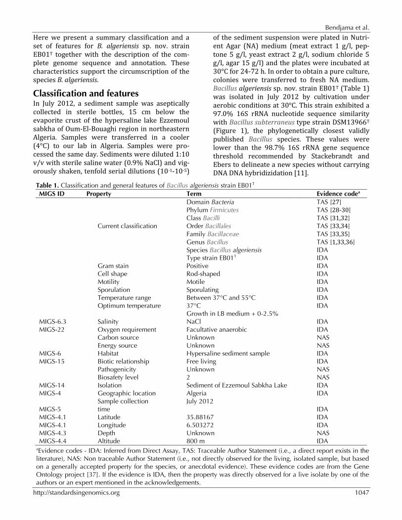
Table 1. Classification and general features of Bacillus algeriensis strain EB01T MIGS ID Property Term Evidence codea
Domain Bacteria TAS [27] Phylum Firmicutes TAS [28-30] Class Bacilli TAS [31,32] Current classification Order Bacillales TAS [33,34] Family Bacillaceae TAS [33,35] Genus Bacillus TAS [1,33,36] Species Bacillus algeriensis IDA Type strain EB01T IDA Gram stain Positive IDA Cell shape Rod-shaped IDA Motility Motile IDA Sporulation Sporulating IDA Temperature range Between 37°C and 55°C IDA Optimum temperature 37°C IDA
MIGS-6.3 Salinity Growth in LB medium + 0-2.5% NaCl IDA
MIGS-22 Oxygen requirement Facultative anaerobic IDA Carbon source Unknown NAS Energy source Unknown NAS MIGS-6 Habitat Hypersaline sediment sample IDA MIGS-15 Biotic relationship Free living IDA
MIGS-14
Pathogenicity Biosafety level Isolation
Unknown 2 Sediment of Ezzemoul Sabkha Lake
NAS NAS IDA
MIGS-4 Geographic location Algeria IDA
MIGS-5 Sample collection time
July 2012 IDA
MIGS-4.1 Latitude 35.88167 IDA MIGS-4.1 Longitude 6.503272 IDA MIGS-4.3 Depth Unknown NAS MIGS-4.4 Altitude 800 m IDA
aEvidence codes - IDA: Inferred from Direct Assay, TAS: Traceable Author Statement (i.e., a direct report exists in the literature), NAS: Non traceable Author Statement (i.e., not directly observed for the living, isolated sample, but based on a generally accepted property for the species, or anecdotal evidence). These evidence codes are from the Gene Ontology project [37]. If the evidence is IDA, then the property was directly observed for a live isolate by one of the authors or an expert mentioned in the acknowledgements.
Bacillus algeriensis
1048 Standards in Genomic Sciences
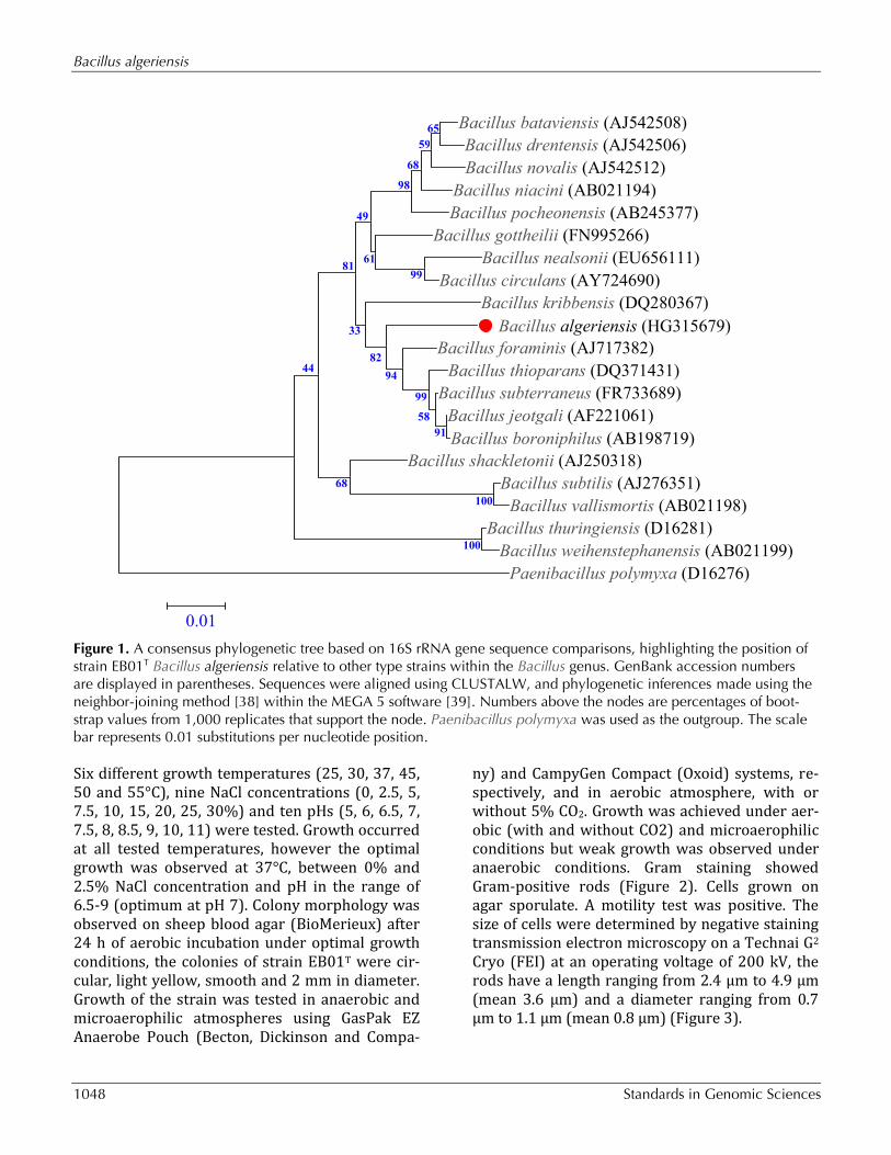
Figure 1. A consensus phylogenetic tree based on 16S rRNA gene sequence comparisons, highlighting the position of strain EB01T Bacillus algeriensis relative to other type strains within the Bacillus genus. GenBank accession numbers are displayed in parentheses. Sequences were aligned using CLUSTALW, and phylogenetic inferences made using the neighbor-joining method [38] within the MEGA 5 software [39]. Numbers above the nodes are percentages of boot-strap values from 1,000 replicates that support the node. Paenibacillus polymyxa was used as the outgroup. The scale bar represents 0.01 substitutions per nucleotide position. Six different growth temperatures (25, 30, 37, 45, 50 and 55°C), nine NaCl concentrations (0, 2.5, 5, 7.5, 10, 15, 20, 25, 30%) and ten pHs (5, 6, 6.5, 7, 7.5, 8, 8.5, 9, 10, 11) were tested. Growth occurred at all tested temperatures, however the optimal growth was observed at 37°C, between 0% and 2.5% NaCl concentration and pH in the range of 6.5-9 (optimum at pH 7). Colony morphology was observed on sheep blood agar (BioMerieux) after 24 h of aerobic incubation under optimal growth conditions, the colonies of strain EB01T were cir-cular, light yellow, smooth and 2 mm in diameter. Growth of the strain was tested in anaerobic and microaerophilic atmospheres using GasPak EZ Anaerobe Pouch (Becton, Dickinson and Compa-
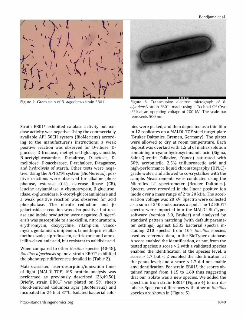
ny) and CampyGen Compact (Oxoid) systems, re-spectively, and in aerobic atmosphere, with or without 5% CO2. Growth was achieved under aer-obic (with and without CO2) and microaerophilic conditions but weak growth was observed under anaerobic conditions. Gram staining showed Gram-positive rods (Figure 2). Cells grown on agar sporulate. A motility test was positive. The size of cells were determined by negative staining transmission electron microscopy on a Technai G2 Cryo (FEI) at an operating voltage of 200 kV, the rods have a length ranging from 2.4 μm to 4.9 μm (mean 3.6 μm) and a diameter ranging from 0.7 μm to 1.1 μm (mean 0.8 μm) (Figure 3).
Bacillus bataviensis (AJ542508)
Bacillus drentensis (AJ542506)
Bacillus novalis (AJ542512)
Bacillus niacini (AB021194)
Bacillus pocheonensis (AB245377)
Bacillus gottheilii (FN995266)
Bacillus nealsonii (EU656111)
Bacillus circulans (AY724690)
Bacillus kribbensis (DQ280367)
Bacillus algeriensis (HG315679)
Bacillus foraminis (AJ717382)
Bacillus thioparans (DQ371431)
Bacillus subterraneus (FR733689)
Bacillus jeotgali (AF221061)
Bacillus boroniphilus (AB198719)
Bacillus shackletonii (AJ250318)
Bacillus subtilis (AJ276351)
Bacillus vallismortis (AB021198)
Bacillus thuringiensis (D16281)
Bacillus weihenstephanensis (AB021199)
Paenibacillus polymyxa (D16276)
100
100
68
99
44
91
58
99
94
65
59
68
98
82
33
81
49
61
0.01
Bendjama et al.
http://standardsingenomics.org 1049
Figure 2. Gram stain of B. algeriensis strain EB01T. Figure 3. Transmission electron micrograph of B.
algeriensis strain EB01T made using a Technai G2 Cryo (FEI) at an operating voltage of 200 kV. The scale bar represents 500 nm.
Strain EB01T exhibited catalase activity but oxi-dase activity was negative. Using the commercially available API 50CH system (BioMerieux) accord-ing to the manufacturer’s instructions, a weak positive reaction was observed for D-ribose, D-glucose, D-fructose, methyl α-D-glucopyranoside, N-acetylglucosamine, D-maltose, D-lactose, D-melibiose, D-saccharose, D-trehalose, D-tagatose, and hydrolysis of starch. Other tests were nega-tive. Using the API ZYM system (BioMerieux), pos-itive reactions were observed for alkaline phos-phatase, esterase (C4), esterase lipase (C8), leucine arylamidase, α-chymotrypsin, β-glucuron-idase, α-glucosidase, N-acetyl-glucosaminidase and a weak positive reaction was observed for acid phosphatase. The nitrate reduction and β-galactosidase reaction was also positive, but ure-ase and indole production were negative. B. algeri-ensis was susceptible to amoxicillin, nitrourantoin, erythromycin, doxycycline, rifampicin, vanco-mycin, gentamicin, imipenem, trimethoprim-sulfa-methoxazole, ciprofloxacin, ceftriaxone and amox-icillin-clavulanic acid, but resistant to nalidixic acid.
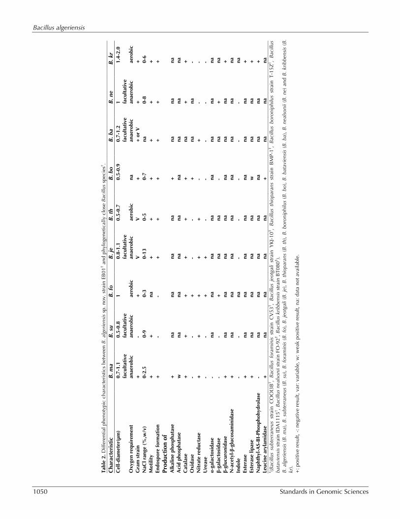
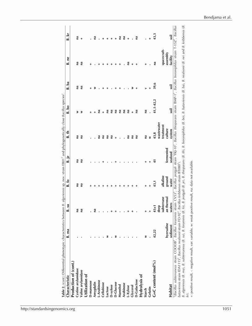
When compared to other Bacillus species [40-48], Bacillus algeriensis sp. nov. strain EB01T exhibited the phenotypic differences detailed in (Table 2).
Matrix-assisted laser-desorption/ionization time-of-flight (MALDI-TOF) MS protein analysis was performed as previously described [26,49,50]. Briefly, strain EB01T was plated on 5% sheep blood-enriched Columbia agar (BioMerieux) and incubated for 24 h at 37°C. Isolated bacterial colo-
nies were picked, and then deposited as a thin film in 12 replicates on a MALDI-TOF steel target plate (Bruker Daltonics, Bremen, Germany). The plates were allowed to dry at room temperature. Each deposit was overlaid with 1.5 µl of matrix solution containing α-cyano-hydroxycinnamic acid (Sigma, Saint-Quentin Fallavier, France) saturated with 50% acetonitrile, 2.5% trifluoroacetic acid and high-performance liquid chromatography (HPLC)-grade water, and allowed to co-crystallize with the sample. Measurements were conducted using the Microflex LT spectrometer (Bruker Daltonics). Spectra were recorded in the linear positive ion mode over a mass range of 2 to 20 kDa. The accel-eration voltage was 20 kV. Spectra were collected as a sum of 240 shots across a spot. The 12 EB01T spectra were imported into the MALDI BioTyper software (version 3.0, Bruker) and analyzed by standard pattern matching (with default parame-ter settings) against 6,335 bacterial spectra in-cluding 210 spectra from 104 Bacillus species, used as reference data, in the BioTyper database. A score enabled the identification, or not, from the tested species: a score > 2 with a validated species enabled the identification at the species level, a score > 1.7 but < 2 enabled the identification at the genus level; and a score < 1.7 did not enable any identification. For strain EB01T, the scores ob-tained ranged from 1.15 to 1.60 thus suggesting that our isolate was a new species. We added the spectrum from strain EB01T (Figure 4) to our da-tabase. Spectrum differences with other of Bacillus species are shown in (Figure 5).
Bacillus algeriensis
1050 Standards in Genomic Sciences
Bendjama et al.
http://standardsingenomics.org 1051

Bacillus algeriensis
1052 Standards in Genomic Sciences
Figure 4. Reference mass spectrum from B. algeriensis strain EB01T. Spectra from 12 individual colonies were compared and a reference spectrum was generated.
Figure 5. Gel view comparing Bacillus algeriensis EB01T spectra with other members of the Bacillus genus (B. weihenstephanensis, B. vallismortis, B. thuringiensis, B. thioparans, B. subtilis subsp. subtilis, B. subterraneus, B. shackletonii, B. novalis, B. niacini, B. nealsonii, B. jeotgali, B. flexus, B. circulans, B. bataviensis and B. asahii). The Gel View displays the raw spectra of all loaded spectrum files as a pseudo-electrophoretic gel. The x-axis records the m/z value. The left y-axis displays the running spectrum number originating from subsequent spectra loading. The peak intensity is expressed by a grey scale scheme code. The grey scale bar on the right y-axis indi-cates the relation between the shade of grey a peak is displayed with and the peak intensity in arbitrary units.
0.0
0.5
1.0
1.5
4x10
Inte
ns. [a
.u.]
2000 4000 6000 8000 10000 12000 14000 16000 18000m/z

Bendjama et al.
http://standardsingenomics.org 1053
Genome sequencing informationGenome project history The organism was selected for sequencing on the basis of its phylogenetic position and 16S rRNA similarity to other members of the genus Bacillus, and is part of a study of Bacillus genus diversity in hypersaline lakes of Algeria. It was the 398th ge-
nome of a Bacillus species and the first genome of Bacillus algeriensis sp. nov. The EMBL accession number is ERP003483 and consists of 46 contigs. Table 3 shows the project information and its as-sociation with MIGS version 2.0 compliance [51].
Table 3. Project information MIGS ID Property Term MIGS-31 Finishing quality High-quality draft MIGS-28 Libraries used Nextera XT library MIGS-29 Sequencing platform Miseq-Illumina MIGS-31.2 Sequencing coverage 34× MIGS-30 Assemblers Velvet MIGS-32 Gene calling method Prodigal EMBL Date of Release January 10, 2014 EMBL ID ERP003483
MIGS-13 Project relevance Study of the Bacillus genus diversity in hypersaline lakes of northeastern Algeria
Growth conditions and DNA isolation Bacillus algeriensis sp. nov strain EB01T, was grown aerobically on 5% sheep blood enriched Columbia agar at 37°C. Three Petri dishes were spread and resuspended in a 2 ml sterile Eppendorf tube containing 1ml of TE buffer with acid-washed glass beads (diameter 106 µm, Sig-ma, Saint-Quentin Fallavier, France). Three cycles of shaking were performed using a FastPrep BIO 101 apparatus (Qbiogene, Strasbourg, France) for 15 sec at level 6.5 (full speed). Then, the superna-tant was placed in a new tube along with one hun-dred μl of 10% SDS and 50 µl of Proteinase K (Qiagen GmbH, Hilden, Germany) and incubated over night at 56°C. The digested mixture was used to perform DNA extraction using the classical phenol-chloroform method. The quality of the DNA was checked on an agarose gel (0.8%) stained with SYBR safe.
Genome sequencing Genomic DNA of B. algeriensis sp. nov. strain EB01T was sequenced on the MiSeq platform (Illumina, Inc, San Diego CA 92121, USA) with a paired end and barcode strategy in order to be mixed with 7 others genomic projects constructed with the Nextera XT library kit (Illumina).
The gDNA was quantified by a Qubit assay with the high sensitivity kit (Life technologies, Carls-bad, CA, USA) to 34.4 ng/µL and dilution was per-formed to provide 1 ng of each small genome as input. The “tagmentation” step fragmented and tagged the DNA to generate an optimum insert
size of 1.6 kb, validated on a high sensitivity labchip Calliper-Perkin Elmer (Caliper Life Scienc-es, Inc, Massachusetts, USA). Then limited cycle PCR amplification completed the tags adapters and introduced dual-index barcodes. After purifi-cation on Ampure beads (Lifetechnolgies, Carls-bad, CA, USA), the libraries were normalized on specific beads according to the Nextera XT proto-col (Illumina). Normalized libraries are pooled in-to a single library for sequencing on the MiSeq. The pooled single strand library was loaded onto the reagent cartridge and then onto the instru-ment along with the flow cell. Automated cluster generation and paired-end sequencing with dual index reads was performed in a single 39-hour run with a 2×250 bp read length. Within this pooled run, the index representation was deter-mined to 7.1%. Total information of 2.4 G bases was obtained from a 320 K/mm2 density with 94.9% (5,757,000 clusters) of the clusters passing quality control (QC) filters. From the genome se-quencing process, the 775,420 produced Illumina reads for B. algeriensis EB01T were filtered ac-cording to the read qualities and sizes using the fastq-mcf program (Ea-utils: command-line tools for processing biological sequencing data) [52]. 714,540 filtered read sequences were kept for ge-nome assembly. The Velvet assembler was used with different kmer values (from 51 to 95) and the best assembly result with kmer value (n=91) pro-ducing 46 contigs with sizes from 872 bp to 409,112 bp, was retained for genome annotation.

Bacillus algeriensis
1054 Standards in Genomic Sciences
Genome annotation Open Reading Frames (ORFs) were predicted us-ing Prodigal [53] with default parameters. The predicted bacterial protein sequences were searched against the Clusters of Orthologous Groups (COG) database and the GenBank database [54] using BLASTP. Ribosomal RNAs were found by using RNAmmer 2.1 server [55,56] and BLASTn against the GenBank database, whereas the tRNAScanSE tool [57] was used to find tRNA genes. Transmembrane helices and lipoprotein signal peptides were predicted using phobius web server [58]. ORFans were identified if their BLASTP E-value was lower than 1e-03 for align-ment length greater than 80 amino acids. If align-ment lengths were smaller than 80 amino acids, we used an E-value of 1e-05. Artemis [59] was used for data management and DNA Plotter [60] was used for visualization of genomic features. To estimate the mean level of nucleotide sequence similarity at the genome level between B. algeriensis sp nov. strain EB01T and seven other Bacillus species, we use the Average Genomic Identity of Orthologous gene Sequences (AGIOS) in-house software. Briefly, this software combines
the Proteinortho software [61] for pairwise com-parison and detection of orthologous proteins be-tween genomes, then retrieves the corresponding genes and determines the mean percentage of nu-cleotide sequence identity among orthologous ORFs using the Needleman-Wunsch global align-ment algorithm.
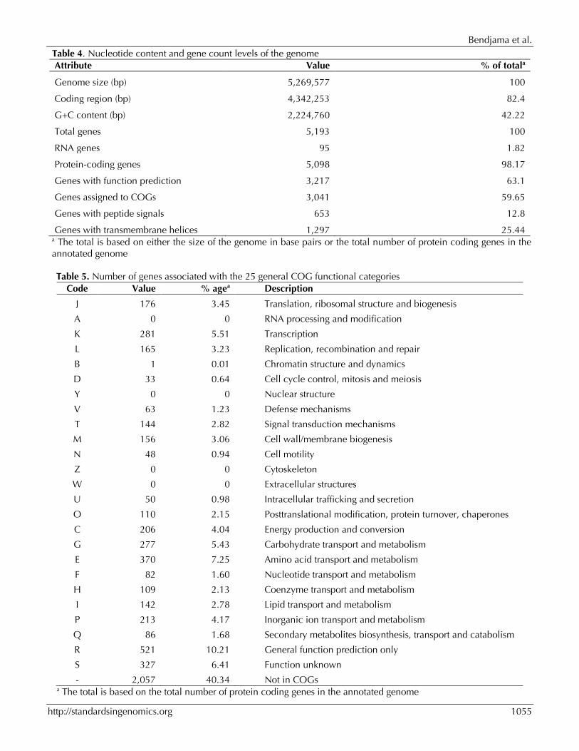
Genome properties The genome is 5,269,577 bp long with 42.22% GC content (Figure 6 and Table 4). It is composed of 46 contigs. Of the 5,193 predicted genes, 5,098 were protein-coding genes, and 95 were RNAs (10 genes encode 5S rRNA, 1 gene encodes 16S rRNA, 1 gene encodes 23S rRNA, 83 genes are tRNA genes). A total of 3,217 genes (63.1%) were as-signed a putative function (by cogs or by NR blast). 457 genes were identified as ORFans (8.96%). The remaining genes were annotated as hypothetical proteins (1,097 genes, 21.52%). The distribution of genes into COGs functional catego-ries is presented in Table 5. The properties and statistics of the genome are summarized in Tables 4 and 5.
Figure 6. Graphical circular map of the chromosome. From outside to the center: Red and gray bars representing contigs, genes on the forward strand colored by COG categories (only genes assigned to COG), genes on the reverse strand colored by COG categories (only genes assigned to COG), RNA genes (tRNAs green, rRNAs red), GC content. The inner-most circle shows the GC skew, purple and olive indicating negative and positive values, respectively.
Bendjama et al.
http://standardsingenomics.org 1055
Table 4. Nucleotide content and gene count levels of the genome Attribute Value % of totala
Genome size (bp) 5,269,577 100
Coding region (bp) 4,342,253 82.4
G+C content (bp) 2,224,760 42.22
Total genes 5,193 100
RNA genes 95 1.82
Protein-coding genes 5,098 98.17
Genes with function prediction 3,217 63.1
Genes assigned to COGs 3,041 59.65
Genes with peptide signals 653 12.8
Genes with transmembrane helices 1,297 25.44 a The total is based on either the size of the genome in base pairs or the total number of protein coding genes in the annotated genome
Table 5. Number of genes associated with the 25 general COG functional categories Code Value % agea Description
J 176 3.45 Translation, ribosomal structure and biogenesis
A 0 0 RNA processing and modification
K 281 5.51 Transcription
L 165 3.23 Replication, recombination and repair
B 1 0.01 Chromatin structure and dynamics
D 33 0.64 Cell cycle control, mitosis and meiosis
Y 0 0 Nuclear structure
V 63 1.23 Defense mechanisms
T 144 2.82 Signal transduction mechanisms
M 156 3.06 Cell wall/membrane biogenesis
N 48 0.94 Cell motility
Z 0 0 Cytoskeleton
W 0 0 Extracellular structures
U 50 0.98 Intracellular trafficking and secretion
O 110 2.15 Posttranslational modification, protein turnover, chaperones
C 206 4.04 Energy production and conversion
G 277 5.43 Carbohydrate transport and metabolism
E 370 7.25 Amino acid transport and metabolism
F 82 1.60 Nucleotide transport and metabolism
H 109 2.13 Coenzyme transport and metabolism
I 142 2.78 Lipid transport and metabolism
P 213 4.17 Inorganic ion transport and metabolism
Q 86 1.68 Secondary metabolites biosynthesis, transport and catabolism
R 521 10.21 General function prediction only
S 327 6.41 Function unknown
- 2,057 40.34 Not in COGs a The total is based on the total number of protein coding genes in the annotated genome

Bacillus algeriensis
1056 Standards in Genomic Sciences
Comparison with other species Bacil-lus genomes Here, we compared the genome of B. algeriensis strain EB01T with those of B. kribbensis strain DSM 17871, B. nealsonii strain AAU1, B. bataviensis strain LMG 21833, B. subtilis subsp. subtilis strain 168, B. vallismortis strain DV1-F-3, B. thuringiensis strain BMB171 and B. weihenstephanensis strain KBAB4 (Table 6). The draft genome of B. algeriensis (5.26Mb) is larger in size than those of B. kribbensis, B. nealsonii, B. subtilis subsp. subtilis and B. vallismortis (5.05, 4.98, 4.22 and 3.88 Mb, respectively) but smaller than those of, B. bataviensis, B. thuringiensis and B. weihenstephanensis (5.37, 5.64 and 5.87 Mb, re-spectively). B. algeriensis has a lower G+C content than B. kribbensis, B. subtilis subsp. subtilis and B. vallismortis (42.22% vs 43%, 43.5% and 43.8%, respectively) but higher than B. nealsonii, B. bataviensis, B. thuringiensis and B. weihenstephanensis (42.22% vs 35.1%, 39.6%,
35.2% and 35.5%, respectively). B. algeriensis has more predicted protein coding genes (5,098) than B. kribbensis, B. nealsonii, B. subtilis subsp. subtilis and B. vallismortis (4,918, 4,789, 4,175 and 4,097, respectively) but fewer protein coding genes than B. bataviensis,B. thuringiensis and B. weihenstephanensis (5,207, 5,352 and 5,653, re-spectively). In addition, B. algeriensis shared 1,804, 1,778, 2,017, 1,768, 1,985, 1,541, 1,863 orthologous genes with B. thuringiensis, B. nealsonii, B. bataviensis, B. subtilis subsp. subtilis, B. kribbensis, B. vallismortis, B. weihenstephanensis respectively.
The average nucleotide sequence identity of orthologous genes ranges from 64.54 to 91.06% among the 8 Bacillus species, and from 64.77 to 69.33% between Bacillus algeriensis and the other compared genomes (Table 7), thus confirming its new species status.
Table 6. Genomic comparison of B. algeriensis sp. nov. strain EB01T with seven other Bacillus species†.
Species Strain Genome accession number
Genome size (Mb)
G+C content
Bacillus algeriensis EB01T ERP003483 5.26 42.22
Bacillus kribbensis DSM 17871 AUMQ00000000.1 5.05 43
Bacillus nealsonii AAU1 ASRU00000000.1 4.98 35.1
Bacillus bataviensis LMG 21833 AJLS00000000.1 5.37 39.6
Bacillus subtilis subsp. subtilis 168 NC_000964.3 4.22 43.5
Bacillus vallismortis DV1-F-3 AFSH00000000.1 3.88 43.8
Bacillus thuringiensis BMB171 NC_014171.1 5.64 35.2
Bacillus weihenstephanensis KBAB4 NC_010184.1 5.87 35.5 † species and strain names, genome accession numbers, sizes and G+C contents.

Bendjama et al.
http://standardsingenomics.org 1057

Bacillus algeriensis
1058 Standards in Genomic Sciences
Conclusion On the basis of phenotypic (Table 2), phylogenetic and genomic analyses (taxonogenomics) (Table 6), we formally propose the creation of Bacillus algeriensis sp. nov. that contains the strain EB01T. This strain has been found in hypersaline lacus-trine sediment sample collected from Algeria.
Description of Bacillus algeriensis sp. nov. Bacillus algeriensis (al.ge.ri.en’sis. NL. masc.adj. algeriensis, of or pertaining to Algeria). Strain EB01T is a facultative anaerobic Gram-positive, endospore-forming, motile and rod shaped bacte-rium with rounded ends. Growth is achieved aer-obically between 30 and 55°C (optimum 37°C), between 0% and 2.5% NaCl concentration and pH in the range of 6.5-9 (optimum at pH 7). Growth is also observed in microaerophilic atmosphere, however, weak growth was observed under an-aerobic conditions. After 24h growth on 5% sheep blood-enriched Columbia agar (BioMerieux) at 37°C, bacterial colonies were smooth, light yellow with 2 mm in diameter. Cells have a length ranging from 2.4 μm to 4.9 μm (mean 3.6 μm) and a diam-eter ranging from 0.7 μm to 1.1 μm (mean 0.8 μm).
Catalase positive but oxidase negative. Using the commercially available API 50CH system (BioMerieux) according to the manufacturer’s in-structions, a weak positive reaction was observed for D-ribose, D-glucose, D-fructose, methyl α-D-glucopyranoside, N-acetylglucosamine, D-maltose,
D-lactose, D-melibiose, D-saccharose, D-trehalose, D-tagatose, and hydrolysis of starch. Other tests were negative. Using the API ZYM system (BioMerieux), positive reactions were observed for alkaline phosphatase, esterase (C4), esterase lipase (C8), leucine arylamidase, α chymotrypsin, β-glucuronidase, α-glucosidase, N-acetyl-glucos-aminidase and a weak positive reaction was ob-served for acid phosphatase. The nitrate reduction and β-galactosidase reaction was also positive, but urease and indole production were negative. B. algeriensis was susceptible to amoxicillin, nitrofurantoin, erythromycin, doxycycline, rifam-pin, vancomycin, gentamycin, imipenem, trime-thoprim-sulfamethoxazole, ciprofloxacin, ceftriax-one and amoxicillin/clavulanic acid, but resistant to nalidixic acid.
The G+C content of the genome is 42.22. The 16S rRNA and genome sequences are deposited in GenBank under accession numbers HG315679 and EMBL database under accession number ERP003483, respectively. The type strain EB01T (= CSUR P857 = DSM 27334) was isolated from sediment sample of the hypersaline lake Ezzemoul sabkha of Oum-El-Bouaghi region in northeastern Algeria.
Acknowledgements The authors thank Linda Hadjadj for technical as-sistance and Xegen company for automating the genome annotation process.
References1. Cohn F. Untersuchungen über Bakterien. Beitr
Biol Pflanz 1872; 1:127-224.
2. Parte AC. LPSN-list of prokaryotic names with standing in nomenclature. Nucleic Acids Res 2013; 42:613-616. PubMed http://dx.doi.org/10.1093/nar/gkt1111
3. Nicholson WL. Roles of Bacillus endospores in the environment. Cell Mol Life Sci 2002; 59:410-416. PubMed http://dx.doi.org/10.1007/s00018-002-8433-7
4. Moshafi MH, Forootanfar H, Ameri A, Shakibaie M, Noudeh G, Razavi M. Antimicrobial activity of Bacillus sp. strain FAS1 isolated from soil. Pak J Pharm Sci 2011; 24:269-275. PubMed
5. Bumpus SB, Evans BS, Thomas PM, Ntai I, Kelle-her NL. A proteomics approach to discovering natural products and their biosynthetic pathways.
Nat Biotechnol 2009; 27:951-960. PubMed http://dx.doi.org/10.1038/nbt.1565
6. Jernigan JA, Stephens DS, Ashford DA, Omenaca C, Topiel MS, Galbraith M, Tapper M, Fisk TL, Zaki S, Popovic T, et al. Bioterrorism-related inha-lational anthrax: the first 10 cases reported in the United States. Emerg Infect Dis 2001; 7:933-944. PubMed http://dx.doi.org/10.3201/eid0706.010604
7. Bottone EJ. Bacillus cereus, a volatile human pathogen. Clin Microbiol Rev 2010; 23:382-398. PubMed http://dx.doi.org/10.1128/CMR.00073-09
8. Mandell GL, Bennett JE, Dolin R. Principles and Practice of Infectious Diseases. Elsevier 2010, 4320p.
9. Stackebrandt E, Frederiksen W, Garrity GM, Grimont PA, Kämpfer P, Maiden MC, Nesme X, Rosselló-Mora R, Swings J, Trüper HG, et al. Re-
Bendjama et al.
http://standardsingenomics.org 1059
port of the ad hoc committee for the re-evaluation of the species definition in bacteriology. Int J Syst Evol Microbiol 2002; 52:1043-1047. PubMed http://dx.doi.org/10.1099/ijs.0.02360-0
10. Tindall BJ, Rossello-Mora R, Busse HJ, Ludwig W, Kampfer P. Notes on the characterization of pro-karyote strains for taxonomic purposes. Int J Syst Evol Microbiol 2010; 60:249-266. PubMed http://dx.doi.org/10.1099/ijs.0.016949-0
11. Stackebrandt E, Ebers J. Taxonomic parameters revisited: tarnished gold standards. Microbial To-day 2006; 33:152-155.
12. Rossello-Mora R. DNA-DNA Reassociation Methods Applied to Microbial Taxonomy and Their Critical Evaluation. In: Stackebrandt E (ed), Molecular Identification, Systematics, and popu-lation Structure of Prokaryotes. Springer, Berlin 2006. p. 23-50.
13. Welker M, Moore ER. Applications of whole-cell matrix-assisted laser-desorption/ionization time-of-flight mass spectrometry in systematic microbi-ology. Syst Appl Microbiol 2011; 34:2-11. PubMed http://dx.doi.org/10.1016/j.syapm.2010.11.013
14. Ramasamy D, Mishra AK, Lagier JC, Padhmanabhan R, Rossi M,Sentausa E, Raoult D, Fournier PE. A polyphasic strategy incorporating genomic data for the taxonomic description of novel bacterial species. Int J Syst Evol Microbiol 2014; 64:384-391.
15. Ramasamy D, Lagier JC, Gorlas A, Raoult D, Fournier PE. Non contiguous-finished genome se-quence and description of Bacillus massiliosenegalensis sp. nov. Stand Genomic Sci 2013; 8:264-278. PubMed http://dx.doi.org/10.4056/sigs.3496989
16. Keita MB, Diene SM, Robert C, Raoult D, Four-nier PE, Bittar F. Non-contiguous finished genome sequence and description of Bacillus massiliogorillae sp. nov. Stand Genomic Sci 2013; 9:93-105. PubMed http://dx.doi.org/10.4056/sigs.4388124
17. Mishra AK, Pfleiderer A, Lagier JC, Robert C, Raoult D, Fournier PE. Non-contiguous finished genome sequence and description of Bacillus massilioanorexius sp. nov. Stand Genomic Sci 2013; 8:465-479. PubMed http://dx.doi.org/10.4056/sigs.4087826
18. Kokcha S, Mishra AK, Lagier JC, Million M, Leroy Q, Raoult D, Fournier PE. Non contiguous-finished genome sequence and description of Ba-cillus timonensis sp. nov. Stand Genomic Sci
2012; 6:346-355. PubMed http://dx.doi.org/10.4056/sigs.2776064
19. Mishra AK, Lagier JC, Rivet O, Raoult D, Fournier PE. Non-contiguous finished genome sequence and description of Paenibacillus senegalensis sp. nov. Stand Genomic Sci 2012; 7:70-81. PubMed http://dx.doi.org/10.4056/sigs.3056450
20. Mishra AK, Lagier JC, Robert C, Raoult D, Four-nier PE. Genome sequence and description of Timonella senegalensis gen. nov., sp. nov., a new member of the suborder Micrococcinae. Stand Genomic Sci 2013; 8:318-335. PubMed http://dx.doi.org/10.4056/sigs.3476977
21. Ramasamy D, Lagier JC, Nguyen TT, Raoult D, Fournier PE. Non contiguous-finished genome se-quence and description of of Dielma fastidiosa gen. nov., sp. nov., a new member of the Family Erysipelotrichaceae. Stand Genomic Sci 2013; 8:336-351. PubMed http://dx.doi.org/10.4056/sigs.3567059
22. Mishra AK, Hugon P, Lagier JC, Nguyen TT, Couderc C, Raoult D, Fournier PE. Non contigu-ous-finished genome sequence and description of Enorma massiliensis gen. nov., sp. nov., a new member of the Family Coriobacteriaceae. Stand Genomic Sci 2013; 8:290-305. PubMed http://dx.doi.org/10.4056/sigs.3426906
23. Hugon P, Mishra AK, Lagier JC, Nguyen TT, Couderc C, Raoult D, Fournier PE. Non contigu-ous-finished genome sequence and description of Brevibacillus massiliensis sp. nov. Stand Genomic Sci 2013; 8:1-14. PubMed http://dx.doi.org/10.4056/sigs.3466975
24. Lagier JC, El Karkouri K, Mishra AK, Robert C, Raoult D, Fournier PE. Non-contiguous finished genome sequence and description of Enterobacter massiliensis sp. nov. Stand Genomic Sci 2013; 7:399-412. PubMed http://dx.doi.org/10.4056/sigs.3396830
25. Mishra AK, Lagier JC, Nguyen TT, Raoult D, Fournier PE. Non-contiguous finished genome se-quence and description of Peptoniphilus senegalensis sp. nov. Stand Genomic Sci 2013; 7:370-381. PubMed http://dx.doi.org/10.4056/sigs.3366764
26. Roux V, ElKarkouri K, Lagier JC, Robert C, Raoult D. Non-contiguous finished genome sequence and description of Kurthia massiliensis sp. nov. Stand Genomic Sci 2012; 7:221-232. PubMed http://dx.doi.org/10.4056/sigs.3206554
27. Woese CR, Kandler O, Wheelis ML. Towards a natural system of organisms: proposal for the do-

Bacillus algeriensis
1060 Standards in Genomic Sciences
mains Archaea, Bacteria, and Eucarya. Proc Natl Acad Sci USA 1990; 87:4576-4579. PubMed http://dx.doi.org/10.1073/pnas.87.12.4576
28. Gibbons NE, Murray RGE. Proposals concerning the higher taxa of bacteria. Int J Syst Bacteriol 1978; 28:1-6. http://dx.doi.org/10.1099/00207713-28-1-1
29. Murray RGE. The Higher Taxa, or, a Place for Everything...? In: Holt JG (ed). Bergey's Manual of Systematic Bacteriology, First Edition, Volume 1, The Williams and Wilkins Co, Baltimore 1984; p. 31-34.
30. Garrity GM, Holt JG. The Road Map to the Man-ual. In: Garrity GM, Boone DR, Castenholz RW (eds), Bergey's Manual of Systematic Bacteriolo-gy, Second Edition, Volume 1, Springer, New York, 2001, p. 119-169.
31. Ludwig W, Schleifer KH, Whitman WB. Class I. Bacilli class nov. In: De Vos P, Garrity GM, Jones D, N.R. Krieg W. Ludwig W, Rainey EA, Schleifer KH, Withman WB. Bergey's Manual of Systematic Bacteriology, second edition, vol 3 (The Firmicutes), Springer, Dordrecht, Heidelberg, London, New York 2009, pp.19-20.
32. List of new names and new combinations previ-ously effectively, but not validly, published. List no. 132. Int J Syst Evol Microbiol 2010; 60:469-472. http://dx.doi.org/10.1099/ijs.0.022855-0
33. Skerman VBD, McGowan V, Sneath PHA, eds. Approved Lists of Bacterial Names. Int J Syst Bacteriol 1980; 30:225-420. http://dx.doi.org/10.1099/00207713-30-1-225
34. Prévot AR. In: Hauduroy P, Ehringer G, Guillot G, Magrou J, Prévot AR, Rosset. Urbain A (eds) Dic-tionnaire des Bactéries Pathogènes, 2nd ed., Mas-son, Paris, 1953, pp. 1-692.
35. Fischer A. Untersuchungen über bakterien. Jahrbücher für Wissenschaftliche Botanik 1985; 27:1-163.
36. Gibson T, Gordon RE. Genus I. Bacillus Cohn 1872; 174; Nom. gen. cons. Nomencl. Comm. In-tern. Soc. Microbiol. 1937, 28; Opin. A. Jud. Comm. 1955, 39. In: Buchanan RE, Gibbons NE (eds), Bergey's Manual of Determinative Bacteri-ology, Eighth Edition, The Williams and Wilkins Co., Baltimore, 1974, p. 529-550.
37. Ashburner M, Ball CA, Blake JA, Botstein D, But-ler H, Cherry JM, Davis AP, Dolinski K, Dwight SS, Eppig J T et al. Gene ontology: tool for the unification of biology. The Gene Ontology Con-sortium. Nat Genet 2000; 25:25-29. PubMed http://dx.doi.org/10.1038/75556
38. Saitou N, Nei M. The neighbor-joining method: a new method for reconstructing phylogenetic trees. Mol Biol Evol 1987; 4:406-425. PubMed
39. Tamura K, Peterson D, Peterson N, Stecher G, Nei M, Kumar S. MEGA5: molecular evolutionary genetics analysis using maximum likelihood, evo-lutionary distance, and maximum parsimony methods. Mol Biol Evol 2011; 28:2731-2739. PubMed http://dx.doi.org/10.1093/molbev/msr121
40. Kanso S, Greene AC, Patel BKC. Bacillus subterraneus sp. nov., an iron- and manganese-reducing bacterium from a deep subsurface Aus-tralian thermal aquifer. Int J Syst Evol Microbiol 2002; 52:869-874. PubMed http://dx.doi.org/10.1099/ijs.0.01842-0
41. Tiago I, Pires C, Mendes V, Morais PV, da Costa MS, Verissimo A. Bacillus foraminis sp. nov., iso-lated from a non-saline alkaline groundwater. Int J Syst Evol Microbiol 2006; 56:2571-2574. PubMed http://dx.doi.org/10.1099/ijs.0.64281-0
42. Yoon JH, Kang SS, Lee KC, Kho YH, Choi SH, Kang KH, Park YH. Bacillus jeotgali sp. nov., iso-lated from jeotgal, Korean traditional fermented seafood. Int J Syst Evol Microbiol 2001; 51:1087-1092. PubMed http://dx.doi.org/10.1099/00207713-51-3-1087
43. Euzeby J. Validation List no. 117. List of new names and new combinations previously effec-tively, but not validly, published. Int J Syst Evol Microbiol 2007; 57:1933-1934. PubMed http://dx.doi.org/10.1099/ijs.0.65495-0
44. Perez-Ibarra BM, Flores ME, Garcia-Varela M. Isolation and characterization of Bacillus thioparus sp. nov., chemolithoautotrophic, thio-sulfate-oxidizing bacterium. FEMS Microbiol Lett 2007; 271:289-296. PubMed http://dx.doi.org/10.1111/j.1574-6968.2007.00729.x
45. Ahmed I, Yokota A, Fujiwara T. A novel highly boron tolerant bacterium, Bacillus boroniphilus sp. nov., isolated from soil, that requires boron for its growth. Extremophiles 2007; 11:217-224. PubMed http://dx.doi.org/10.1007/s00792-006-0027-0
46. Heyrman J, Vanparys B, Logan NA, Balcaen A, Rodrıguez-Dıaz M, Felske A, De Vos P. Bacillus novalis sp. nov., Bacillus vireti sp. nov., Bacillus soli sp. nov., Bacillus bataviensis sp. nov. and Ba-cillus drentensis sp. nov., from the Drentse A grasslands. Int J Syst Evol Microbiol 2004; 54:47-
http://www.ncbi.nlm.nih.gov/entrez/query.fcgi?cmd=Retrieve&db=PubMed&list_uids=2112744&dopt=Abstract
Bendjama et al.
http://standardsingenomics.org 1061
57. PubMed http://dx.doi.org/10.1099/ijs.0.02723-0
47. Venkateswaran K, Kempf M, Chen F, Satomi M, Nicholson W, Kern R. Bacillus nealsonii sp. nov., isolated from a spacecraft-assembly facility, whose spores are gamma-radiation resistant. Int J Syst Evol Microbiol 2003; 53:165-172. PubMed http://dx.doi.org/10.1099/ijs.0.02311-0
48. Lim JM, Jeon CO, Lee JR, Park DJ, Kim CJ. Bacil-lus kribbensis sp. nov., isolated from a soil sample in Jeju, Korea. Int J Syst Evol Microbiol 2007; 57:2912-2916. PubMed http://dx.doi.org/10.1099/ijs.0.65227-0
49. Seng P, Rolain JM, Fournier PE, La Scola B, Drancourt M, Raoult D. MALDI-TOF-mass spec-trometry applications in clinical microbiology. Fu-ture Microbiol 2010; 5:1733-1754. PubMed http://dx.doi.org/10.2217/fmb.10.127
50. Seng P, Drancourt M, Gouriet F, La Scola B, Fournier PE, Rolain JM, Raoult D. Ongoing revo-lution in bacteriology: routine identification of bacteria by matrix-assisted laser desorption ioni-zation time-of-flight mass spectrometry. Clin In-fect Dis 2009; 49:543-551. PubMed http://dx.doi.org/10.1086/600885
51. Field D, Garrity G, Gray T, Morrison N, Selengut J, Sterk P, Tatusova T, Thomson N, Allen MJ, Angiuoli SV, et al. The minimum information about a genome sequence (MIGS) specification. Nat Biotechnol 2008; 26:541-547. PubMed http://dx.doi.org/10.1038/nbt1360
52. fastq-mcf program. http://code.google.com/p/ea-utils
53. Prodigal. http://prodigal.ornl.gov
54. GenBank database. http://www.ncbi.nlm.nih.gov/genbank
55. RNAmmer 1.2 Server. http://www.cbs.dtu.dk/services/RNAmmer/
56. Lagesen K, Hallin P, Rodland EA, Staerfeldt HH, Rognes T, Ussery DW. RNAmmer: consistent and rapid annotation of ribosomal RNA genes. Nucle-ic Acids Res 2007; 35:3100-3108. PubMed http://dx.doi.org/10.1093/nar/gkm160
57. Lowe TM, Eddy SR. tRNAscan-SE: a program for improved detection of transfer RNA genes in ge-nomic sequence. Nucleic Acids Res 1997; 25:955-964. PubMed http://dx.doi.org/10.1093/nar/25.5.0955
58. Kall L, Krogh A, Sonnhammer EL. Advantages of combined transmembrane topology and signal peptide prediction--the Phobius web server. Nu-cleic Acids Res 2007; 35:W429-W432. PubMed http://dx.doi.org/10.1093/nar/gkm256
59. Rutherford K, Parkhill J, Crook J, Horsnell T, Rice P, Rajandream MA, Barrell B. Artemis: sequence visualization and annotation. Bioinformatics 2000; 16:944-945. PubMed http://dx.doi.org/10.1093/bioinformatics/16.10.944
60. Carver T, Thomson N, Bleasby A, Berriman M, Parkhill J. DNAPlotter: circular and linear interac-tive genome visualization. Bioinformatics 2009; 25:119-120. PubMed http://dx.doi.org/10.1093/bioinformatics/btn578
61. Lechner M, Findeiss S, Steiner L, Marz M, Stadler PF, Prohaska SJ. Proteinortho: detection of (co-)orthologs in large-scale analysis. BMC Bioinfor-matics 2011; 12:124-133. PubMed http://dx.doi.org/10.1186/1471-2105-12-124