Nasal-Septal Fractures Francis B. Quinn, M.D. Herve’ J. LeBoeuf, M.D.
26
Nasal-Septal Fractures Francis B. Quinn, M.D. Herve’ J. LeBoeuf, M.D.
-
Upload
jasmin-fort -
Category
Documents
-
view
222 -
download
0
Transcript of Nasal-Septal Fractures Francis B. Quinn, M.D. Herve’ J. LeBoeuf, M.D.
Nasal-Septal Fractures
Francis B. Quinn, M.D.Herve’ J. LeBoeuf, M.D.
Anatomy
Bones - Frontal process of maxilla,
nasal spine of frontal bone Paired nasal bones Vomer Perpendicular plate of the
ethmoid
Anatomy (cont.)
Cartilage- Lower lateral cartilage Upper lateral (Alar) cartilage Septal cartilage Sesamoid cartilages
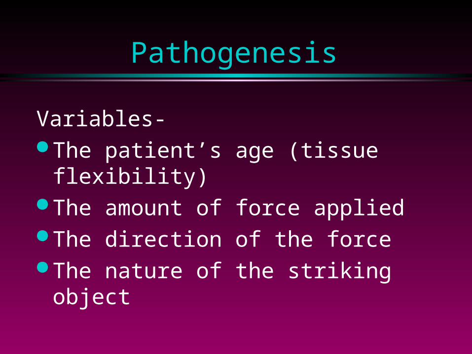
Pathogenesis
Variables- The patient’s age (tissue
flexibility) The amount of force applied The direction of the force The nature of the striking object
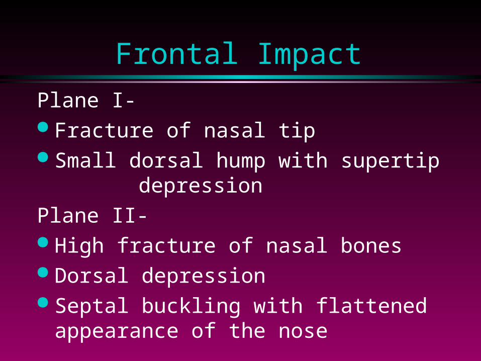
Frontal Impact
Plane I- Fracture of nasal tip Small dorsal hump with supertip
depressionPlane II- High fracture of nasal bones Dorsal depression Septal buckling with flattened
appearance of the nose
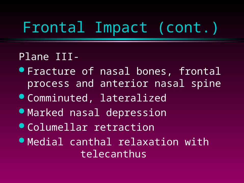
Frontal Impact (cont.)
Plane III- Fracture of nasal bones, frontal
process and anterior nasal spine Comminuted, lateralized Marked nasal depression Columellar retraction Medial canthal relaxation with
telecanthus
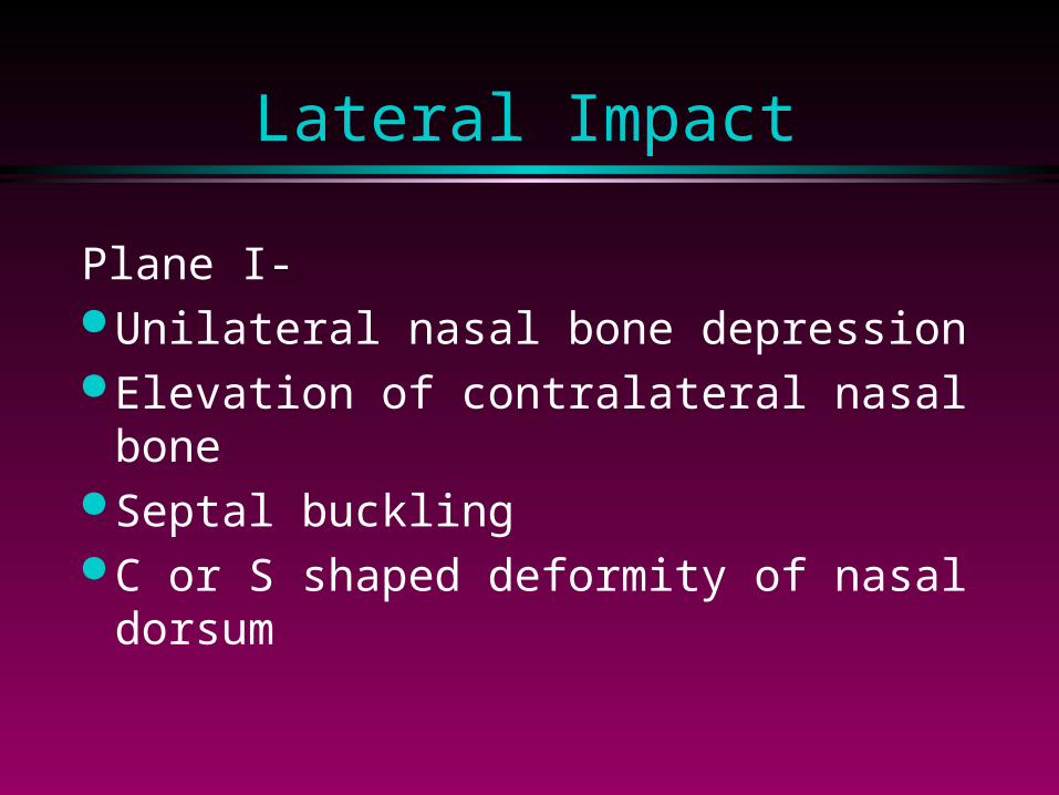
Lateral Impact
Plane I- Unilateral nasal bone depression Elevation of contralateral nasal
bone Septal buckling C or S shaped deformity of nasal
dorsum
Lateral Impact (cont.)
Plane II/III- Fracture extension to frontal
process Marked displacement of
septum and dorsum Medial maxillary wall
depression
Septal Fracture
Vertical with anterior fracture Horizontal with posterior fracture S and C shaped deformities with
healing Telescoping of segments prevents
closed reduction
History
Force, direction of impact Epistaxis External deformity Prior nasal injury, dysfunction Pre-injury photographs

Exam
Nasal deviation Mucosal or skin lacerations Ecchymosis, hematoma Lid edema, chemosis Subconjunctival hemorrhage Telecanthus, CSF rhinorrhea
Exam (cont.)
Topical decongestion Debridement of clots Internal and external palpation Exam of cartilaginous nose Roentgenograms Photographic documentation
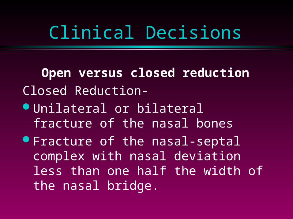
Clinical Decisions
Open versus closed reductionClosed Reduction- Unilateral or bilateral fracture of the
nasal bones Fracture of the nasal-septal complex
with nasal deviation less than one half the width of the nasal bridge.
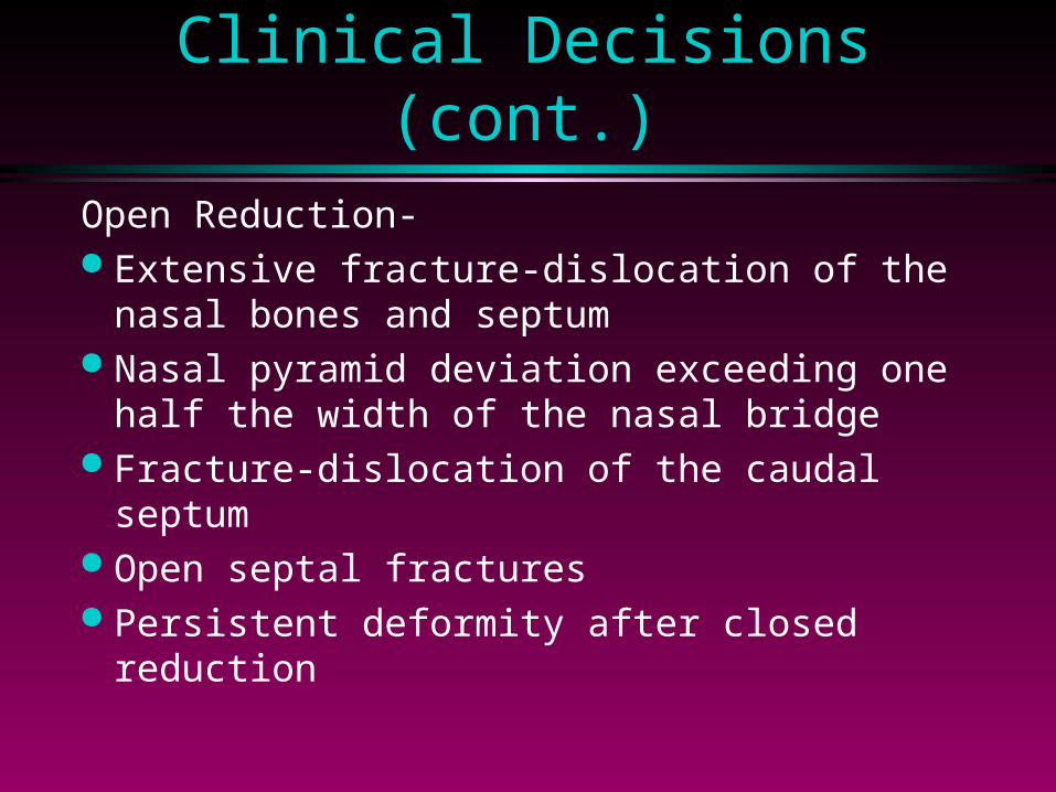
Clinical Decisions (cont.)Open Reduction- Extensive fracture-dislocation of the nasal
bones and septum Nasal pyramid deviation exceeding one
half the width of the nasal bridge Fracture-dislocation of the caudal septum Open septal fractures Persistent deformity after closed reduction
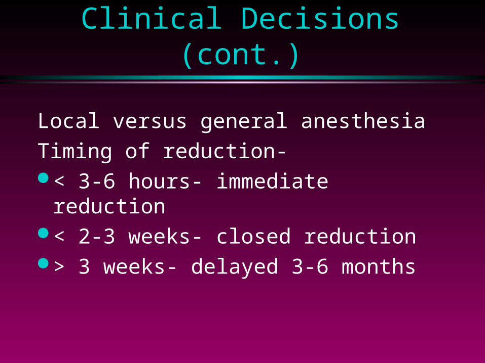
Clinical Decisions (cont.)
Local versus general anesthesiaTiming of reduction- < 3-6 hours- immediate
reduction < 2-3 weeks- closed reduction > 3 weeks- delayed 3-6 months
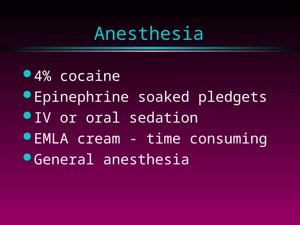
Anesthesia
4% cocaine Epinephrine soaked pledgets IV or oral sedation EMLA cream - time consuming General anesthesia
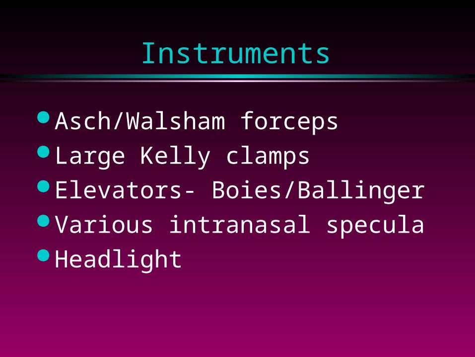
Instruments
Asch/Walsham forceps Large Kelly clamps Elevators- Boies/Ballinger Various intranasal specula Headlight
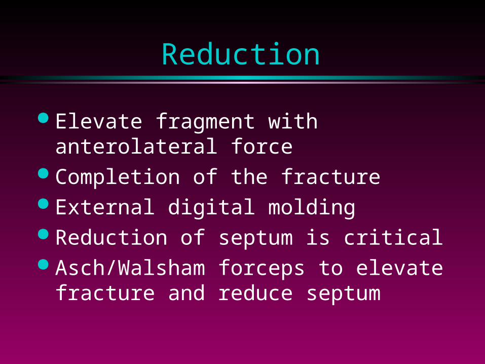
Reduction
Elevate fragment with anterolateral force
Completion of the fracture External digital molding Reduction of septum is critical Asch/Walsham forceps to elevate
fracture and reduce septum
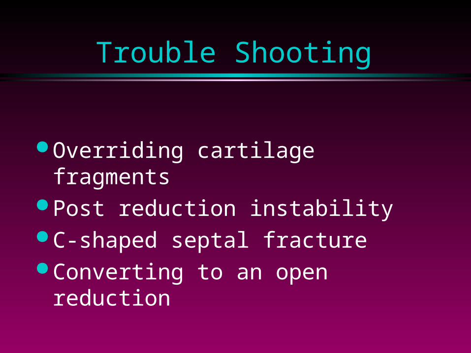
Trouble Shooting
Overriding cartilage fragments Post reduction instability C-shaped septal fracture Converting to an open
reduction
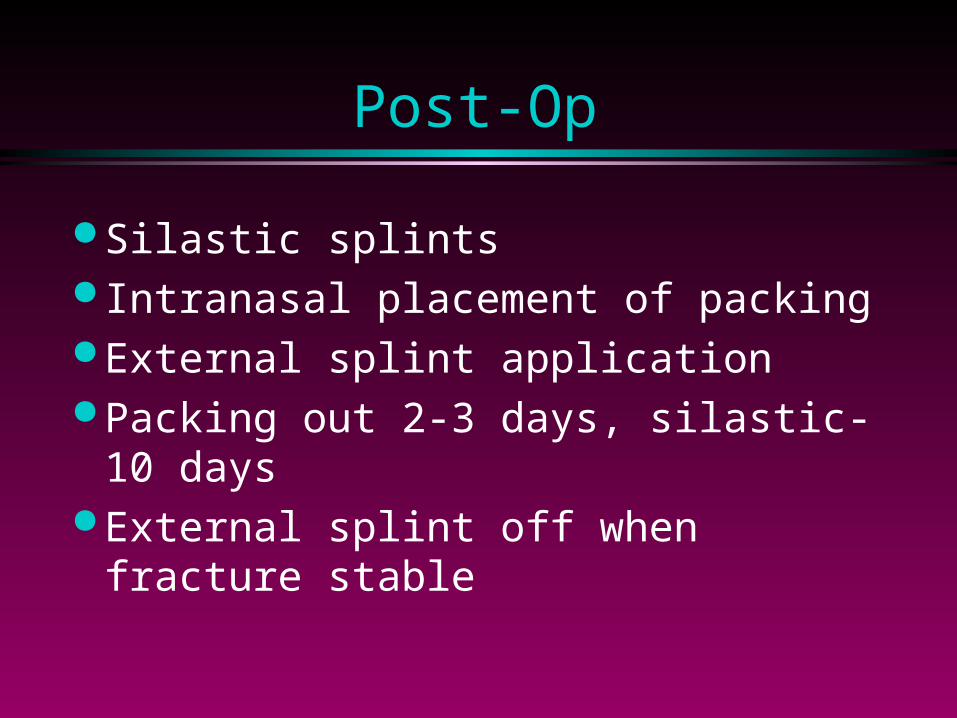
Post-Op
Silastic splints Intranasal placement of packing External splint application Packing out 2-3 days, silastic-10
days External splint off when fracture
stable
Subacute Open Reduction
Hemitransfixion, lateral intercartilaginous incisions
Elevation of dorsal skin and periosteum
Exposure of cartilage segments Reduction of cartilage- scoring, suture Maxillary crest involvement-
“trapdoor”
Complicated Fractures
“Open sky” approach Use preexisting lacerations when
possible Depressed comminuted fractures-
wires versus miniplates Wound closure Prophylactic antibiotics
Delayed Repair
Complicated due to scarring, fibrosis Common problems: Dorsal hump,
C/S shaped septum, saddle deformities, septal displacement, fallen or deviated tip
Common solutions: Excision of hump, cartilage grafting, calvarial grafts, osteotomies
Children
Physical differences- projection, cartilage: bone, growth centers
Small fracture--- obstruction with age Edema, anxiety tend to obscure fracture Operative intervention- cosmesis,
obstruction Digital compression Neonatal fracture-dislocation
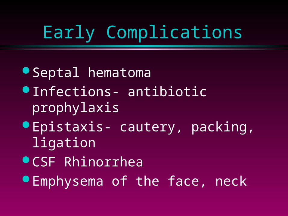
Early Complications
Septal hematoma Infections- antibiotic prophylaxis Epistaxis- cautery, packing,
ligation CSF Rhinorrhea Emphysema of the face, neck
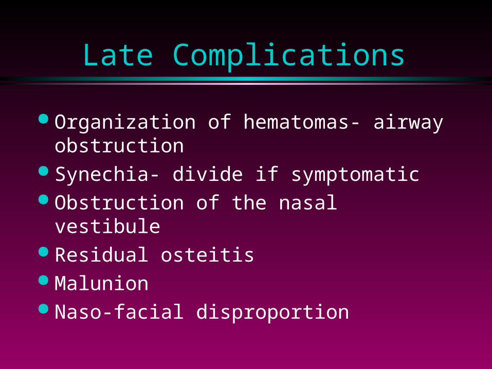
Late Complications
Organization of hematomas- airway obstruction
Synechia- divide if symptomatic Obstruction of the nasal vestibule Residual osteitis Malunion Naso-facial disproportion