Membranes basales glomérulaires minces: une lésion courante.
53
Membranes basales glomérulaires minces: une lésion courante. Marie Claire Gubler/ Laurence Heidet INSERM U574 / MARHEA Hôpital Necker-Enfants Malades Université Paris Descartes Paris Actualités Néphrologiques Jean Hamburger Paris 28-30 avril 2008
Transcript of Membranes basales glomérulaires minces: une lésion courante.

Membranes basales glomérulaires minces: une lésion courante.
Marie Claire Gubler/ Laurence HeidetINSERM U574 / MARHEAHôpital Necker-Enfants MaladesUniversité Paris DescartesParis
Actualités Néphrologiques Jean HamburgerParis 28-30 avril 2008

Thin glomerular basement membrane
1973 : Thin GBM> Familial Benign
Essential HematuriaPM Rogers
1972-1973 : Thick and split GBM
Alport SyndromeN Hinglais, GS Spear, J Churg

The nonspecificity of the ultrastructural alterations in hereditary nephritis and benign
familial hematuria (G Hill, Lab Invest 1974)
• No single morphologic parameter (GBM splitting or attenuation) or group of parameters is diagnostic of either hereditary nephritis or benign familial hematuria.

Thin glomerular basement membrane
• Normal GBM thickness• Thin GBM and familial (or sporadic) benign hematuria• Thin GBM, always a benign condition ?• Thin GBM and associated glomerular diseases
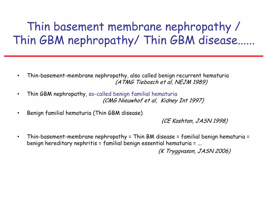
Thin basement membrane nephropathy /Thin GBM nephropathy/ Thin GBM disease......
• Thin-basement-membrane nephropathy, also called benign recurrent hematuria (ATMG Tiebosch et al, NEJM 1989)
• Thin GBM nephropathy, so-called benign familial hematuria(CMG Nieuwhof et al, Kidney Int 1997)
• Benign familial hematuria (Thin GBM disease)(CE Kashtan, JASN 1998)
• Thin-basement-membrane nephropathy = Thin BM disease = familial benign hematuria = benign hereditary nephritis = familial benign essential hematuria = ...
(K Tryggvason, JASN 2006)

Normal GBM Thickness
• Birth: 100 nm• Childhood: 100 > 300 nm (3-5 ans)
• Adulthood : 315 +/-98 nm (Osawa, 1966) (Normal kidneys) 356 - 464 nm (FE Dische, 1990) (IgA Glomerulopathies)418 +/- 48 nm (S Saxena, 1990) (Normal renal biopsies)
According to sex :Male: 330 +/- 50 nm (M Haas, 2006) (Nephrosis, AIN)Female: 305 +/- 45 nm
Male: 343 +/- 39 nm (CS Rayat, 2005) (Normal kidneys)Female: 300 +/- 12 nm
Male: 364 +/- 56 nm (TMG Tiebosch, 1989) (Normal renal biopies Female: 350 +/- 45 nm IgA Glomerulopathies)
Male: 373 +/- 42 nm (MW Steffes, 1983) (Kidneys from donors Female: 326 +/- 45 nm 118 cases)
Normal GBM ThicknessWhy so different?
• Differences in the processing of specimens (fixation, inclusion) for electron microscopy.
• Differences in the number of measures, and the methods for measuring GBM thickness (distance between the endothelial and epithelial cell membranes apposed to the GBM)
– Measures performed at regular intervals along the GBM without any selection
– Measures avoiding tangential sections of the GBM
– Measures made where the lines of an overlaid grid cut the endothelium, with or without exclusion of the tangential sections.
• Each laboratory has to establish its own standard.
• At least two glomeruli have to be systematically examined.
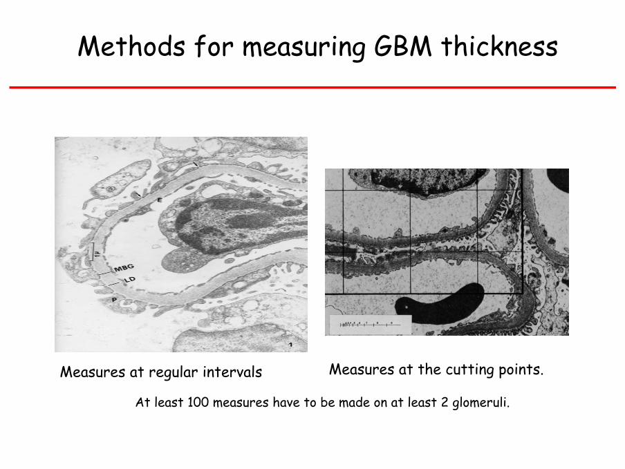
Methods for measuring GBM thickness
At least 100 measures have to be made on at least 2 glomeruli.
Measures at regular intervals Measures at the cutting points.

Normal GBM Thickness (G Osawa, 1966)
• Nine renal biopsies from healthy volunteers and 14 autopsy specimens• Individuals aged 22 to 92 years. No relationship between age and sex
and GBM thickness• “There was great irregularity in width”

Thin GBM. Definition criteria, prevalence
• Definition of thin GBM.Thickness: In adults < 200 nm
< 250 nm (most studies)< 300nm
In children <180 nm
Extension : Diffuse> 50%between 10 and 50%
• PrevalenceIn the general population : 1-10%Among renal biopsies : 0.8-11%In hematuric patients : 10-50%

Ultrastuctural study of the GBM
Normal GBM Thin GBM
GBM in “TBMD” is remarkably thin (G Basta-Jovanovic, J Churg.1990)
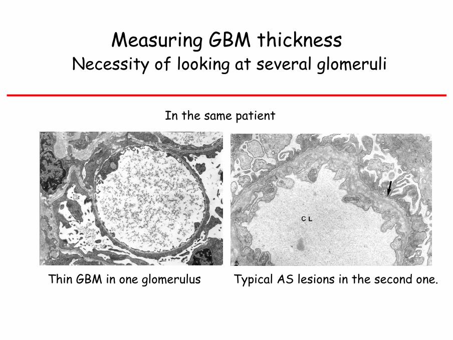
Measuring GBM thickness Necessity of looking at several glomeruli
In the same patient
Thin GBM in one glomerulus Typical AS lesions in the second one.
Thin glomerular basement membrane
• Normal GBM thickness• Thin GBM and familial (or sporadic) benign hematuria• Thin GBM, always a benign condition ?• Thin GBM and associated glomerular diseases

Familial benign essential hematuriaPW Rogers et al. Arch Intern Med, 1973
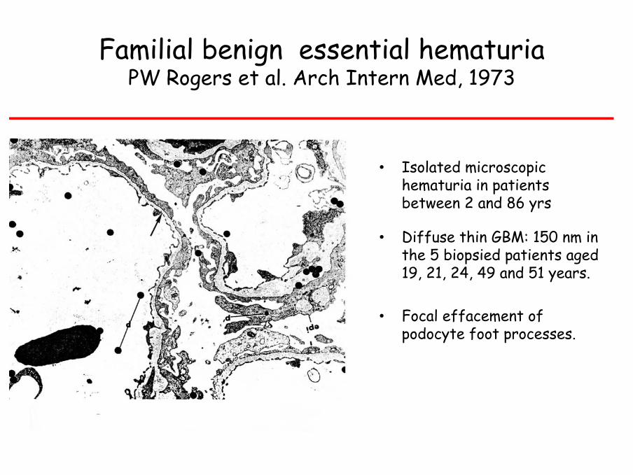
Familial benign essential hematuriaPW Rogers et al. Arch Intern Med, 1973
• Isolated microscopic hematuria in patients between 2 and 86 yrs
• Diffuse thin GBM: 150 nm in the 5 biopsied patients aged 19, 21, 24, 49 and 51 years.
• Focal effacement of podocyte foot processes.
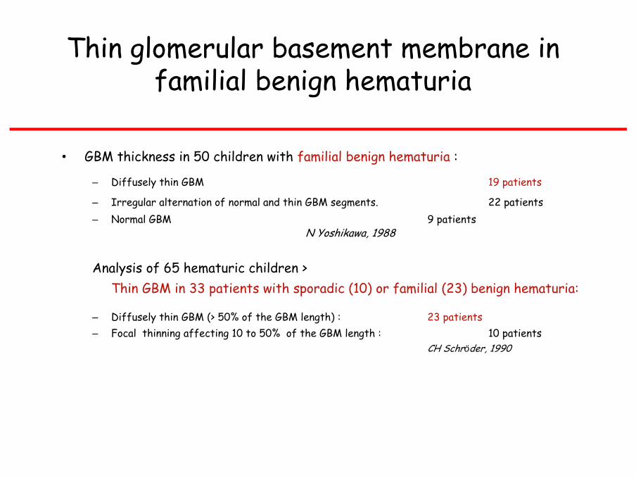
Thin glomerular basement membrane in familial benign hematuria
• GBM thickness in 50 children with familial benign hematuria :
– Diffusely thin GBM 19 patients
– Irregular alternation of normal and thin GBM segments. 22 patients – Normal GBM 9 patients
N Yoshikawa, 1988
Analysis of 65 hematuric children > Thin GBM in 33 patients with sporadic (10) or familial (23) benign hematuria:
– Diffusely thin GBM (> 50% of the GBM length) : 23 patients– Focal thinning affecting 10 to 50% of the GBM length : 10 patients
CH Schröder, 1990

Benign familial hematuria with thin GBM
C3
α5

Thin glomerular basement membrane

Prevalence of thin GBM in the general population (FE Dische, 1990)
• According to their definition of normal GBM thickness (330-460 nm), the incidence of thin GBM in the general population lies between 5.2 and 9.2 %.

Incidence of “thin-membrane nephropathy ” in non-transplant renal biopsies (Aarons et al, 1989)
• Patients (8-63 yrs) presenting with hematuria and/or proteinuria were studied during a seven-year period.
• >>>> 110 patients (11%) (81F, 30M) with GBM attenuation – Mild in 39 pts 241 nm– Moderate/severe in 71 pts 214 nm
Controls 320 nm
Proteinuria (45%), familial kidney disease (40%), deafness in the family (10%)
“Long-term prospective follow-up is necessary to evaluate the nature and effects of this entity”

Incidence of “thin-membrane nephropathy ” in hematuric patients (GJ Perry, 1989)
“Thin-membrane nephropathy”diagnosis in 26 (28%) hematuric patients (Mean age: 42 yrs)
“Limited follow-up has confirmed the good pronostic of the condition”
But 4 patients developed Pu 4 patients have a family history of nephritis.
NB: controls: MCD or AIN)
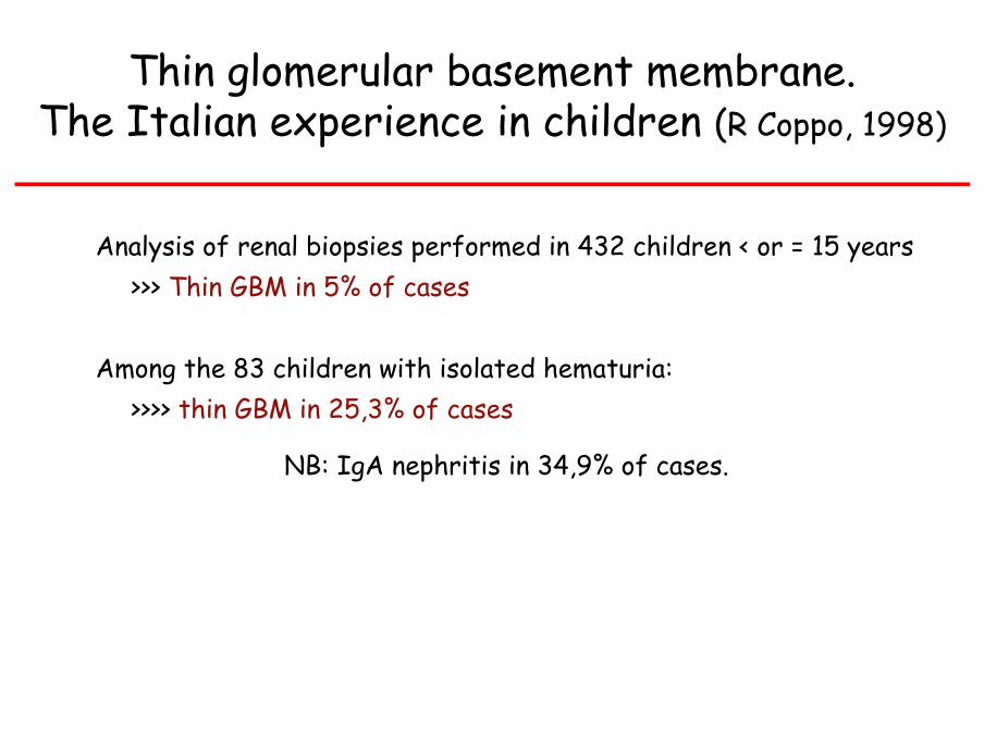
Thin glomerular basement membrane.The Italian experience in children (R Coppo, 1998)
Analysis of renal biopsies performed in 432 children < or = 15 years>>> Thin GBM in 5% of cases
Among the 83 children with isolated hematuria:>>>> thin GBM in 25,3% of cases
NB: IgA nephritis in 34,9% of cases.

Benign familial hematuria is due to mutation of the type IV collagen α4 gene.
Lemmink, 1996
HH Lemmink, 1996

• Heterozygous COL4A3 or COL4A4 mutations are associated with familial benign hematuria in autosomal recessive Alport syndromefamilies (Lamprecht, 1996; Boye, 1998; Heidet, 2000, Buzza, 2001, Longo, 2002 )
• Heterozygous COL4A3 or COL4A4 mutations are associated with familial benign hematuria in families without autosomal recessive Alport syndrome (Badenas, 2002; Buzza, 2003; Gross, 2003)
• > So, a certain proportion of patients with BFM and TGBM have a type IV collagen disease, and are carriers of autosomal recessive Alport syndrome.
Benign familial hematuria, a type IV collagen disorder

Benign familial hematuria with thin GBM not linked to the COL4A3/COL4A4 locus.
Yamazaki, 1995; Piccini 1999
8 unrelated kindreds from eastern Sicilia.
Hematuric pts: 4.5-61 years.
Exclusion ofCOL4A3,
COL4A4,COL4A5.

Autosomal recessive Alport syndrome Phenotype of heterozygous carriers
• No symptom
• Persistent or intermittent microscopic hematuria :
> Clinical features of familial benign hematuria.
Rare cases of slowly progressive nephropathy.
Thin GBM in all studied carriers
COL4A4 mutation

Thin glomerular basement membrane
• Normal GBM thickness• Thin GBM and familial (or sporadic) benign hematuria• Thin GBM, always a benign condition ?• Thin GBM and associated glomerular diseases

Thin basement membrane nephropathy /Thin GBM nephropathy/ Thin GBM disease......
Always a benign condition ?
• Proteinuria > 500mg/24h: 6/18 patients180-500mg/24h: 5/18 patients
AT�MG Tiebosch, 1989
• Proteinuria 200-500mg/24h: 50/110 patientsRF (2 pts), HTA (21 pts) Family history of nephropathy or deafness 44/110 patients
I Aarons, 1989
• Proteinuria and/or RF and/orfamily history of progressive nephropathy 7/14 patients
FE Dische 1985
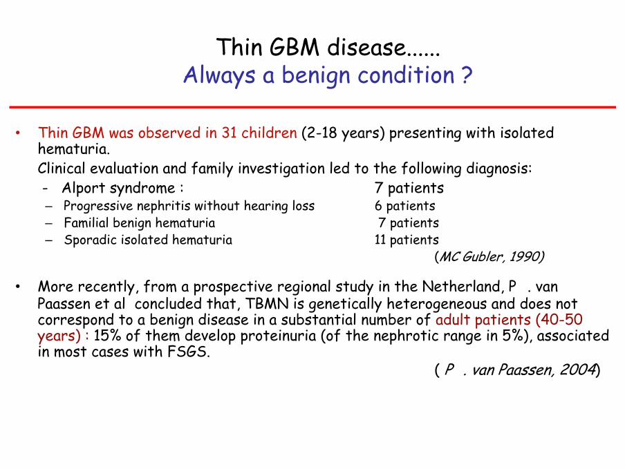
Thin GBM disease......Always a benign condition ?
• Thin GBM was observed in 31 children (2-18 years) presenting with isolated hematuria. Clinical evaluation and family investigation led to the following diagnosis:- Alport syndrome : 7 patients – Progressive nephritis without hearing loss 6 patients– Familial benign hematuria 7 patients– Sporadic isolated hematuria 11 patients
(MC Gubler, 1990)
• More recently, from a prospective regional study in the Netherland, P�. van Paassen et al concluded that, TBMN is genetically heterogeneous and does not correspond to a benign disease in a substantial number of adult patients (40-50 years) : 15% of them develop proteinuria (of the nephrotic range in 5%), associated in most cases with FSGS.
( P�. van Paassen, 2004)

Thin GBM disease......Always a benign condition ?
• Thin GBM is not a specific lesion.• It may be observed in progressive hereditary nephritis
– X-linked Alport syndrome– Autosomal recessive Alport syndrome– Autosomal dominant Alport syndrome– Progressive hematuric nephritis without extra-renal symptoms.
• In these conditions, the finding of thin GBM is more frequent in children than in adults but may be the only GBM lesion observed all along the life.

Thin GBM in X-linked Alport syndrome.
One 4 year-old hematuric boy without extrarenal symptoms or family history
De novo COL4A5 mutation α1
α5
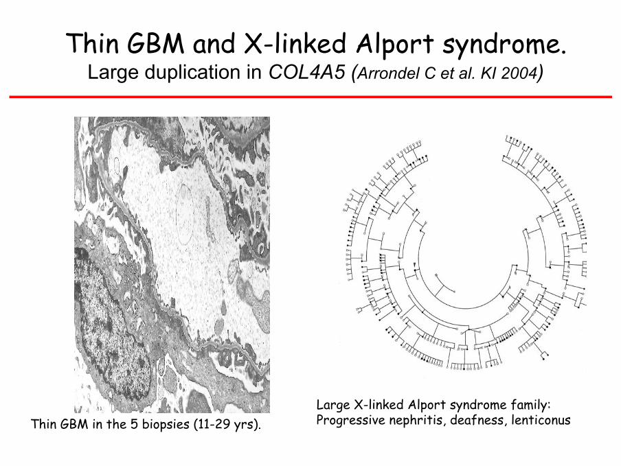
Thin GBM and X-linked Alport syndrome.Large duplication in COL4A5 (Arrondel C et al. KI 2004)
Thin GBM in the 5 biopsies (11-29 yrs).Large X-linked Alport syndrome family:Progressive nephritis, deafness, lenticonus

Thin GBM in X-linked Alport syndrome.
α5

Thin GBM in autosomal recessive Alport syndrome.
2 yrs: hematuria + Pu 100 mgno extrarenal symptomsno family history
15 yrs: nephrotic syndrome 20 yrs: CRI22 yrs: ESRDMutations COL4A3
5yrs
16yrs

Autosomal recessive Alport syndrome Expression of type IV collagen chains
Control Patient
α5 α5
α5α5

A/G A/G A/G A/G A/G A/G
Autosomal dominant Alport syndromeHeterozygous COL4A3 mutation :
Large family with autosomal dominant Alport syndrome
Jefferson et al. 1997: Linkage to COL4A3-COL4A4. van der Loop et al. 2000: Heterozygous splice mutation in COL4A3
GBM : irregular thickeningIF : α3−α5 +

• Analysis of 25 individuals in 4 families with heterozygous mutation in COL4A3 (2 families) or COL4A4 (2 families)
Phenotypic heterogeneity:– Renal invovement:
– ESRD between 41 and 70 years in 5 patients– Chronic renal failure between 46 and 65 years in 4 patients– Isolated hematuria between 22 and 51 in 12 patients– No symptom in 3 patients 11, 22 and 90 year-old
– Hearing involvement :• Hearing defect in 4 patients after 45 years
– No ocular involvement– Normal expression of type IV collagen chains
GBM: thick and split in three familiesThin in one family
Pescucci et al. KI 2004
Autosomal dominant Alport syndromeSpectrum of phenotypes associated with heterozygous
COL4A3-COL4A4 mutation :
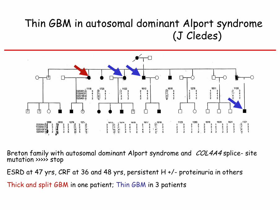
Thin GBM in autosomal dominant Alport syndrome(J Cledes)
Breton family with autosomal dominant Alport syndrome and COL4A4 splice- site mutation >>>>> stop
ESRD at 47 yrs, CRF at 36 and 48 yrs, persistent H +/- proteinuria in others
Thick and split GBM in one patient; Thin GBM in 3 patients

COL4A3/COL4A4 mutations producing focal segmental glomerulosclerosis and renal failure in thin
basement membrane nephropathyK Voskarides et al, JASN 2007
• 13 Cypriot families (82 patients).• Microscopic hematuria in adolescence and early adult life• Follow-up of 3 decades.• Progressive development of proteinuria, nephrotic syndrome and
CRF (37.8%) and ESRD (19.5%).• Absence of extrarenal symptoms.• Mutations COL4A3: G1334E (7 families) and G871C (3 families)
COL4A4: 2854delG (1 family)
• Renal biopsies >>> dual diagnosis of“Thin basement membrane nephropathy”and “focal segmental glomerular sclerosis”

COL4A3/COL4A4 mutations producing focal segmental glomerulosclerosis and renal failure in thin basement
membrane nephropathyK Voskarides et al, JASN 2008

.
COL4A3/COL4A4 mutations producing focal segmental glomerulosclerosis and renal failure in thin basement
membrane nephropathyK Voskarides et al, JASN 2008
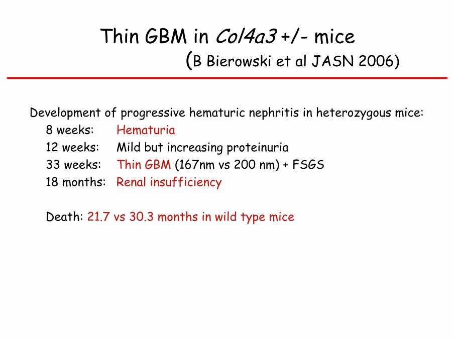
Thin GBM in Col4a3 +/- mice(B Bierowski et al JASN 2006)
Development of progressive hematuric nephritis in heterozygous mice:8 weeks: Hematuria12 weeks: Mild but increasing proteinuria33 weeks: Thin GBM (167nm vs 200 nm) + FSGS18 months: Renal insufficiency
Death: 21.7 vs 30.3 months in wild type mice
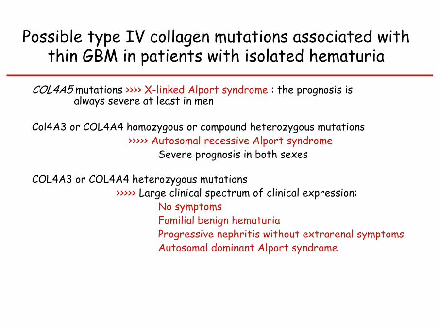
Possible type IV collagen mutations associated with thin GBM in patients with isolated hematuria
COL4A5 mutations >>>> X-linked Alport syndrome : the prognosis is always severe at least in men
Col4A3 or COL4A4 homozygous or compound heterozygous mutations>>>>> Autosomal recessive Alport syndrome
Severe prognosis in both sexes
COL4A3 or COL4A4 heterozygous mutations >>>>> Large clinical spectrum of clinical expression:
No symptomsFamilial benign hematuriaProgressive nephritis without extrarenal symptomsAutosomal dominant Alport syndrome
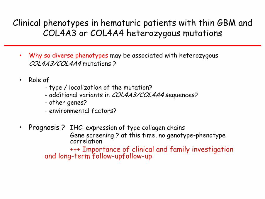
Clinical phenotypes in hematuric patients with thin GBM and COL4A3 or COL4A4 heterozygous mutations
• Why so diverse phenotypes may be associated with heterozygousCOL4A3/COL4A4 mutations ?
• Role of- type / localization of the mutation?- additional variants in COL4A3/COL4A4 sequences?- other genes?- environmental factors?
• Prognosis ? IHC: expression of type collagen chainsGene screening ? at this time, no genotype-phenotype correlation+++ Importance of clinical and family investigation
and long-term follow-upfollow-up

Thin GBM and kidney donationin families with progressive hematuric nephritis
• Difficult ethical question when heterozygous relatives (asymptomatic or hematuric) are willing to serve as kidney donors, usually for their children.
• They should not be accepted if they have proteinuria, HTA or sensorineural deafness.
• They may be accepted if they are strongly motivated, well informed and asymptomatic.
• Or if they have isolated hematuria over a certain age (for ex 40-50 years)

Thin glomerular basement membrane
• Normal GBM thickness• Thin GBM and familial (or sporadic) benign hematuria• Thin GBM, always a benign condition ?• Thin GBM and associated glomerular diseases

Thin GBM observed in other glomerulopathies
• Thin GBM has been observed in patients with:
– Steroid sensitive nephrotic syndrome with minimal glomerular changes– Nephrotic syndrome with FSGS– Diabetic nephropathy– Membranous nephropathy– Mesangioproliferative glomerulopathy……
In patients with this association, microscopic hematuria is more frequent than in controls
But no difference in the clinical course of the associated disease has been observed
“Thin GBM is a developmental abnormality of little or no significance”. (Sue, Nephrology 2004)
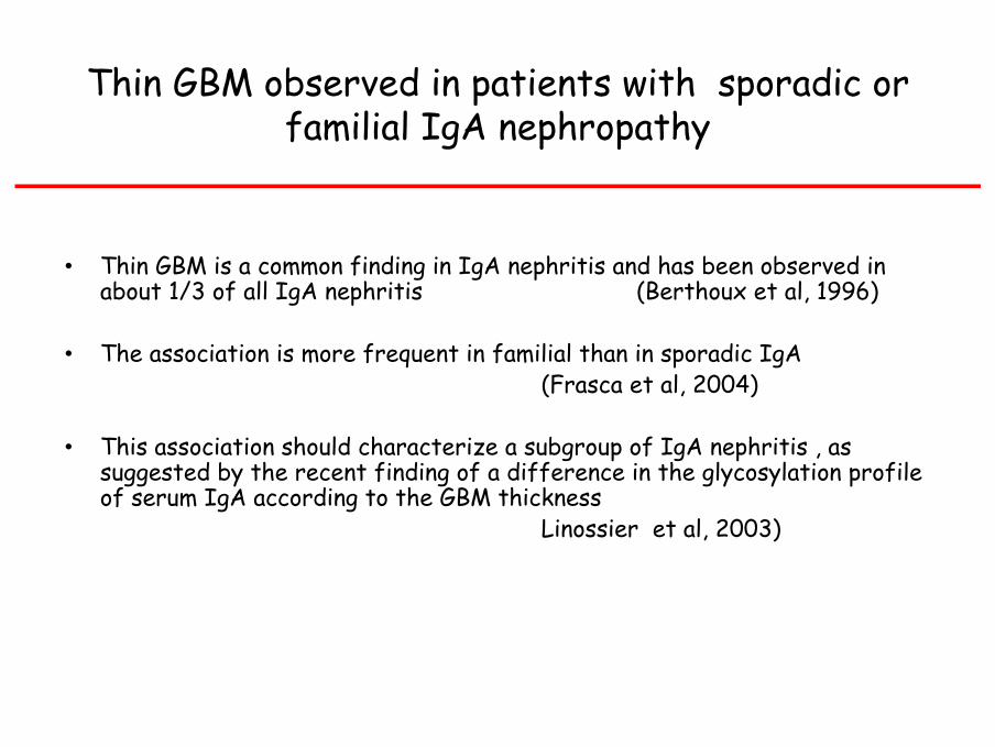
Thin GBM observed in patients with sporadic or familial IgA nephropathy
• Thin GBM is a common finding in IgA nephritis and has been observed in about 1/3 of all IgA nephritis (Berthoux et al, 1996)
• The association is more frequent in familial than in sporadic IgA(Frasca et al, 2004)
• This association should characterize a subgroup of IgA nephritis , as suggested by the recent finding of a difference in the glycosylation profile of serum IgA according to the GBM thickness
Linossier et al, 2003)
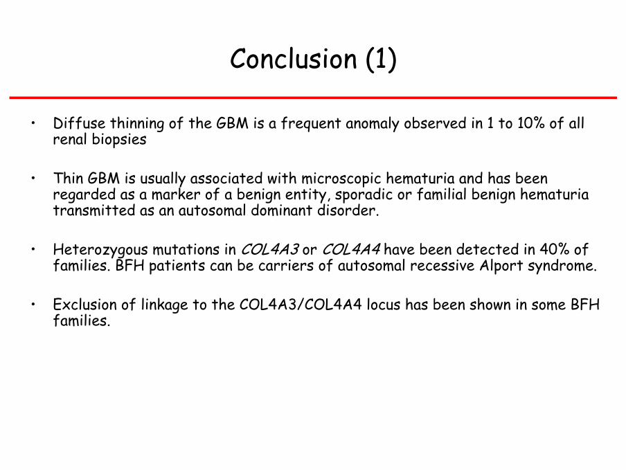
Conclusion (1)
• Diffuse thinning of the GBM is a frequent anomaly observed in 1 to 10% of all renal biopsies
• Thin GBM is usually associated with microscopic hematuria and has been regarded as a marker of a benign entity, sporadic or familial benign hematuria transmitted as an autosomal dominant disorder.
• Heterozygous mutations in COL4A3 or COL4A4 have been detected in 40% of families. BFH patients can be carriers of autosomal recessive Alport syndrome.
• Exclusion of linkage to the COL4A3/COL4A4 locus has been shown in some BFH families.
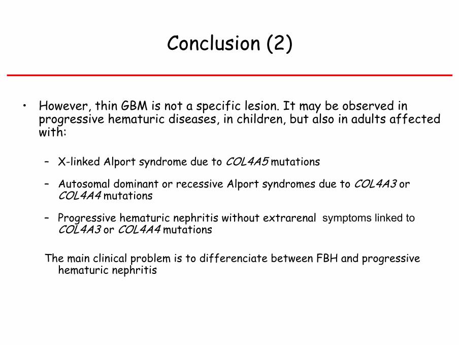
Conclusion (2)
• However, thin GBM is not a specific lesion. It may be observed in progressive hematuric diseases, in children, but also in adults affected with:
– X-linked Alport syndrome due to COL4A5 mutations
– Autosomal dominant or recessive Alport syndromes due to COL4A3 or COL4A4 mutations
– Progressive hematuric nephritis without extrarenal symptoms linked to COL4A3 or COL4A4 mutations
The main clinical problem is to differenciate between FBH and progressive hematuric nephritis

Conclusion (3)

Conclusion (4)
• The unsolved questions:
– What is the meaning of the effacement of foot processes along the thin GBM, in non proteinuric patients ?
– And the classical dilemma: how to explain the contrast between the presence of persistent hematuria and the absence of proteinuria ?

Laurence Heidet Patrick NiaudetChristelle Arondel Michel BroyerOlivier Gribouval Rémi Salomon Mireille SichCorinne Antignac
INSERM U574Département de Néphrologie PédiatriqueHôpital Necker-Enfants Malades