Manuel concernant les audits cliniques de radioprotection ...
47
Schweizerische Gesellschaft für Kardiologie Société Suisse de Cardiologie Società Svizzera di Cardiologia Swiss Society of Cardiology Manuel concernant les audits cliniques de radioprotection en cardiologie et recommandations pour l’élaboration d’un manuel de qualité destiné aux institutions Recommandations de la Société suisse de cardiologie (SSC), la Société suisse de radiobiologie et physique médicale (SSRPM) et l’Association suisse des techniciens en radiologie médicale (ASTRM) élaborées sur mandat de l’Office fédéral de la santé publique (OFSP) 21 Février 2020
Transcript of Manuel concernant les audits cliniques de radioprotection ...
Schweizerische Gesellschaft für Kardiologie Société Suisse de Cardiologie Società Svizzera di Cardiologia Swiss Society of Cardiology
Manuel concernant les audits cliniques de
radioprotection en cardiologie et
recommandations pour l’élaboration d’un
manuel de qualité destiné aux institutions
Recommandations de
la Société suisse de cardiologie (SSC), la Société suisse de
radiobiologie et physique médicale (SSRPM) et
l’Association suisse des techniciens en radiologie
médicale (ASTRM)
élaborées sur mandat de l’Office fédéral de la santé
publique (OFSP)
21 Février 2020

Ardan M. Saguner et al.
Dufourstrasse 30 • CH-3005 Bern, + 41 31 388 80 90, Fax 031 388 80 98, [email protected], www.swisscardio.ch
Impressum Éditeur Comité de pilotage des audits cliniques en radioprotection Contact OFFICE FÉDÉRAL DE LA SANTÉ PUBLIQUE - OFSP Schwarzenburgstrasse 157, 3003 Berne Tel. +41 58 467 16 24 [email protected] www.auditclinique.ch Société suisse de cardiologie (SSC) Dr. med. Marjam Rüdiger-Stürchler Directrice Dufourstrasse 30, 3005 Berne [email protected] +41 76 324 44 13 www.swisscardio.ch Auteurs
• Représentants de la SSC dans le comité de pilotage des audits cliniques o Prof. Dr. Christoph Kaiser o Suppléant : Prof. Dr. med. Tobias Reichlin, groupe de travail stimulation
cardiaque et électrophysiologie de la SSC
• Commission d’experts o Prof. Dr. med. Mario Togni, Fribourg/Berne o Prof. Dr. med. Burkhard Hornig, Aarau/Bâle (président suppléant) o Prof. Dr. med. Lorenz Räber, Berne o Prof. Dr. med. Tobias Reichlin, Berne o PD Dr. med. Ardan Saguner, Zurich (président) o Andreas Franke, infirmier, Aarau o Hansjörg Salomon, infirmier, Ermatingen o Severine Dziergwa, TRM, Bâle o Natalia Saltybaeva, physicienne médicale, Zurich o Dr. Roman Menz, physicien médical, Bâle
• Direction SSC o Dr. med. Marjam Rüdiger-Stürchler
Ardan M. Saguner et al.
Dufourstrasse 30 • CH-3005 Bern, + 41 31 388 80 90, Fax 031 388 80 98, [email protected], www.swisscardio.ch
Préambule
Les audits cliniques en cardiologie interventionnelle (domaine des doses élevées :
notamment coronarographie/PCI, TAVI/Mitra-CLIP ; EEP/traitement ablatif ;
implantation PM, ICD et CRT) sont obligatoires en Suisse depuis le 1er janvier 2018,
date de l’entrée en vigueur de la révision de l’ordonnance sur la radioprotection. Ils
visent à améliorer la radioprotection des patients, des médecins appliquant la
radiation et du personnel paramédical. Ils ont lieu sous forme de « peer review » et
ne sont pas des examens. Lors d’un audit clinique, l’équipe d’auditeurs (comprenant
un médecin, un physicien médical et un représentant du personnel paramédical), sur
la base d’une discussion avec leurs confrères de l’institution auditée, se fait une idée
sur la situation actuelle dans l’institution et la compare avec un standard prédéfini de
bonnes pratiques cliniques (« good clinical practice »). En formulant des propositions
d’amélioration basées sur un regard externe neutre, l’audit doit générer une valeur
ajoutée pour l’institution auditée.
Le groupe de travail « Audits cliniques en cardiologie » de la Société suisse de
cardiologie (SSC) a élaboré le premier projet d’un concept de mise en œuvre des
audits cliniques en cardiologie. Au terme des audits pilotes, la commission d’experts
l’a adapté. Ce concept résume les points principaux nécessaires au succès de la
mise en œuvre des audits cliniques en cardiologie.
Ce manuel doit servir d’instruction pour l’organisation, la planification, l’exécution et
l’évaluation des audits cliniques en cardiologie.
Il s’agit concrètement de fixer les compétences et les responsabilités en
radioprotection, notamment concernant la formation du personnel dans ce domaine,
les mesures visant au respect des prescriptions, la justification de l’application à un
patient donné, les protocoles d’examen et de traitement, l’information du patient,
l’enregistrement des doses ainsi que l’établissement et la documentation du
diagnostic.
A cet effet, le manuel peut être adapté aux données individuelles de l’institution en
question. Il apparaît opportun que les contenus du manuel soient disponibles sous
forme numérique afin d'être accessibles aux collaborateurs de manière rapide,
simple et immédiate.
Ce document, ainsi que le manuel de qualité des instituts concernés, servent de
base aux auditeurs pour la réalisation de leur mandat. En outre une check-list
permettant de simplifier la préparation de l’audit est donnée en annexe.
Ardan M. Saguner et al.
Dufourstrasse 30 • CH-3005 Bern, + 41 31 388 80 90, Fax 031 388 80 98, [email protected], www.swisscardio.ch
Table des matières Impressum
Préambule
1. Compétences et responsabilités
1.1. Compétences dans le cadre de l’organisation et de l’exécution des audits cliniques par „peer-review“ en radioprotection dans la cardiologie
1.2. Compétences en radioprotection de l’institut audité
2. Parc d’appareils pour les examens et les interventions sous radioscopie
3. Formation du personnel
3.1 Instruction
3.2 Formation continue
3.2.1 Formation en radioprotection des cardiologues/électrophysiologistes réalisant des interventions
3.2.2 Formation en radioprotection du personnel spécialisé
3.2.3 Formation en radioprotection des physiciens médicaux
4. Mesures visant au respect de la pratique de justification
4.1 Recommandations à l’intention des prescripteurs
4.2 Indication justificative par le médecin responsable
4.3 Patients exigeant une évaluation minutieuse du risque
4.4 Exigences concernant l’examen
5. Protocoles d’examen et de traitement et informations à l’intention du patient
5.1 Protocoles d’examen
5.2 Informations et explications données au patient
6. Documentation des doses de rayonnements
7. Établissement et communication du diagnostic, enregistrement et transmission des données
7.1 Établissement et communication du diagnostic
7.2 Enregistrement et transmission des données
Ardan M. Saguner et al.
Dufourstrasse 30 • CH-3005 Bern, + 41 31 388 80 90, Fax 031 388 80 98, [email protected], www.swisscardio.ch
8. Assurance de la qualité
9. Autoévaluation
10. Audits cliniques
10.1. Déroulement des audits cliniques
10.2. Contenus possibles des audits cliniques
10.2.1 Indicateurs quantitatifs
10.2.2 Indicateurs qualitatifs
10.2.3 Aides pour les instituts
10.2.4 Prochain audit
10.3 Instituts audités
10.4. Auditeurs
10.4.1 Composition de l’équipe d’auditeurs
10.4.2 Conditions pour l’agrément comme auditeur
10.4.3 Recrutement et administration du pool des auditeurs
10.5. Financement des audits cliniques
11. Annexe
11.1 Check-list à l’intention des instituts en vue de la préparation de l’audit clinique
11.2 Check-list à l’intention des auditeurs lors des audits cliniques en cardiologie
11.3. Exemple d’un plan d’audit
11.4 Modèle de rapport d’audit en cardiologie
11.5 Figure 1. Organigramme des audits cliniques
11.6 Tableau 1. Liste des indications de cardiologie interventionnelle
11.7 Tableau 2. Liste des indications en rythmologie
11.8 Tableau 3. Niveaux de référence diagnostiques pour les procédures cardiologiques
Ardan M. Saguner et al.
Dufourstrasse 30 • CH-3005 Bern, + 41 31 388 80 90, Fax 031 388 80 98, [email protected], www.swisscardio.ch
1. Compétences et responsabilités
1.1. Compétences dans le cadre de l’organisation et de l’exécution des audits
cliniques par « peer-review » en radioprotection dans la cardiologie
Deux représentants de la SSC en tant qu’experts médicaux dans le Comité de pilotage des
Audits cliniques, dont le secrétariat se trouve à l’Office fédéral de la santé publique (OFSP) :
Tâches des représentants :
- Expertise au profit de l’OFSP sur des questions spécifiques touchant à la radioprotection en
cardiologie
- Sauvegarde des intérêts de la SSC et de ses membres
Au moins un membre du comité de pilotage sur les audits cliniques de l’OFSP :
Tâches du comité de pilotage :
- définir la stratégie de mise en œuvre des audits cliniques - définir les exigences concernant le manuel de qualité en cardiologie - définir le déroulement des audits cliniques - approuver les propositions de la commission d’experts - tenue d’un bureau des réclamations pour les entreprises auditées - sélection et nomination des auditeurs
Commission d’experts
Comprenant au moins six membres, parmi lesquels des cardiologues (avec au moins un représentant de la cardiologie interventionnelle, respectivement de l’électrophysiologie (EEE)), des personnes soignantes, un technicien en radiologie médicale (TRM) et un physicien médical disposant d’une longue expérience en cardiologie.
Tâches de la commission d’experts
- développement du présent manuel à l’aide de contenus et de critères pour les audits cliniques de radioprotection en cardiologie
- proposition pour les critères de sélection des auditeurs (p. ex. participation active comme médecin ou infirmier dans un laboratoire de cathétérisme/EEE qui utilise les rayonnements ionisants; expérience professionnelle de plusieurs années dans le domaine à auditer ; expertise dans l’utilisation de la radioscopie et en radioprotection, dernière activité professionnelle datant de moins de huit ans ; pour les physiciens médicaux : spécialisation SSRPM ou formation équivalente ; pour les cardiologues : titre de spécialiste FMH en cardiologie, expertise en cardiologie interventionnelle ou en rythmologie et au moins activité comme chef de clinique, formation de base en technique d’audit (cours d’auditeur souhaitable))
- Proposition pour la composition de l’équipe de l’audit clinique (p. ex. au moins un cardiologue, un soignant/technicien en radiologie médicale, un physicien médical)
- proposition pour le déroulement des audits cliniques - proposition pour la check-list des audits cliniques - proposition pour le modèle du rapport d’audit - proposition pour les critères de sélection des institutions qui seront auditées - définition de critères qui représentent un « écart important par rapport aux standards »
(conformément à l’art. 42, al. 4, ORaP) ; leur élaboration se base sur les expériences acquises lors des audits pilotes
Auditeurs
- planification et réalisation des audits cliniques en cardiologie - documentation des audits cliniques
Ardan M. Saguner et al.
Dufourstrasse 30 • CH-3005 Bern, + 41 31 388 80 90, Fax 031 388 80 98, [email protected], www.swisscardio.ch
- rédaction et téléchargement dans les 21 jours d’un rapport d’audit sur la plate-forme en ligne mise à disposition par l‘OFSP
- une notification à l’OFSP n’intervient que lorsque des infractions légales sont constatées lors de l’audit clinique (p. ex. dosimétrie individuelle manquante, manque de documentation sur la durée de radioscopie, sur les doses de rayonnements et les produits dose – surface, moyens de protection pas contrôlés ou absents, etc.)
1.2. Compétences en radioprotection de l’institut audité
Avant l’audit clinique, une liste contenant les informations suivantes doit être mise à
disposition des auditeurs :
• titulaire de l’autorisation
• médecin responsable de la radioprotection (« expert en radioprotection » avec la
qualification, les tâches et les devoirs correspondants, conformément à la directive L-
03-04 de l’OFSP)
• personnel paramédical responsable de la radioprotection (direction des soins en
cardiologie interventionnelle ou en rythmologie)
• physicien médical responsable conformément à l’art. 36 ORaP
• le cas échéant, collaborateur responsable du manuel de qualité
• le cas échéant, certains collaborateurs ayant des missions particulières (p. ex.
responsable de la formation en radioprotection des cardiologues et du personnel
soignant en formation dans le laboratoire de cathétérisme, responsable de la
dosimétrie individuelle)
2. Parc d’appareils pour les examens et les interventions sous radioscopie Le parc des appareils pour les examens et les interventions sous radioscopie de l’institution en question doit être mentionné dans le manuel de qualité. Les informations suivantes doivent être indiquées pour toutes les installations à rayons X en
service :
désignation du type
date de la première installation ainsi que des mises à jour du matériel
autorisation valable délivrée par l’OFSP pour l’utilisation de l’installation (copie ou
PDF)
contrats de révision (copie ou PDF)
documentation des révisions et des contrôles de qualité exigés par la loi
3. Formation du personnel
3.1 Instruction
Le présent manuel s’adresse en premier lieu à tous les collaborateurs de cardiologie qui sont
régulièrement exposés au rayonnement ionisant ou qui les utilisent.
Avant le début de son activité, chaque collaborateur pouvant être exposé durant l’exercice
de sa profession aux rayonnements ionisants, devrait recevoir une instruction de la part de
l’expert en radioprotection ou d’un représentant que celui-ci a désigné. Cette instruction
devrait être actualisée régulièrement (au moins tous les 5 ans). Les modalités pratiques de
son déroulement et de son enregistrement devraient être précisées dans le manuel de
qualité de l’institut.
Ardan M. Saguner et al.
Dufourstrasse 30 • CH-3005 Bern, + 41 31 388 80 90, Fax 031 388 80 98, [email protected], www.swisscardio.ch
3.2 Formation continue
L’obligation de suivre des formations continues en radioprotection (selon l’art. 175 ORaP
ainsi que selon l’ordonnance sur la formation) implique que de telles formations, internes ou
externes à l’entreprise, soient suivies à raison de 8 heures en l’espace de cinq ans et que
cette participation soit documentée par l’institution. Le responsable de la radioprotection est
chargé d'établir le concept de formation de l’entreprise. Un module de formation en ligne
représente aussi une possibilité de formation continue (un module de ce type est en cours de
préparation par les hôpitaux universitaires de Suisse et le groupe Hirslanden).
3.2.1 Formation en radioprotection des cardiologues/électrophysiologistes réalisant
des interventions
Les responsabilités concernant la radioprotection en radioscopie, notamment l’instruction des nouveaux collaborateurs médicaux, doivent être réglementées et consignées par écrit. L’institut devrait de préférence disposer d’un programme structuré pour l’instruction de nouveaux collaborateurs.
3.2.2 Formation en radioprotection du personnel spécialisé
Dans le cas des techniciens en radiologie, la formation pratique de radioprotection en
radioscopie est acquise dans le cadre de la formation professionnelle. Pour le personnel
soignant, il est recommandé d’élaborer un programme de formation en radioprotection que
chaque nouveau collaborateur devrait suivre.
• Concept d’instruction des nouveaux collaborateurs
3.2.3 Formation en radioprotection des physiciens médicaux
La formation en radioprotection est effectuée dans le cadre de la spécialisation SSRMP en
physique médicale. Un nombre de cours de formation continue, notamment sur le thème de
la radioprotection, est défini pour maintenir la reconnaissance de cette spécialisation. Le lien
suivant permet d’obtenir des informations complémentaires sur la spécialisation SSRPM en
physique médicale : http://www.sgsmp.ch/certification-for-medical-physicists/rules.
4. Mesures visant au respect de la pratique de justification
Selon l’art. 29 ORaP, l’indication touchant à une procédure dans le domaine de la cardiologie
interventionnelle doit être documentée, ressortir du rapport et correspondre aux directives
nationales (voir les tableaux 1 et 2).
4.1 Recommandations à l’intention des prescripteurs
La SSC a rassemblé les indications principales pour la prise de décision à l’intention des
prescripteurs dans l’annexe au présent document (voir les tableaux 1 et 2).
4.2 Indication justificative par le médecin responsable
La SSC a établi, dans le cadre du présent manuel, des tableaux séparés pour les domaines
de la cardiologie et de la rythmologie (tableaux 1 et 2) présentant les indications courantes
visant à justifier une intervention diagnostique ou thérapeutique ayant recours au
rayonnement ionisant en cardiologie.
4.3 Patients exigeant une évaluation particulièrement minutieuse du risque
Une appréciation minutieuse des bénéfices et des risques lors du recours à la radioscopie,
tenant compte des modalités alternatives d’examen n’utilisant pas de rayonnement ionisant,
Ardan M. Saguner et al.
Dufourstrasse 30 • CH-3005 Bern, + 41 31 388 80 90, Fax 031 388 80 98, [email protected], www.swisscardio.ch
telles que l’ultrasonographie, la tomographie par résonance magnétique ou la procédure de
cartographie électro-anatomique 3D, doit être réalisée pour les patients suivants :
• les adolescents et les jeunes adultes
• les femmes enceintes (notamment au 1er trimestre de la grossesse)
• les patients qui subissent régulièrement des examens impliquant du rayonnement
ionisant
4.4 Exigences concernant l’examen
Pour des raisons de sécurité et de qualité, les informations suivantes devraient figurer dans
un rapport d’intervention de cardiologie interventionnelle ou de rythmologie :
• données de base du patient
• indication justifiant l’examen
• degré d‘urgence (électif, pressant, urgent)
• produit dose-surface, durée de scopie, dose accumulée au point de référence (dose
qui représente une estimation de la dose attendue à la peau).
5. Protocoles d’examen et de traitement et informations à l’intention du patient
5.1 Protocoles d’examen
Il est judicieux que les protocoles actuels pour les différents examens soient consignés dans
le manuel de qualité et soient disponibles sous forme numérique.
Il apparaît raisonnable que chaque institution définisse des processus clairs fixant comment
réagir à des dépassements de certaines valeurs de dose (p. ex. faire appel à un opérateur
expérimenté, changer l’angulation du tube à rayons X).
Les protocoles utilisés doivent autant que possible être optimisés vis-à-vis de l’exposition du
patient au rayonnement (p. ex. choix de la fréquence des impulsions lors de la radioscopie
ou de la fréquence des images lors de l’acquisition de séries, choix d’un mode à faible dose,
dose par impulsion).
5.2 Informations et explications données au patient
Le manuel de qualité de l’institution devrait contenir des informations concernant le patient,
notamment celles qui sont indiquées ci-dessous :
• indication de la présence de rayonnement ionisant lors de l’explication donnée au
patient
• contenu de l’explication (p. ex. la préparation à l’examen, son déroulement, le cas
échéant le type de produit de contraste utilisé, les possibles complications telles que
les réactions d’hypersensibilité et d’allergie, l’établissement d’une éventuelle
grossesse, le faible risque d’érythème radio-induit)
• emplacement d’archivage et documentation numérique de la déclaration écrite de
consentement
6. Documentation des doses de rayonnements
Selon l’art. 33 ORaP, les expositions diagnostiques et thérapeutiques doivent être
documentées de façon à pouvoir déterminer ultérieurement la dose de rayonnements reçue
par le patient (selon l’art. 20 de l’ordonnance sur les rayons X, le produit dose-surface, la
Ardan M. Saguner et al.
Dufourstrasse 30 • CH-3005 Bern, + 41 31 388 80 90, Fax 031 388 80 98, [email protected], www.swisscardio.ch
dose accumulée au point de référence ainsi que la durée de scopie et le nombre d’images
doivent être consignés). Les données doivent être enregistrées et conservées durant dix
ans.
Une analyse périodique et une communication/discussion sur les doses moyennes et les
durées de scopie pour les divers examens, ainsi qu’une comparaison avec les niveaux
nationaux de référence diagnostiques (s’ils sont disponibles) ou avec les niveaux de
référence propres à l’institution, est souhaitable (p. ex. coronarographie, PCI, CTO, TAVI,
Mitra Clip, fermeture de FOP, fermeture de l’auricule gauche (LAA), stimulateur cardiaque,
CRT, thermo-ablations).
A cet effet, on utilisera de préférence un logiciel de gestion des doses.
7. Établissement et communication du diagnostic, enregistrement et transmission des données 7.1 Établissement et communication du diagnostic
Les directives utilisées dans l’institut pour l’établissement du diagnostic sont à consigner.
Elles doivent comprendre les points suivants :
• indication
• date de l’examen
• données de base du patient
• médecin établissant le diagnostic / médecin responsable
• complications aiguës
Données recommandées dans le rapport médical (si applicable) :
• durée de scopie et produit dose-surface, le cas échéant dose accumulée au point de
référence
7.2 Enregistrement et transmission des données
• La conservation des clichés radiologiques et l’enregistrement des données sont
traités conformément aux bases légales (loi fédérale et lois cantonales sur la
protection des données). Durant les dix années du délai de conservation, les
données, non corrompues, doivent être accessibles à tout instant. Leur protection
contre des dommages naturels est assurée par un enregistrement redondant.
• Il existe une description de la procédure d’enregistrement des données et de leur
sécurisation (p. ex. par RIS/PACS).
• Il existe une description de la procédure de transmission des données (images de
documentation) aux partenaires et aux prescripteurs.
8. Assurance de la qualité
La radioprotection des patients, d’éventuels accompagnants et du personnel exposé durant
l’exercice de leur profession aux rayonnements devrait toujours être prise en compte. Un
soin particulier est à apporter aux situations d’urgence, telles que l’incendie, l’urgence
médicale, les dérangements sur l’installation de radioscopie et autres événements
extraordinaires.
Le manuel de qualité doit comprendre des informations sur les aspects suivants :
Ardan M. Saguner et al.
Dufourstrasse 30 • CH-3005 Bern, + 41 31 388 80 90, Fax 031 388 80 98, [email protected], www.swisscardio.ch
- garantie d’application de la radioprotection opérationnelle aux cas particuliers (p. ex.
réduction de l’examen)
- existence de directives pour des groupes particuliers de patients (p. ex. enfants,
femmes enceintes)
- type de moyens de protection disponibles (p. ex. moyens physiques de
radioprotection tels que tabliers de protection, protections des organes génitaux)
- existence d’une directive interne à l’institution concernant l’utilisation des moyens de
protection
- collaborateurs en charge de la vérification périodique des moyens de protection
- fréquence et mode de vérification des moyens de protection vis-à-vis d’éventuelles
défaillances ainsi que la méthode et le lieu de la documentation correspondante
9. Autoévaluation
Les activités et les projets visant à l’amélioration de la qualité, par autoévaluation et par mise
en application des propositions d’audit, sont souhaitables et constituent des éléments
importants dans le cadre du processus continu d’amélioration.
En voici quelques exemples :
- rapports d’audits cliniques précédents
- existence, au niveau de la clinique, d’un CIRS (critical incidence reporting system)
visant à saisir les événements radiologiques médicaux conformément à l’art. 49
ORaP
- utilisation d’un système de gestion des doses en vue de l’analyse périodique des
doses individuelles délivrées aux patients et comme système interne d’avertissement
de l’examinateur en cas de surexposition dans un cas d’espèce
- formations internes périodiques sur le thème de la radioprotection

Ardan M. Saguner et al.
Dufourstrasse 30 • CH-3005 Bern, + 41 31 388 80 90, Fax 031 388 80 98, [email protected], www.swisscardio.ch
10. Audits cliniques
10.1 Déroulement des audits cliniques
Avant l’audit, l’extension, le déroulement ainsi que les thèmes prioritaires sont établis par le
responsable de l’audit, en accord avec l’équipe à auditer, et sont communiqués.
10.2. Contenus possibles des audits cliniques
10.2.1 Indicateurs quantitatifs
Les indicateurs quantitatifs suivants ont été définis par le groupe de travail comme
composants possibles des audits cliniques en cardiologie/électrophysiologie interventionnelle
:
• Comparaison des paramètres dosimétriques de types d’intervention sélectionnés
avec les niveaux nationaux de référence (NRD) ou avec des valeurs de référence
proposées par le groupe de travail et reposant sur des publications dans ce
domaine :
- coronarographie diagnostique
- PTCA
- TAVI
- MITRA-Clip
- fermeture de FOP
- fermeture de l’auricule gauche (LAA)
- occlusion de shunt
- biopsie endomyocardique
- implantation d’un stimulateur cardiaque/ICD
- implantation d’une CRT
- examen diagnostique électrophysiologique
- thermo-ablation de tachycardies supraventriculaires (sans système de
cartographie 3D)
- thermo-ablation de tachycardies supraventriculaires (avec système de
cartographie 3D)
- traitement ablatif de fibrillations auriculaires
• prélèvement d’échantillons, p. ex. d’un ou de plusieurs des interventions citées ci-
dessus, réalisés lors du dernier mois, afin de vérifier l’indication justificative, le produit
dose-surface et la durée de la radioscopie
• contrôle des données de la dosimétrie individuelle
10.2.2 Indicateurs qualitatifs
Les indicateurs qualitatifs suivants sont à vérifier quant à leur mise en application :
• respect des indications pour les différentes procédures selon les tableaux 1 et 2 (voir
annexe)
• existence et observance des protocoles radiologiques spécifiques aux indications (p.
ex. réglages de scopie spécifiques à la procédure, images/s, dose par impulsion,
etc.)
• mesures de radioprotection opérationnelle
• état des moyens de protection (tabliers de protection, etc.)
• première instruction en radioprotection des nouveaux collaborateurs réalisée en
temps utile :
o organisation de la radioprotection dans la clinique
o dosimétrie individuelle
Ardan M. Saguner et al.
Dufourstrasse 30 • CH-3005 Bern, + 41 31 388 80 90, Fax 031 388 80 98, [email protected], www.swisscardio.ch
o moyens de protection pour le patient /le personnel
o comportement correct dans la salle d’examen (distance, blindage, durée de
séjour)
L’évaluation des différents indicateurs qualitatifs s’effectue, dans le cadre de l’audit, à l’aide
de critères subjectifs.
10.2.3 Aides pour les instituts
Les aides mises à disposition des instituts sont le présent manuel et ses annexes « Check-
list à l’intention des instituts en vue de la préparation de l’audit clinique » et « Exemple d’un
plan d’audit ». Le manuel de qualité devrait être adapté individuellement par les
institutions/établissements et actualisé régulièrement.
10.2.4 Prochain audit
Un nouvel audit peut avoir lieu après cinq ans.
10.3. Institutions/établissements audités
Le choix des institutions/établissements à auditer s’effectue de façon aléatoire (sur la base
d’une clef de répartition entre les trois régions linguistiques) par le secrétariat scientifique de
l’OFSP. La première série d’audits (2020 à 2025) concernera les institutions/établissements
qui effectuent des interventions aussi bien en électrophysiologie (y.c. les traitements ablatifs
par cathétérisme) qu’en cardiologie interventionnelle.
Le calendrier des audits ainsi que l’ordre dans lequel auditer les centres sont organisés par
le secrétariat scientifique de l’OFSP.
L’attribution de l’auditeur principal et des autres auditeurs aux différents
institutions/établissements peut être refusée par les auditeurs ou par les
institutions/établissements pour des motifs valables. Elle est alors effectuée à nouveau par le
secrétariat scientifique de l’OFSP.
10.4. Auditeurs
10.4.1 Composition de l’équipe d’auditeurs
En principe l’équipe d’auditeurs est composée de trois personnes, une de chaque groupe
professionnel (cardiologie, physique médicale, personnel soignant/technicien en radiologie).
Une de ces personnes fonctionne comme auditeur principal. Le nombre de personnes et la
composition de l’équipe dépendent de l’extension et du thème de l’audit (dans le cas où la
clinique auditée dispose aussi bien d’un laboratoire de cardiologie que d’électrophysiologie
interventionnelle, au moins deux cardiologues issus de chacune des sous-disciplines doivent
être présents). Les auditeurs devraient de préférence ne pas venir de la même institution.
La composition de l’équipe d’auditeurs est déterminée par le secrétariat scientifique de
l’OFSP. Concrètement, l’auditeur principal définit la taille et la composition de l’équipe
d’auditeurs, paramètres qui devraient être adaptés à la dimension de la clinique auditée.
Le groupe de travail part de l’idée qu’un auditeur est en mesure d’effectuer en moyenne
deux audits par année. Il renonce délibérément à fixer une limite minimale et maximale au
nombre d’audits par auditeur et par année.
10.4.2 Conditions pour l’agrément comme auditeur
Font partie de la formation technique nécessaire :
• une expérience professionnelle de plusieurs années dans le domaine à auditer, dont
au moins deux en Suisse
Ardan M. Saguner et al.
Dufourstrasse 30 • CH-3005 Bern, + 41 31 388 80 90, Fax 031 388 80 98, [email protected], www.swisscardio.ch
• une expertise en cardiologie interventionnelle et/ou en électrophysiologie
• une dernière activité professionnelle datant de moins de huit ans
• pour les physiciens médicaux, la spécialisation SSRPM ou une formation équivalente
• pour les cardiologues, le titre de spécialiste FMH en cardiologie et au moins une
activité comme chef de clinique
• pour le personnel soignant/technicien en radiologie, plusieurs années d’expérience
dans le domaine de la cardiologie interventionnelle et/ou de l’électrophysiologie
fait partie de la formation spécifique des candidats auditeurs :
• une formation de base en technique d’audit
10.4.3 Recrutement et administration du pool des auditeurs
Le recrutement des auditeurs est effectué par la commission d’experts et est approuvé par le
comité de pilotage. L’administration et l’organisation des audits sont pris en charge par le
secrétariat scientifique de l‘OFSP, en accord avec les auditeurs et l’institut à auditer. Dans le
courant d’un cycle d’audits, des auditeurs nouveaux s’ajoutant à l’équipe peuvent être
formés « sur le terrain » dans le cadre de l’audit et être accompagnés par un auditeur
chevronné.
10.5. Financement des audits cliniques et de l’organisation
Les coûts de l’audit clinique incombent en principe à l’institution auditée. Ils comprennent le
travail des auditeurs.
L’émolument d’audit est forfaitaire et s’élève pour les institutions à CHF 7‘100.-
Les auditeurs sont indemnisés de manière forfaitaire au tarif suivant :
- médecin spécialiste en cardiologie CHF 2'000.-
- physicien médical CHF 1'500.-
- personnel soignant / technicien en radiologie : CHF 1’100.-
- auditeur principal : complément de CHF 500.-
Ardan M. Saguner et al.
Dufourstrasse 30 • CH-3005 Bern, + 41 31 388 80 90, Fax 031 388 80 98, [email protected], www.swisscardio.ch
11. Annexe
11.1 Check-list à l’intention des instituts en vue de la préparation de l’audit clinique
Préparation
☐ actualisation des documents pertinents du manuel de qualité (de préférence sous forme
numérique)
☐ information des collaborateurs
☐ accord/coordination avec l’auditeur principal
☐ préparation des documents préalablement demandés
Déroulement
☐ garantir l’accessibilité aux dossiers techniques des installations
☐ garantir l’accessibilité au manuel de qualité
☐ garantir l’accessibilité à la documentation dosimétrique
☐ garantir la disponibilité des personnes responsables
☐ garantir l’accès aux locaux

Ardan M. Saguner et al.
Dufourstrasse 30 • CH-3005 Bern, + 41 31 388 80 90, Fax 031 388 80 98, [email protected], www.swisscardio.ch
11.2 Check-list à l’intention des auditeurs lors des audits cliniques en cardiologie
1. Mandat de l’audit
Quel est le mandat de l’audit (p. ex. optimisation du produit dose-surface) ?
2. Type de l’institution auditée
Quel est le type de l’institution auditée (clinique A ou B, cabinet médical) ? Qui est le
propriétaire des installations de radioscopie ? Y a-t-il un expert en radioprotection ?
3. Interventions réalisées par l’institut
Quelles interventions sont-elles réalisées et combien par année ?
Combien de personnes sont employées de manière permanente dans le laboratoire de
cathétérisme ?
4. Composition de l’équipe d’audits
Comment l’équipe d’audit est-elle composée ? L’institution auditée dispose-t-elle d’une
d’expertise suffisante ?
5. Manuel de qualité
Y a-t-il un manuel local de qualité, ou des SOP, pour la radioprotection en cardiologie ? Si
oui, sous quelle forme, où est-il conservé, qui y a accès et avec quelle périodicité est-il
actualisé ?
6. Formation en radioprotection
Y a-t-il, dans le manuel de qualité, une check-list interne « Introduction dans la
radioprotection en cardiologie » ? Tous les opérateurs responsables (qui ont le FMH de
cardiologie depuis le 1er janvier 2003) ont-ils acquis le certificat de formation complémentaire
« Qualifications pour les examens radiologiques à forte dose en cardiologie (SSC) » ? Le
responsable local en radioprotection dispose-t-il des « qualifications techniques en
radioprotection » ou d’une qualification équivalente ? Des formations continues périodiques
sont-elles suivies (au moins huit heures de radioprotection tous les cinq ans) ? Qui réalise
ces formations continues et comment la participation est-elle contrôlée et accréditée ?
Comment et par qui les nouveaux collaborateurs sont-ils instruits en radioprotection ? Tous
les opérateurs responsables, les collaborateurs et les experts doivent pouvoir présenter leur
certificat de qualification aux auditeurs.
7. Dose de rayonnements
Chaque service audité devrait rassembler, p. ex. sous forme d’un tableau Excel, un choix
d’interventions en cardiologie dans lesquelles la radioscopie est appliquée (par exemple
toutes les coronarographies et PTCA du dernier mois). Les données concernant les
stimulateurs cardiaques et les ICD peuvent être reprises de « Paceweb ». La liste devrait
contenir les informations suivantes :
• le type de l’intervention (p. ex. coronarographie diagnostique)
• l’indication
• le produit dose - surface
• la durée de radioscopie
• la dose accumulée au point de référence
Ardan M. Saguner et al.
Dufourstrasse 30 • CH-3005 Bern, + 41 31 388 80 90, Fax 031 388 80 98, [email protected], www.swisscardio.ch
• l’opérateur responsable
Où et par qui ces données sont-elles documentées ? Qui les consulte ? Combien de temps
sont-elles archivées ?
Que fait-on des valeurs de mesure : évaluation, discussion, mesures d’amélioration ?
Combien de ces procédures se situent-elles hors des NRD correspondants pour la
Suisse/l’Europe ? (pour les niveaux de référence, voir OFSP 2017)
Dans le cas où le niveau de référence par patient et par intervention est dépassé, un
deuxième opérateur expérimenté est-il consulté ? La gestion de telles situations est-elle
enseignée ? L’angulation du tube à rayons X est-elle modifiée ?
Un opérateur particulier peut-il vérifier comment il se situe à l’interne ou au niveau national ?
8. Mesures de radioprotection pour le bien-être du patient
L’exposition aux rayonnements et les conséquences que celle-ci peut avoir sont-elles
mentionnées lors de l’explication donnée au patient ainsi que dans le formulaire de
consentement ? Une grossesse est-elle exclue et si oui comment ? Dans le cas contraire,
quel moyen de protection est appliqué à la patiente ? Lorsque des NRD sont
significativement dépassés par intervention, réalise-t-on des contrôles de suivi de la peau (p.
ex. après deux à quatre semaines) ?
Les mesures techniques de réduction de l’exposition au rayonnement, tels que la réduction
de la fréquence des impulsions/des images, la focalisation du faisceau, l’optimisation des
angulations, la réduction de la durée de scopie, etc., sont-elles suffisamment appliquées ?
Quels moyens de protection sont appliqués au patient ?
9. Mesures concrètes de radioprotection pour le personnel
• Y a-t-il suffisamment de tabliers de protection pour les opérateurs et le personnel ?
• Ces tabliers sont-ils régulièrement contrôlés quant à leur intégrité et si oui à quelle
fréquence et par qui ? Où cela est-il documenté ? Où les tabliers sont-ils conservés
et comment sont-ils entreposés ?
• Utilise-t-on des lunettes de protection et des dosimètres sur et sous le tablier ?
Dispose-t-on de dosimètres actifs ? Que fait-on des dosimètres et des données ? Où
les données sont-elles évaluées, transmises et documentées ? Qui en porte la
responsabilité ? Que se passe-t-il en cas de dépassement des doses de référence ?
L’équipe reçoit-elle un retour concernant l’évaluation des dosimètres ?
• Contrôle de l’état des moyens personnels de protection
10. Sélection des cas
Au moins deux cas devraient être sélectionnés par les auditeurs et discutés en détail avec
les responsables. Outre la dose de rayonnements et la durée de scopie, l’indication
justificative devrait aussi être thématisée.
11. Défaillance / événement radiologique médical
Que se passe-t-il en cas de défaillances radiologiques ? Utilise-t-on un CIRS pour les
enregistrer ? Quelle est la voie de communication ?
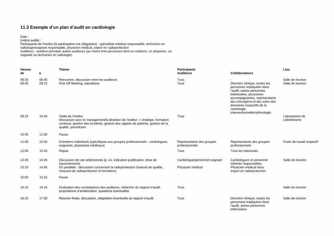
11.3 Exemple d’un plan d’audit en cardiologie Date :
Institut audité : Participants de l’institut (la participation est obligatoire) : spécialiste médical responsable, technicien en radiologie/soignant responsable, physicien médical, expert en radioprotection
Auditeurs : auditeur principal, autres auditeurs (au moins trois personnes dont un médecin, un physicien, un soignant ou technicien en radiologie)
Heures Thème Participants Lieu de à Auditeurs Collaborateurs
08:30 08:45 Rencontre, discussion entre les auditeurs Tous - Salle de réunion 08:45 09:15 Kick-Off Meeting, salutations Tous Direction clinique, toutes les
personnes impliquées dans l’audit, autres personnes intéressées, personnes accompagnantes, représentants des chirurgiens et des soins des domaines respectifs de la cardiologie interventionnelle/rythmologie
Salle de réunion
09:15 10:45 Visite de l’institut Discussion avec le management/la direction de l’institut -> stratégie, formation continue, gestion des incidents, gestion des rappels de patients, gestion de la qualité, procédures
Tous Laboratoires de cathétérisme
10:45
11:00
Pause
11:00 12:00 Entretiens individuels (spécifiques aux groupes professionnels : cardiologues, soignants, physiciens médicaux)
Représentants des groupes professionnels
Représentants des groupes professionnels
Poste de travail respectif
12:00 12:45 Repas Tous Tous les intéressés
12:45 13:15
14:45 14:45
Discussion de cas sélectionnés (p. ex. indication justificative, dose de rayonnements) En parallèle : discussion concernant la radioprotection (manuel de qualité, mesures de radioprotection et formation)
Cardiologue/personnel soignant Physicien médical
Cardiologues et personnel infirmier responsables Physicien médical et/ou expert en radioprotection
Salle de réunion
15:00 15:15 Pause
15:15 16:15 Evaluation des constatations des auditeurs, rédaction du rapport d’audit, propositions d‘amélioration, questions éventuelles
Tous Salle de réunion
16:15 17:00 Réunion finale, discussion, adaptation éventuelle du rapport d‘audit
Tous Direction clinique, toutes les personnes impliquées dans l’audit, autres personnes intéressées
Salle de réunion
Ardan M. Saguner et al.
Dufourstrasse 30 • CH-3005 Bern, + 41 31 388 80 90, Fax 031 388 80 98, [email protected], www.swisscardio.ch
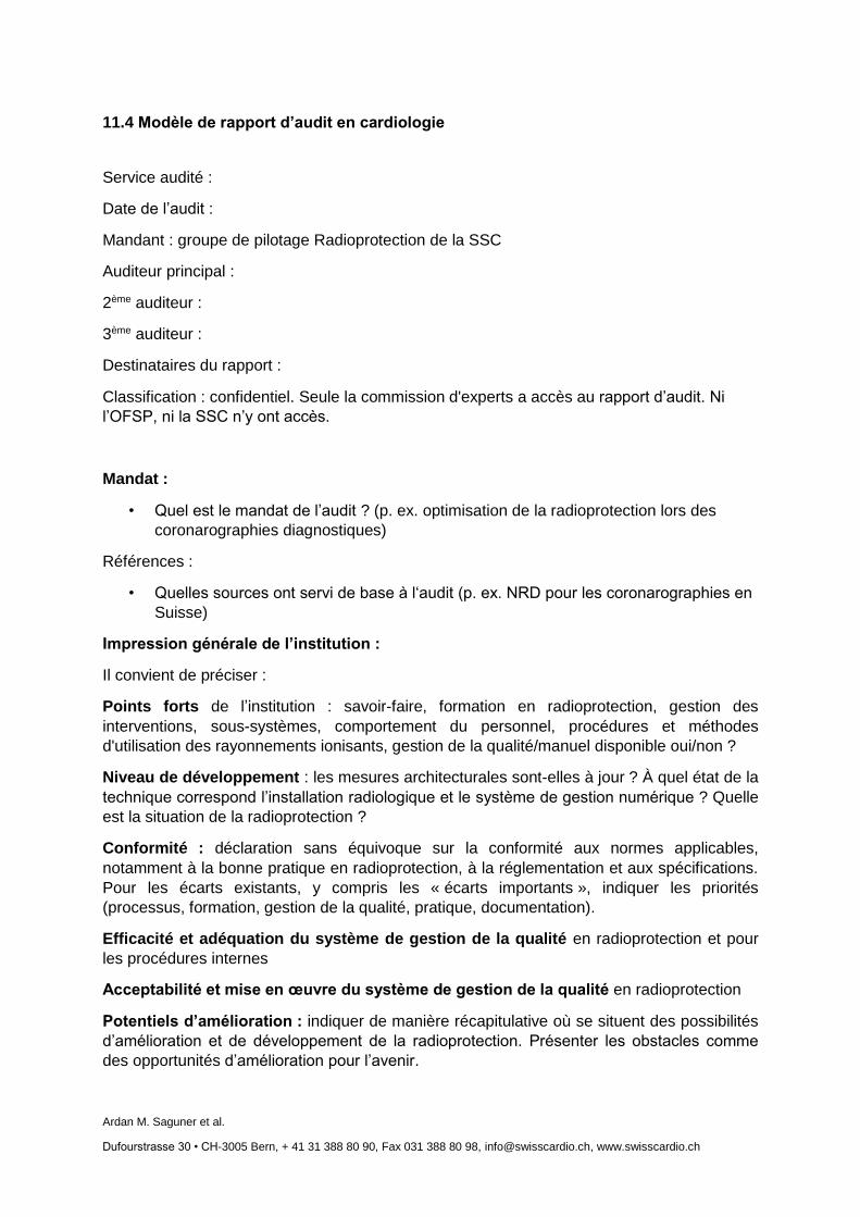
11.4 Modèle de rapport d’audit en cardiologie
Service audité :
Date de l’audit :
Mandant : groupe de pilotage Radioprotection de la SSC
Auditeur principal :
2ème auditeur :
3ème auditeur :
Destinataires du rapport :
Classification : confidentiel. Seule la commission d'experts a accès au rapport d’audit. Ni
l’OFSP, ni la SSC n’y ont accès.
Mandat :
• Quel est le mandat de l’audit ? (p. ex. optimisation de la radioprotection lors des
coronarographies diagnostiques)
Références :
• Quelles sources ont servi de base à l‘audit (p. ex. NRD pour les coronarographies en
Suisse)
Impression générale de l’institution :
Il convient de préciser :
Points forts de l’institution : savoir-faire, formation en radioprotection, gestion des
interventions, sous-systèmes, comportement du personnel, procédures et méthodes
d'utilisation des rayonnements ionisants, gestion de la qualité/manuel disponible oui/non ?
Niveau de développement : les mesures architecturales sont-elles à jour ? À quel état de la
technique correspond l’installation radiologique et le système de gestion numérique ? Quelle
est la situation de la radioprotection ?
Conformité : déclaration sans équivoque sur la conformité aux normes applicables,
notamment à la bonne pratique en radioprotection, à la réglementation et aux spécifications.
Pour les écarts existants, y compris les « écarts importants », indiquer les priorités
(processus, formation, gestion de la qualité, pratique, documentation).
Efficacité et adéquation du système de gestion de la qualité en radioprotection et pour
les procédures internes
Acceptabilité et mise en œuvre du système de gestion de la qualité en radioprotection
Potentiels d’amélioration : indiquer de manière récapitulative où se situent des possibilités
d’amélioration et de développement de la radioprotection. Présenter les obstacles comme
des opportunités d’amélioration pour l’avenir.
Ardan M. Saguner et al.
Dufourstrasse 30 • CH-3005 Bern, + 41 31 388 80 90, Fax 031 388 80 98, [email protected], www.swisscardio.ch
Remerciements
L’équipe d’auditeurs remercie les participants de xxxx pour leur collaboration efficace et pour
l’organisation de l’audit. La transmission fluide des informations et l’accès à la documentation
et aux procédures de la division ont été des conditions préalables importantes pour
l’acquisition des résultats de l’audit.
Lieu et date
Nom et signature
Auditeur principal
Destinataires du rapport et des pièces jointes
Ardan M. Saguner et al.
Dufourstrasse 30 • CH-3005 Bern, + 41 31 388 80 90, Fax 031 388 80 98, [email protected], www.swisscardio.ch
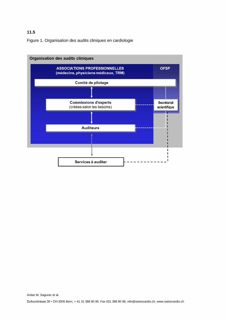
11.5
Figure 1. Organisation des audits cliniques en cardiologie
Ardan M. Saguner et al.
Dufourstrasse 30 • CH-3005 Bern, + 41 31 388 80 90, Fax 031 388 80 98, [email protected], www.swisscardio.ch
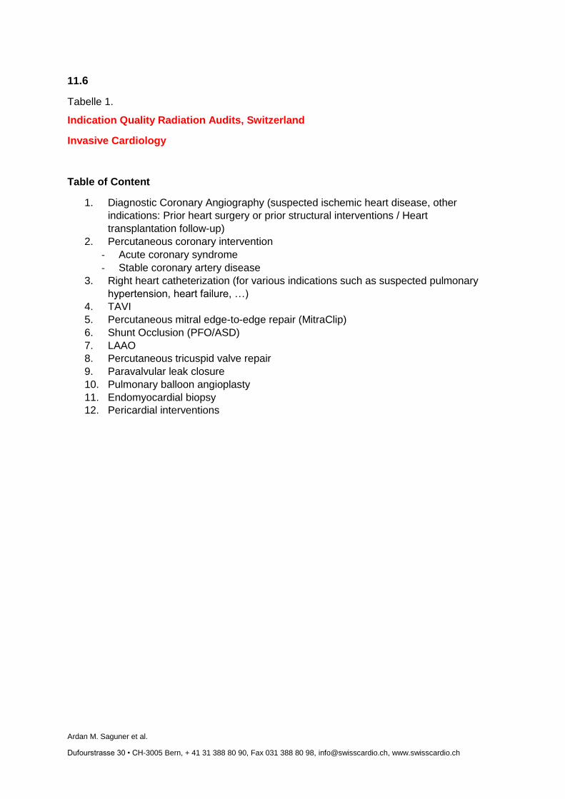
11.6
Tabelle 1.
Indication Quality Radiation Audits, Switzerland
Invasive Cardiology
Table of Content
1. Diagnostic Coronary Angiography (suspected ischemic heart disease, other
indications: Prior heart surgery or prior structural interventions / Heart
transplantation follow-up)
2. Percutaneous coronary intervention
- Acute coronary syndrome
- Stable coronary artery disease
3. Right heart catheterization (for various indications such as suspected pulmonary
hypertension, heart failure, …)
4. TAVI
5. Percutaneous mitral edge-to-edge repair (MitraClip)
6. Shunt Occlusion (PFO/ASD)
7. LAAO
8. Percutaneous tricuspid valve repair
9. Paravalvular leak closure
10. Pulmonary balloon angioplasty
11. Endomyocardial biopsy
12. Pericardial interventions
Ardan M. Saguner et al.
Dufourstrasse 30 • CH-3005 Bern, + 41 31 388 80 90, Fax 031 388 80 98, [email protected], www.swisscardio.ch
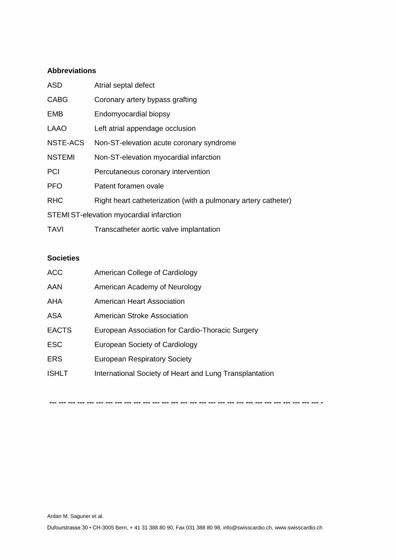
Abbreviations
ASD Atrial septal defect
CABG Coronary artery bypass grafting
EMB Endomyocardial biopsy
LAAO Left atrial appendage occlusion
NSTE-ACS Non-ST-elevation acute coronary syndrome
NSTEMI Non-ST-elevation myocardial infarction
PCI Percutaneous coronary intervention
PFO Patent foramen ovale
RHC Right heart catheterization (with a pulmonary artery catheter)
STEMI ST-elevation myocardial infarction
TAVI Transcatheter aortic valve implantation
Societies
ACC American College of Cardiology
AAN American Academy of Neurology
AHA American Heart Association
ASA American Stroke Association
EACTS European Association for Cardio-Thoracic Surgery
ESC European Society of Cardiology
ERS European Respiratory Society
ISHLT International Society of Heart and Lung Transplantation
--- --- --- --- --- --- --- --- --- --- --- --- --- --- --- --- --- --- --- --- --- --- --- --- --- --- --- --- --- -
Ardan M. Saguner et al.
Dufourstrasse 30 • CH-3005 Bern, + 41 31 388 80 90, Fax 031 388 80 98, [email protected], www.swisscardio.ch
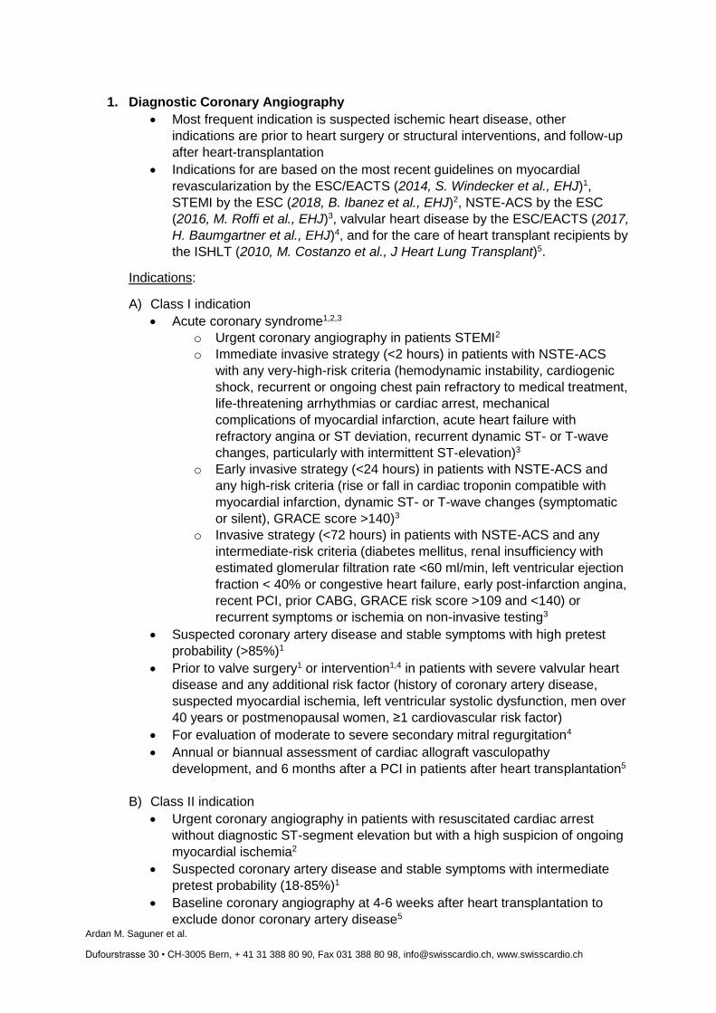
1. Diagnostic Coronary Angiography
Most frequent indication is suspected ischemic heart disease, other
indications are prior to heart surgery or structural interventions, and follow-up
after heart-transplantation
Indications for are based on the most recent guidelines on myocardial
revascularization by the ESC/EACTS (2014, S. Windecker et al., EHJ)1,
STEMI by the ESC (2018, B. Ibanez et al., EHJ)2, NSTE-ACS by the ESC
(2016, M. Roffi et al., EHJ)3, valvular heart disease by the ESC/EACTS (2017,
H. Baumgartner et al., EHJ)4, and for the care of heart transplant recipients by
the ISHLT (2010, M. Costanzo et al., J Heart Lung Transplant)5.
Indications:
A) Class I indication
Acute coronary syndrome1,2,3
o Urgent coronary angiography in patients STEMI2
o Immediate invasive strategy (<2 hours) in patients with NSTE-ACS
with any very-high-risk criteria (hemodynamic instability, cardiogenic
shock, recurrent or ongoing chest pain refractory to medical treatment,
life-threatening arrhythmias or cardiac arrest, mechanical
complications of myocardial infarction, acute heart failure with
refractory angina or ST deviation, recurrent dynamic ST- or T-wave
changes, particularly with intermittent ST-elevation)3
o Early invasive strategy (<24 hours) in patients with NSTE-ACS and
any high-risk criteria (rise or fall in cardiac troponin compatible with
myocardial infarction, dynamic ST- or T-wave changes (symptomatic
or silent), GRACE score >140)3
o Invasive strategy (<72 hours) in patients with NSTE-ACS and any
intermediate-risk criteria (diabetes mellitus, renal insufficiency with
estimated glomerular filtration rate <60 ml/min, left ventricular ejection
fraction < 40% or congestive heart failure, early post-infarction angina,
recent PCI, prior CABG, GRACE risk score >109 and <140) or
recurrent symptoms or ischemia on non-invasive testing3
Suspected coronary artery disease and stable symptoms with high pretest
probability (>85%)1
Prior to valve surgery1 or intervention1,4 in patients with severe valvular heart
disease and any additional risk factor (history of coronary artery disease,
suspected myocardial ischemia, left ventricular systolic dysfunction, men over
40 years or postmenopausal women, ≥1 cardiovascular risk factor)
For evaluation of moderate to severe secondary mitral regurgitation4
Annual or biannual assessment of cardiac allograft vasculopathy
development, and 6 months after a PCI in patients after heart transplantation5
B) Class II indication
Urgent coronary angiography in patients with resuscitated cardiac arrest
without diagnostic ST-segment elevation but with a high suspicion of ongoing
myocardial ischemia2
Suspected coronary artery disease and stable symptoms with intermediate
pretest probability (18-85%)1
Baseline coronary angiography at 4-6 weeks after heart transplantation to
exclude donor coronary artery disease5
Ardan M. Saguner et al.
Dufourstrasse 30 • CH-3005 Bern, + 41 31 388 80 90, Fax 031 388 80 98, [email protected], www.swisscardio.ch
2. Percutaneous coronary intervention
Percutaneous coronary intervention is a life-saving procedure that
allows prompt revascularization of occluded or severely narrowed
coronary arteries in acute coronary syndromes, and is indicated for
prognostic and/or symptomatic treatment in patients with stable
coronary artery disease
Indications for are based on the most recent guidelines on myocardial
revascularization by the ESC/EACTS (2014, S. Windecker et al., EHJ)1,
STEMI by the ESC (2018, B. Ibanez et al., EHJ)2, and NSTE-ACS by the ESC
(2016, M. Roffi et al., EHJ)3.
Indications:
A) Class I indication
Acute coronary syndrome1,2,3
o Primary PCI in patients with STEMI presenting within 12 hours of
symptom onset1,2
o Primary PCI in patients with STEMI in the presence of continuing
ischemia or pain, life-threatening arrhythmias, severe acute heart
failure or cardiogenic shock independent from time delay of symptom
onset1,2
o Coronary angiography with the intent to revascularize the infarct
related artery in patients with STEMI initially treated with fibrinolysis
(within 24 hours in case of successful fibrinolysis; urgently if fibrinolysis
has failed, or in cardiogenic shock)2
o PCI in patients with NSTE-ACS with significant coronary artery
stenosis amendable to percutaneous revascularization and not
considered to be treated with CABG (based on clinical status,
comorbidities and disease severity according to the local Heart Team
protocol)3
Stable coronary artery disease
o For prognosis in patients with stable angina or silent ischemia in the
presence of left main diseasea and SYNTAX score ≤ 22, any
significant proximal left anterior descending artery stenosisa, two-
vessel disease with significant stenosisa and with impaired left
ventricular function (ejection fraction <40%), three-vessel disease
(SYNTAX score ≤ 22) with significant stenosisa and with impaired left
ventricular function (ejection fraction <40%), or single remaining
patent coronary artery with significant stenosis1 a>50% stenosis and documented ischemia or fractional flow reserve
≤0.80
o For symptoms in patients any coronary stenosis >50% and
documented ischemia and limiting angina or angina equivalent,
unresponsive to medical therapy1
B) Class II indication
Acute coronary syndrome1,2,3
o Primary PCI in patients with STEMI presenting 12-48 hours of
symptom onset1,2
Stable coronary artery disease1

Ardan M. Saguner et al.
Dufourstrasse 30 • CH-3005 Bern, + 41 31 388 80 90, Fax 031 388 80 98, [email protected], www.swisscardio.ch
o For prognosis in patients with stable angina or silent ischemia in the
presence of left main disease (>50% stenosis and documented
ischemia or fractional flow reserve ≤0.80) and SYNTAX score 23-321
3. Right heart catheterization
Right heart catheterization (and pulmonary artery catheter) allows the
assessment of intracardiac and pulmonary artery pressures and shunt lesions.
It has various indications such as suspected pulmonary hypertension, heart
failure and congenital heart disease.
Indications for are based on the most recent guidelines on the diagnosis and
treatment of acute and chronic heart failure by the ESC (2016, P. Ponikowski,
EHJ)6, the diagnosis and treatment of pulmonary hypertension by the
ESC/ERS (2015, N. Galiè, EHJ)7, the diagnosis and management of
pericardial disease by the ESC (2015, Y. Adler, EHJ)8, and for the
management of grown-up congenital heart disease by the ESC (2010, H.
Baumgartner, EHJ)9.
Indications:
A) Class I indication
Heart failure
o RHC is recommended in patients with severe heart failure being
evaluated for heart transplantation or mechanical circulatory support.6
Pulmonary hypertension
o RHC is recommended to confirm the diagnosis of pulmonary arterial
hypertension (group 1) and to support treatment decisions7
o RHC is recommended in patients with congenital cardiac shunts to
support decisions on correction7
o RHC is recommended in patients with pulmonary hypertension due to
left heart disease (group 2) or lung disease (group 3) if organ
transplantation is considered7
o RHC is indicated in patients with chronic thromboembolic pulmonary
hypertension (group 4) to confirm the diagnosis and support treatment
decisions7
B) Class II indication
Heart failure
o RHC should be considered in patients with probable pulmonary
hypertension assessed by echocardiography in order to confirm
pulmonary hypertension and its reversibility before the correction of
valve/structural heart disease6
o RHC may be considered in order to adjust therapy in patients with
heart failure who remain severely symptomatic despite initial standard
therapies and whose hemodynamic status is unclear6
Pulmonary hypertension
o RHC should be considered in pulmonary arterial hypertension (group
1) to assess the treatment effect of drugs
o Pulmonary angiography should be considered in the workup of
patients with chronic thromboembolic pulmonary hypertension (group
4)7
Ardan M. Saguner et al.
Dufourstrasse 30 • CH-3005 Bern, + 41 31 388 80 90, Fax 031 388 80 98, [email protected], www.swisscardio.ch
C) Other indications
RHC is required for the assessment of valvular disease when non-invasive
evaluation is inconclusive or discordant with clinical findings. When elevated
pulmonary pressure is the only criterion to support the indication for surgery,
confirmation of echo data by invasive measurement is recommended.4
RHC is required for the assessment of congenital heart lesions when non-
invasive evaluation leaves uncertainity. It is essential for therapeutic decision
making in shunt lesions in patients with echocardiographically documented
pulmonary hypertension.9
4. Transcatheter Aortic Valve Implantation
Transcatheter aortic valve implantation is a treatment option of severe aortic
stenosis in patients that are inoperable or at high surgical risk. Decision
between TAVI and surgery should be made by the Heart Team.
TAVI is usually performed via a femoral arterial access. Other approaches are
transapical, transsubclavial, or transcaval.
Indications for are based on the most recent guidelines on valvular heart
disease by the ESC/EACTS (2017, H. Baumgartner et al., EJH)4.
Indications:
A) Class I indication
TAVI is recommended in patients with symptomatic aortic stenosisa who are
not suitable for surgical aortic valve replacement as assessed by the Heart
Team4
In patients with symptomatic aortic stenosisa who are at increased surgical
risk (STS or EuroSCORE II ≥4% or logistic EuroSCORE ≥10% or other risk
factors, such as frailty, porcelain aorta, sequelae of chest radiation), the
decision between surgery and TAVI should be made by the Heart Team. TAVI
is favoured in elderly patients suitable for transfemoral access.4
aIndication for aortic valve intervention (surgical or TAVI) is given in the presence
of severe high-gradient (mean gradient ≥40 mmHg or peak velocity ≥4.0 m/s)
aortic stenosis with (class I), severe low-flow, low-gradient (mean gradient < 40
mmHg) aortic stenosis with reduced ejection fraction and evidence of flow reserve
(class I), low-flow, low-gradient (<40 mmHg) aortic stenosis with normal ejection
fraction after careful confirmation of severe aortic stenosis (class II), and low-flow,
low-gradient (<40 mmHg) aortic stenosis and reduced ejection fraction without
flow reserve (class II).
B) Class II indication
Balloon aortic valvotomy may be considered as a bridge to surgical aortic
valve replacement or TAVI in hemodynamically unstable patients or in
patients with symptomatic severe aortic stenosis who require urgent major
non-cardiac surgery.4
5. Percutaneous mitral edge-to-edge repair (MitraClip)
Mitral regurgitation (MR) is a highly prevalent condition among elderly patients
affecting more than 10% of the general population aged 75 and older. From a
Ardan M. Saguner et al.
Dufourstrasse 30 • CH-3005 Bern, + 41 31 388 80 90, Fax 031 388 80 98, [email protected], www.swisscardio.ch
mechanistic point of view, MR is usually classified into two different
categories: primary (degenerative) and secondary (functional) MR.
Degenerative mitral valve (MV) disease is the most frequent mechanism of
MR (60-70%) and is related to structural modification of the valve leaflet tissue
including prolapse, flail leaflet and annular calcification and the supporting
apparatus.
According to current guidelines, surgical treatment remains the first-line
therapy of symptomatic severe MR, in particular for patients presenting with a
degenerative etiology. However, a high proportion of patients with mitral valve
disease are turned down for open-heart surgery, mainly due to advanced age,
diminished left ventricular function and comorbidities.
Edge-to-edge repair (MitraClip) is considered an alternative for anatomically
suitable patients. Due to lack of data, no mention is made in the current
guidelines of other repair procedures, despite CE-approval obtained by some
of them (e.g. Mitralign or Edwards Cardioband).
Indications for the percutaneous treatment of mitral regurgitation are based on
the most recent guidelines on valvular heart disease by the ESC/EACTS
(2017, H. Baumgartner et al., EJH)4.
Indications:
A) Class IIb/C
Edge-to-edge repair may be considered in symptomatic patients presenting
with primary severe mitral valve regurgitation who are deemed inoperable or
at high surgical risk by the Heart Team, avoiding futility.
In patients with secondary mitral regurgitation, edge-to-edge repair may be
considered by the Heart Team in patients who remain symptomatic despite
optimal medical management (including CRT, if indicated) and who have no
option for revascularization. Careful evaluation for a ventricular assist device
or heart transplant should also have been previously performed.
6. Shunt Occlusion (PFO/ASD)
6.1 Patent foramen ovale (PFO) percutaneous closure
PFO closure is most frequently performed in patients with history of embolic-
appearing cryptogenic stroke. The procedure consists of the implantation of a
2 self-expanding discs system device on PFO preventing paradoxical
embolism and thereby reducing the risk of recurrent stroke.
The most frequently used transcatheterdevices are the Amplatzer PFO
Occluder, the Starflex Septal Occluder and the Gore Helex/Cardioform Septal
Occluder. PFO closure is usually performed via a femoral venous access. The
implantation is fluoroscopy guided, with or without TEE guidance.
Indication for PFO closure in Recurrent Cryptogenic Stroke Prevention is
based on the most recent randomized controlled trials (2017, JL. Saver et al.,
NEJM)10, (2017, JL. Mas et al., NEJM)11, (2017, L. Søndergaard et al.,
NEJM)12 and meta-analysis (2018, G. Tsivgoulis et al., Neurology)13, each of
which has demonstrated significant decreases in recurrent stroke after PFO
device closure over medical therapy alone. However national and society
Ardan M. Saguner et al.
Dufourstrasse 30 • CH-3005 Bern, + 41 31 388 80 90, Fax 031 388 80 98, [email protected], www.swisscardio.ch
guidelines (AAN, AHA and ASA) regarding the management of PFO in
patients with cryptogenic stroke are outdated as they were published prior to
the 2017 (2016, R. Messé et al., Neurology)14, (2014, Kernan et al, Stroke)15
and do not agree with the current expert opinion (2018, DN. Feldman et al.,
JACC)16 and approach to PFO closure for selected patients with cryptogenic
stroke. Updated guidelines are expected.
Indications:
A) Class I indication
All patients age ≤60 years with an embolic-appearing cryptogenic ischemic
stroke who have a PFO with a right-to-left shunt detected by bubble study,
should be considered for percutaneous PFO closure in addition to antiplatelet
therapy, rather than antiplatelet therapy alone.
6.2 Atrial septal defect (ASD) percutaneous closure
ASD closure is most frequently performed in patients with a significant left-to-
right shunt from the ASD or in patients with history of paradoxical embolism.
The procedure consists of the implantation of a 2 self-expanding discs system
device to close the intracardiac shunt.
Most frequently used transcatheterdevices are Amplatzer Septal Occluder,
Amplatzer Multi-Fenestrated (Cribriform) Septal Occluder, Gore
Helex/Cardioform Occluders. ASD closure is usually performed via a femoral
venous access. The implantation is fluoroscopy guided, with or without TEE
guidance.
Indications for ASD closure are based on the most recent guidelines about the
management of Grown-Up Congenital Heart Disease by the ESC (2010, H.
Baumgartner, EHJ)9 and the ACC-AHA (2008, ACC-AHA, Circulation)17.
Indications:
A) Class I indication
Patients with a significant shunt ASD linked as evidenced by right ventricular
volume overload and without pulmonary arterial hypertension (pulmonary
vascular resistance <5 Wood units) should undergo ASD closure regardless of
symptoms.
B) Class II indication
All ASDs regardless of size in patients with suspicion of paradoxical embolism
should be considered for ASD closure.
For patients with ASD who have an embolic-appearing cryptogenic ischemic
stroke (ie, no evidence of source of stroke despite a comprehensive
evaluation) an ASD closure should be considered.
All patients with an ASD with documented orthodeoxia-platypnea should be
considered for intervention.
7. Left atrial appendage occlusion (LAAO)
LAAO is most frequently performed in patients who are not suitable, for
various reasons, to anticoagulant therapy. The procedure consists of a self-
Ardan M. Saguner et al.
Dufourstrasse 30 • CH-3005 Bern, + 41 31 388 80 90, Fax 031 388 80 98, [email protected], www.swisscardio.ch
expanding device implantation at the LAA ostium to mechanically prevent
embolization of LAA thrombi and thereby reducing the risk of stroke.
Most frequently used transcatheterdevices are Watchman System, Amplatzer
Amulet and WaveCrest device. The procedure is performed via a femoral
venous access and the device’s implantation is fluoroscopy guided, usually
with TEE guidance as well. Unlike the endovascular devices, another kind of
system (Lariat system), that requires access to both the endocardial and the
epicardial space, uses a magnetic guide placed within the LAA to allow the
epicardially placed lasso to tie off the LAA.
Indications for LAA closure are based on the most recent guidelines by ESC
(2016, P. Kirchhof et al., EHJ)18 and on a recent consensus document
(2017,A. Tzikas et al., Europace)19.
Indications:
A) Class I indication
In non valvular atrial fibrillation patients at high risk for ischemic strokes who
should be treated with long-term oral anticoagulation but for whom such
therapy poses an unacceptably high risk of bleeding or inconvenience (history
of major or recurrent bleeding, concomitant dual antiplatelet therapy, recurrent
falls, diffuse intracranial amyloid angiopathy, bowel angiodysplasia, blood cell
dyscrasia, severe renal insufficiency, intolerance or poor adherence to
medication, labil INR) a LAA closure intervention is indicated.
B) Class II indication
In non valvular AF patients with history of thromboembolic event or
documented presence of thrombus in the LAA despite adequate OAC therapy,
a LAA closure intervention should be considered.
8. Percutaneous tricuspid valve repair
Severe tricuspid regurgitation is a frequent disease (approximately 4% of the
adult population) and has been identified as a predictor of hospitalization for
cardiac decompensation and death. Secondary tricuspid regurgitation is
present in the majority of patients.
The first line treatment of symptomatic tricuspid regurgitation consists of open-
heart surgery. In the 2017 guidelines, only symptomatic patients with severe
primary tricuspid regurgitation or patients with severe tricuspid regurgitation
(primary or secondary) undergoing left-sided surgery have a class I (level C)
indication for surgical valve repair/replacement.
Although already in clinical use in specialized centers, transcatheter tricuspid
valve repair is not mentioned in the latest guidelines on valvular heart disease.
9. Paravalvular leaks closure
Transcatheter closure may be considered for paravalvular leaks with clinically
significant regurgitation in surgical high-risk patients (Heart Team decision)4

Ardan M. Saguner et al.
Dufourstrasse 30 • CH-3005 Bern, + 41 31 388 80 90, Fax 031 388 80 98, [email protected], www.swisscardio.ch
10. Pulmonary balloon angioplasty
Pulmonary balloon angioplasty is a treatment option in patients with chronic
thromboembolic pulmonary hypertension aiming at the reduction of the
pulmonary vascular resistance by dilatation chronically obstructed pulmonary
arteries. An average of 4.8 sessions is needed per patient to improve
parameters of right ventricular function.
Indications for are based on the most recent guidelines on the diagnosis and
treatment of pulmonary hypertension by the ESC/ERS (2015, N. Galiè, EHJ)7.
Indications:
A) Class II indication
11. Interventional pulmonary balloon angioplasty may be considered in patients with
chronic thromboembolic pulmonary hypertension who are technically non-operable or
carry an unfavourable risk:benefit ratio for pulmonary endarterectomy7
12. Endomyocardial biopsy
EMB of the right ventricle is most frequently performed in patients after heart
transplantation for routine surveillance or during rejection. Furthermore, it may
be required for diagnostic reasons in heart failure. EMB is usually performed
either via a femoral or jugular venous access.
Indications for are based on the most recent guidelines on the diagnosis and
treatment of acute and chronic heart failure by the ESC (2016, P. Ponikowski,
EHJ)7, and for the care of heart transplant recipients by the ISHLT (2010, M.
Costanzo et al., J Heart Lung Transplant)5.
Indications:
A) Class I indication
13. For diagnosis and treatment guidance of symptomatic acute allograft rejection in
heart transplant recipients
An EMB should be performed as early as possible if there is suspicion of
symptomatic acute heart allograft rejection5
Follow-up EMB should be done 1-2 weeks after initiation of therapy for
symptomatic acute cellular rejection5
B) Class II indication
14. For diagnosis in heart failure
EMB should be considered in patients with rapidly progressive heart failure
despite standard therapy, when there is a specific diagnosis which can be
confirmed only in myocardial samples and specific therapy is available and
effective.7
15. Surveillance of heart transplant rejection in adult (and adolescent) heart transplant
recipients
Routine periodic EMB during the first 6-12 post-operative months5
Extended EMB surveillance in patients at higher risk for late acute rejection up
to 5 years (every 4-6 months)5
Extended EMB surveillance of heart transplant rejection in heart transplant
recipients later than 5 years after heart transplantation5

Ardan M. Saguner et al.
Dufourstrasse 30 • CH-3005 Bern, + 41 31 388 80 90, Fax 031 388 80 98, [email protected], www.swisscardio.ch
16. For treatment guidance of asymptomatic acute allograft rejection in heart transplant
recipients
Follow-up EMB should be considered 2-4 weeks after initiation of therapy for
asymptomatic acute cellular allograft rejection in heart transplant recipients5
17. Pericardial interventions
Pericardiocentesis is usually performed urgently for cardiac tamponade, or for
diagnostic or symptomatic reasons for pericardial effusion
Indications for are based on the most recent guidelines on the diagnosis and
management of pericardial disease by the ESC (2015, Y. Adler, EHJ)8.
Indications:
B) Class I indication
18. Pericardiocentesis is indicated for cardiac tamponade or for symptomatic moderate to
large pericardial effusions not responsive to medical therapy, and for suspicion of
unknown bacterial or neoplastic etiology8
C) Class II indication
19. Percutaneous pericardial biopsy may be considered in selected cases of suspected
neoplastic or tuberculous pericarditis8
20. Percutaneous balloon pericardiotomy may be considered for the prevention of
recurrences of neoplastic pericardial effusions8

Ardan M. Saguner et al.
Dufourstrasse 30 • CH-3005 Bern, + 41 31 388 80 90, Fax 031 388 80 98, [email protected], www.swisscardio.ch
Cited Guidelines and recent papers
1) Myocardial revascularization by the ESC/EACTS, 2014
S. Windecker et al., European Heart Journal (2014) 35, 2541–2619
2) STEMI by the ESC, 2018
B. Ibanez et al., European Heart Journal (2018) 39, 119-177
3) NSTE-ACS by the ESC, 2016
M. Roffi et al., European Heart Journal (2016) 37, 267–315
4) Valvular heart disease by the ESC/EACTS, 2017
H. Baumgartner et al, European Heart Journal (2017) 36, 2739-2791
5) Heart transplantation by the ISHLT, 2010
M. Costanzo et al, Journal of Heart and Lung Transplantation (2010) 29, 914-956
6) Heart failure by the ESC, 2016
P. Ponikowski et al., European Heart Journal (2016) 37, 2129-2200
7) Pulmonary hypertension by the ESC/ERS (2015)
N. Galiè et al., European Heart Journal (2016), 37, 67-119
8) Pericardial Disease by the ESC, 2015
Y. Adler et al., European Heart Journal (2015) 36, 2921-2964
9) Grown-up congenital heart disease by the ESC (2010)
H. Baumgartner et al., European Heart Journal (2010) 31, 2915-2957
10) RESPECT Trial - JL. Saver et al., NEJM (2017) 377:1022-32.
11) CLOSE Trial- JL. Mas et al., NEJM (2017) 377:1011-21.
12) GORE REDUCE Trial - L. Søndergaard et al., NEJM (2017) 377:1033-42.
13) Metanalysis - G. Tsivgoulis et al., Neurology (2018) 3;91(1):e8-e18.
14) Recurrent stroke with patent foramen ovale by the AAN, 2016
R. Messé et al., Neurology (2016); 87:815–821.
15) Prevention of stroke in patients with stroke and TIA by the AHA/ASA, 2014
WN. Kernan et al., Stroke (2014); 45:2160.
16) Expert opinion - DN. Feldman et al., JACC (2018) 71; 2343-5.
17) Management of adults with congenital heart disease by the ACC/AHA
CA. Warnes et al., Circulation (2008); 118:e714.
18) Atrial fibrillation by the ESC (2016)
P. Kirchhof et al., European Heart Journal (2016) 38: 2893–2962.
19) Munich Consensus Document (2017)
A.Tzikas et al., Europace (2017) 19, 4–15

Ardan M. Saguner et al.
Dufourstrasse 30 • CH-3005 Bern, + 41 31 388 80 90, Fax 031 388 80 98, [email protected], www.swisscardio.ch
11.7
Tabelle 2.
Indication Quality Radiation Audits, Switzerland
Cardiac Interventional Electrophysiology/Cardiac Rhythm Management
Abbreviations
AV atrioventricular
AVRT atrioventricular reentry tachycardia
AVNRT atrioventricular nodal reentry tachycardia
CRT cardiac resynchronization therapy
EPS electrophysiologic study
3D three-dimensional
ICD implantable cardioverter defibrillator
JACC Journal of the American College of Cardiology
KERP Kent effective refractory period
LV left ventricle
LVOT left ventricular outflow tract
PVS Premature ventricular ectopy
RR R wave to R wave interval
VA ventricular arrhythmias
WPW Wolff-Parkinson-White syndrome
Societies
ACC American College of Cardiology
AHA American Heart Association
EHRA European Heart Rhythm Association
ESC European Society of Cardiology
HRS Heart Rhythm Society
--- --- --- --- --- --- --- --- --- --- --- --- --- --- --- --- --- --- --- --- --- --- --- --- --- --- --- --- --- --

Ardan M. Saguner et al.
Dufourstrasse 30 • CH-3005 Bern, + 41 31 388 80 90, Fax 031 388 80 98, [email protected], www.swisscardio.ch
I. Interventional Electrophysiology
1. Left- and right atrial ablation procedures (Pulmonary vein isolation +/-
additional left and right atrial lesions including cavotricuspid isthmus-
dependent atrial flutter)
- most frequently used current techniques are radiofrequency ablation with 3-
dimensional (3-D) mapping systems and cryoballoon technology
- Indications for these types of procedures are based on the most recent
guidelines by the EHRA (2017, H. Calkins et al., Europace) developed in
collaboration with other major international arrhythmia societies and
ACC/AHA/HRS guidelines (2015, R. Page et al., JACC)
Indications:
A. Class I indication
- Symptomatic paroxysmal atrial fibrillation/atrial tachycardia/atypical atrial flutter
refractory or intolerant to at least one Class I or III antiarrhythmic medication
- Symptomatic or medical-therapy refractory cavotricuspid isthmus-dependent
atrial flutter
B. Class II indication
- Symptomatic persistent atrial fibrillation/atypical atrial flutter refractory or
intolerant to at least one Class I or III antiarrhythmic medication
- Symptomatic paroxysmal/persistent atrial fibrillation/atypical atrial flutter as first-
line therapy (patient’s preference)
- Treatment of tachycardia-bradycardia syndrome
2. Supraventricular arrhythmias other than atrial fibrillation/atrial flutter
- These types of arrhythmias include atrioventricular nodal reentrant tachycardia
(AVNRT), atrioventricular reentrant tachycardia (AVRT), WPW ECG with overt
pre-excitation, junctional tachycardia, and focal atrial tachycardia
- A diagnostic electrophysiologic study (EPS) is the first step in establishing the
correct diagnosis and therefore usually precedes the ablation procedure
- The most frequently used ablation technique is radiofrequency ablation +/- 3-D
mapping systems
- Indications for these types of procedures are based on the most recent
guidelines by the ACC/AHA/HRS (2015, R. Page et al., JACC)
A. Class I indication
- All forms of symptomatic supraventricular tachycardias mentioned in this
paragraph if patient prefers ablation vs. medical therapy or if medical therapy is
ineffective/not well tolerated

Ardan M. Saguner et al.
Dufourstrasse 30 • CH-3005 Bern, + 41 31 388 80 90, Fax 031 388 80 98, [email protected], www.swisscardio.ch
B. Class IIa indication
- An EPS is reasonable in asymptomatic patients with pre-excitation to risk-stratify
for arrhythmic events
- Catheter ablation of the accessory pathway is reasonable in asymptomatic
patients with pre-excitation if an EPS identifies a high risk of arrhythmic events
(KERP<250ms), including rapidly conducting pre-excited AF (shortest RR
≤250ms)
- Catheter ablation of the accessory pathway is reasonable in asymptomatic
patients if the presence of preexcitation precludes specific employment (such as
with pilots or other forms of public transportation)
3. Ventricular arrhythmias
- These types of arrhythmias include idiopathic premature ventricular ectopy (PVS)
and ventricular tachycardia as well as ventricular tachycardia/fibrillation in
structural heart disease or ion-channel disorders/idiopathic ventricular
fibrillation/polymorphic ventricular tachycardia
- A diagnostic EPS is the first step in establishing the correct diagnosis and
therefore usually precedes the ablation procedure
- The most frequently used ablation technique is radiofrequency ablation +/- 3-D
mapping systems
- Indications for these types of procedures are based on the most recent
guidelines by the ESC (2015, S. Priori et al. EHJ) and AHA/ACC/HRS (2017, S.
Al-Khatib et al., Heart Rhythm)
A. Class I indication
- Symptomatic idiopathic ventricular arrhythmias (VA) (including outflow tract,
annular, papillary muscle, fascicular) or polymorphic VT/idiopathic VF induced by
PVCs if medication is ineffective, not tolerated or not the patient’s preference
- Asymptomatic PVC with declining LV function (PVC-induced cardiomyopathy,
generally >15% PVC burden) if medication is ineffective, not tolerated or not the
patient’s preference
- Catheter ablation as first-line therapy is recommended in patients presenting with
bundle branch re-entrant tachycardia
- Symptomatic VT or electrical storm due to recurrent VT/VF in patients with prior
myocardial infarction if medication is ineffective or not tolerated
- In patients with Brugada syndrome experiencing recurrent ICD shocks for
polymorphic VT, catheter ablation is recommended
- In patients with spontaneous type 1 Brugada electrocardiographic pattern
and symptomatic VA who either are not candidates for or decline an ICD,
catheter ablation is recommended

Ardan M. Saguner et al.
Dufourstrasse 30 • CH-3005 Bern, + 41 31 388 80 90, Fax 031 388 80 98, [email protected], www.swisscardio.ch
B. Class II indication
- Symptomatic VT or electrical storm due to recurrent VT/VF in patients with non-
ischemic cardiomyopathy/arrhythmogenic right ventricular cardiomyopathy
(ARVC) if medication is ineffective or not tolerated
- Catheter ablation should be considered after a first episode of sustained VT in
patients with ischemic heart disease and an ICD
- In patients with a spontaneous (not-induced) type 1 Brugada
electrocardiographic pattern, ARVC or dilated cardiomyopathy (DCM), an EPS
with programmed ventricular stimulation may be considered for further risk
stratification
- Catheter ablation should be considered as an alternative to drug therapy for
symptomatic sustained monomorphic VT in patients with congenital heart
disease and an implantable cardioverter defibrillator.
II. Cardiac Rhythm Management
1. Permanent Pacemakers (Single-chamber VVI including leadless pacing or dual
chamber DDD)
- The implantation of permanent pacemakers including leadless pacers
necessitates fluoroscopic guidance, although under special circumstances
implantation without fluoroscopy with echocardiography guidance or 3D mapping
may be possible
- Indications for these types of procedures are based on the most recent
guidelines by the ESC (2013, M Brignole et al., EHJ and 2015, S. Priori et al.
EHJ)
Indications:
A. Class I indication
- Sinus node disease without a treatable reversible cause: Pacing is indicated
when symptoms (e.g. (pre-)syncope, fatigue, dyspnea) can clearly be attributed
to bradycardia, or when the treatment of tachycardia-bradycardia symptoms
requires rate slowing or antiarrhythmic drugs
- Acquired AV block (including atrial fibrillation with a slow ventricular rate) without
a treatable reversible cause: Pacing is indicated in patients with third- or second-
degree type 2 AV block irrespective of symptoms
- Pacing is indicated in patients with syncope, complete bundle branch block
(BBB) and HV interval of ≥70 ms, or second- or third-degree His-Purkinje block
demonstrated during incremental atrial pacing or with pharmacological challenge
- Pacing is indicated in patients with alternating BBB irrespective of symptoms
- Pacing is indicated in patients with dominant cardioinhibitory carotid sinus
syndrome and recurrent unpredictable syncope
- High degree or complete AV block after cardiac surgery and TAVI: A period
Ardan M. Saguner et al.
Dufourstrasse 30 • CH-3005 Bern, + 41 31 388 80 90, Fax 031 388 80 98, [email protected], www.swisscardio.ch
of clinical observation up to 7 days is usually indicated in order to assess whether
the rhythm disturbance is transient and resolves. However, in case of complete
AV block with low rate of escape rhythm this observation period can be
shortened since resolution is unlikely
- Sinus node dysfunction after cardiac surgery and heart transplantation:
A period of clinical observation from 5 days up to some weeks is indicated in
order to assess if the rhythm disturbance resolves
B. Class II indication
- Sinus node disease: Pacing may be indicated when symptoms are likely to be
due to bradycardia, even if the evidence is not conclusive.
- Acquired AV block: Pacing should be considered in patients with second-degree
type 1 AV block (Wenckebach) which causes symptoms or is found to be ocated
at intra- or infra-His levels at EPS
- Reflex asystolic syncope: Pacing should be considered in patients ≥40 years with
syncopes and documented symptomatic pause/s due to sinus arrest or AV block
or the combination of the two
- Asymptomatic pauses (sinus arrest or AV block). Pacing should be considered in
patients with history of syncope and documentation of asymptomatic pauses >6 s
due to sinus arrest, sinus-atrial block or AV block
- BBB, unexplained syncope, non-diagnostic investigations. Pacing may be
considered in selected patients with unexplained syncope and BBB
- Chronotropic incompetence after heart transplantation: Cardiac pacing should be
considered for chronotropic incompetence impairing the quality of life late in the
post-transplant period
- Permanent pacemaker implantation should be considered for patients with
persistent symptoms similar to those of pacemaker syndrome and attributable to
first-degree AV block (PR >0.3 s)
- Pacing may be considered in patients with hypertrophic cardiomyopathy to
decrease LVOT obstruction or in those who are at high risk of developing AV
block following septal alcohol ablation or septal myectomy
- Permanent pacemaker implantation may be considered in patients with myotonic
dystrophy type 1 (Steinert disease), Kearns–Sayre syndrome or limb-girdle,
muscular dystrophy with any degree of AV block (including first-degree) in
consideration of the risk of rapid progression
Note: Pacing is not indicated in reversible causes of bradycardia (e.g. myocardial ischemia or
hypothyreodism) and not for sinus node disease or BBB in asymptomatic patients
2. Implantable cardioverter defibrillators (ICD)
- The implantation of ICDs including subcutaneous devices (S-ICD) necessitates
fluoroscopic guidance, although under rare special circumstances implantation
without fluoroscopy with echocardiography guidance or 3D mapping may be
possible
Ardan M. Saguner et al.
Dufourstrasse 30 • CH-3005 Bern, + 41 31 388 80 90, Fax 031 388 80 98, [email protected], www.swisscardio.ch
- Indications for these types of procedures are based on the most recent
guidelines by the ESC (2015, S. Priori et al. EHJ)
A. Class I indication
Secondary prevention
- ICD implantation is recommended in patients with documented VF or
haemodynamically not tolerated VT in the absence of reversible causes or within
48 h after myocardial infarction who are receiving chronic optimal medical
therapy and have a reasonable expectation of survival with a good
functional status > 1 year
- An ICD is recommended in patients with DCM, HCM, ARVC, restrictive
cardiomyopathy, Channelopathies (Brugada syndrome, LQTS or SQTS) or
congenital heart disease (CHD) and survived sudden cardiac arrest or sustained
VT/VF, who are expected to survive for > 1 year with good functional status
- ICD implantation is recommended in survivors of idiopathic VF
- ICD is recommended in patients with conclusive diagnosis of short-coupled
Torsade de pointes
Primary prevention
- ICD therapy is recommended to reduce sudden cardiac death (SCD) in patients
with symptomatic heart failure (HF) (NYHA class II–III) and LVEF ≤35% after ≥3
months of optimal medical therapy (OMT) who are expected to survive for at
least 1 year with good functional status for both
Ischemic etiology (at least 6 weeks after myocardial infarction)
Non-ischemic etiology
- The implantation of an ICD is recommended in patients with valvular heart
disease who, after surgical repair, satisfy the criteria for primary and secondary
prevention of SCD
- ICD therapy is recommended in adults with CHD and a systemic LVEF <35%,
biventricular physiology, symptomatic HF despite OMT and NYHA functional
class II or III.
B. Class II indication
Secondary prevention
- ICD implantation should be considered in patients with ischemic heart disease,
recurrent sustained VT (not within 48 h after myocardial infarction) who are
receiving chronic OMT, have a normal LVEF and have a reasonable expectation
of survival with good functional status for > 1 year
- An ICD should be considered in patients with light-chain amyloidosis or
hereditary transthyretin associated cardiac amyloidosis and VA causing
haemodynamic instability who are expected to survive >1 year with good
functional status
- ICD implantation may be considered <40 days after myocardial infarction in
selected patients (incomplete revascularization, pre-existing LVEF dysfunction,
occurrence of arrhythmias >48 h after the onset of ACS, polymorphic VT or VF)
Ardan M. Saguner et al.
Dufourstrasse 30 • CH-3005 Bern, + 41 31 388 80 90, Fax 031 388 80 98, [email protected], www.swisscardio.ch
- ICD implantation should be considered for primary and secondary prevention of
SCD in patients who are listed for heart transplant
- An ICD should be considered in patients with DCM and a confirmed disease-
causing LMNA mutation and clinical risk factors
- ICD implantation should be considered in patients with HCM with an estimated 5-
year risk of sudden death ≥6% and a life expectancy >1 year following detailed
clinical assessment that takes into account the lifelong risk of complications and
the impact of an ICD on lifestyle, socioeconomic status and psychological health
- ICD implantation may be considered in patients with HCM with an estimated 5-
year risk of SCD of ≥4 to <6% or even <4% (but other proven prognostic factors),
and a life expectancy >1 year following detailed clinical assessment that takes
into account the lifelong risk of complications and the impact of an ICD on
lifestyle, socioeconomic status and psychological health
- ICD implantation should be considered in ARVC patients who have
haemodynamically well-tolerated sustained VT, balancing the risk of ICD therapy,
including long-term complications, and the benefit for the patient
- ICD implantation may be considered in ARVC patients with one or more
recognized risk factors for VA in adult patients with a life expectancy >1 year
following detailed clinical assessment that takes into account the lifelong risk of
complications and the impact of an ICD on lifestyle, socioeconomic status and
psychological health
- An ICD should be considered in patients with Chagas cardiomyopathy and an
LVEF <40% when they are expected to survive >1 year with good functional
status
- ICD implantation in addition to beta-blockers should be considered in LQTS
patients who experienced syncope and/or sustained VT while receiving an
adequate dose of beta-blockers
- Implant of an ICD may be considered in addition to beta-blocker therapy in
asymptomatic carriers of a pathogenic mutation in KCNH2 or SCN5A when QTc
is >500 ms
- ICD implantation should be considered in patients with a spontaneous diagnostic
type I ECG pattern and history of syncope
- ICD implantation may be considered in patients with a diagnosis of Brugada
syndrome who develop VF during PVS with two or three extrastimuli at two sites.
- ICD implantation should be considered in patients with congenital heart disease
with syncope of unknown origin in the presence of either advanced ventricular
dysfunction or inducible sustained VT or VF on PVS.
- ICD implantation should be considered in selected patients with tetralogy of
Fallot and multiple risk factors for SCD, including LV dysfunction, non-sustained
VT, QRS duration >180 ms during sinus rhythm or inducible sustained VT on
PVS.
- ICD therapy may be considered in patients with advanced single or systemic RV
dysfunction in the presence of other risk factors such as non-sustained VT,
NYHA functional class II or III or severe systemic AV valve regurgitation.

Ardan M. Saguner et al.
Dufourstrasse 30 • CH-3005 Bern, + 41 31 388 80 90, Fax 031 388 80 98, [email protected], www.swisscardio.ch
3. Cardiac resynchronization therapy (CRT-P/CRT-D)
- The implantation of CRTs necessitates fluoroscopic guidance
- Indications for these types of procedures are based on the most recent
guidelines by the ESC (2016, P. Ponikowski et al. EHJ)
A. Class I indication
- CRT is recommended for symptomatic patients with HF in sinus rhythm with a
QRS duration ≥150 msec and LBBB QRS morphology and with LVEF ≤35%
despite OMT in order to improve symptoms and reduce morbidity and mortality.
- CRT is recommended for symptomatic patients with HF in sinus rhythm with a
QRS duration of 130–149 msec and LBBB QRS morphology and with LVEF
≤35% despite OMT in order to improve symptoms and reduce morbidity and
mortality.
- CRT rather than RV pacing is recommended for patients with HF with reduced
ejection fraction (LVEF ≤45%) regardless of NYHA class who have an indication
for ventricular pacing and high degree AV block in order to reduce morbidity. This
includes patients with AF.
B. Class II indication
- CRT should be considered for symptomatic patients with HF in sinus rhythm with
a QRS duration ≥150 msec and non-LBBB
- CRT may be considered for symptomatic patients with HF in sinus rhythm with a
QRS duration of 130–149 msec and non-LBBB
- CRT should be considered for patients with LVEF ≤35% in NYHA Class III–IV
despite OMT in order to improve symptoms and reduce morbidity and mortality, if
they are in AF and have a QRS duration ≥130 msec provided a strategy to
ensure biventricular capture is in place or the patient is expected to return to
sinus rhythm
- Patients with HF with an LVEF ≤45% who have received a conventional
pacemaker or an ICD and subsequently develop worsening HF despite OMT and
who have a high proportion of RV pacing may be considered for upgrade to CRT.
Note: CRT is contra-indicated in patients with a QRS duration < 130 msec.
4. General comments for use of fluoroscopy in cardiac electrophysiology/cardiac
rhythm management (CRM)
Ardan M. Saguner et al.
Dufourstrasse 30 • CH-3005 Bern, + 41 31 388 80 90, Fax 031 388 80 98, [email protected], www.swisscardio.ch
Fluoroscopy has historically been the primary imaging modality used for EPS and CRM
implantation. The use of ionizing radiation puts patient, operator, and laboratory staff at risk
of the short- and long-term effects of radiation exposure. Attention to optimal fluoroscopic
technique and adoption of radiation-reducing strategies can minimize radiation dose to the
patient and operator. This is particularly important in obese, repeat procedures, children and
pregnant women. The current standard is to use the “as low as reasonably achievable”
(ALARA) principle on the assumption that there is no threshold below which ionizing radiation
is free from harmful biological effect. Alternative imaging systems, such as 3D
electroanatomic mapping and intracardiac echocardiography have led to the ability to
perform catheter ablation with no or minimal fluoroscopy, sometime with shorter procedural
duration, and success and complication rates similar to standard techniques. Acute radiation
injury is seen in <0.1% of procedures. Minimizing fluoroscopy exposure with modern X-ray
equipment can help to reduce the incidence of this rare acute complication. The risk of
radiation exposure to the fetus is a concern with catheter ablation and CRM implantation in
pregnant patients, because high-dose ionizing radiation has been linked to excess
malignancy and congenital malformations. However, the fetal radiation dose for most
common cardiovascular interventions is not likely to exceed the 50-mGy negligible-risk
threshold dose for excess malignancy. One study that used phantoms to simulate pregnancy
estimated a low lifetime risk of malignancies from radiation exposure to the conceptus during
a typical ablation procedure (Damilakis J et al. Conceptus radiation dose and risk from
cardiac catheter ablation procedures. Circulation. 2001;104:893–7.) Furthermore, with
current technologies such as 3D electroanatomic mapping systems, catheter ablation
procedures using minimal or even zero fluoroscopy have been described in pregnant
women. Thus, if a catheter ablation procedure is required in a pregnant woman, radiation-
reduction technologies should be used, and the procedure should be avoided in the first
trimester when the teratogenic risk is greatest. Of note, shielding the fetus by covering the
mother with a lead apron does not eliminate radiation to the fetus because most of the
radiation to the fetus comes from scatter.
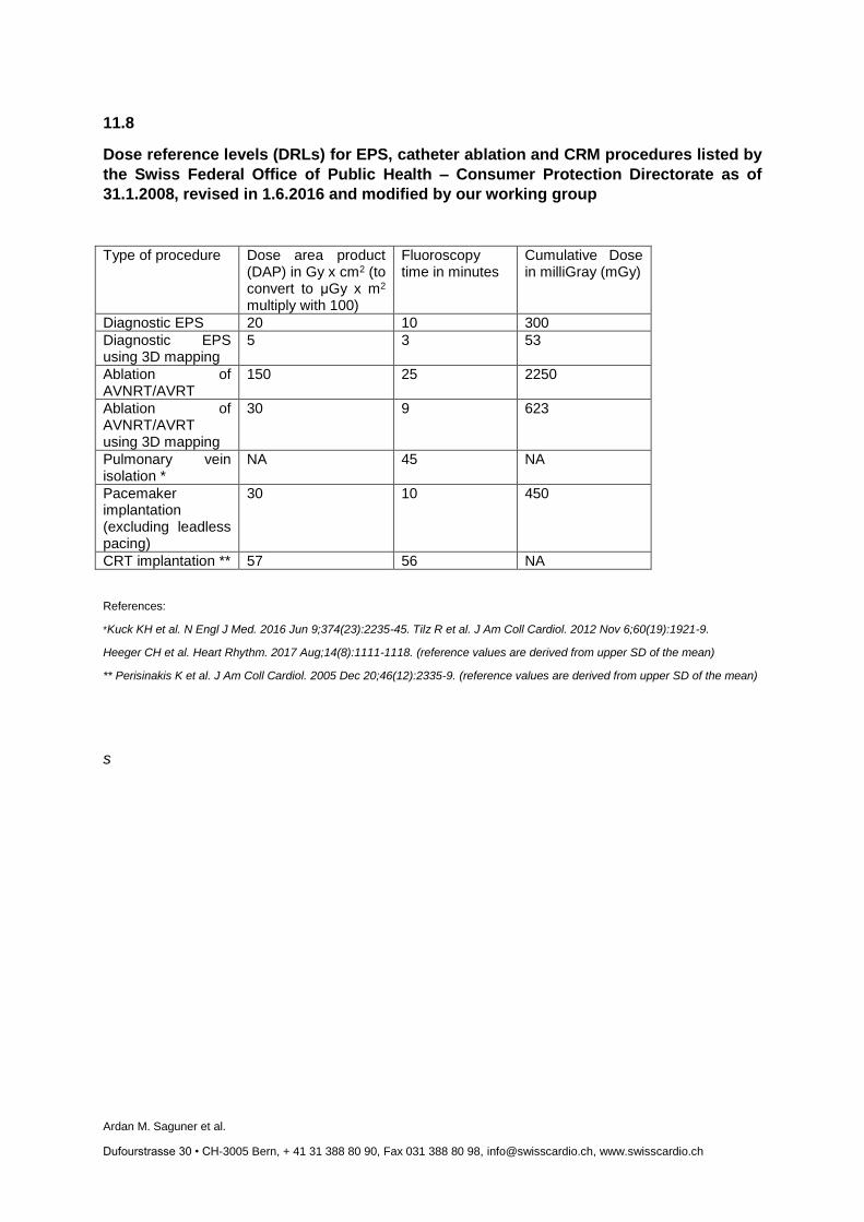
Ardan M. Saguner et al.
Dufourstrasse 30 • CH-3005 Bern, + 41 31 388 80 90, Fax 031 388 80 98, [email protected], www.swisscardio.ch
11.8
Dose reference levels (DRLs) for EPS, catheter ablation and CRM procedures listed by
the Swiss Federal Office of Public Health – Consumer Protection Directorate as of
31.1.2008, revised in 1.6.2016 and modified by our working group
Type of procedure Dose area product (DAP) in Gy x cm2 (to convert to μGy x m2 multiply with 100)
Fluoroscopy time in minutes
Cumulative Dose in milliGray (mGy)
Diagnostic EPS 20 10 300
Diagnostic EPS using 3D mapping
5 3 53
Ablation of AVNRT/AVRT
150 25 2250
Ablation of AVNRT/AVRT using 3D mapping
30 9 623
Pulmonary vein isolation *
NA 45 NA
Pacemaker implantation (excluding leadless pacing)
30 10 450
CRT implantation ** 57 56 NA
References:
*Kuck KH et al. N Engl J Med. 2016 Jun 9;374(23):2235-45. Tilz R et al. J Am Coll Cardiol. 2012 Nov 6;60(19):1921-9.
Heeger CH et al. Heart Rhythm. 2017 Aug;14(8):1111-1118. (reference values are derived from upper SD of the mean)
** Perisinakis K et al. J Am Coll Cardiol. 2005 Dec 20;46(12):2335-9. (reference values are derived from upper SD of the mean)
s
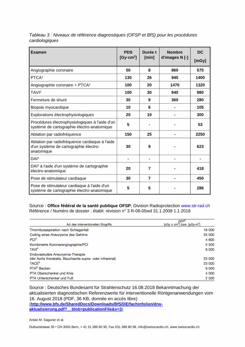
Ardan M. Saguner et al.
Dufourstrasse 30 • CH-3005 Bern, + 41 31 388 80 90, Fax 031 388 80 98, [email protected], www.swisscardio.ch
Tableau 3 : Niveaux de référence diagnostiques (OFSP et BfS) pour les procédures
cardiologiques
Examen PDS
[Gy·cm2]
Durée t
[min]
Nombre
d'images N [-]
DC
[mGy]
Angiographie coronaire 50 8 860 575
PTCA1 130 26 940 1400
Angiographie coronaire + PTCA1 100 20 1470 1320
TAVI2 100 30 940 980
Fermeture de shunt 30 9 360 280
Biopsie myocardique 10 6 - 105
Explorations électrophysiologiques 20 10 - 300
Procédures électrophysiologiques à l'aide d'un système de cartographie électro-anatomique
5 - - 53
Ablation par radiofréquence 150 25 - 2250
Ablation par radiofréquence cardiaque à l'aide d'un système de cartographie électro-anatomique
30 9 - 623
DAI3 - - - -
DAI3 à l'aide d'un système de cartographie électro-anatomique
20 7 - 418
Pose de stimulateur cardiaque 30 7 - 450
Pose de stimulateur cardiaque à l'aide d'un système de cartographie électro-anatomique
5 5 - 286
Source : Office fédéral de la santé publique OFSP, Division Radioprotection www.str-rad.ch
Référence / Numéro de dossier : établi: révision n° 3 R-06-05wd 31.1.2008 1.1.2018
Source : Deutsches Bundesamt für Strahlenschutz 16.08.2018 Bekanntmachung der
aktualisierten diagnostischen Referenzwerte für interventionelle Röntgenanwendungen vom
16. August 2018 (PDF, 36 KB, donnée en accès libre) (http://www.bfs.de/SharedDocs/Downloads/BfS/DE/fachinfo/ion/drw-
aktualisierung.pdf?__blob=publicationFile&v=3)
Modèle pour l’établissement d’un
MANUEL DE QUALITÉ
Radioprotection en cardiologie
Institution xx Auteurs xx Version xx.xx.xxxx Une version détaillée de ce manuel de qualité peut être obtenue à l’adresse suivante [email protected]. La version détaillée concerne le manuel de qualité de radioprotection en radiologie du centre universitaire de cardiologie de Zurich. Il est expressément autorisé de l’utiliser comme modèle pour vos propres manuels de qualité. Dans ce cas, la remarque suivante doit être ajoutée : « Édition du MANUEL DE QUALITÉ de radioprotection en cardiologie de l’USZ, Ruth Kofler, 2019 ».
1. PRÉAMBULE
2. COMPÉTENCES ET RESPONSABILITÉS 2.1. Organigramme 2.2. Titulaire de l’autorisation 2.3. Organigramme concernant la radioprotection 2.4. Direction médicale 2.5. Direction des soins 2.6. Responsabilités 2.7. Manuel de qualité
3. Parc d’appareils pour les examens et les traitements
4. Formation du personnel 4.1. Instruction
4.1.1 Réalisation
4.1.2 Contenu
4.1.3 Documentation
4.2. Formation continue
4.2.1 Formation continue en radioprotection pour les cardiologues actifs en radiologie interventionnelle et les rythmologues
4.2.2 Formation continue en radioprotection du personnel paramédical
5. Dosimétrie des collaborateurs professionnellement exposés aux rayonnements
5.1. Dosimètres individuels et manière de les porter
5.2. Dosimètres non individuels
5.2.1 Médecins invités, stagiaires, personnel temporaire
5.2.2 Visiteurs, pré-apprentis et étudiants
5.3. Administration et organisation de la dosimétrie
5.4. Gestion des données à la fin des relations de travail
6. Moyens et mesures de protection pour le personnel 6.1. Moyens de protection
6.2. Mesures de protection
6.3. Mesures de protection en cas de grossesse pour les personnes professionnellement exposées aux rayonnements
7. Contrôle de qualité des moyens de protection
8. Documentation des doses de rayonnements
9. Mesures de protection des patients

10. Mesures visant au respect de la pratique de justification 10.1. Indication justifiée par le médecin responsable
10.2. Patients nécessitant une évaluation minutieuse du risque
10.3. Exigences concernant l’examen
11. Protocoles d’examen et de traitement et informations destinées au patient
11.1. Protocoles d’examen et de traitement
11.2. Information à l’intention du patient / consentement éclairé
12. Établissement et communication du diagnostic, enregistrement et transmission des données
12.1. Établissement du diagnostic
12.2. Communication du diagnostic
12.3. Enregistrement et transmission des données
13. Assurance de la qualité
14. Autoévaluation
15. Liste des références
16. Annexe