Lincomycin at Subinhibitory Concentrations Potentiates ... · Lincomycin at Subinhibitory...
11
Lincomycin at Subinhibitory Concentrations Potentiates Secondary Metabolite Production by Streptomyces spp. Yu Imai, a Seizo Sato, b Yukinori Tanaka, c Kozo Ochi, c Takeshi Hosaka d,e Interdisciplinary Graduate School of Science and Technology, Shinshu University, Nagano, Japan a ; Central Research Laboratory, Nippon Suisan Kaisha, Ltd., Tokyo Innovation Center, Tokyo, Japan b ; Hiroshima Institute of Technology, Department of Life Science, Hiroshima, Japan c ; Department of Bioscience of Biotechnology, Faculty of Agriculture, Shinshu University, Nagano, Japan d ; Department of Interdisciplinary Genome Sciences and Cell Metabolism, Institute for Biomedical Sciences, Shinshu University, Nagano, Japan e Antibiotics have either bactericidal or bacteriostatic activity. However, they also induce considerable gene expression in bacteria when used at subinhibitory concentrations (below the MIC). We found that lincomycin, which inhibits protein synthesis by binding to the ribosomes of Gram-positive bacteria, was effective for inducing the expression of genes involved in secondary metabolism in Streptomyces strains when added to medium at subinhibitory concentrations. In Streptomyces coelicolor A3(2), lincomycin at 1/10 of its MIC markedly increased the expression of the pathway-specific regulatory gene actII-ORF4 in the blue- pigmented antibiotic actinorhodin (ACT) biosynthetic gene cluster, which resulted in ACT overproduction. Intriguingly, S. livi- dans 1326 grown in the presence of lincomycin at a subinhibitory concentration (1/12 or 1/3 of its MIC) produced abundant an- tibacterial compounds that were not detected in cells grown in lincomycin-free medium. Bioassay and mass spectrometry analysis revealed that some antibacterial compounds were novel congeners of calcium-dependent antibiotics. Our results indi- cate that lincomycin at subinhibitory concentrations potentiates the production of secondary metabolites in Streptomyces strains and suggest that activating these strains by utilizing the dose-response effects of lincomycin could be used to effectively induce the production of cryptic secondary metabolites. In addition to these findings, we also report that lincomycin used at concentrations for markedly increased ACT production resulted in alteration of the cytoplasmic protein (F o F 1 ATP synthase and subunits, etc.) profile and increased intracellular ATP levels. A fundamental mechanism for these unique phenomena is also discussed. S treptomyces is the largest genus of Gram-positive filamentous actinomycetes, and members of this genus produce abundant amounts of numerous bioactive metabolites, including antitumor agents, immunosuppressants, and antibiotics in particular (1, 2). Whole-genome sequencing studies of Streptomyces strains have shown that each species could produce many more secondary me- tabolites than were expected. For example, Streptomyces coelicolor A3(2), S. avermitilis MA-4680, and S. griseus IFO 13350 each pro- duce several secondary metabolites, although they have more than 20 gene clusters that can encode a number of known or predicted biosynthetic pathways for secondary metabolites (3–5). This indi- cates that the vast majority of secondary metabolites remain un- expressed or barely expressed under standard laboratory condi- tions. Thus, there is considerable interest in exploring practical means to induce this genetic potential in streptomycetes, which could result in the isolation of novel bioactive secondary metabo- lites. Recent developments in new methodologies, including physi- ological and genetic engineering approaches, have opened the door for the discovery of novel secondary metabolites by activat- ing cryptic biosynthetic pathways in streptomycetes (6–13). The improvements and modifications of culture conditions, such as the one strain-many compounds approach (which changes fer- mentation conditions like medium compositions and culture ves- sel types), coculture (bacterial-bacterial or fungal-bacterial phys- ical interactions), and utilizing inducing molecules (siderophores, -butyrolactones, N-acetylglucosamine, rare earth elements, an- tibiotic-remodeling compounds, and antibiotics), are simple, ef- fective methods of inducing cryptic gene cluster expression and increasing the production of secondary metabolites (14–19). Molecular genetic manipulation, including directed mutagen- esis, expression of antibiotic activators, and heterologous expres- sion, is also an efficient approach for activating target gene clusters and improving the yields of secondary metabolites (20, 21). In particular, altering the expression of transcription factors that control biosynthetic pathways and amplifying biosynthetic gene clusters have resulted in the dramatically increased production of secondary metabolites and the isolation of novel compounds and their congeners (22–24). We have alternatively developed a novel method for activating poorly expressed Streptomyces genes to produce antibiotics via a mutation that confers resistance to drugs by targeting RNA poly- merase or ribosomes, such as rifampin, streptomycin, gentamicin, and erythromycin (25–29). This method, a so-called ribosome engineering approach, led to the discovery of a novel antibiotic, piperidamycin, produced by Streptomyces sp. strain 631689, Received 27 December 2014 Accepted 23 March 2015 Accepted manuscript posted online 27 March 2015 Citation Imai Y, Sato S, Tanaka Y, Ochi K, Hosaka T. 2015. Lincomycin at subinhibitory concentrations potentiates secondary metabolite production by Streptomyces spp. Appl Environ Microbiol 81:3869 –3879. doi:10.1128/AEM.04214-14. Editor: H. Nojiri Address correspondence to Takeshi Hosaka, [email protected]. Supplemental material for this article may be found at http://dx.doi.org/10.1128 /AEM.04214-14. Copyright © 2015, American Society for Microbiology. All Rights Reserved. doi:10.1128/AEM.04214-14 June 2015 Volume 81 Number 11 aem.asm.org 3869 Applied and Environmental Microbiology on June 11, 2020 by guest http://aem.asm.org/ Downloaded from
Transcript of Lincomycin at Subinhibitory Concentrations Potentiates ... · Lincomycin at Subinhibitory...
Lincomycin at Subinhibitory Concentrations Potentiates SecondaryMetabolite Production by Streptomyces spp.
Yu Imai,a Seizo Sato,b Yukinori Tanaka,c Kozo Ochi,c Takeshi Hosakad,e
Interdisciplinary Graduate School of Science and Technology, Shinshu University, Nagano, Japana; Central Research Laboratory, Nippon Suisan Kaisha, Ltd., TokyoInnovation Center, Tokyo, Japanb; Hiroshima Institute of Technology, Department of Life Science, Hiroshima, Japanc; Department of Bioscience of Biotechnology, Facultyof Agriculture, Shinshu University, Nagano, Japand; Department of Interdisciplinary Genome Sciences and Cell Metabolism, Institute for Biomedical Sciences, ShinshuUniversity, Nagano, Japane
Antibiotics have either bactericidal or bacteriostatic activity. However, they also induce considerable gene expression in bacteriawhen used at subinhibitory concentrations (below the MIC). We found that lincomycin, which inhibits protein synthesis bybinding to the ribosomes of Gram-positive bacteria, was effective for inducing the expression of genes involved in secondarymetabolism in Streptomyces strains when added to medium at subinhibitory concentrations. In Streptomyces coelicolor A3(2),lincomycin at 1/10 of its MIC markedly increased the expression of the pathway-specific regulatory gene actII-ORF4 in the blue-pigmented antibiotic actinorhodin (ACT) biosynthetic gene cluster, which resulted in ACT overproduction. Intriguingly, S. livi-dans 1326 grown in the presence of lincomycin at a subinhibitory concentration (1/12 or 1/3 of its MIC) produced abundant an-tibacterial compounds that were not detected in cells grown in lincomycin-free medium. Bioassay and mass spectrometryanalysis revealed that some antibacterial compounds were novel congeners of calcium-dependent antibiotics. Our results indi-cate that lincomycin at subinhibitory concentrations potentiates the production of secondary metabolites in Streptomycesstrains and suggest that activating these strains by utilizing the dose-response effects of lincomycin could be used to effectivelyinduce the production of cryptic secondary metabolites. In addition to these findings, we also report that lincomycin used atconcentrations for markedly increased ACT production resulted in alteration of the cytoplasmic protein (FoF1 ATP synthase �and � subunits, etc.) profile and increased intracellular ATP levels. A fundamental mechanism for these unique phenomena isalso discussed.
Streptomyces is the largest genus of Gram-positive filamentousactinomycetes, and members of this genus produce abundant
amounts of numerous bioactive metabolites, including antitumoragents, immunosuppressants, and antibiotics in particular (1, 2).Whole-genome sequencing studies of Streptomyces strains haveshown that each species could produce many more secondary me-tabolites than were expected. For example, Streptomyces coelicolorA3(2), S. avermitilis MA-4680, and S. griseus IFO 13350 each pro-duce several secondary metabolites, although they have more than20 gene clusters that can encode a number of known or predictedbiosynthetic pathways for secondary metabolites (3–5). This indi-cates that the vast majority of secondary metabolites remain un-expressed or barely expressed under standard laboratory condi-tions. Thus, there is considerable interest in exploring practicalmeans to induce this genetic potential in streptomycetes, whichcould result in the isolation of novel bioactive secondary metabo-lites.
Recent developments in new methodologies, including physi-ological and genetic engineering approaches, have opened thedoor for the discovery of novel secondary metabolites by activat-ing cryptic biosynthetic pathways in streptomycetes (6–13). Theimprovements and modifications of culture conditions, such asthe one strain-many compounds approach (which changes fer-mentation conditions like medium compositions and culture ves-sel types), coculture (bacterial-bacterial or fungal-bacterial phys-ical interactions), and utilizing inducing molecules (siderophores,�-butyrolactones, N-acetylglucosamine, rare earth elements, an-tibiotic-remodeling compounds, and antibiotics), are simple, ef-fective methods of inducing cryptic gene cluster expression andincreasing the production of secondary metabolites (14–19).
Molecular genetic manipulation, including directed mutagen-esis, expression of antibiotic activators, and heterologous expres-sion, is also an efficient approach for activating target gene clustersand improving the yields of secondary metabolites (20, 21). Inparticular, altering the expression of transcription factors thatcontrol biosynthetic pathways and amplifying biosynthetic geneclusters have resulted in the dramatically increased production ofsecondary metabolites and the isolation of novel compounds andtheir congeners (22–24).
We have alternatively developed a novel method for activatingpoorly expressed Streptomyces genes to produce antibiotics via amutation that confers resistance to drugs by targeting RNA poly-merase or ribosomes, such as rifampin, streptomycin, gentamicin,and erythromycin (25–29). This method, a so-called ribosomeengineering approach, led to the discovery of a novel antibiotic,piperidamycin, produced by Streptomyces sp. strain 631689,
Received 27 December 2014 Accepted 23 March 2015
Accepted manuscript posted online 27 March 2015
Citation Imai Y, Sato S, Tanaka Y, Ochi K, Hosaka T. 2015. Lincomycin atsubinhibitory concentrations potentiates secondary metabolite production byStreptomyces spp. Appl Environ Microbiol 81:3869–3879. doi:10.1128/AEM.04214-14.
Editor: H. Nojiri
Address correspondence to Takeshi Hosaka, [email protected].
Supplemental material for this article may be found at http://dx.doi.org/10.1128/AEM.04214-14.
Copyright © 2015, American Society for Microbiology. All Rights Reserved.
doi:10.1128/AEM.04214-14
June 2015 Volume 81 Number 11 aem.asm.org 3869Applied and Environmental Microbiology
on June 11, 2020 by guesthttp://aem
.asm.org/
Dow
nloaded from
which rarely produced antibiotics in any culture medium withoutstrain improvement (30–32). Using antibiotic-overproducingmutants that harbored mutations in RNA polymerase and/or ri-bosomes, we previously demonstrated the importance of alteringthe promoter selectivity of RNA polymerase and maintaining ahigh level of protein synthesis during the late growth phase forinducing antibiotic production. Although the detailed mecha-nisms have not been fully elucidated, the important finding wasthat modulating the transcriptional or translational apparatus orboth (i.e., RNA polymerase and ribosomes) could dramaticallyalter gene expression in Streptomyces strains, which reflected theirpotential to produce novel secondary metabolites (29, 33).
On the basis of the rational approaches described above, wesought more effective methods of developing cryptic gene clustersfor secondary metabolic pathways in Streptomyces strains. Wefound that secondary metabolite production could be dramati-cally potentiated by using a ribosome-targeting antibiotic at con-centrations below its MIC. This appeared to agree with the con-cept of antibiotic hormesis, for which antibiotics at subinhibitoryconcentrations have pleiotropic effects on bacterial gene expres-sion, as previously reported by Davies et al. (34, 35). Thus, in thepresent study, we used genetically best-characterized strains ofStreptomyces, primarily S. coelicolor A3(2) and its close relative S.lividans 66, and conducted a detailed investigation of the dose-response effects of the ribosome-targeting antibiotic lincomycinon secondary metabolite production. We also investigated thepossibility that approaches that utilize the hormetic effects of ri-bosome-targeting antibiotics could be used effectively to improvethe yields of cryptic secondary metabolites. Additionally, as a firststep toward understanding the mechanisms by which lincomycininduces the production of secondary metabolites, we attempted toidentify those proteins that may have been involved in the markeddifferences between cells grown with or without lincomycin. Stud-ies by Shibl (36) and Herbert et al. (37) have demonstrated effectsof subinhibitory lincosamide antibiotics on virulence, adherence,and toxin production, etc. in Staphylococcus aureus. These previ-ous studies showed lincosamide antibiotics at subinhibitory con-centrations to have very little effect on cytoplasmic protein pro-files. In contrast, the present study clearly demonstrated that thelevels of several cytoplasmic proteins (FoF1 ATP synthase � and �subunits, proteins involved in antibiotic biosynthesis, etc.) wereconsiderably different between the S. coelicolor A3(2) cells thatwere grown with or without lincomycin. A fundamental mecha-nism for these unique phenomena raised by lincomycin is dis-cussed on the basis of the results of the present work.
MATERIALS AND METHODSChemicals. Chloramphenicol, erythromycin, streptomycin, and tetracy-cline were purchased from Nacalai Tesque, Inc. (Japan). Clindamycin,
gentamicin, lincomycin, and tylosin were purchased from Wako PureChemicals (Japan). Thiostrepton was purchased from Sigma-Aldrich.
Bacterial strains and growth conditions. The actinomycete strainsused in this study are shown in Table 1. Spores of these strains wereprepared from cultures grown at 30°C for 7 days on GYM agar (27).Modified R5 (MR5) medium (both agar and liquid medium) (38) wasused for phenotypic characterizations of S. coelicolor A3(2) and S. lividansstrains. MR5 liquid medium was also used to grow cells to prepare totalRNA and proteins. Typically, agar and liquid cultures were as follows. Foragar culture, approximately 1 � 105 to 5 � 105 spores were inoculatedonto MR5 agar (25 ml per culture plate [90 mm in diameter by 15 mmdeep]) and incubated at 30°C. For liquid culture, approximately 1 � 106
spores were inoculated into 20 ml of MR5 liquid medium in a 100-mlErlenmeyer flask and incubated at 30°C with shaking at 200 rpm. SPY (39)liquid medium was used for streptomycin production by S. griseus IFO13189. As required, media were supplemented with appropriate drug con-centrations.
MIC determinations. To determine the MICs of drugs for S. coelicolorA3(2), S. lividans, and S. griseus, their spores were directly inoculated ontoor into media that contained different concentrations of a particular drugand incubated at 30°C. The minimum drug concentration that nearlycompletely inhibited growth was defined as the MIC.
Dry cell weights. A culture broth was filtered through filter paper or amembrane filter under vacuum and washed two times with reverse osmo-sis water to remove any residual medium remaining on the cell surfaces.The remaining cells were then dried to a constant weight in an oven at55°C (for filter paper) or at room temperature (for a membrane filter) for2 days. Dry cell weights were determined by subtracting the weight of thefilter paper (membrane filter) from the weight of the filter paper (mem-brane filter) plus the cells.
Antibiotic assays. Actinorhodin (ACT) production on MR5 agar wasdirectly assessed by blue color intensity. The production of ACT and un-decylprodigiosin (RED) in liquid culture with MR5 medium was deter-mined as described previously (2, 25). A bioassay to detect antibacterialcompound produced by S. lividans was performed as follows. Strain 1326was grown on MR5 agar. At the times indicated, agar plugs (diameter, 8mm) were cut from agar plates and transferred to an assay plate preparedwith 2-fold-diluted Mueller-Hinton agar (20 ml per no. 2 square plate)that contained cells (1 � 107 CFU) of the lincomycin-resistant mutant ofBacillus subtilis ATCC 6633 used as a test organism. To assess the zone ofinhibition of antibacterial compounds in these agar plugs, the assay plateswere incubated at 37°C for an additional 24 h. Streptomycin productionin liquid SPY medium was determined as described previously (39).
HPLC analysis of culture extracts. Samples used to assess the high-performance liquid chromatography (HPLC) profiles of S. lividans cul-ture extracts were prepared as follows. Strain 1326 was grown for 7 days onMR5 agar covered with a cellophane sheet. Next, agar without cells wasextracted with 25 ml of methanol by gentle agitation for 10 h. The crudeextract was dried in a rotary evaporator and lyophilized. The resultingsample was dissolved in 25 ml of ultrapure water and separated into or-ganic (ethyl acetate) and aqueous phases. The aqueous phase was lyoph-ilized, dissolved in 5 ml of 50% aqueous methanol, and filtered through aFILTSTAR syringe filter (hydrophilic nylon, 0.22-�m pore size; Starlab
TABLE 1 Actinomycete strains used in this study
Strain Relevant characteristic Reference(s)
Streptomyces coelicolor A3(2) 1147 Prototrophic wild type (SCP1� SCP2�) 2Streptomyces lividans 1326 Prototrophic wild type (SLP2� SLP3�) 2Streptomyces lividans SLE299 Spontaneous lincomycin-resistant mutant isolated from 1326 with a mutation in the
23S rRNA gene (rrnC-A2281G)25
Streptomyces griseus IFO 13189 Prototrophic streptomycin-producing wild type 39Saccharopolyspora erythraea NRRL 2338 Prototrophic erythromycin-producing wild type 32, 56
Imai et al.
3870 aem.asm.org June 2015 Volume 81 Number 11Applied and Environmental Microbiology
on June 11, 2020 by guesthttp://aem
.asm.org/
Dow
nloaded from
Science Co. Ltd., China). Aliquots (20 �l) of each sample were subjectedto HPLC with an InertSustain C18 reverse-phase column (4.6 by 250 mm,5-�m particle size; GL Science Inc., Japan) and eluted at a flow rate of 1ml/min. The HPLC apparatus included a Shimadzu Prominence HPLCsystem equipped with an SPD-M20A diode array detector (SHIMADZUCo. Ltd., Japan). The solvent and conditions used were 0 to 5 min of 30%acetonitrile (ACN) that contained 0.01% formic acid (FA) and 5 to 30 minof a linear gradient of 30 to 80% ACN that contained 0.01% FA. Multiple-wavelength monitoring was performed at 190 to 800 nm.
Isolation and analysis of antibacterial compounds. To isolate anyantibacterial compounds produced by S. lividans, strain 1326 was inocu-lated onto MR5 agar (20 plates) that contained 20 �g/ml of lincomycinand covered with cellophane sheets. After incubation for 7 days, cells on acellophane sheet were removed and the agar was combined. A cultureextract was prepared as described above, except that a Millex-HP syringe-driven filter unit (0.45 �m; Millipore) was used, and further purified bysemipreparative HPLC with an InertSustain C18 reverse-phase column(10 by 250 mm, 5-�m particle size; GL Science Inc., Japan). Antibacterialcompounds that were eluted by a gradient elution (flow rate, 4 ml/min) of30 to 60% ACN that contained 0.01% FA were collected and dried in arotary evaporator and lyophilized. Purified antibacterial compounds wereanalyzed by electrospray ionization time of flight mass spectrometry (ESI-TOF-MS). ESI-MS was performed with a Micromass LCT-Premier XEinstrument (Waters). Mass data were acquired in the negative-ion modewith a scan range of 100 to 2,500 Da. The electrospray capillary voltagewas 2.2 kV, and the cone voltage was 80 V. The source temperature wasmaintained at 100°C. Mass measurements were made at a resolution of10,000 resolving power (RP).
Transcription analysis. Total RNA was isolated from cells (approxi-mately 50 mg wet weight) grown in MR5 liquid medium or on an MR5agar plate with Sepasol-RNA I super G (Nacalai Tesque, Inc., Japan). Aftertreatment with RNase-free Recombinant DNase I (TaKaRa Bio. Inc., Ja-pan), each sample was further purified with a High Pure RNA isolation kit(Roche). Total RNA (2 �g) was used as the template for a reverse tran-scription (RT) reaction (20 �l) at 55°C for 30 min with a TranscriptorFirst Strand cDNA synthesis kit (Roche). Real-time quantitative RT-PCR(qRT-PCR) analysis for actII-ORF4, redD, SLI3570, atpA, and atpD geneexpression was performed as described previously (40). The results ofthese assays were normalized relative to the corresponding transcriptionallevel of hrdB, the gene that encodes the principal sigma factor. For theprimers used for real-time qPCR, see Table S1 in the supplemental mate-rial.
Protein gel electrophoresis and Western blot analysis. Total cell pro-teins for sodium dodecyl sulfate-polyacrylamide gel electrophoresis(SDS-PAGE) and Western blot analysis were prepared as follows. Cells(approximately 50 to 350 mg [wet weight]) of S. coelicolor A3(2) grown inMR5 liquid medium were suspended in 1 ml of TMA-I buffer (10 mMTris-HCl [pH 7.7], 30 mM NH4 acetate [OAc], 10 mM MgOAc2, 1 mMdithiothreitol, 10% [vol/vol] glycerol) that contained 1 mM phenylmeth-ylsulfonyl fluoride and disrupted by homogenizing with 2 g of zirconiaballs (0.5 mm). After centrifugation at 15,000 � g for 10 min at 4°C, the
supernatant was dialyzed twice for 3 h against more than 50 volumes ofTMA-I buffer and used as a total cell protein sample. Samples (5 �g) oftotal cellular protein were subjected to 12% (wt/vol) SDS-PAGE, afterwhich the separated proteins was electroblotted onto a polyvinylidenedifluoride membrane (Immobilon-P; Millipore). Blots were developedwith polyclonal antibodies and an ECL Select Western blotting detectionsystem (GE Healthcare) for chemiluminescence detection according tothe manufacturer’s instructions. Polyclonal antibodies against ActII-ORF4 was prepared from rabbits by immunizing them with a syntheticoligopeptide [211-MGRRAEALESYRNL-224, numbers represent aminoacid residues in S. coelicolor A3(2)]. These were used as the primary anti-body at a dilution of 1:5,000.
Protein identification. Total cellular proteins that had been separatedby SDS-PAGE were stained with CBB Stain One (Nacalai Tesque, Inc.,Japan). The protein bands of interest were cut out and subjected to pep-tide mass fingerprinting analysis. Mascot software (Matrix Science) wasused to analyze mass data and identify proteins.
Measurements of intracellular nucleotide pools. Intracellular nucle-otides, including ATP, GTP, UTP, and CTP, were extracted with 1 M FAfrom cells that had been grown in MR5 liquid medium and assayed with aShimadzu Prominence HPLC system equipped with a Partisil-10 SAXion-exchange column (4.6 by 250 mm, 5-�m particle size; Hichrom Co.,United Kingdom) as described previously (39). Nucleotide concentra-tions were expressed in nanomoles per milligram of dry cell weight.
RESULTSLincomycin inducing effects on antibiotic production. To assessthe effects of subinhibitory concentrations of the ribosome-tar-geting antibiotics (clindamycin, chloramphenicol, erythromycin,gentamicin, lincomycin, streptomycin, tetracycline, thiostrepton,and tylosin) on ACT production by S. coelicolor A3(2) and S. livi-dans, we first determined the MICs of these drugs for each strain.The MICs of each antibiotic are shown in Table 2. Except forgentamicin, lincomycin, and streptomycin, the MICs of these forS. lividans were similar to those for S. coelicolor A3(2). We thenassessed each antibiotic’s effectiveness at enhancing ACT produc-tion at concentrations ranging from 1/10,000 of the antibiotic’sMIC to its MIC. The results obtained with the optimal concentra-tion of each antibiotic are shown in Fig. 1A. It is clear that all ofthese antibiotics were more or less effective at enhancing ACTproduction by S. coelicolor A3(2). The effects of lincomycin, chlor-amphenicol, and tetracycline were especially pronounced, andlincomycin had the greatest effect. Clindamycin, an antibiotic ofthe lincosamide class, had no effect on ACT production by S.lividans. Figure 1B shows the dose-dependent effect of lincomycinon ACT production. In S. coelicolor A3(2), lincomycin effectivelyenhanced ACT production over a wide concentration range, withan optimal concentration of 5 �g/ml (1/16 of its MIC). In S. livi-
TABLE 2 MICs of ribosome-targeting antibiotics for Streptomyces strains
Strain
MIC (�g/ml)a
Cli Cm Ery Gen Lin Sm Tet Tsp Tyl
S. coelicolor A3(2) 1147 30 20 8 0.5 80 2.5 500 2 20S. lividans 1326 30 20 8 0.7 60 4 500 2 20S. lividans SLE299b ND ND ND ND �1,000 ND ND ND NDS. griseus IFO 13189 ND ND ND ND 50 ND ND ND NDa Determined after incubation on an MR5 agar plate for 7 days [S. coelicolor A3(2), S. lividans 1326, and S. lividans SLE299] or a GYM agar plate for 3 days (S. griseus IFO 13189).Abbreviations: Cli, clindamycin; Cm, chloramphenicol; Ery, erythromycin; Gen, gentamicin; Lin, lincomycin; Sm, streptomycin; Tet, tetracycline; Tsp, thiostrepton; Tyl, tylosin;ND, not determined.b Lincomycin-resistant mutant strain (25).
Lincomycin Induction of Secondary Metabolism
June 2015 Volume 81 Number 11 aem.asm.org 3871Applied and Environmental Microbiology
on June 11, 2020 by guesthttp://aem
.asm.org/
Dow
nloaded from
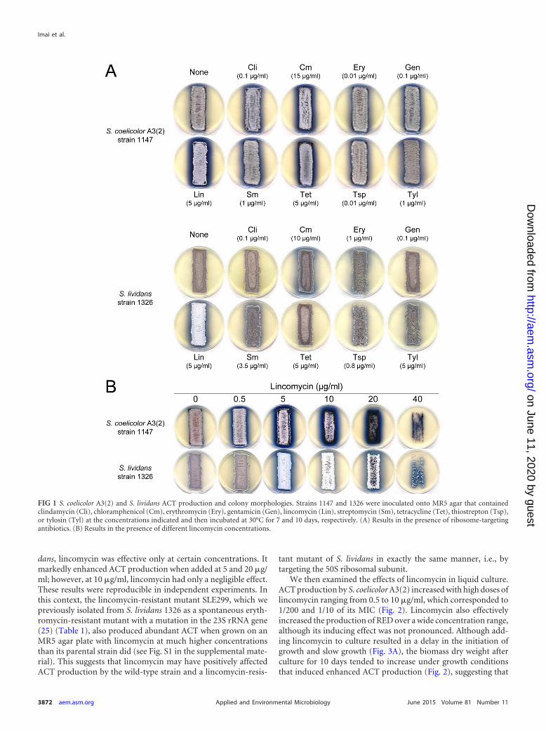
dans, lincomycin was effective only at certain concentrations. Itmarkedly enhanced ACT production when added at 5 and 20 �g/ml; however, at 10 �g/ml, lincomycin had only a negligible effect.These results were reproducible in independent experiments. Inthis context, the lincomycin-resistant mutant SLE299, which wepreviously isolated from S. lividans 1326 as a spontaneous eryth-romycin-resistant mutant with a mutation in the 23S rRNA gene(25) (Table 1), also produced abundant ACT when grown on anMR5 agar plate with lincomycin at much higher concentrationsthan its parental strain did (see Fig. S1 in the supplemental mate-rial). This suggests that lincomycin may have positively affectedACT production by the wild-type strain and a lincomycin-resis-
tant mutant of S. lividans in exactly the same manner, i.e., bytargeting the 50S ribosomal subunit.
We then examined the effects of lincomycin in liquid culture.ACT production by S. coelicolor A3(2) increased with high doses oflincomycin ranging from 0.5 to 10 �g/ml, which corresponded to1/200 and 1/10 of its MIC (Fig. 2). Lincomycin also effectivelyincreased the production of RED over a wide concentration range,although its inducing effect was not pronounced. Although add-ing lincomycin to culture resulted in a delay in the initiation ofgrowth and slow growth (Fig. 3A), the biomass dry weight afterculture for 10 days tended to increase under growth conditionsthat induced enhanced ACT production (Fig. 2), suggesting that
FIG 1 S. coelicolor A3(2) and S. lividans ACT production and colony morphologies. Strains 1147 and 1326 were inoculated onto MR5 agar that containedclindamycin (Cli), chloramphenicol (Cm), erythromycin (Ery), gentamicin (Gen), lincomycin (Lin), streptomycin (Sm), tetracycline (Tet), thiostrepton (Tsp),or tylosin (Tyl) at the concentrations indicated and then incubated at 30°C for 7 and 10 days, respectively. (A) Results in the presence of ribosome-targetingantibiotics. (B) Results in the presence of different lincomycin concentrations.
Imai et al.
3872 aem.asm.org June 2015 Volume 81 Number 11Applied and Environmental Microbiology
on June 11, 2020 by guesthttp://aem
.asm.org/
Dow
nloaded from
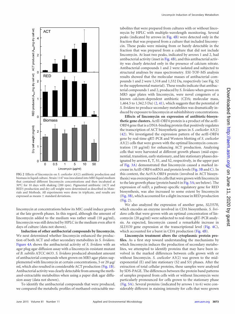
lincomycin at concentrations below its MIC could induce growthat the late growth phases. In this regard, although the amount oflincomycin added to the medium was rather small (10 �g/ml),lincomycin was still detected by HPLC in the medium even after 6days of culture (data not shown).
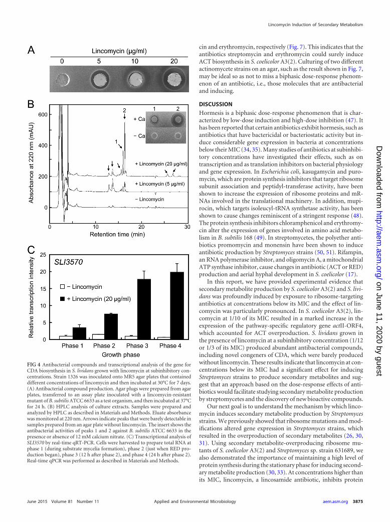
Induction of other antibacterial compounds by lincomycin.We next determined whether lincomycin enhanced the produc-tion of both ACT and other secondary metabolites in S. lividans.Figure 4A shows the antibacterial activity of S. lividans with anagar plug agar diffusion assay with a lincomycin-resistant mutantof B. subtilis ATCC 6633. S. lividans produced abundant amountof antibacterial compounds when grown on MR5 agar plates sup-plemented with lincomycin at certain concentrations, 5 or 20 �g/ml, which also resulted in considerable ACT production (Fig. 1B).Antibacterial activity was clearly detectable from among the meth-anol-extractable metabolites when using a paper disk agar diffu-sion assay (data not shown).
To identify the antibacterial compounds that were produced,we compared the metabolic profiles of methanol-extractable me-
tabolites that were prepared from cultures with or without linco-mycin by HPLC with multiple-wavelength monitoring. Severalpeaks (indicated by arrows in Fig. 4B) were detected only in thefraction that was prepared from a culture that included lincomy-cin. These peaks were missing from or barely detectable in thefraction that was prepared from a culture that did not includelincomycin. At least two peaks, indicated by arrows 1 and 2, hadantibacterial activity (inset in Fig. 4B), and this antibacterial activ-ity was clearly detected only in the presence of calcium nitrate.Antibacterial compounds 1 and 2 were isolated and subjected tostructural analyses by mass spectrometry. ESI-TOF-MS analysisresults showed that the molecular masses of antibacterial com-pounds 1 and 2 were 1,518 and 1,532 Da, respectively (see Fig. S2in the supplemental material). These results indicate that antibac-terial compounds 1 and 2, produced by S. lividans when grown onMR5 agar plates with lincomycin, were novel congeners of aknown calcium-dependent antibiotic (CDA; molecular mass,1,464.5 to 1,562.5 Da) (2, 41), which suggests that the potential ofS. lividans to produce secondary metabolites was dramatically in-duced by exposure to lincomycin at subinhibitory concentrations.
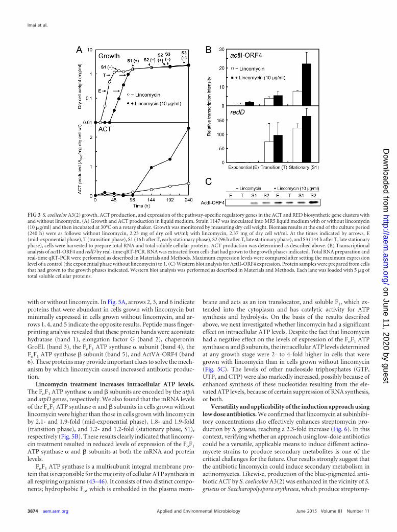
Effects of lincomycin on expression of antibiotic-biosyn-thetic gene clusters. ActII-ORF4 protein is a product of the actII-ORF4 gene that is a DNA-binding protein that positively regulatesthe transcription of ACT biosynthetic genes in S. coelicolor A3(2)(42). We investigated the expression pattern of the actII-ORF4gene by real-time qRT-PCR and Western blotting of S. coelicolorA3(2) cells that were grown with the optimal lincomycin concen-tration (10 �g/ml) for enhancing ACT production. Analyzingcells that were harvested at different growth phases (mid-expo-nential, transition, early stationary, and late stationary phases des-ignated by arrows E, T, S1, and S2, respectively, in the upper partof Fig. 3A) demonstrated that lincomycin caused a marked in-crease in ActII-ORF4 mRNA and protein levels (Fig. 3B and C). Inthis context, the ActVA-ORF4 protein (involved in ACT biosyn-thesis) was overexpressed in cells that were grown with lincomycinto the late growth phase (protein band 6 in Fig. 5A; see below). Theexpression of redD, a pathway-specific regulatory gene for REDbiosynthesis, was also increased to some extent by lincomycin(Fig. 3B), which accounted for a slight increase in RED production(Fig. 2).
We also analyzed the expression of another gene, SLI3570,which encodes an enzyme involved in CDA biosynthesis. S. livi-dans cells that were grown with an optimal concentration of lin-comycin (20 �g/ml) were subjected to real-time qRT-PCR analy-sis. As expected, lincomycin caused a remarkable increase inSLI3570 gene expression at the transcriptional level (Fig. 4C),which accounted for a burst in CDA production (Fig. 4B).
Lincomycin treatment alters the cytoplasmic protein pro-files. As a first step toward understanding the mechanisms bywhich lincomycin induces the production of secondary metabo-lites, we attempted to identify proteins that may have been in-volved in the marked differences between cells grown with orwithout lincomycin. S. coelicolor A3(2) was grown to the mid-exponential (E) and late stationary (S2 and S3) phases. After theextraction of total cellular proteins, these samples were analyzedby SDS-PAGE. The differences between the protein band patternsof samples prepared from cells with or without lincomycin wereparticularly pronounced for cells grown to the stationary phase(Fig. 5A). Several proteins (indicated by arrows 1 to 6) were con-siderably different in staining intensity for cells that were grown
FIG 2 Effects of lincomycin on S. coelicolor A3(2) antibiotic production andbiomass in liquid culture. Strain 1147 was inoculated into MR5 liquid mediumthat contained different lincomycin concentrations and then incubated at30°C for 10 days with shaking (200 rpm). Pigmented antibiotic (ACT andRED) production and dry cell weight were determined as described in Mate-rials and Methods. All experiments were done in triplicate, and results areexpressed as means standard deviations.
Lincomycin Induction of Secondary Metabolism
June 2015 Volume 81 Number 11 aem.asm.org 3873Applied and Environmental Microbiology
on June 11, 2020 by guesthttp://aem
.asm.org/
Dow
nloaded from
with or without lincomycin. In Fig. 5A, arrows 2, 3, and 6 indicateproteins that were abundant in cells grown with lincomycin butminimally expressed in cells grown without lincomycin, and ar-rows 1, 4, and 5 indicate the opposite results. Peptide mass finger-printing analysis revealed that these protein bands were aconitatehydratase (band 1), elongation factor G (band 2), chaperoninGroEL (band 3), the FoF1 ATP synthase � subunit (band 4), theFoF1 ATP synthase � subunit (band 5), and ActVA-ORF4 (band6). These proteins may provide important clues to solve the mech-anism by which lincomycin caused increased antibiotic produc-tion.
Lincomycin treatment increases intracellular ATP levels.The FoF1 ATP synthase � and � subunits are encoded by the atpAand atpD genes, respectively. We also found that the mRNA levelsof the FoF1 ATP synthase � and � subunits in cells grown withoutlincomycin were higher than those in cells grown with lincomycinby 2.1- and 1.9-fold (mid-exponential phase), 1.8- and 1.9-fold(transition phase), and 1.2- and 1.2-fold (stationary phase, S1),respectively (Fig. 5B). These results clearly indicated that lincomy-cin treatment resulted in reduced levels of expression of the FoF1
ATP synthase � and � subunits at both the mRNA and proteinlevels.
FoF1 ATP synthase is a multisubunit integral membrane pro-tein that is responsible for the majority of cellular ATP synthesis inall respiring organisms (43–46). It consists of two distinct compo-nents; hydrophobic Fo, which is embedded in the plasma mem-
brane and acts as an ion translocator, and soluble F1, which ex-tended into the cytoplasm and has catalytic activity for ATPsynthesis and hydrolysis. On the basis of the results describedabove, we next investigated whether lincomycin had a significanteffect on intracellular ATP levels. Despite the fact that lincomycinhad a negative effect on the levels of expression of the FoF1 ATPsynthase � and � subunits, the intracellular ATP levels determinedat any growth stage were 2- to 4-fold higher in cells that weregrown with lincomycin than in cells grown without lincomycin(Fig. 5C). The levels of other nucleoside triphosphates (GTP,UTP, and CTP) were also markedly increased, possibly because ofenhanced synthesis of these nucleotides resulting from the ele-vated ATP levels, because of certain suppression of RNA synthesis,or both.
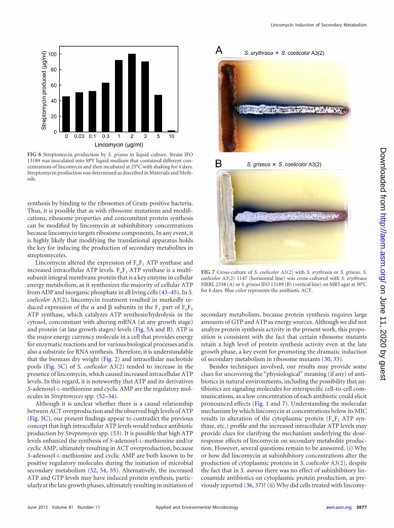
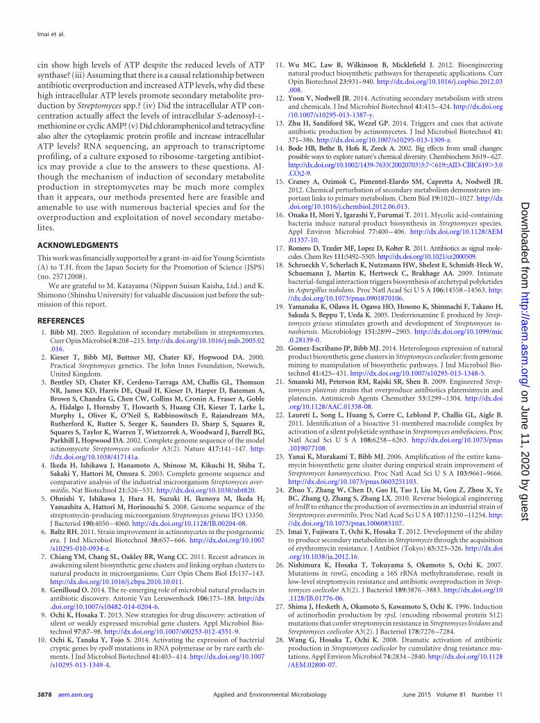
Versatility and applicability of the induction approach usinglow dose antibiotics. We confirmed that lincomycin at subinhibi-tory concentrations also effectively enhances streptomycin pro-duction by S. griseus, reaching a 2.3-fold increase (Fig. 6). In thiscontext, verifying whether an approach using low-dose antibioticscould be a versatile, applicable means to induce different actino-mycete strains to produce secondary metabolites is one of thecritical challenges for the future. Our results strongly suggest thatthe antibiotic lincomycin could induce secondary metabolism inactinomycetes. Likewise, production of the blue-pigmented anti-biotic ACT by S. coelicolor A3(2) was enhanced in the vicinity of S.griseus or Saccharopolyspora erythraea, which produce streptomy-
FIG 3 S. coelicolor A3(2) growth, ACT production, and expression of the pathway-specific regulatory genes in the ACT and RED biosynthetic gene clusters withand without lincomycin. (A) Growth and ACT production in liquid medium. Strain 1147 was inoculated into MR5 liquid medium with or without lincomycin(10 �g/ml) and then incubated at 30°C on a rotary shaker. Growth was monitored by measuring dry cell weight. Biomass results at the end of the culture period(240 h) were as follows: without lincomycin, 2.23 mg of dry cell wt/ml; with lincomycin, 2.37 mg of dry cell wt/ml. At the times indicated by arrows, E(mid-exponential phase), T (transition phase), S1 (16 h after T, early stationary phase), S2 (96 h after T, late stationary phase), and S3 (144 h after T, late stationaryphase), cells were harvested to prepare total RNA and total soluble cellular proteins. ACT production was determined as described above. (B) Transcriptionalanalysis of actII-ORF4 and redD by real-time qRT-PCR. RNA was extracted from cells that had grown to the growth phases indicated. Total RNA preparation andreal-time qRT-PCR were performed as described in Materials and Methods. Maximum expression levels were compared after setting the maximum expressionlevel of a control (the exponential phase without lincomycin) to 1. (C) Western blot analysis for ActII-ORF4 expression. Protein samples were prepared from cellsthat had grown to the growth phases indicated. Western blot analysis was performed as described in Materials and Methods. Each lane was loaded with 5 �g oftotal soluble cellular proteins.
Imai et al.
3874 aem.asm.org June 2015 Volume 81 Number 11Applied and Environmental Microbiology
on June 11, 2020 by guesthttp://aem
.asm.org/
Dow
nloaded from
cin and erythromycin, respectively (Fig. 7). This indicates that theantibiotics streptomycin and erythromycin could surely induceACT biosynthesis in S. coelicolor A3(2). Culturing of two differentactinomycete strains on an agar, such as the result shown in Fig. 7,may be ideal so as not to miss a biphasic dose-response phenom-enon of an antibiotic, i.e., those molecules that are antibacterialand inducing.
DISCUSSION
Hormesis is a biphasic dose-response phenomenon that is char-acterized by low-dose induction and high-dose inhibition (47). Ithas been reported that certain antibiotics exhibit hormesis, such asantibiotics that have bactericidal or bacteriostatic activity but in-duce considerable gene expression in bacteria at concentrationsbelow their MIC (34, 35). Many studies of antibiotics at subinhibi-tory concentrations have investigated their effects, such as ontranscription and as translation inhibitors on bacterial physiologyand gene expression. In Escherichia coli, kasugamycin and puro-mycin, which are protein synthesis inhibitors that target ribosomesubunit association and peptidyl-transferase activity, have beenshown to increase the expression of ribosome proteins and mR-NAs involved in the translational machinery. In addition, mupi-rocin, which targets isoleucyl-tRNA synthetase activity, has beenshown to cause changes reminiscent of a stringent response (48).The protein synthesis inhibitors chloramphenicol and erythromy-cin alter the expression of genes involved in amino acid metabo-lism in B. subtilis 168 (49). In streptomycetes, the polyether anti-biotics promomycin and monensin have been shown to induceantibiotic production by Streptomyces strains (50, 51). Rifampin,an RNA polymerase inhibitor, and oligomycin A, a mitochondrialATP synthase inhibitor, cause changes in antibiotic (ACT or RED)production and aerial hyphal development in S. coelicolor (17).
In this report, we have provided experimental evidence thatsecondary metabolite production by S. coelicolor A3(2) and S. livi-dans was profoundly induced by exposure to ribosome-targetingantibiotics at concentrations below its MIC and the effect of lin-comycin was particularly pronounced. In S. coelicolor A3(2), lin-comycin at 1/10 of its MIC resulted in a marked increase in theexpression of the pathway-specific regulatory gene actII-ORF4,which accounted for ACT overproduction. S. lividans grown inthe presence of lincomycin at a subinhibitory concentration (1/12or 1/3 of its MIC) produced abundant antibacterial compounds,including novel congeners of CDA, which were barely producedwithout lincomycin. These results indicate that lincomycin at con-centrations below its MIC had a significant effect for inducingStreptomyces strains to produce secondary metabolites and sug-gest that an approach based on the dose-response effects of anti-biotics would facilitate studying secondary metabolite productionby streptomycetes and the discovery of new bioactive compounds.
Our next goal is to understand the mechanism by which linco-mycin induces secondary metabolite production by Streptomycesstrains. We previously showed that ribosome mutations and mod-ifications altered gene expression in Streptomyces strains, whichresulted in the overproduction of secondary metabolites (26, 30,31). Using secondary metabolite-overproducing ribosome mu-tants of S. coelicolor A3(2) and Streptomyces sp. strain 631689, wealso demonstrated the importance of maintaining a high level ofprotein synthesis during the stationary phase for inducing second-ary metabolite production (30, 33). At concentrations higher thanits MIC, lincomycin, a lincosamide antibiotic, inhibits protein
FIG 4 Antibacterial compounds and transcriptional analysis of the gene forCDA biosynthesis in S. lividans grown with lincomycin at subinhibitory con-centrations. Strain 1326 was inoculated onto MR5 agar plates that containeddifferent concentrations of lincomycin and then incubated at 30°C for 7 days.(A) Antibacterial compound production. Agar plugs were prepared from agarplates, transferred to an assay plate inoculated with a lincomycin-resistantmutant of B. subtilis ATCC 6633 as a test organism, and then incubated at 37°Cfor 24 h. (B) HPLC analysis of culture extracts. Samples were prepared andanalyzed by HPLC as described in Materials and Methods. Eluate absorbancewas monitored at 220 nm. Arrows indicate peaks that were barely detectable insamples prepared from an agar plate without lincomycin. The insert shows theantibacterial activities of peaks 1 and 2 against B. subtilis ATCC 6633 in thepresence or absence of 12 mM calcium nitrate. (C) Transcriptional analysis ofSLI3570 by real-time qRT-PCR. Cells were harvested to prepare total RNA atphase 1 (during substrate mycelia formation), phase 2 (just when RED pro-duction began), phase 3 (12 h after phase 2), and phase 4 (24 h after phase 2).Real-time qPCR was performed as described in Materials and Methods.
Lincomycin Induction of Secondary Metabolism
June 2015 Volume 81 Number 11 aem.asm.org 3875Applied and Environmental Microbiology
on June 11, 2020 by guesthttp://aem
.asm.org/
Dow
nloaded from
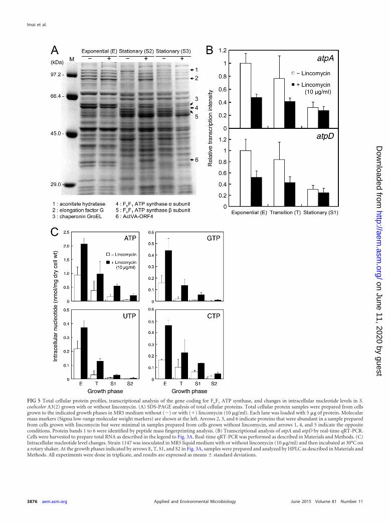
FIG 5 Total cellular protein profiles, transcriptional analysis of the gene coding for FoF1 ATP synthase, and changes in intracellular nucleotide levels in S.coelicolor A3(2) grown with or without lincomycin. (A) SDS-PAGE analysis of total cellular proteins. Total cellular protein samples were prepared from cellsgrown to the indicated growth phases in MR5 medium without () or with (�) lincomycin (10 �g/ml). Each lane was loaded with 5 �g of protein. Molecularmass markers (Sigma low-range molecular weight markers) are shown at the left. Arrows 2, 3, and 6 indicate proteins that were abundant in a sample preparedfrom cells grown with lincomycin but were minimal in samples prepared from cells grown without lincomycin, and arrows 1, 4, and 5 indicate the oppositeconditions. Protein bands 1 to 6 were identified by peptide mass fingerprinting analysis. (B) Transcriptional analysis of atpA and atpD by real-time qRT-PCR.Cells were harvested to prepare total RNA as described in the legend to Fig. 3A. Real-time qRT-PCR was performed as described in Materials and Methods. (C)Intracellular nucleotide level changes. Strain 1147 was inoculated in MR5 liquid medium with or without lincomycin (10 �g/ml) and then incubated at 30°C ona rotary shaker. At the growth phases indicated by arrows E, T, S1, and S2 in Fig. 3A, samples were prepared and analyzed by HPLC as described in Materials andMethods. All experiments were done in triplicate, and results are expressed as means standard deviations.
Imai et al.
3876 aem.asm.org June 2015 Volume 81 Number 11Applied and Environmental Microbiology
on June 11, 2020 by guesthttp://aem
.asm.org/
Dow
nloaded from
synthesis by binding to the ribosomes of Gram-positive bacteria.Thus, it is possible that as with ribosome mutations and modifi-cations, ribosome properties and concomitant protein synthesiscan be modified by lincomycin at subinhibitory concentrationsbecause lincomycin targets ribosome components. In any event, itis highly likely that modifying the translational apparatus holdsthe key for inducing the production of secondary metabolites instreptomycetes.
Lincomycin altered the expression of FoF1 ATP synthase andincreased intracellular ATP levels. FoF1 ATP synthase is a multi-subunit integral membrane protein that is a key enzyme in cellularenergy metabolism, as it synthesizes the majority of cellular ATPfrom ADP and inorganic phosphate in all living cells (43–45). In S.coelicolor A3(2), lincomycin treatment resulted in markedly re-duced expression of the � and � subunits in the F1 part of FoF1
ATP synthase, which catalyzes ATP synthesis/hydrolysis in thecytosol, concomitant with altering mRNA (at any growth stage)and protein (at late growth stages) levels (Fig. 5A and B). ATP isthe major energy currency molecule in a cell that provides energyfor enzymatic reactions and for various biological processes and isalso a substrate for RNA synthesis. Therefore, it is understandablethat the biomass dry weight (Fig. 2) and intracellular nucleotidepools (Fig. 5C) of S. coelicolor A3(2) tended to increase in thepresence of lincomycin, which caused increased intracellular ATPlevels. In this regard, it is noteworthy that ATP and its derivativesS-adenosyl-L-methionine and cyclic AMP are the regulatory mol-ecules in Streptomyces spp. (52–54).
Although it is unclear whether there is a causal relationshipbetween ACT overproduction and the observed high levels of ATP(Fig. 5C), our present findings appear to contradict the previousconcept that high intracellular ATP levels would reduce antibioticproduction by Streptomyces spp. (53). It is possible that high ATPlevels enhanced the synthesis of S-adenosyl-L-methionine and/orcyclic AMP, ultimately resulting in ACT overproduction, becauseS-adenosyl-L-methionine and cyclic AMP are both known to bepositive regulatory molecules during the initiation of microbialsecondary metabolism (52, 54, 55). Alternatively, the increasedATP and GTP levels may have induced protein synthesis, partic-ularly at the late growth phases, ultimately resulting in initiation of
secondary metabolism, because protein synthesis requires largeamounts of GTP and ATP as energy sources. Although we did notanalyze protein synthesis activity in the present work, this propo-sition is consistent with the fact that certain ribosome mutantsretain a high level of protein synthesis activity even at the lategrowth phase, a key event for promoting the dramatic inductionof secondary metabolism in ribosome mutants (30, 33).
Besides techniques involved, our results may provide someclues for uncovering the “physiological” meaning (if any) of anti-biotics in natural environments, including the possibility that an-tibiotics are signaling molecules for interspecific cell-to-cell com-munications, as a low concentration of each antibiotic could elicitpronounced effects (Fig. 1 and 7). Understanding the molecularmechanism by which lincomycin at concentrations below its MICresults in alteration of the cytoplasmic protein (FoF1 ATP syn-thase, etc.) profile and the increased intracellular ATP levels mayprovide clues for clarifying the mechanism underlying the dose-response effects of lincomycin on secondary metabolite produc-tion. However, several questions remain to be answered. (i) Whyor how did lincomycin at subinhibitory concentrations alter theproduction of cytoplasmic proteins in S. coelicolor A3(2), despitethe fact that in S. aureus there was no effect of subinhibitory lin-cosamide antibiotics on cytoplasmic protein production, as pre-viously reported (36, 37)? (ii) Why did cells treated with lincomy-
FIG 7 Cross-culture of S. coelicolor A3(2) with S. erythraea or S. griseus. S.coelicolor A3(2) 1147 (horizontal line) was cross-cultured with S. erythraeaNRRL 2338 (A) or S. griseus IFO 13189 (B) (vertical line) on MR5 agar at 30°Cfor 8 days. Blue color represents the antibiotic ACT.
FIG 6 Streptomycin production by S. griseus in liquid culture. Strain IFO13189 was inoculated into SPY liquid medium that contained different con-centrations of lincomycin and then incubated at 25°C with shaking for 4 days.Streptomycin production was determined as described in Materials and Meth-ods.
Lincomycin Induction of Secondary Metabolism
June 2015 Volume 81 Number 11 aem.asm.org 3877Applied and Environmental Microbiology
on June 11, 2020 by guesthttp://aem
.asm.org/
Dow
nloaded from
cin show high levels of ATP despite the reduced levels of ATPsynthase? (iii) Assuming that there is a causal relationship betweenantibiotic overproduction and increased ATP levels, why did thesehigh intracellular ATP levels promote secondary metabolite pro-duction by Streptomyces spp.? (iv) Did the intracellular ATP con-centration actually affect the levels of intracellular S-adenosyl-L-methionine or cyclic AMP? (v) Did chloramphenicol and tetracyclinealso alter the cytoplasmic protein profile and increase intracellularATP levels? RNA sequencing, an approach to transcriptomeprofiling, of a culture exposed to ribosome-targeting antibiot-ics may provide a clue to the answers to these questions. Al-though the mechanism of induction of secondary metaboliteproduction in streptomycetes may be much more complexthan it appears, our methods presented here are feasible andamenable to use with numerous bacterial species and for theoverproduction and exploitation of novel secondary metabo-lites.
ACKNOWLEDGMENTS
This work was financially supported by a grant-in-aid for Young Scientists(A) to T.H. from the Japan Society for the Promotion of Science (JSPS)(no. 25712008).
We are grateful to M. Katayama (Nippon Suisan Kaisha, Ltd.) and K.Shimono (Shinshu University) for valuable discussion just before the sub-mission of this report.
REFERENCES1. Bibb MJ. 2005. Regulation of secondary metabolism in streptomycetes.
Curr Opin Microbiol 8:208 –215. http://dx.doi.org/10.1016/j.mib.2005.02.016.
2. Kieser T, Bibb MJ, Buttner MJ, Chater KF, Hopwood DA. 2000.Practical Streptomyces genetics. The John Innes Foundation, Norwich,United Kingdom.
3. Bentley SD, Chater KF, Cerdeno-Tarraga AM, Challis GL, ThomsonNR, James KD, Harris DE, Quail H, Kieser D, Harper D, Bateman A,Brown S, Chandra G, Chen CW, Collins M, Cronin A, Fraser A, GobleA, Hidalgo J, Hornsby T, Howarth S, Huang CH, Kieser T, Larke L,Murphy L, Oliver K, O’Neil S, Rabbinowitsch E, Rajandream MA,Rutherford K, Rutter S, Seeger K, Saunders D, Sharp S, Squares R,Squares S, Taylor K, Warren T, Wietzorrek A, Woodward J, Barrell BG,Parkhill J, Hopwood DA. 2002. Complete genome sequence of the modelactinomycete Streptomyces coelicolor A3(2). Nature 417:141–147. http://dx.doi.org/10.1038/417141a.
4. Ikeda H, Ishikawa J, Hanamoto A, Shinose M, Kikuchi H, Shiba T,Sakaki Y, Hattori M, Omura S. 2003. Complete genome sequence andcomparative analysis of the industrial microorganism Streptomyces aver-mitilis. Nat Biotechnol 21:526 –531. http://dx.doi.org/10.1038/nbt820.
5. Ohnishi Y, Ishikawa J, Hara H, Suzuki H, Ikenoya M, Ikeda H,Yamashita A, Hattori M, Horinouchi S. 2008. Genome sequence of thestreptomycin-producing microorganism Streptomyces griseus IFO 13350.J Bacteriol 190:4050 – 4060. http://dx.doi.org/10.1128/JB.00204-08.
6. Baltz RH. 2011. Strain improvement in actinomycetes in the postgenomicera. J Ind Microbiol Biotechnol 38:657– 666. http://dx.doi.org/10.1007/s10295-010-0934-z.
7. Chiang YM, Chang SL, Oakley BR, Wang CC. 2011. Recent advances inawakening silent biosynthetic gene clusters and linking orphan clusters tonatural products in microorganisms. Curr Opin Chem Biol 15:137–143.http://dx.doi.org/10.1016/j.cbpa.2010.10.011.
8. Genilloud O. 2014. The re-emerging role of microbial natural products inantibiotic discovery. Antonie Van Leeuwenhoek 106:173–188. http://dx.doi.org/10.1007/s10482-014-0204-6.
9. Ochi K, Hosaka T. 2013. New strategies for drug discovery: activation ofsilent or weakly expressed microbial gene clusters. Appl Microbiol Bio-technol 97:87–98. http://dx.doi.org/10.1007/s00253-012-4551-9.
10. Ochi K, Tanaka Y, Tojo S. 2014. Activating the expression of bacterialcryptic genes by rpoB mutations in RNA polymerase or by rare earth ele-ments. J Ind Microbiol Biotechnol 41:403– 414. http://dx.doi.org/10.1007/s10295-013-1349-4.
11. Wu MC, Law B, Wilkinson B, Micklefield J. 2012. Bioengineeringnatural product biosynthetic pathways for therapeutic applications. CurrOpin Biotechnol 23:931–940. http://dx.doi.org/10.1016/j.copbio.2012.03.008.
12. Yoon V, Nodwell JR. 2014. Activating secondary metabolism with stressand chemicals. J Ind Microbiol Biotechnol 41:415– 424. http://dx.doi.org/10.1007/s10295-013-1387-y.
13. Zhu H, Sandiford SK, Wezel GP. 2014. Triggers and cues that activateantibiotic production by actinomycetes. J Ind Microbiol Biotechnol 41:371–386. http://dx.doi.org/10.1007/s10295-013-1309-z.
14. Bode HB, Bethe B, Hofs R, Zeeck A. 2002. Big effects from small changes:possible ways to explore nature’s chemical diversity. Chembiochem 3:619–627.http://dx.doi.org/10.1002/1439-7633(20020703)3:7�619::AID-CBIC619�3.0.CO;2-9.
15. Craney A, Ozimok C, Pimentel-Elardo SM, Capretta A, Nodwell JR.2012. Chemical perturbation of secondary metabolism demonstrates im-portant links to primary metabolism. Chem Biol 19:1020 –1027. http://dx.doi.org/10.1016/j.chembiol.2012.06.013.
16. Onaka H, Mori Y, Igarashi Y, Furumai T. 2011. Mycolic acid-containingbacteria induce natural-product biosynthesis in Streptomyces species.Appl Environ Microbiol 77:400 – 406. http://dx.doi.org/10.1128/AEM.01337-10.
17. Romero D, Traxler MF, Lopez D, Kolter R. 2011. Antibiotics as signal mole-cules. Chem Rev 111:5492–5505. http://dx.doi.org/10.1021/cr2000509.
18. Schroeckh V, Scherlach K, Nutzmann HW, Shelest E, Schmidt-Heck W,Schuemann J, Martin K, Hertweck C, Brakhage AA. 2009. Intimatebacterial-fungal interaction triggers biosynthesis of archetypal polyketidesin Aspergillus nidulans. Proc Natl Acad Sci U S A 106:14558 –14563. http://dx.doi.org/10.1073/pnas.0901870106.
19. Yamanaka K, Oilawa H, Ogawa HO, Hosono K, Shinmachi F, Takano H,Sakuda S, Beppu T, Ueda K. 2005. Desferrioxamine E produced by Strep-tomyces griseus stimulates growth and development of Streptomyces ta-nashiensis. Microbiology 151:2899 –2905. http://dx.doi.org/10.1099/mic.0.28139-0.
20. Gomez-Escribano JP, Bibb MJ. 2014. Heterologous expression of naturalproduct biosynthetic gene clusters in Streptomyces coelicolor: from genomemining to manipulation of biosynthetic pathways. J Ind Microbiol Bio-technol 41:425– 431. http://dx.doi.org/10.1007/s10295-013-1348-5.
21. Smanski MJ, Peterson RM, Rajski SR, Shen B. 2009. Engineered Strep-tomyces platensis strains that overproduce antibiotics platensimycin andplatencin. Antimicrob Agents Chemother 53:1299 –1304. http://dx.doi.org/10.1128/AAC.01358-08.
22. Laureti L, Song L, Huang S, Corre C, Leblond P, Challis GL, Aigle B.2011. Identification of a bioactive 51-membered macrolide complex byactivation of a silent polyketide synthase in Streptomyces ambofaciens. ProcNatl Acad Sci U S A 108:6258 – 6263. http://dx.doi.org/10.1073/pnas.1019077108.
23. Yanai K, Murakami T, Bibb MJ. 2006. Amplification of the entire kana-mycin biosynthetic gene cluster during empirical strain improvement ofStreptomyces kanamyceticus. Proc Natl Acad Sci U S A 103:9661–9666.http://dx.doi.org/10.1073/pnas.0603251103.
24. Zhuo Y, Zhang W, Chen D, Gao H, Tao J, Liu M, Gou Z, Zhou X, YeBC, Zhang Q, Zhang S, Zhang LX. 2010. Reverse biological engineeringof hrdB to enhance the production of avermectins in an industrial strain ofStreptomyces avermitilis. Proc Natl Acad Sci U S A 107:11250 –11254. http://dx.doi.org/10.1073/pnas.1006085107.
25. Imai Y, Fujiwara T, Ochi K, Hosaka T. 2012. Development of the abilityto produce secondary metabolites in Streptomyces through the acquisitionof erythromycin resistance. J Antibiot (Tokyo) 65:323–326. http://dx.doi.org/10.1038/ja.2012.16.
26. Nishimura K, Hosaka T, Tokuyama S, Okamoto S, Ochi K. 2007.Mutations in rsmG, encoding a 16S rRNA methyltransferase, result inlow-level streptomycin resistance and antibiotic overproduction in Strep-tomyces coelicolor A3(2). J Bacteriol 189:3876 –3883. http://dx.doi.org/10.1128/JB.01776-06.
27. Shima J, Hesketh A, Okamoto S, Kawamoto S, Ochi K. 1996. Inductionof actinorhodin production by rpsL (encoding ribosomal protein S12)mutations that confer streptomycin resistance in Streptomyces lividans andStreptomyces coelicolor A3(2). J Bacteriol 178:7276 –7284.
28. Wang G, Hosaka T, Ochi K. 2008. Dramatic activation of antibioticproduction in Streptomyces coelicolor by cumulative drug resistance mu-tations. Appl Environ Microbiol 74:2834 –2840. http://dx.doi.org/10.1128/AEM.02800-07.
Imai et al.
3878 aem.asm.org June 2015 Volume 81 Number 11Applied and Environmental Microbiology
on June 11, 2020 by guesthttp://aem
.asm.org/
Dow
nloaded from
29. Xu J, Tozawa Y, Lai C, Hayashi H, Ochi K. 2002. A rifampicin resistancemutation in the rpoB gene confers ppGpp-independent antibiotic produc-tion in Streptomyces coelicolor A3(2). Mol Genet Genomics 268:179 –189.http://dx.doi.org/10.1007/s00438-002-0730-1.
30. Hosaka T, Ohnishi-Kameyama M, Muramatsu H, Murakami K, Tsu-rumi Y, Kodani S, Yoshida M, Fujie A, Ochi K. 2009. Antibacterialdiscovery in actinomycetes strains with mutations in RNA polymerase orribosomal protein S12. Nat Biotechnol 27:462– 464. http://dx.doi.org/10.1038/nbt.1538.
31. Ochi K, Okamoto S, Tozawa Y, Inaoka T, Hosaka T, Xu J, Kurosawa K.2004. Ribosome engineering and secondary metabolite production. Adv ApplMicrobiol 56:155–184. http://dx.doi.org/10.1016/S0065-2164(04)56005-7.
32. Tanaka Y, Kasahara K, Hirose Y, Murakami K, Kugimiya R, Ochi K. 2013.Activation and products of the cryptic secondary metabolite biosynthetic geneclusters by rifampicin resistance (rpoB) mutations in actinomycetes. J Bacte-riol 195:2959–2970. http://dx.doi.org/10.1128/JB.00147-13.
33. Hosaka T, Xu J, Ochi K. 2006. Increased expression of ribosome recy-cling factor is responsible for the enhanced protein synthesis during thelate growth phase in an antibiotic-overproducing Streptomyces coelicolorribosomal rpsL mutant. Mol Microbiol 61:883– 897. http://dx.doi.org/10.1111/j.1365-2958.2006.05285.x.
34. Davies J, Spiegelman GB, Yim G. 2006. The world of subinhibitoryantibiotic concentrations. Curr Opin Microbiol 9:445– 453. http://dx.doi.org/10.1016/j.mib.2006.08.006.
35. Yim G, Wang HH, Davies J. 2006. The truth about antibiotics. Int J MedMicrobiol 296:163–170. http://dx.doi.org/10.1016/j.ijmm.2006.01.039.
36. Shibl MA. 1993. Effect of antibiotics on production of enzymes and toxinsby microorganisms. Rev Infect Dis 5:865– 875.
37. Herbert S, Barry P, Novick PR. 2001. Subinhibitory clindamycin differ-entially inhibits transcription of exoprotein genes in Staphylococcus au-reus. Infect Immun 69:2996 –3003. http://dx.doi.org/10.1128/IAI.69.5.2996-3003.2001.
38. Huang J, Lih CJ, Pan KH, Cohen SN. 2001. Global analysis of growthphase responsive gene expression and regulation of antibiotic biosyntheticpathways in Streptomyces coelicolor using DNA microarrays. Genes Dev15:3183–3192. http://dx.doi.org/10.1101/gad.943401.
39. Ochi K. 1987. Metabolic initiation of differentiation and secondary me-tabolism by Streptomyces griseus: significance of the stringent response(ppGpp) and GTP content in relation to A factor. J Bacteriol 169:3608 –3616.
40. Tanaka Y, Hosaka T, Ochi K. 2010. Rare earth elements activate the second-ary metabolite-biosynthetic gene clusters in Streptomyces coelicolor A3(2). JAntibiot (Tokyo) 63:477– 481. http://dx.doi.org/10.1038/ja.2010.53.
41. Neary JM, Powell A, Gordon L, Milne C, Flett F, Wilkinson B, SmithCP, Micklefield J. 2007. An asparagine oxygenase (AsnO) and a 3-hy-droxyasparaginyl phosphotransferase (HasP) are involved in the biosyn-thesis of calcium-dependent lipopeptide antibiotics. Microbiology 153:768 –776. http://dx.doi.org/10.1099/mic.0.2006/002725-0.
42. Gramajo HC, Takano E, Bibb MJ. 1993. Stationary-phase production ofthe antibiotic actinorhodin in Streptomyces coelicolor A3(2) is transcrip-tionally regulated. Mol Microbiol 7:837– 845. http://dx.doi.org/10.1111/j.1365-2958.1993.tb01174.x.
43. Capaldi RA, Aggeler R. 2002. Mechanism of the F(1)F(0)-type ATPsynthase, a biological rotary motor. Trends Biochem Sci 27:154 –160. http://dx.doi.org/10.1016/S0968-0004(01)02051-5.
44. Senior AE. 2007. ATP synthase: motoring to the finish line. Cell 130:220 –221. http://dx.doi.org/10.1016/j.cell.2007.07.004.
45. Senior AE. 1990. The proton-translocating ATPase of Escherichia coli.Annu Rev Biophys Biophys Chem 19:7– 41. http://dx.doi.org/10.1146/annurev.bb.19.060190.000255.
46. Soga N, Kinosita K, Jr, Yoshida M, Suzuki T. 2011. Efficient ATPsynthesis by thermophilic Bacillus FoF1-ATP synthase. FEBS J 278:2647–2654. http://dx.doi.org/10.1111/j.1742-4658.2011.08191.x.
47. Calabrese EJ, Baldwin LA. 2003. Hormesis: the dose-response revo-lution. Annu Rev Pharmacol Toxicol 43:175–197. http://dx.doi.org/10.1146/annurev.pharmtox.43.100901.140223.
48. Sabina J, Dover N, Templeton LJ, Smulski DR, Soll D, LaRossa RA.2003. Interfering with different steps of protein synthesis explored by tran-scriptional profiling of Escherichia coli K-12. J Bacteriol 185:6158 – 6170.http://dx.doi.org/10.1128/JB.185.20.6158-6170.2003.
49. Lin JT, Connelly MB, Amolo C, Otani S, Yaver DS. 2005. Globaltranscriptional response of Bacillus subtilis to treatment with subinhibi-tory concentrations of antibiotics that inhibit protein synthesis. Antimi-crob Agents Chemother 49:1915–1926. http://dx.doi.org/10.1128/AAC.49.5.1915-1926.2005.
50. Amano S, Morota T, Kano YK, Narita H, Hashidzume T, Yamamoto S,Mizutani K, Sakuda S, Furihata K, Takano-Shiratori H, Takano H,Beppu T, Ueda K. 2010. Promomycin, a polyether promoting antibioticproduction in Streptomyces spp. J Antibiot 63:486 – 491. http://dx.doi.org/10.1038/ja.2010.68.
51. Amano S, Sakurai T, Keisuke E, Takano H, Beppu T, Furihata K,Sakuda S, Ueda K. 2011. A cryptic antibiotic triggered by monensin. JAntibiot 64:703. http://dx.doi.org/10.1038/ja.2011.69.
52. Horinouchi S, Ohnishi Y, Kang DK. 2001. The A-factor regulatorycascade and cAMP in the regulation of physiological and morphologicaldevelopment in Streptomyces griseus. J Ind Microbiol Biotechnol 27:177–182. http://dx.doi.org/10.1038/sj.jim.7000068.
53. Meng L, Li M, Yang SH, Kim TJ, Suh JW. 2011. Intracellular ATP levelsaffect secondary metabolite production in Streptomyces spp. Biosci Bio-technol Biochem 75:1576 –1581. http://dx.doi.org/10.1271/bbb.110277.
54. Okamoto S, Lezhava A, Hosaka T, Okamoto-Hosoya Y, Ochi K. 2003.Enhanced expression of S-adenosylmethionine synthetase causes over-production of actinorhodin in Streptomyces coelicolor A3(2). J Bacteriol185:601– 609. http://dx.doi.org/10.1128/JB.185.2.601-609.2003.
55. Tojo S, Kim JY, Tanaka Y, Inaoka T, Hiraga Y, Ochi K. 2014. The mthAmutation conferring low-level resistance to streptomycin enhances anti-biotic production in Baci l lus subti l i s by increasing the S-adenosylmethionine pool size. J Bacteriol 196:1514 –1524. http://dx.doi.org/10.1128/JB.01441-13.
56. Oliynyk M, Samborskyy M, Lester JB, Mironenko T, Scott N, Dickens S,Haydock SF, Leadlay PF. 2007. Complete genome sequence of the erythro-mycin-producing bacterium Saccharopolyspora erythraea NRRL23338. NatBiotechnol 25:447– 453. http://dx.doi.org/10.1038/nbt1297.
Lincomycin Induction of Secondary Metabolism
June 2015 Volume 81 Number 11 aem.asm.org 3879Applied and Environmental Microbiology
on June 11, 2020 by guesthttp://aem
.asm.org/
Dow
nloaded from