Karyogamy after Electrofusion of Single Egg and …Karyogamy after Electrofusion of Single Egg and...
10
The Plant Cell, Vol. 5, 747-755, July 1993 O 1993 American Society of Plant Physiologists Karyogamy after Electrofusion of Single Egg and Sperm Cell Protoplasts from Maize: Cytological Evidence and Time Couke Jean-Emmanuel Faure,’ H. Lloyd Mogensen,b Christian Dumas,’” Horst Lijrz,’ and Erhard Kranz‘ a Reconnaissance Cellulaire et Amélioration des Plantes, UMR 9938 CNRS-INRA, Ecole Normale Supérieure de Lyon, 69364 Lyon Cedex 07, France Department of Biological Sciences, Northern Arizona University, Flagstaff, Arizona 8601 1 lnstitut für Allgemeine Botanik, Universitat Hamburg, 22609 Hamburg, Germany In maize, in vitro fusion of isolated male gametes with isolated egg cell protoplasts can be induced by electric pulses. Until now, karyogamy has not been demonstrated. In this study, we cytologically examined fusion products fixed at differ- ent times after electrofusion with phase contrast microscopy and transmission electron microscopy. We obtained a precise timetable from 23 samples studied during the first 3 hr. The sperm nucleus was integrated within the egg cell protoplast, migrated toward the egg cell nucleus, and fused with it within 1 hr, as demonstrated by ultrastructural observations, three-dimensional reconstructions of nuclei, and subsequent nuclear volume estimates. Fusion of nuclei occurred be- fore zygotic mitosis, as is the case in vivo. These findings demonstrate karyogamy during in vitro fertilization of maize. INTRODUCTION The fertilizationof egg and central cells, which are deeply en- closed in maternal tissues, by male gametes issued from the pollen tube was discovered in flowering plants approximately one century ago (Nawaschin, 1898; Guignard, 1899) and termed “double fertilization.” However, the mechanisms under- lying this process are still largely unknown in comparison with fertilization in animals. Severa1questions remain unanswered, such as the mechanisms of a possible intergametic recogni- tion, the precise process of gametic fusion, the block to polyspermy, and the early metabolism of the zygote following fertilization. Our lack of knowledge is due mainly to the inac- cessibility of the gametes. Hence, the performance of fertilization in vitro should allow detailed study of the fertiliza- tion event under controlled and accessible conditions, as has been done in animals (Date, 1991). Maize is a suitable plant with which to perform in vitro fertil- ization. Many genetic and cytogenetic analyses have been conducted (Sheridan, 1988), and the partners involved in fer- tilization have been well studied, as will be presented by Dumas and Mogensen (1993). In particular, both male (Dupuis et al., 1987; Cass and Fabi, 1988) and female (Wagner et al., 1989a) gametes have been isolated from maize, and the first in vitro fusion of an isolated egg cell with an isolated sperm cell by electrofusion was performedin maize (Kranz et al., 1990,1991). However, as discussed by Chasan (1992), nuclear fusion To whom correspondence should be addressed. following the cytoplasmicfusion remains to be demonstrated, and regeneration of plants from the fusion products has yet to be performed. Embryogenesis and regeneration of fertile plants are now possible (Kranz and Lorz, 1993). In this article, we describe the fate of the male nucleus after electrofusion and demonstrate the occurrence of complete karyogamy. To follow the fate of the male nucleus, we have chosen trans- mission electron microscopy, which is a more precise tool to follow small nuclei than is fluorescence or confocal laser scanning microscopy and which allows complementary ultra- structural observations.We conducted the fixation of cells at precise time intervals after electrofusion. RESULTS Penetration of the Male Nucleus into the Egg Cell Protoplast We used the general procedure described in Figure 1 for the study of electrofusionproducts from isolated male and female gametes. In each of three samples fixed 7 to 8 min after fu- sion, a dark object of *4 wm in diameter was observed on semithin sections, as shown in Figure 2A. We interpretedthis object to be the male nucleus. We confirmed this analysis with transmission electron microscopy and by reembedding and ultrathin sectioning the semithin sections containing the
Transcript of Karyogamy after Electrofusion of Single Egg and …Karyogamy after Electrofusion of Single Egg and...

The Plant Cell, Vol. 5, 747-755, July 1993 O 1993 American Society of Plant Physiologists
Karyogamy after Electrofusion of Single Egg and Sperm Cell Protoplasts from Maize: Cytological Evidence and Time Couke
Jean-Emmanuel Faure,’ H. Lloyd Mogensen,b Christian Dumas,’” Horst Lijrz,’ and Erhard Kranz‘ a Reconnaissance Cellulaire et Amélioration des Plantes, UMR 9938 CNRS-INRA, Ecole Normale Supérieure de Lyon, 69364 Lyon Cedex 07, France
Department of Biological Sciences, Northern Arizona University, Flagstaff, Arizona 8601 1 lnstitut für Allgemeine Botanik, Universitat Hamburg, 22609 Hamburg, Germany
In maize, in vitro fusion of isolated male gametes with isolated egg cell protoplasts can be induced by electric pulses. Until now, karyogamy has not been demonstrated. In this study, we cytologically examined fusion products fixed at differ- ent times after electrofusion with phase contrast microscopy and transmission electron microscopy. We obtained a precise timetable from 23 samples studied during the first 3 hr. The sperm nucleus was integrated within the egg cell protoplast, migrated toward the egg cell nucleus, and fused with it within 1 hr, as demonstrated by ultrastructural observations, three-dimensional reconstructions of nuclei, and subsequent nuclear volume estimates. Fusion of nuclei occurred be- fore zygotic mitosis, as is the case in vivo. These findings demonstrate karyogamy during in vitro fertilization of maize.
INTRODUCTION
The fertilization of egg and central cells, which are deeply en- closed in maternal tissues, by male gametes issued from the pollen tube was discovered in flowering plants approximately one century ago (Nawaschin, 1898; Guignard, 1899) and termed “double fertilization.” However, the mechanisms under- lying this process are still largely unknown in comparison with fertilization in animals. Severa1 questions remain unanswered, such as the mechanisms of a possible intergametic recogni- tion, the precise process of gametic fusion, the block to polyspermy, and the early metabolism of the zygote following fertilization. Our lack of knowledge is due mainly to the inac- cessibility of the gametes. Hence, the performance of fertilization in vitro should allow detailed study of the fertiliza- tion event under controlled and accessible conditions, as has been done in animals (Date, 1991).
Maize is a suitable plant with which to perform in vitro fertil- ization. Many genetic and cytogenetic analyses have been conducted (Sheridan, 1988), and the partners involved in fer- tilization have been well studied, as will be presented by Dumas and Mogensen (1993). In particular, both male (Dupuis et al., 1987; Cass and Fabi, 1988) and female (Wagner et al., 1989a) gametes have been isolated from maize, and the first in vitro fusion of an isolated egg cell with an isolated sperm cell by electrofusion was performed in maize (Kranz et al., 1990,1991). However, as discussed by Chasan (1992), nuclear fusion
To whom correspondence should be addressed.
following the cytoplasmic fusion remains to be demonstrated, and regeneration of plants from the fusion products has yet to be performed. Embryogenesis and regeneration of fertile plants are now possible (Kranz and Lorz, 1993). In this article, we describe the fate of the male nucleus after electrofusion and demonstrate the occurrence of complete karyogamy.
To follow the fate of the male nucleus, we have chosen trans- mission electron microscopy, which is a more precise tool to follow small nuclei than is fluorescence or confocal laser scanning microscopy and which allows complementary ultra- structural observations. We conducted the fixation of cells at precise time intervals after electrofusion.
RESULTS
Penetration of the Male Nucleus into the Egg Cell Protoplast
We used the general procedure described in Figure 1 for the study of electrofusion products from isolated male and female gametes. In each of three samples fixed 7 to 8 min after fu- sion, a dark object of *4 wm in diameter was observed on semithin sections, as shown in Figure 2A. We interpreted this object to be the male nucleus. We confirmed this analysis with transmission electron microscopy and by reembedding and ultrathin sectioning the semithin sections containing the
748 The Plant Cell
A
F I
C Electric-induced fusion
( time t = O min.)
D 1 Fixation of the fusion
product ( time t )
'Ihick wtioning Phase contrast microscopy
Reembeddq Serial ultrathin sectioning
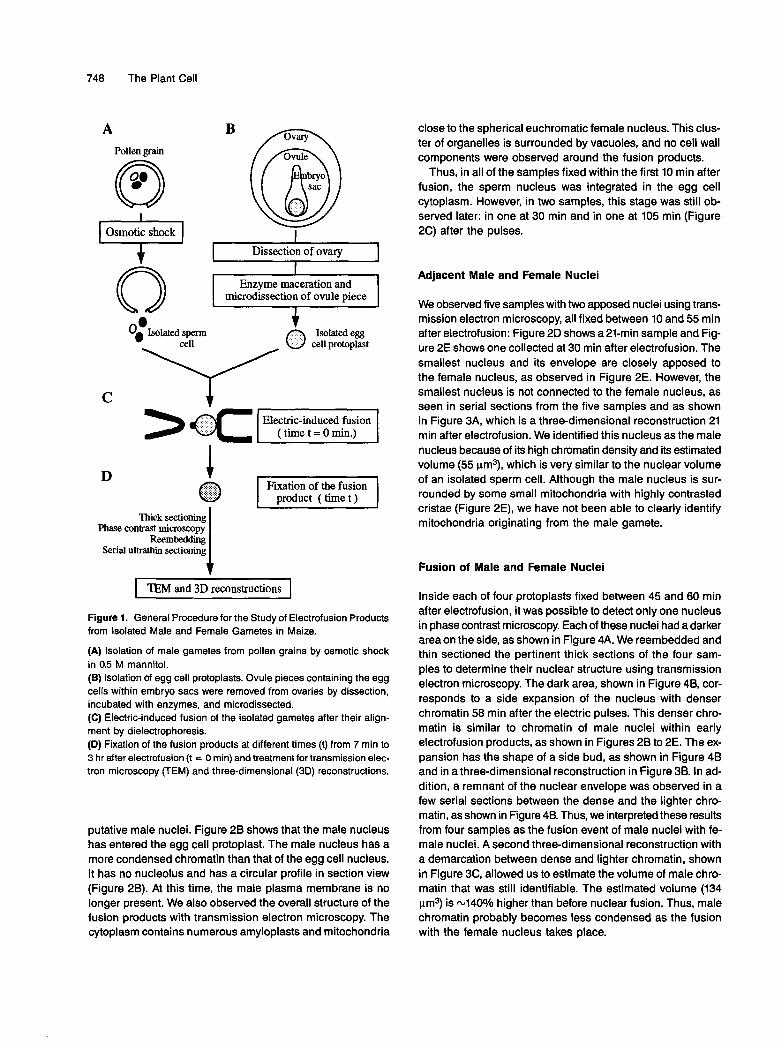
I TEM and 3D reconstructions I Figure 1. General Procedure for the Study of Electrofusion Products from lsolated Male and Female Gametes in Maize.
(A) lsolation of male gametes from pollen grains by osmotic shock in 0.5 M mannitol. (B) lsolation of egg cell protoplasts. Ovule pieces containing the egg cells within embryo sacs were removed from ovaries by dissection, incubated with enzymes, and microdissected. (C) Electric-induced fusion of the isolated gametes after their align- ment by dielectrophoresis. (D) Fixation of the fusion products at different times (t) from 7 min to 3 hr after electrofusion (t = O min) and treatment for transmission elec- tron microscopy (TEM) and three-dimensional (3D) reconstructions.
putative male nuclei. Figure 28 shows that the male nucleus has entered the egg cell protoplast. The male nucleus has a more condensed chromatin than that of the egg cell nucleus. It has no nucleolus and has a circular profile in section view (Figure 28). At this time, the male plasma membrane is no longer present. We also observed the overall structure of the fusion products with transmission electron microscopy. The cjrtoplasm contains numerous amyloplasts and mitochondria
close to the spherical euchromatic female nucleus. This clus- ter of organelles is surrounded by vacuoles, and no cell wall components were observed around the fusion products.
Thus, in all of the samples fixed within the first 10 min after fusion, the sperm nucleus was integrated in the egg cell cytoplasm. However, in mo samples, this stage was still ob- served later: in one at 30 min and in one at 105 min (Figure 2C) after the pulses.
Adjacent Male and Female Nuclei
We observed five samples with two apposed nuclei using trans- mission electron microscopy, all fixed between 10 and 55 min after electrofusion: Figure 2D shows a 21-min sample and Fig- ure 2E shows one collected at 30 min after electrofusion. The smallest nucleus and its envelope are closely apposed to the female nucleus, as observed in Figure 2E. However, the smallest nucleus is not connected to the female nucleus, as seen in serial sections from the five samples and as shown in Figure 3A, which is a three-dimensional reconstruction 21 min after electrofusion. We identified this nucleus as the male nucleus because of its high chromatin density and its estimated volume (55 pm3), which is very similar to the nuclear volume of an isolated sperm cell. Although the male nucleus is sur- rounded by some small mitochondria with highly contrasted cristae (Figure 2E), we have not been able to clearly identify mitochondria originating from the male gamete.
Fusion of Male and Female Nuclei
lnside each of four protoplasts fixed between 45 and 60 min after electrofusion, it was possible to detect only one nucleus in phase contrast microscopy. Each of these nuclei had a darker areaon the side, as shown in Figure 4A. We reembedded and thin sectioned the pertinent thick sections of the four sam- ples to determine their nuclear structure using transmission electron microscopy. The dark area, shown in Figure 48, cor- responds to a side expansion of the nucleus with denser chromatin 58 min after the electric pulses. This denser chro- matin is similar to chromatin of male nuclei within early electrofusion products, as shown in Figures 28 to 2E. The ex- pansion has the shape of a side bud, as shown in Figure 48 and in a three-dimensional reconstruction in Figure 38. In ad- dition, a remnant of the nuclear envelope was observed in a few serial sections between the dense and the lighter chro- matin, as shown in Figure 48. Thus, we interpreted these results from four samples as the fusion event of male nuclei with fe- male nuclei. A second three-dimensional reconstruction with a demarcation between dense and lighter chromatin, shown in Figure 3C, allowed us to estimate the volume of male chro- matin that was still identifiable. The estimated volume (134 pm3) is 44O0/o higher than before nuclear fusion. Thus, male chromatin probably becomes less condensed as the fusion with the female nucleus takes place.
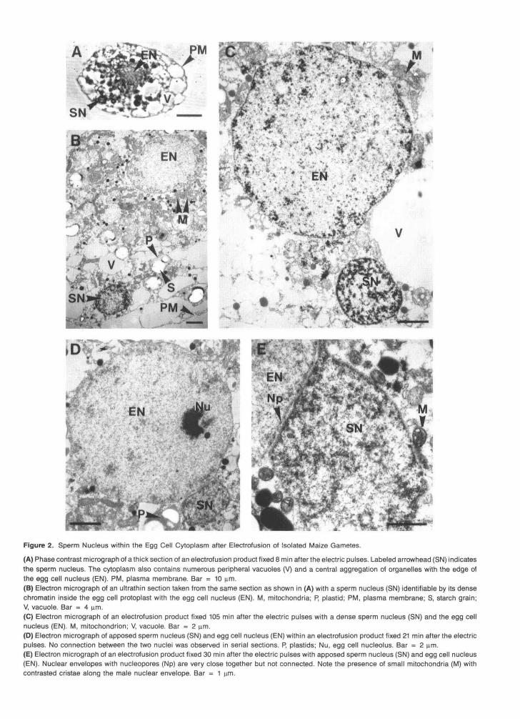
Figure 2. Sperm Nucleus within the Egg Cell Cytoplasm after Electrofusion of Isolated Maize Gametes.
(A) Phase contrast micrograph of a thick section of an electrofusion product fixed 8 min after the electric pulses. Labeled arrowhead (SN) indicatesthe sperm nucleus. The cytoplasm also contains numerous peripheral vacuoles (V) and a central aggregation of organelles with the edge ofthe egg cell nucleus (EN). PM, plasma membrane. Bar = 10 urn.(B) Electron micrograph of an ultrathin section taken from the same section as shown in (A) with a sperm nucleus (SN) identifiable by its densechromatin inside the egg cell protoplast with the egg cell nucleus (EN). M, mitochondria; P, plastid; PM, plasma membrane; S, starch grain;V, vacuole. Bar = 4 urn.(C) Electron micrograph of an electrofusion product fixed 105 min after the electric pulses with a dense sperm nucleus (SN) and the egg cellnucleus (EN). M, mitochondrion; V, vacuole. Bar = 2 urn.(D) Electron micrograph of apposed sperm nucleus (SN) and egg cell nucleus (EN) within an electrofusion product fixed 21 min after the electricpulses. No connection between the two nuclei was observed in serial sections. P, plastids; Nu, egg cell nucleolus. Bar = 2 nm.(E) Electron micrograph of an electrofusion product fixed 30 min after the electric pulses with apposed sperm nucleus (SN) and egg cell nucleus(EN). Nuclear envelopes with nucleopores (Np) are very close together but not connected. Note the presence of small mitochondria (M) withcontrasted cristae along the male nuclear envelope. Bar = 1 urn.

750 The Plant Cell
Figure 3. Stereoscopic Pairs of Reconstructed Nuclei in Electrofusion Products.
(A) Reconstruction of nuclei 21 min after electrofusion. The sperm cell nucleus (yellow-green), the egg cell nucleus (light blue), and the eggcell nucleolus (dark blue) are outlined in every section. The sample used for the three-dimensional reconstruction is shown in Figure 2D. Approxi-mate magnification x2610.(B) Reconstruction of the nucleus (light blue) with its nucleolus (dark blue) in every section 58 min after electrofusion. Note the budlike shapein the upper part of the nucleus, which corresponds to the fusing sperm nucleus. The sample used for the three-dimensional reconstructionis shown in Figures 4A and 4B. Approximate magnification x3130.(C) Reconstruction 58 min after electrofusion from the same sample shown in (B). Additional outlines have been used (yellow-green) to representthe male chromatin, which is still identifiable. Approximate magnification x3130.

Karyogamy after Electrofusion of Gametes 751
B
Figure 4. Sperm Nucleus Fusing with the Egg Cell Nucleus after Electrofusion of Isolated Maize Gametes.
(A) Phase contrast micrograph of a thick section from a sample fixed 58 min after electrofusion. The sperm nucleus (SN) is fusing with the eggcell nucleus (EN). Nu, egg cell nucleolus. Bar = 10 u.m.(B) Electron micrograph of an ultrathin section taken from the same section shown in (A) depicting the fusion of the sperm nucleus (SN) withthe egg cell nucleus (EN). Note the difference in chromatm density and a nuclear envelope remnant (unlabeled arrowheads) close to the malechromatin. L, lipid body; M, mitochondrion; Nu, egg cell nucleolus. Bar = 1 \im.
In the 10 samples fixed later, between 60 and 195 min afterelectrofusion, with one exception (already described in Fig-ure 2C), only one nucleus was observed in each protoplastby means of phase contrast microscopy. In five of these sam-ples, the unique nucleus contained two nucleoli.
shown in Figure 5. The male nucleus penetrated the cytoplasmof the egg cell protoplast within the first 20 min, became ap-posed to the female nucleus between 20 and 45 min, fusedwith it between 45 and 60 min, and was integrated into theegg nucleus after 60 min.
Chronology
Results from the 23 samples described above, all of which wereexamined within the first 3 hr after electrofusion, were quiteconsistent, thereby enabling us to establish the chronology
DISCUSSION
The following observations indicated that karyogamy occursafter the electrofusion of isolated maize gametes. (1) Both
2 nuclei 2 apposednuclei
ID
IDIDID
1
0Time
€
C ®OCID C
1 1 '
30
2 fusingnuclei®
@@C ®1 1 1 1
60between electrofusion and
••ID
1 ' '
90
i
••••1 '
120fixation of fusion
nucleus
•
• •i | i i |
150 180products (min.)
•
Figure 5. Chronology of the Fate of Male and Female Nuclei after the Electrofusion of Isolated Maize Gametes.
Each of the 23 samples studied is put on a time scale and is symbolized by one of the four different patterns described in the upper part ofthe figure. Patterns are given according to nuclear stages, i.e., two nuclei, two apposed nuclei, two fusing nuclei, or only one nucleus.

752 The Plant Cell
closely apposed and fusing male and female nuclei were visi- ble with transmission electron microscopy (Figures 2 and 4). Male and female nuclei were identified by comparing their mor- phology with those of isolated sperm cells (Wagner et al., 1989b; Mogensen et ai., 1990) and nonfertilized isolated egg cell protoplasts (Faure et al., 1992). In addition, the volume of male chromatin estimated from three-dimensional recon- structions (Figure 3) was in agreement with that of isolated sperm cells (Mogensen et al., 1990). (2) A consistent chronol- ogy was obtained when the results from the 23 samples fixed at different times after electrofusion were compiled (Figure 5).
Time Course of Karyogamy
The time course from plasmogamy to karyogamy is difficult to estimate in vivo. Indeed, the fusion of a sperm cell with a central cell was shown once in Plumbago zeylanica (Russell, 1983) with transmission electron microscopy, but, to our knowl- edge, fusion between egg and sperm cells has never been shown at this level. Therefore, there is no precise reference time in vivo. Nevertheless, some estimates have been pub- lished in which male gametes have been observed close to egg cells or male nuclei have been seen within the egg cell cytoplasm after gametic fusion. Karyogamy seems to occur within -1 to 2 hr after plasmogamy in cereals, such as wheat (vou and Jensen, 1985; Guo-Wei and Jia-Heng, 1992), Triticale (Hause and Schroder, 1987), and probably barley (Mogensen, 1982). No data are available in maize, although up to 22 hr may elapse between pollination and fertilization, depending on silk length and environmental conditions (Diboll, 1968; R. MOI, E. Matthys-Rochon, and C. Dumas, manuscript in preparation).
By comparison with in vivo studies, we have obtained a pre- cise timetable in vitro, in which the time of gametic fusion is well defined and controlled. Karyogamy occurs 45 to 60 min after gametic fusion under our experimental conditions. There- fore, maize electrofusion products appear to behave like zygotes obtained in vivo in other grasses with regard to the time course of karyogamy.
Fate of the Male Nucleus
The male nucleus first migrates toward the female nucleus, as it does in animals (for review, see Longo, 1988). The use of colcemid, an inhibiting factor of microtubule formation, prevents pronuclear movement in sea urchin (Zimmerman and Zimmerman, 1967), rabbit (Longo, 1976), and mouse (Schatten et al., 1985). Therefore, microtubules are apparently involved in the movement of the male pronucleus and its apposition to the female pronucleus. However, the mechanism of this migration is not known and is highly speculative in animals (for review, see Longo, 1988). Microtubules have been detected by immunocytochemistry in the mature egg cells of some
higher plants (see Huang and Russell, 1992), but to our knowl- edge, no study has demonstrated their involvement in nuclear migration. The accessibility of gametes and the possible con- trol of fertilization parameters, such as time, under in vitro conditions should permit studies of the role of the plant cytoskeleton during fertilization in the near future.
Karyogamy after in vivo gametic fusion was first shown using transmission electron microscopy in cotton (Jensen, 1964) and later in other plants, including grasses (Van Went, 1970; Wilms, 1981; Mogensen, 1982; You and Jensen, 1985; Hause and Schroder, 1987; Guo-Wei and Jia-Heng, 1992). Three main stepk were described and are as follows: (1) the external nu- clear membranes become closely apposed or in contact via the endoplasmic reticulum and then fuse; (2) the inner nuclear membranes fuse and bridges are formed between nuclei; and (3) the bridges enlarge and can entrap some cytoplasm. The male chromatin then becomes more diffuse. We have not been able to observe the second step, which probably occurs very rapidly, in the in vitro gametic fusion system. However, as un- der in vivo fertilization conditions, we observed the apposition of the external nuclear membranes (Figures 2D and 2E). Later, we observed a remnant of the nuclear envelope (Figure 48) and noticed that the male chromatin becomes less condensed, as it does under in vivo fertilization conditions. The male chro- matin volume estimate was higher than before the fusion of nuclei: 134 pm3 compared to 55 pm3 before nuclear fusion. Nuclei within isolated sperm cells range in volume from 35.6 to 88.5 pm3 (Mogensen et al., 1990). After these three main steps, the nucleus of some samples contained two nucleoli, as is probably the case transiently under in vivo fertilization conditions in some grasses, such as wheat, maize, and bar- ley (see Batygina, 1974; Mogensen, 1982).
Thus, gametic fusion products generated in vitro behaved like zygotes generated in vivo with regard to the ultrastructure of their karyogamy. Fusion of nuclei occurred before the zygotic division, as is the case under in vivo conditions in grasses and in some animals, including sea urchin (see Longo, 1973). Thus, fusion was premitotic rather than postmitotic, in which nuclei remain distinct until after the first mitotic division (see Longo, 1973; Hu and Zhu, 1979). In addition, in vitro karyogamy seems to be similar to nuclear fusion in in vitro somatic pro- toplast fusion systems when studied with transmission electron microscopy (Fowke et al., 1977).
Two important questions about nuclear fusion remain un- solved. First, when does DNA replication take place? In a nonflowering plant, Ephedra, which exhibits a double fertilization process (Friedman, 1990), DNA measurements demonstrated that male DNA replication occurs when the male and female nuclei are apposed just before karyogamy (Friedman, 1991). In flowering plants, few data are available. It was demonstrated by flowcytometric DNA measurement from maize pollen grains that sperm cells are in the G1 phase (Bino et al., 1990). These results are not sufficient to determine the DNA replication time in sperm cell nuclei. However, according to Woodard (1956), sperm and egg nuclei can fuse in the G1 phase in Tradescantia.

Karyogamy after Electrofusion of Gametes 753
These results have to be confirmed, at least in maize. The in vitro fertilization system could be used to answer this ques- tion by obtaining DNA measurements before and after karyogamy, according to the timetable we have established. Second, is there nuclear recognition? Hexaploid wheat x maize crosses lead to a very efficient karyogamy in wheat egg cells, according to Laurie and Bennett (1990). However, it is not possible to determine from these results whether a nuclear recognition involving conserved signals in maize and wheat takes place or not. lnterspecific crosses in vitro should allow the analysis of interspecific barriers.
Fate of the Male Cytoplasmic Organelles
What is the fate of mitochondria seen in sperm cells within pollen grains (McConchie et a1.,1987) or after isolation (Wagner et al., 1989b)? Because no externa1 cytoplasmic bodies were observed after electrofusion, there is apparently no exclusion of sperm cytoplasm during gametic fusion as occurs in vivo in barley (Mogensen, 1988) and probably in cotton (Jensen and Fisher, 1968). Thus, mitochondria seem to be transmitted under our experimental system; however, it is then hard to fol- low their fate because male and female mitochondria are difficult to distinguish from each other. Some mitochondria with highly contrasted cristae, as seen in isolated maize sperm cells (Wagner et al., 1989b), were observed in some samples (Fig- ure 2E). However, even if these mitochondria originated from the male side, they could have degenerated later (see Mogensen, 1988). Therefore, genetic studies are needed with crosses of different lines or species to determine whether inheritance is uniparental or biparental, as is the case in some animals (Gyllensten et al., 1991; Zouros et al., 1992). It seems that in vivo maize mitochondrial inheritance is strictly maternal, ac- cording to genetic analysis (Levings and Pring, 1976; Conde et al., 1979).
On the other hand, plastid inheritance should be strictly maternal, as it is under in vivo fertilization conditions (for re- view, see Hagemann and Schroder, 1989), because sperm cells have not been seen to contain plastids either within pollen grains (McConchie et al., 1987; Rusche and Mogensen, 1988) or after isolation (Wagner et al., 1989b; Mogensen et al., 1990).
Conclusion
Our results demonstrate the occurrence of karyogamy in the unique in vitro fertilization system performed in flowering plants. As discussed above, the ultrastructure and the time course of karyogamy seem to be similar to in vivo fertilization. In addition, the overall ultrastructure of the fusion products com- pared with isolated egg cell protoplasts (Faure et al., 1992) does not seem to be altered, and the fusion of both gametes proba- bly occurs by fusion of their plasma membranes, as is probably the case in vivo (see Russell, 1992). Thus, the cytological data we obtained, together with embryogenesis and regeneration
of fertile plants that are now possible (Kranz and Lorz, 1993), indicate that this gametic fusion system is very promising. Adhesion and cell-to-cell recognition cannot be studied be- cause the gametic fusion is forced. However, such a system should allow studies of early events of fertilization, such as nuclear migration or DNA replication, and embryogenesis. It should also be useful for genetic manipulations.
METHODS
Plant Material, lsolation of Gametes, and Electrofusion of Gametes
The plant material and the methods used for gamete isolation and electrofusion were the same as described by Kranz and Lorz (1993).
Chemical Fixation
Electrofusion products of isolated sperm and egg cell protoplasts were washed in two agarose droplets of -2 pL (1% [whr] agarose type IX [Sigma], in 0.4 M mannitol, ultralow-gelling temperature) and then in- troduced by means of a hydraulic system into 5-pL agarose droplets deposited on plastic pieces measuring 0.5 x 0.5 cm. The agarose was solidified rapidly by cooling the samples for 5 to 10 min at 4OC. These steps allowed the individual cells to be rapidly carried at precise times after electric pulses in fixative (3% [whr] glutaraldehyde in sodium cace dylic acid buffer) and postfixative (1% [w/v] osmium tetroxide in the same buffer) solutions, as described by Faure et al. (1992). The fixed electrofusion products inside agarose droplets were then dehydrated in a graded ethanol series (25, 50, 70, 95, and 100% [v/v]), infiltrated by Spurr's resin (Spurr, 1969), removed from plastic pieces, and em- bedded in the same resin polymerized at 7OoC for 24 hr.
Microscopy
Twenty-three samples were serially semithin sectioned (3 pm) with glass knives on a Reichert Ultracut E micrdome at the Centre de Microscopie Appliquee B Ia Biologie et Ia Geologie, Universite Lyon I. The sections were observed with a microscope equipped with phase contrast ob- jectives (Laborlux S; Leitz, Wetzlar, Germany). Pertinent sections were photographed and reembedded at the surface of an epoxy block (see Mogensen, 1971). These reembedded sections were serially ultrathin sectioned with a Microstar diamond knife on a Reichert Ultracut E micro- tome at the Biology Department of Northern Arizona University. Sections were collected on formvar-coated and carbon-stabilized sin- gle slot grids and stained with uranyl acetate for 30 min at 4OoC and lead citrate for 30 min at 2OoC in an LKB Ultrostainer (LKB, Bromma, Sweden). Stained sections were observed at the Biology Department of Northern Arizona University under a JEOL 1200 EX I I electron mi- croscope (JEOL LTD., Tokyo, Japan) at 60 kV.
Three-Dlmensional Reconstructions
Three-dimensional reconstructions were made according to the methods of Mogensen et al. (1990) and Faure et al. (1992). Seria1

754 The Plant Cell
photographic prints were prepared from seria1 ultrathin sections. Trac- ingsof the nuclei were then prepared on plasticoverlays and digitized with a digitizing tablet (Summasketch; Summagraphics Corp., Fair- field, CT) and an IBM PC-AT computer. Three-dimensional reconstructions were obtained on a color monitor with the computer program written by Young et al. (1987), and volumes were estimated using a program written by M. Rusche (unpublished results).
ACKNOWLEDGMENTS
We gratefully acknowledge Maren Kohler for her excellent technical help, Drs. Tong Zhu and Rafal MOI for helpful discussions, Dr. Annie Chaboud and Chantal Mariac-Kleman for critical reading of the manu- script, and Pierre Audenis for photographic work. This research was supported by the National Science Foundation-Centre National de Ia Recherche Scientifique joint program Grant No. INT-8815251; the France-Germany PROCOPE joint program from the lnstitut National de Ia Recherche Agronomique and Deutscher Akademischer Austauschdienst Grant No. 3ll-pro/ca; the Organized Research Fund of Northern Arizona University and the Deutsche Forschungsgemein- schaft Grant No. Kr 1256/1.
Received April 5, 1993; accepted June 1, 1993.
REFERENCES
Batygina, T.B. (1974). Fertilization process of cereals. In Fertilization in Higher Plants, H.F. Linskens, ed (Amsterdam: North-Holland),
Bino, R.J., Van Tuyl, J.M., and De Vrles, J.N. (1990). Flow cytomet- ric determination of relative nuclear DNA contents in bicellulate and tricellulate pollen. Ann. Bot. 65, 3-8.
Cass, D.D., and Fabi, G.C. (1988). Structure and properties of sperm cells isolated from the pollen of Zea mays. Can. J. Bot. 66,819-825.
Chasan, R. (1992). Frontiers in fertilization. Plant Cell 4, 369-372. Conde, M.F., Pring, D.F., and Levings, C.S. (1979). Maternal in-
heritance of organelle DNAs in Zea mays-Zea perennis reciproca1 crosses. J. Hered. 70, 2-4.
Dele, B. (1991). Mechanism of fertilization: Plants to humans. In Cell to Cell Signals in Plants and Animals, V. Neuhoff and J. Friend, eds, NATO AS1 Series Vol. H51 (Berlin: Springer-Verlag), pp. 83-90.
Diboll, A.G. (1968). Fine structural development of the megagameto- phyte of Zea mays following fertilization. Am. J. Bot. 55, 787-806.
Dumas, C., and Mogensen, H.L. (1993). Gametes and fertilization: Maize as a model system for experimental embryogenesis in flower- ing plants. Plant Cell 5, in press.
Dupuis, I., Roeckel, P., Matthys-Rochon, E., and Dumas, C. (1987). Procedure to isolate viable sperm cells from corn (Zea mays L.) pol- len grains. Plant Physiol. 85, 876-878.
Faure, J.-E., Mogensen, H.L., Kranz, E., Digonnet, C., and Dumas, C. (1992). Ultrastructural characterization and three-dimensional reconstruction of isolated maize (Zea mays L.) egg cell protoplasts. Protoplasma 171, 97-103.
pp. 205-220.
Fowke, L.C., Constabel, F., and Gamborg, O.L. (1977). Fine struc- ture of fusion products from soybean cell culture and pea leaf protoplasts. Planta 135, 257-266.
Friedman, W.E. (1990). Double fertilization in Ephedra, a nonflower- ing seed plant: Its bearing on the origin of angiosperms. Science
Friedman, W.E. (1991). Double fertilization in Ephedra trifufca, a non- flowering seed plant: The relationship between fertilization events and the cell cycle. Protoplasma 165, 106-120.
Guignard, L. (1899). Sur les anthérozoides et Ia double copulation sexuelle chez les végétaux angiospermes. Rev. Gen. Bot. 11,
Guo-Wei, T., and Jia-Heng, S. (1992). Ultrastructural obsewations of fertilization in wheat (Triticum aestivum). I. The fusion of the sperm and egg nuclei. Chinese J. Bot. 4, 87-91.
Gyllensten, U., Wharton, D., Josefsson, A., and Wilson, A.C. (1991). Paternal inheritance of mitochondrial DNA in mice. Nature 352,
Hagemann, R., and Schriider, M.-B. (1989). The cytological basis of the plastid inheritance in angiosperms. Protoplasma 152,57-64.
Hause, G., and Schriider, M.-B. (1987). Reproduction in Triticale. 2. Karyogamy. Protoplasma 139, 100-104.
Hu, S.Y., and Zhu, C. (1979). The fusion of male and female nuclei in fertilization of higher plants. Acta Bot. Sin. 21, 1-15.
Huang, B.-Q., and Russell, S.D. (1992). Female germ unit: Organiza- tion, isolation, and function. Int. Rev. Cytol. 140, 233-293.
Jensen, W.A. (1964). Observations on the fusion of nuclei in plants. J. Cell Biol. 23, 669-672.
Jensen, W.A., and Fisher, D.B. (1968). Cotton embryogenesis: The entrance and discharge of the pollen tube in the embryo sac. Planta
Kranz, E., and Urz , H. (1993). In vitro fertilization with isolated, sin- gle gametes results in zygotic embryogenesis and fertile maize plants. Plant Cell 5, 739-746.
Kranz, E., Bautor, J., and Urz , H. (1990). In vitro fertilization of sin- gle, isolated gametes, transmission of cytoplasmic organelles and cell reconstitution of maize (Zea mays L.). In Progress in Plant Cel- lular and Molecular Biology. Proceedings of the Vllth lnternational Congress on Plant Tissue and Cell Culture, Amsterdam, The Nether- lands, 24-29 June 1990, H.J.J. Nijkamp, L.H.W. Van der Plas, and J. Van Aartrijk, eds (Dordrecht: Kluwer Academic Publishers), pp. 252-257.
Kranz, E., Bautor, J., and Urz , H. (1991). In vitro fertilization of sin- gle, isolated gametes of maize by electrofusion. Sex. Plant Reprod.
Laurie, D.A., and Bennett, M.D. (1990). Early post-pollination events in hexaploid wheat x maize crosses. Sex. Plant Reprod. 3,70-76.
Levings, C.S., and Pring, D.R. (1976). Restriction endonuclease anal- ysis of mitochondrial DNA from normal and Texas cytoplasmic male-sterile maize. Science 193, 158-160.
Longo, F.J. (1973). Fertilization: A comparative ultrastructural review. Biol. Reprod. 9, 149-215.
Longo, F.J. (1976). Sperm aster in rabbit zygotes: Its structure and function. J. Cell Biol. 69, 539-547.
Longo, F.J. (1988). Development of the sperm aster and pronuclear migration. In Fertilization, W.J. Brammar and M. Edidin, eds (Lon- don: Chapman and Hall), pp. 135-140.
247, 951-954.
129-135.
255-257.
78, 158-183.
4, 12-16.

Karyogamy after Electrofusion of Gametes 755
McConchie, C.A., Hough, T., and Knox, R.B. (1987). Ultrastructural analysis of the sperm cells of mature pollen of maize, Zea mays. Protoplasma 139, 9-19.
Mogensen, H.L. (1971). A modified method for re-embedding thick epoxy sections for ultramicrotomy. J. Arizona Acad. Sci. 6,249-250.
Mogensen, H.L. (1982). Double fertilization in barley and the cylolog- ical explanation for haploid embryo formation, embryoless caryopses, and ovule abortion. Carlsberg Res. Commun. 47, 313-354.
Mogensen, H.L. (1988). Exclusion of male mitochondria and plastids during syngamy in barley as a basis for maternal inheritance. Proc. Natl. Acad. Sci. USA 85, 2594-2597.
Mogensen, H.L., Wagner, V.T., and Dumas, C. (1990). Quantitative three-dimensional ultrastructure of isolated maize (Zea mays) sperm cells. Protoplasma 153, 136-140.
Nawaschin, S.G. (1898). Über das Verhalten des Pollenschlauches bei der Ulme. Bul. Acad. Imp. des Sci. St. Petersburg 8, 345-357.
Rusche, M.L., and Mogensen, H.L. (1988). The male germ unit of Zea mays L.: Three-dimensional reconstruction and quantitative anal- ysis. In Sexual Reproduction in Higher Plants, M. Cresti, P. Gori, and E. Pacini, eds (Berlin: Springer-Verlag), pp. 221-226.
Russell, S.D. (1983). Fertilization in Plumbago zeylanica: Gametic fu- sion and fate of the male cytoplasm. Am. J. Bot. 70, 416-434.
Russell, S.D. (1992). Double fertilization. Int. Rev. Cytol. 140,357-388.
Schatten, G., Simerly, C., and Schatten, H. (1985). Microtubule con- figurations during fertilization, mitosis, and early development in the mouse and the requirement for egg microtubule-mediated mo- tility during mammalian fertilization. Proc. Natl. Acad. Sci. USA 82, 41 52-41 56.
Sheridan, W.F. (1988). Maize developmental genetics: Genes of mor- phogenesis. Annu. Rev. Genet. 22, 353-385.
Spurr, A.R. (1969). A new viscosity resin embedding medium for elec- tron microscopy. J. Ultrastruct. Res. 26, 31-43.
Van Went, J.L. (1970). The ultrastructure of the fertilized embryo sac of petunia. Acta Bot. Neerl. 19, 468-480.
Wagner, V.T., Song, Y.C., Matthys-Rochon, E., and Dumas, C. (1989a). Observations on the isolated embryo sac of Zea mays L. Plant Sci. 59, 127-132.
Wagner, V.T., Dumas, C., and Mogensen, H.L. (1989b). Morphomet- ric analysis of isolated Zea mays sperm. J. Cell Sci. 93, 179-184.
Wilms, H.J. (1981). Pollen tube penetration and fertilization in spinach. Acta Bot. Neerl. 30, 101-122.
Woodard, J.W. (1956). DNA in gametogenesis and embryogeny in Tradescantia. J. Biophys. Biochem. Cytol. 2, 765-776.
You, R., and Jensen, W.A. (1985). Ultrastructural observations of the mature megagametophyte and the fertilization in wheat (Triticum aes- tivum). Can. J. Bot. 63, 163-170.
Young, S.J., Royer, S.M., Goves, EM., and Kinnamon, J.C. (1987). Three dimensional reconstructions from seria1 micrographs using the IBM PC. J. Electron Microsc. Tech. 6, 207-218.
Zimmerman, A.M., and Zimmerman, S. (1967). Action of colcemid in sea urchin eggs. J. Cell Biol. 34, 483-488.
Zouros, E., Freeman, K.R., Oberhauser Ball, A., and Pogson, G.H. (1992). Direct evidence for extensive paternal mitochondrial DNA inheritance in the marine mussel Mytilus. Nature 359, 412-414.

DOI 10.1105/tpc.5.7.747 1993;5;747-755Plant Cell
J. E. Faure, H. L. Mogensen, C. Dumas, H. Lorz and E. KranzCytological Evidence and Time Course.
Karyogamy after Electrofusion of Single Egg and Sperm Cell Protoplasts from Maize:
This information is current as of May 13, 2020
Permissions 8X
https://www.copyright.com/ccc/openurl.do?sid=pd_hw1532298X&issn=1532298X&WT.mc_id=pd_hw153229
eTOCs http://www.plantcell.org/cgi/alerts/ctmain
Sign up for eTOCs at:
CiteTrack Alerts http://www.plantcell.org/cgi/alerts/ctmain
Sign up for CiteTrack Alerts at:
Subscription Information http://www.aspb.org/publications/subscriptions.cfm
is available at:Plant Physiology and The Plant CellSubscription Information for
ADVANCING THE SCIENCE OF PLANT BIOLOGY © American Society of Plant Biologists