Immunosuppressive capacity of mesenchymal stem cells ...
8
RESEARCH Open Access Immunosuppressive capacity of mesenchymal stem cells correlates with metabolic activity and can be enhanced by valproic acid Madeleine C. Killer 1† , Philipp Nold 1† , Katharina Henkenius 1 , Lea Fritz 1 , Tabea Riedlinger 2 , Christina Barckhausen 3 , Miriam Frech 1 , Holger Hackstein 4 , Andreas Neubauer 3 and Cornelia Brendel 3* Abstract Background: Mesenchymal stem cells (MSCs) have entered the clinic as an Advanced Therapy Medicinal Product and are currently evaluated in a wide range of studies for tissue regeneration or in autoimmune disorders. Various efforts have been made to standardize and optimize expansion and manufacturing processes, but until now reliable potency assays for the final MSC product are lacking. Because recent findings suggest superior therapeutic efficacy of freshly administered MSCs in comparison with frozen cells, we sought to correlate the T-cell suppressive capacity of MSCs with their metabolic activity. Methods: Human MSCs were obtained from patients’ bone fragments and were employed in coculture with peripheral blood mononuclear cells (PBMCs) in an allogeneic T-cell proliferation assay to measure immunosuppressive function. Metabolic activity of MSCs was measured in real time in terms of aerobic glycolysis quantified by the extracellular acidification rate and mitochondrial respiration quantified by the oxygen consumption rate. Results: We show that MSC-induced suppression of T-cell proliferation was highly dependent on individual healthy donors’ lymphocytes. Moreover, coculture with PBMCs increased the glycolytic and respiratory activity of MSCs considerably in a PBMC donor-dependent manner. The twofold to threefold enhancement of cell metabolism was accompanied by higher T-cell suppressive capacities of MSCs. The cryoprotectant dimethyl sulfoxide decreased metabolic and immunosuppressive performances of MSCs while valproic acid (VPA) increased their glycolytic, respiratory and T-cell suppressive capacity. Conclusions: Functional fitness of MSCs can be determined by measuring metabolic activity and can be enhanced by exposure to VPA. Pretesting the increment of metabolic activity upon interaction of donor MSCs with patient T- cells provides a rational approach for an individualized potency assay prior to MSC therapy. Keywords: Mesenchymal stem cells, Immunosuppression, T-cell, Cryopreservation, Valproic acid * Correspondence: [email protected] † Equal contributors 3 Department of Hematology, Oncology and Immunology, Philipps-University Marburg, Baldingerstraße, 35043 Marburg, Germany Full list of author information is available at the end of the article © The Author(s). 2017 Open Access This article is distributed under the terms of the Creative Commons Attribution 4.0 International License (http://creativecommons.org/licenses/by/4.0/), which permits unrestricted use, distribution, and reproduction in any medium, provided you give appropriate credit to the original author(s) and the source, provide a link to the Creative Commons license, and indicate if changes were made. The Creative Commons Public Domain Dedication waiver (http://creativecommons.org/publicdomain/zero/1.0/) applies to the data made available in this article, unless otherwise stated. Killer et al. Stem Cell Research & Therapy (2017) 8:100 DOI 10.1186/s13287-017-0553-y
Transcript of Immunosuppressive capacity of mesenchymal stem cells ...
RESEARCH Open Access
Immunosuppressive capacity ofmesenchymal stem cells correlates withmetabolic activity and can be enhanced byvalproic acidMadeleine C. Killer1†, Philipp Nold1†, Katharina Henkenius1, Lea Fritz1, Tabea Riedlinger2, Christina Barckhausen3,Miriam Frech1, Holger Hackstein4, Andreas Neubauer3 and Cornelia Brendel3*
Abstract
Background: Mesenchymal stem cells (MSCs) have entered the clinic as an Advanced Therapy Medicinal Productand are currently evaluated in a wide range of studies for tissue regeneration or in autoimmune disorders. Variousefforts have been made to standardize and optimize expansion and manufacturing processes, but until now reliablepotency assays for the final MSC product are lacking. Because recent findings suggest superior therapeutic efficacyof freshly administered MSCs in comparison with frozen cells, we sought to correlate the T-cell suppressive capacityof MSCs with their metabolic activity.
Methods: Human MSCs were obtained from patients’ bone fragments and were employed in coculture withperipheral blood mononuclear cells (PBMCs) in an allogeneic T-cell proliferation assay to measureimmunosuppressive function. Metabolic activity of MSCs was measured in real time in terms of aerobic glycolysisquantified by the extracellular acidification rate and mitochondrial respiration quantified by the oxygenconsumption rate.
Results: We show that MSC-induced suppression of T-cell proliferation was highly dependent on individual healthydonors’ lymphocytes. Moreover, coculture with PBMCs increased the glycolytic and respiratory activity of MSCsconsiderably in a PBMC donor-dependent manner. The twofold to threefold enhancement of cell metabolism wasaccompanied by higher T-cell suppressive capacities of MSCs. The cryoprotectant dimethyl sulfoxide decreasedmetabolic and immunosuppressive performances of MSCs while valproic acid (VPA) increased their glycolytic,respiratory and T-cell suppressive capacity.
Conclusions: Functional fitness of MSCs can be determined by measuring metabolic activity and can be enhancedby exposure to VPA. Pretesting the increment of metabolic activity upon interaction of donor MSCs with patient T-cells provides a rational approach for an individualized potency assay prior to MSC therapy.
Keywords: Mesenchymal stem cells, Immunosuppression, T-cell, Cryopreservation, Valproic acid
* Correspondence: [email protected]†Equal contributors3Department of Hematology, Oncology and Immunology, Philipps-UniversityMarburg, Baldingerstraße, 35043 Marburg, GermanyFull list of author information is available at the end of the article
© The Author(s). 2017 Open Access This article is distributed under the terms of the Creative Commons Attribution 4.0International License (http://creativecommons.org/licenses/by/4.0/), which permits unrestricted use, distribution, andreproduction in any medium, provided you give appropriate credit to the original author(s) and the source, provide a link tothe Creative Commons license, and indicate if changes were made. The Creative Commons Public Domain Dedication waiver(http://creativecommons.org/publicdomain/zero/1.0/) applies to the data made available in this article, unless otherwise stated.
Killer et al. Stem Cell Research & Therapy (2017) 8:100 DOI 10.1186/s13287-017-0553-y
BackgroundBecause of their high ex-vivo expansion potential and theirimmunomodulatory capacity, the therapeutic benefits ofmesenchymal stem cells (MSCs) are currently assessed innumerous clinical trials [1, 2]. Promising therapeutic effectshave been reported in autoimmune disorders [3], inparticular for treatment of multiple sclerosis and graft-versus-host disease (GvHD) [4–7].While most studies on MSCs as an immunosuppressive
cellular therapy product raised new hope for treatment ofotherwise refractory patients [5, 8], outcomes of other stud-ies were below expectations [9, 10]. These differences couldbe explained by the highly varying manufacturing protocolsemployed for MSC expansion in different studies. Effortshave been made to harmonize and standardize theseprocesses under good manufacturing practice (GMP)-com-pliant conditions [11, 12]. Moreover, expansion protocolswere optimized in order to improve the immunosuppres-sive performances of MSCs, paving the way for a reliablecellular product that can be administered safely and evalu-ated in clinical trials [13, 14]. However, an in-vitro potencyassay that reliably determines the immunomodulatorycapabilities of MSCs is still lacking [15].Recent studies indicate that freshly administered MSCs
may have a superior therapeutic impact compared withfrozen cells [16, 17]. In order to elucidate this observation,we aimed to identify the metabolic properties of MSCs ingeneral and under cryopreservative conditions. By con-ducting simultaneous T-cell proliferation assays and meta-bolic measurements, we were able to relate the T-cellsuppressive capacity of MSCs to their glycolytic andrespiratory activity. Interestingly, we found a significantdependency on the peripheral blood mononuclear cell(PBMC) source in these allogeneic MSC–PBMC inter-action assays. Furthermore, metabolic activity and alsoT-cell suppressive capacity of MSCs were consistentlyreduced by the cryoprotectant dimethyl sulfoxide(DMSO). In contrast, both metabolism and T-cell sup-pressive capacity were enhanced by exposure of MSCsto valproic acid (VPA).Our data thus indicate the requirement of a matching
MSC–PBMC pair for optimal immunosuppression andprovide evidence that metabolic activity is of crucial im-portance for the immunosuppressive capabilities of MSCs.
MethodsIsolation and cultivation of human MSCsHuman MSCs were obtained from bone fragments of pa-tients undergoing hip replacement surgery as approved bythe ethics committee at the Philipps-University Marburg(study no. 64/01 and 25/10). MSCs were isolated and culti-vated as described previously [11, 14]. Dulbecco’s ModifiedEagle Medium (DMEM) containing 1 g/l D-glucose (Gibcoby Life Technologies, Carlsbad, CA, USA) supplemented
with 1% penicillin/streptomycin (P/S (100×) P11-010; PAALaboratories GmbH, Pasching, Austria) was supplementedwith either 10% fetal calf serum (FCS; Sera Plus; PAN Bio-tech GmbH, Aidenbach, Germany) or 10% human plateletlysate (HPL). Cells were incubated at 37 °C with 5% CO2.MSCs were passaged when they reached ~80% confluence.The immunophenotype and differentiation potential ofMSCs were tested as recommended [18] and describedpreviously [11]. MSCs at low passage numbers were frozenin 10% DMSO at –80 °C and stored in liquid nitrogen.Cells were thawed and allowed to equilibrate at least 3 daysbefore being used for further experiments.
T-cell proliferation assayThe immunomodulatory capacities of human MSCs wereinvestigated using T-cell proliferation assays as describedpreviously [11, 14]. Briefly, MSCs were seeded at densitiesof 2.5 × 10⁴–1 × 10⁵ cells per well in a 24-well plate. After24 h of equilibration, PBMCs from healthy donors wereisolated from buffy coats via density gradient centrifuga-tion. Subsequently PBMCs were labeled with 1 μM 5,6-carboxyfluorescein succinimidyl ester (CFSE; MolecularProbes, Eugene, OR, USA) and 1 × 10⁶ PBMCs wereadded per well. T-cell proliferation was induced byaddition of CD3 and CD28 antibodies (0.1 μg/ml each; BDBiosciences, Franklin Lakes, NJ, USA). After 5 days of in-cubation at 37 °C with 5% CO2, PBMCs were collectedand measured using a BD FACS LSR II with FACS Divasoftware (BD Biosciences). Results were evaluated usingFlowJo™ software (Ashland, OR, USA).The negative control, in which cells remained unstimu-
lated, was used to define a threshold of the CFSE signal ofnonproliferating T-cells. A lower amount of CFSE per cell(in comparison with the negative control) indicates in-creased proliferation of the respective cells. Percentages ofT-cell proliferation after MSC coculture were defined bythe CFSE threshold of the respective negative control. Allvalues were calculated as a percentage of the respectivepositive control. Suppression of T-cell proliferation wasthen calculated as follows:
100%�ðT‐cell proliferation after coculture ð%of positive controlÞÞ¼ T‐cell suppression ð%Þ:
For DMSO pretreatment, MSCs were incubated with1–5% DMSO for 24 h. VPA pretreatment was carriedout with 1 mM VPA for 6 days prior to onset of theassay. DMSO and VPA for pretreatment were removedby media exchange before addition of PBMCs. Additionof 1 mM VPA to the coculture was performed directlyafter seeding of PBMCs.
Metabolic analysesMetabolic studies were performed using the XF96 Extracel-lular Flux Analyzer (Seahorse Bioscience, North Billerica,
Killer et al. Stem Cell Research & Therapy (2017) 8:100 Page 2 of 8
MA, USA) which enables the simultaneous real-time meas-urement of aerobic glycolysis quantified by the extracellularacidification rate (ECAR) and mitochondrial respirationquantified by the oxygen consumption rate (OCR). HumanMSCs were seeded at a density of 1 × 10⁴ cells in 80 μl perwell in a 96-well plate and measured after 24 h. In the caseof measurements of MSCs after coculture with PBMCs,1 × 105 PBMCs were added to the MSCs after 24 h and themeasurement was performed after a further 24 h. One hourbefore the measurement, cells were washed and culturemedia were replaced with low-glucose media. DMSO orVPA pretreatment of MSCs was performed as alreadydescribed. Six to eight replicates were performed for eachcondition within each measurement. Basal measurementsof ECAR and OCR as well as measurements after additionof glucose (final concentration 10 mM; Sigma Aldrich,St. Louis, MO, USA), oligomycin (final concentration5 μM; Sigma Aldrich) and 2-deoxy-D-glucose (2DG,final concentration 100 mM; Seahorse Bioscience) wereperformed as described in the XF Glycolysis Stress TestKit User Manual (Seahorse Bioscience). All media andsolutions were prepared and applied as recommendedby the manufacturer. Oligomycin was dissolved to5 mM in DMSO.To exclude tampering of metabolic data by varying cell
numbers, ECAR and OCR values were normalized to theprotein content of the respective well. Protein concentra-tion was determined with the Pierce BCA Protein Assay(Thermo Scientific, Waltham, MA, USA). Absorbance at540 nm was measured with a microplate absorbance
reader provided with Magellan data analysis software(Tecan sunrise; Tecan, Männedorf, Switzerland). A bovineserum albumin (BSA) standard curve was included in eachmeasurement.
Statistical analysesAll statistical data analyses were performed using Graph-Pad Prism 5 software (San Diego, CA, USA). Error barsindicate mean ± SEM. For analyses of correlation thePearson’s r value was determined. Group comparisonwas calculated employing the Student’s t test or one-wayANOVA with Bonferroni correction.
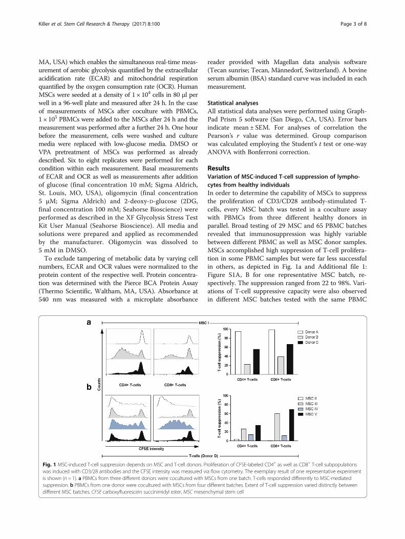
ResultsVariation of MSC-induced T-cell suppression of lympho-cytes from healthy individualsIn order to determine the capability of MSCs to suppressthe proliferation of CD3/CD28 antibody-stimulated T-cells, every MSC batch was tested in a coculture assaywith PBMCs from three different healthy donors inparallel. Broad testing of 29 MSC and 65 PBMC batchesrevealed that immunosuppression was highly variablebetween different PBMC as well as MSC donor samples.MSCs accomplished high suppression of T-cell prolifera-tion in some PBMC samples but were far less successfulin others, as depicted in Fig. 1a and Additional file 1:Figure S1A, B for one representative MSC batch, re-spectively. The suppression ranged from 22 to 98%. Vari-ations of T-cell suppressive capacity were also observedin different MSC batches tested with the same PBMC
Fig. 1 MSC-induced T-cell suppression depends on MSC and T-cell donors. Proliferation of CFSE-labeled CD4+ as well as CD8+ T-cell subpopulationswas induced with CD3/28 antibodies and the CFSE intensity was measured via flow cytometry. The exemplary result of one representative experimentis shown (n = 1). a PBMCs from three different donors were cocultured with MSCs from one batch. T-cells responded differently to MSC-mediatedsuppression. b PBMCs from one donor were cocultured with MSCs from four different batches. Extent of T-cell suppression varied distinctly betweendifferent MSC batches. CFSE carboxyfluorescein succinimidyl ester, MSC mesenchymal stem cell
Killer et al. Stem Cell Research & Therapy (2017) 8:100 Page 3 of 8
donor. Figure 1b and Additional file 1: Figure S1C, Dillustrate results for a single donor PBMC and four MSCbatches, respectively. Here, the suppression was between0.5 and 69%. Monitoring cell proliferation of CD4+ andCD8+ cells gave similar results. The highest variation,however, was observed when CD8+ T-cell proliferationwas determined after coculture with MSCs from differ-ent donors. Thus, potency for in-vitro immunosuppres-sive activity of MSCs does not simply rely on specificMSC batch performances but highly depends on thepredisposition of lymphocytes from different donors. Acorrelation between donor age and T-cell suppressionwas not observed (Additional file 1: Figure S2).
Interaction with PBMCs enhances MSC metabolismFurthermore, we sought to identify factors that deter-mine the donor dependency of PBMC and MSC inter-actions in coculture. As shown previously, senescenceof MSCs is correlated with poor T-cell suppressivecapacity [14]. Because senescent MSCs exhibit lowmetabolic activity [19], we sought to analyze the
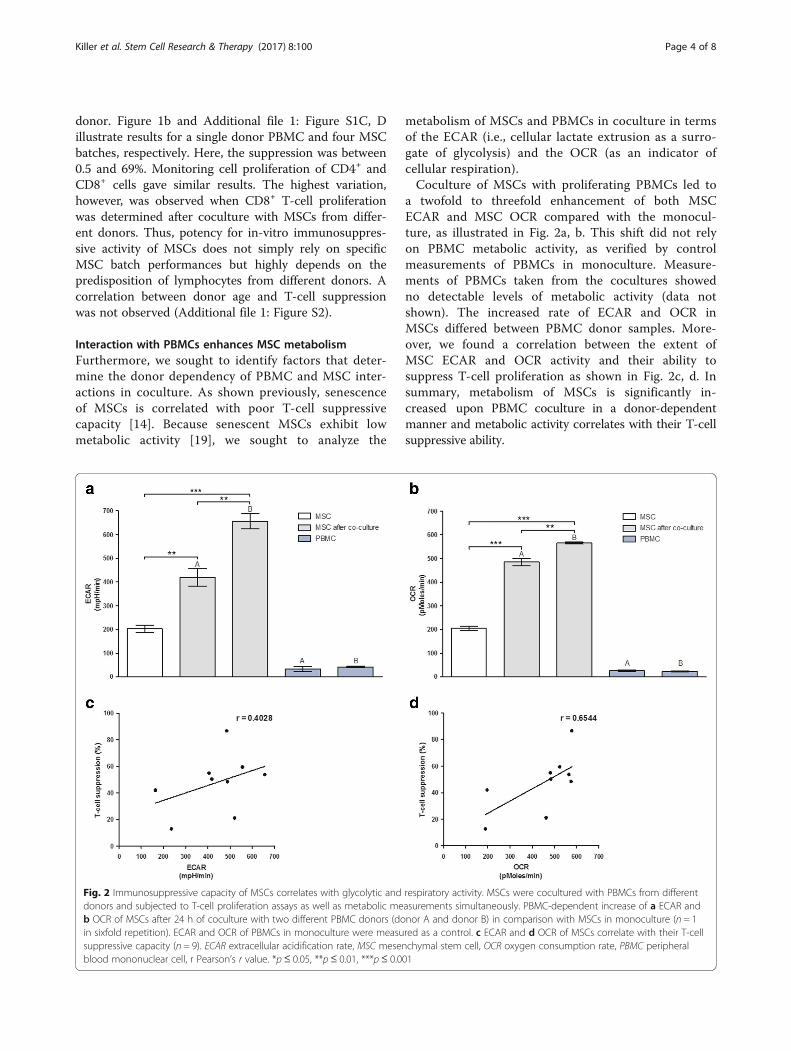
metabolism of MSCs and PBMCs in coculture in termsof the ECAR (i.e., cellular lactate extrusion as a surro-gate of glycolysis) and the OCR (as an indicator ofcellular respiration).Coculture of MSCs with proliferating PBMCs led to
a twofold to threefold enhancement of both MSCECAR and MSC OCR compared with the monocul-ture, as illustrated in Fig. 2a, b. This shift did not relyon PBMC metabolic activity, as verified by controlmeasurements of PBMCs in monoculture. Measure-ments of PBMCs taken from the cocultures showedno detectable levels of metabolic activity (data notshown). The increased rate of ECAR and OCR inMSCs differed between PBMC donor samples. More-over, we found a correlation between the extent ofMSC ECAR and OCR activity and their ability tosuppress T-cell proliferation as shown in Fig. 2c, d. Insummary, metabolism of MSCs is significantly in-creased upon PBMC coculture in a donor-dependentmanner and metabolic activity correlates with their T-cellsuppressive ability.
Fig. 2 Immunosuppressive capacity of MSCs correlates with glycolytic and respiratory activity. MSCs were cocultured with PBMCs from differentdonors and subjected to T-cell proliferation assays as well as metabolic measurements simultaneously. PBMC-dependent increase of a ECAR andb OCR of MSCs after 24 h of coculture with two different PBMC donors (donor A and donor B) in comparison with MSCs in monoculture (n = 1in sixfold repetition). ECAR and OCR of PBMCs in monoculture were measured as a control. c ECAR and d OCR of MSCs correlate with their T-cellsuppressive capacity (n = 9). ECAR extracellular acidification rate, MSC mesenchymal stem cell, OCR oxygen consumption rate, PBMC peripheralblood mononuclear cell, r Pearson’s r value. *p≤ 0.05, **p≤ 0.01, ***p≤ 0.001
Killer et al. Stem Cell Research & Therapy (2017) 8:100 Page 4 of 8
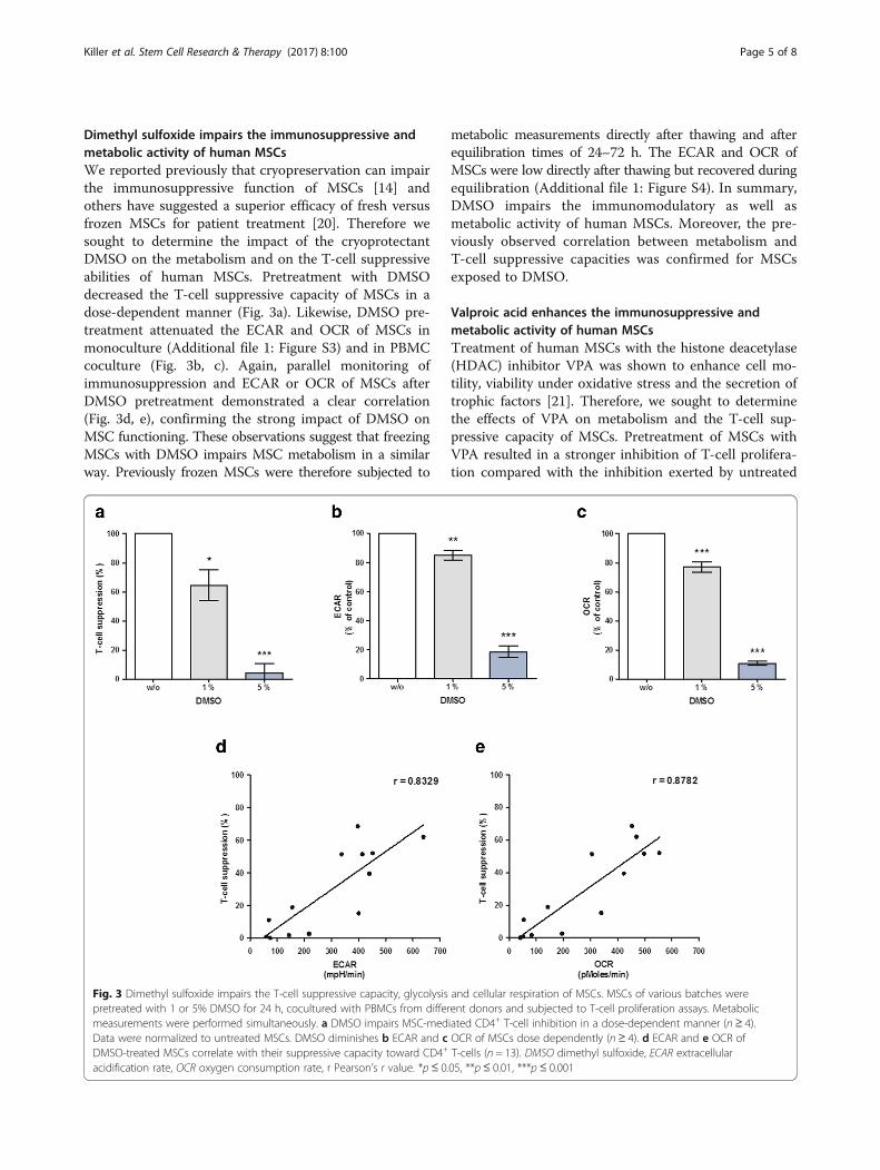
Dimethyl sulfoxide impairs the immunosuppressive andmetabolic activity of human MSCsWe reported previously that cryopreservation can impairthe immunosuppressive function of MSCs [14] andothers have suggested a superior efficacy of fresh versusfrozen MSCs for patient treatment [20]. Therefore wesought to determine the impact of the cryoprotectantDMSO on the metabolism and on the T-cell suppressiveabilities of human MSCs. Pretreatment with DMSOdecreased the T-cell suppressive capacity of MSCs in adose-dependent manner (Fig. 3a). Likewise, DMSO pre-treatment attenuated the ECAR and OCR of MSCs inmonoculture (Additional file 1: Figure S3) and in PBMCcoculture (Fig. 3b, c). Again, parallel monitoring ofimmunosuppression and ECAR or OCR of MSCs afterDMSO pretreatment demonstrated a clear correlation(Fig. 3d, e), confirming the strong impact of DMSO onMSC functioning. These observations suggest that freezingMSCs with DMSO impairs MSC metabolism in a similarway. Previously frozen MSCs were therefore subjected to
metabolic measurements directly after thawing and afterequilibration times of 24–72 h. The ECAR and OCR ofMSCs were low directly after thawing but recovered duringequilibration (Additional file 1: Figure S4). In summary,DMSO impairs the immunomodulatory as well asmetabolic activity of human MSCs. Moreover, the pre-viously observed correlation between metabolism andT-cell suppressive capacities was confirmed for MSCsexposed to DMSO.
Valproic acid enhances the immunosuppressive andmetabolic activity of human MSCsTreatment of human MSCs with the histone deacetylase(HDAC) inhibitor VPA was shown to enhance cell mo-tility, viability under oxidative stress and the secretion oftrophic factors [21]. Therefore, we sought to determinethe effects of VPA on metabolism and the T-cell sup-pressive capacity of MSCs. Pretreatment of MSCs withVPA resulted in a stronger inhibition of T-cell prolifera-tion compared with the inhibition exerted by untreated
Fig. 3 Dimethyl sulfoxide impairs the T-cell suppressive capacity, glycolysis and cellular respiration of MSCs. MSCs of various batches werepretreated with 1 or 5% DMSO for 24 h, cocultured with PBMCs from different donors and subjected to T-cell proliferation assays. Metabolicmeasurements were performed simultaneously. a DMSO impairs MSC-mediated CD4+ T-cell inhibition in a dose-dependent manner (n ≥ 4).Data were normalized to untreated MSCs. DMSO diminishes b ECAR and c OCR of MSCs dose dependently (n ≥ 4). d ECAR and e OCR ofDMSO-treated MSCs correlate with their suppressive capacity toward CD4+ T-cells (n = 13). DMSO dimethyl sulfoxide, ECAR extracellularacidification rate, OCR oxygen consumption rate, r Pearson’s r value. *p≤ 0.05, **p≤ 0.01, ***p ≤ 0.001
Killer et al. Stem Cell Research & Therapy (2017) 8:100 Page 5 of 8
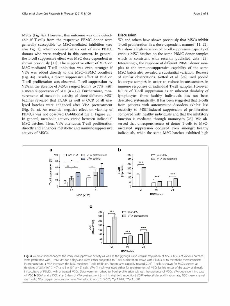
MSCs (Fig. 4a). However, this outcome was only detect-able if T-cells from the respective PBMC donor weregenerally susceptible to MSC-mediated inhibition (seealso Fig. 1), which occurred in six out of nine PBMCdonors who were analyzed in this context. In general,the T-cell suppressive effect was MSC dose dependent asshown previously [11]. The supportive effect of VPA onMSC-mediated T-cell inhibition was even stronger ifVPA was added directly to the MSC–PBMC coculture(Fig. 4a). Besides, a direct suppressive effect of VPA onT-cell proliferation was observed. T-cell suppression byVPA in the absence of MSCs ranged from 7 to 77%, witha mean suppression of 31% (n = 12). Furthermore, mea-surements of metabolic activity of three different MSCbatches revealed that ECAR as well as OCR of all ana-lyzed batches were enhanced after VPA pretreatment(Fig. 4b, c). An essential negative effect on viability ofPBMCs was not observed (Additional file 1: Figure S5).In general, metabolic activity varied between individualMSC batches. Thus, VPA attenuates T-cell proliferationdirectly and enhances metabolic and immunosuppressiveactivity of MSCs.
DiscussionWe and others have shown previously that MSCs inhibitT-cell proliferation in a dose-dependent manner [11, 22].We show a high variation of T-cell suppressive capacity ofvarious MSC batches on the same PBMC donor sampleswhich is consistent with recently published data [23].Interestingly, the response of different PBMC donor sam-ples to the immunosuppressive capability of the sameMSC batch also revealed a substantial variation. Becauseof similar observations, Ketterl et al. [24] used pooledleukocyte samples in order to reduce inconsistencies inimmune responses of individual T-cell samples. However,failure of T-cell suppression as an inherent disability oflymphocytes from healthy individuals has not beendescribed systematically. It has been suggested that T-cellsfrom patients with autoimmune disorders exhibit lessreactivity to MSC-induced suppression of proliferationcompared with healthy individuals and that the inhibitoryfunction is mediated through monocytes [25]. We ob-served that unresponsiveness of donor T-cells to MSC-mediated suppression occurred even amongst healthyindividuals, while the same MSC batches exhibited high
Fig. 4 Valproic acid enhances the immunosuppressive activity as well as the glycolysis and cellular respiration of MSCs. MSCs of various batcheswere pretreated with 1 mM VPA for 6 days and were either subjected to T-cell proliferation assays with PBMCs or to metabolic measurementsin monoculture. a VPA increases the MSC-mediated T-cell inhibition. Suppressive capacity toward CD4+ T-cells is shown for MSCs seeded atdensities of 2.5 × 10⁴ (n = 7) and 5 × 10⁴ (n = 3) cells. VPA (1 mM) was used either for pretreatment of MSCs before onset of the assay or directlyin coculture of PBMCs with untreated MSCs. Data were normalized to T-cell proliferation without the presence of MSCs. VPA-dependent increaseof MSC b ECAR and c OCR after 6 days of VPA pretreatment (n = 1 in eightfold repetition). ECAR extracellular acidification rate, MSC mesenchymalstem cells, OCR oxygen consumption rate, VPA valproic acid. *p≤ 0.05, **p≤ 0.01, ***p≤ 0.001
Killer et al. Stem Cell Research & Therapy (2017) 8:100 Page 6 of 8
immunosuppressive action toward T-cells from otherdonors. Additionally, MSCs from different individuals actdifferently at the level of direct interaction with PBMCs.We further showed that different PBMC batches enhanceMSC ECAR and OCR to variable degrees. MSCs that arehighly capable of suppressing T-cell proliferation reactwith a significant enhancement of metabolism in responseto PBMC coculture, suggesting a dependency of T-cellsuppressive capacity of MSCs on metabolic activity.Accordingly, a linear correlation of metabolic activity andT-cell suppressive capacity of MSCs was observed. In linewith the observation of low glycolytic activity in senescentMSCs [26], we could previously link diminished T-cellsuppression of MSCs with senescence [14].Advanced Therapy Medicinal Products (ATMPs) are
often frozen employing 5–10% DMSO [27]. We there-fore corroborated our hypothesis by demonstrating aDMSO dose-dependent impairment of metabolic activityand immunosuppressive function of MSCs. In contrast,we showed that pretreatment of MSCs with VPA inducedmetabolic activity. Furthermore, we found that VPA dir-ectly reduced T-cell proliferation. Anti-proliferative andapoptosis-inducing effects of VPA on T-cells have beendescribed in-vitro and in-vivo [28]. Beyond this T-cellmodulating effect, MSCs pretreated with VPA displayed asuperior function to suppress T-cell proliferation com-pared with untreated MSCs. When adding VPA dir-ectly to the MSC–PBMC coculture, we observed afurther increase of MSC-mediated T-cell suppression.Because HDAC inhibitors exhibit an immunosuppres-sing effect when applied for treatment of GvHD [29],our data support the notion that combined applicationof MSCs plus VPA could be a very active treatmentregimen for GvHD. In line with this, VPA was shownto increase frequency and function of regulatory T-cells (T-regs) in a mouse model of immune-mediatedarthritis which correlated with reduced incidence andseverity of the disease [30]. In-vitro immunosuppres-sion via increased amounts of T-regs is a mechanismalso described for MSCs [31].In search of a suitable potency assay for MSCs, T-cell
proliferation assays with pooled donor T-cells havebeen proposed [32]. In contrast, our data indicate thatmeasurement of individual patient T-cell response toMSCs could be more relevant in assessment of effi-ciency of MSC therapy. Based on the correlation ofMSC metabolism with their immunosuppressive poten-tial, we suggest that MSC functionality can be pre-dicted via metabolic measurements such as lactate acidproduction or oxygen consumption after patient PBMCand donor MSC coculture. Taken together, our findingspoint to a feasible and informative potency assay thatconsiders interindividual variations in the interactionof MSCs with patient immune effector cells.
ConclusionOur data pave the way for an individualized potencyassay for donor MSC and recipient T-cell interactionbased on metabolic measurements. Enhancement ofT-cell suppressive function through VPA supports thenotion that the immunosuppressive activity of MSCs canbe further enhanced in-vitro and potentially in-vivo.Whether metabolic parameters could be useful to predictthe efficacy of MSC therapy in-vivo needs to be deter-mined in prospective clinical trials.
Additional files
Additional file 1: Figure S1. Showing that T-cell suppression inducedby MSCs is heterogeneous, Figure S2. Showing that PBMC predisposition toMSC-mediated suppression does not correlate with donor age, Figure S3.Showing that DMSO pretreatment attenuates the ECAR and OCR of MSCs,Figure S4. Showing that freezing with DMSO attenuates MSC metabolismand Figure S5. Showing the influence of VPA and DMSO treatment onPBMC survival. (DOCX 2025 kb)
Abbreviations2DG: 2-Deoxy-D-glucose; ATMP: Advanced Therapy Medicinal Product;CFSE: 5,6-Carboxyfluorescein succinimidyl ester; DMSO: Dimethyl sulfoxide;ECAR: Extracellular acidification rate; GMP: Good manufacturing practice;GvHD: Graft-versus-host disease; HDAC: Histone deacetylase;MSC: Mesenchymal stem cell; OCR: Oxygen consumption rate;PBMC: Peripheral blood mononuclear cell; T-reg: Regulatory T-cell;VPA: Valproic acid
AcknowledgementsThe authors would like to thank Kathleen Stabla and Gavin Giel for excellenttechnical assistance.
FundingThis work was supported by the §2 Abs. 3 Kooperationsvertrag (project no. 3/2014, to CBr and HH), the von-Behring-Röntgen-Stiftung (project no. 60-0038, toCBr and HH), the German Carreras Leukemia Foundation (AH06-01, to AN) andthe Flow Cytometry Core Facility Marburg.
Availability of data and materialsNot applicable.
Authors’ contributionsMK and PN were responsible for conception and design, collection of data,data analysis and interpretation, and manuscript writing. MK, LF and TRconducted the experiments. KH and CBa were responsible for conceptionand design, administrative support and data analysis and interpretation. MFwas responsible for data analysis and interpretation. CBr was responsible forconception and design, administrative support, provision of study materialand data analysis and interpretation. HH and AN were responsible foradministrative support, provision of study material and data analysis andinterpretation. All authors were involved in drafting and/or critical revision ofthe manuscript and approved the final submitted version. MK and PNcontributed equally to this article.
Competing interestsThe authors declare that they have no competing interests.
Consent for publicationNot applicable.
Ethics approval and consent to participateThe procedures had been approved by the local ethics committee at thePhilipps-University Marburg (study no. 64/01 and 25/10) and patients hadgiven written informed consent.
Killer et al. Stem Cell Research & Therapy (2017) 8:100 Page 7 of 8
Publisher’s NoteSpringer Nature remains neutral with regard to jurisdictional claims inpublished maps and institutional affiliations.
Author details1Zentrum für Tumor- und Immunbiologie (ZTI), Hans-Meerwein-Straße 3,35043 Marburg, Germany. 2Biochemisches Institut, Friedrichstraße 24, 35392Giessen, Germany. 3Department of Hematology, Oncology and Immunology,Philipps-University Marburg, Baldingerstraße, 35043 Marburg, Germany.4Institute for Clinical Immunology and Transfusion Medicine, Justus-LiebigUniversity Giessen, Langhansstraße 7, 35385 Giessen, Germany.
Received: 14 December 2016 Revised: 16 March 2017Accepted: 4 April 2017
References1. Uccelli A, Moretta L, Pistoia V. Mesenchymal stem cells in health and
disease. Nat Rev Immunol. 2008;8:726–36.2. Trounson A, McDonald C. Stem cell therapies in clinical trials: progress and
challenges. Cell Stem Cell. 2015;17:11–22.3. Tyndall A. Mesenchymal stromal cells and rheumatic disorders. Immunol
Lett. 2015;168:201–7.4. Zafranskaya MM, Nizheharodova DB, Yurkevich MY, Lamouskaya NV,
Motuzova YM, Bagatka SS, Ivanchik HI, Fedulov AS. In vitro assessment ofmesenchymal stem cells immunosuppressive potential in multiple sclerosispatients. Immunol Lett. 2013;149:9–18.
5. Le Blanc K, Frassoni F, Ball L, Locatelli F, Roelofs H, Lewis I, Lanino E,Sundberg B, Bernardo ME, Remberger M, Dini G, Egeler RM, Bacigalupo A,Fibbe W, Ringdén O. Mesenchymal stem cells for treatment of steroid-resistant, severe, acute graft-versus-host disease: a phase II study. Lancet.2008;371:1579–86.
6. Amorin B, Alegretti AP, Valim V, Pezzi A, Laureano AM, da Silva MAL, WieckA, Silla L. Mesenchymal stem cell therapy and acute graft-versus-hostdisease: a review. Hum Cell. 2014;27:137–50.
7. Simonson OE, Mougiakakos D, Heldring N, Bassi G, Johansson HJ, Dalén M,Jitschin R, Rodin S, Corbascio M, El Andaloussi S, Wiklander OPB, Nordin JZ,Skog J, Romain C, Koestler T, Hellgren-Johansson L, Schiller P, JoachimssonP-O, Hägglund H, Mattsson M, Lehtiö J, Faridani OR, Sandberg R, KorsgrenO, Krampera M, Weiss DJ, Grinnemo K-H, Le Blanc K. In vivo effects ofmesenchymal stromal cells in two patients with severe acute respiratorydistress syndrome. Stem Cells Transl Med. 2015;4:1199–213.
8. Kebriaei P, Isola L, Bahceci E, Holland K, Rowley S, McGuirk J, Devetten M,Jansen J, Herzig R, Schuster M, Monroy R, Uberti J. Adult humanmesenchymal stem cells added to corticosteroid therapy for thetreatment of acute graft-versus-host disease. Biol Blood MarrowTransplant. 2009;15:804–11.
9. von Bonin M, Stölzel F, Goedecke A, Richter K, Wuschek N, Hölig K,Platzbecker U, Illmer T, Schaich M, Schetelig J, Kiani A, Ordemann R,Ehninger G, Schmitz M, Bornhäuser M. Treatment of refractory acute GVHDwith third-party MSC expanded in platelet lysate-containing medium. BoneMarrow Transplant. 2009;43:245–51.
10. Martin PJ, Uberti JP, Soiffer RJ, Klingemann H, Waller EK, Daly AS, HerrmannRP, Kebriaei P. Prochymal improves response rates in patients with steroid-refractory acute graft versus host disease (SR-GVHD) involving the liver andgut: results of a randomized, placebo-controlled, multicenter phase III trial inGVHD. Biol Blood Marrow Transplant. 2010;16:S169–70.
11. Nold P, Brendel C, Neubauer A, Bein G, Hackstein H. Good manufacturingpractice-compliant animal-free expansion of human bone marrow derivedmesenchymal stroma cells in a closed hollow-fiber-based bioreactor.Biochem Biophys Res Commun. 2013;430:325–30.
12. Rojewski MT, Fekete N, Baila S, Nguyen K, Fürst D, Antwiler D, Dausend J,Kreja L, Ignatius A, Sensebé L, Schrezenmeier H. GMP-compliant isolationand expansion of bone marrow-derived MSCs in the closed, automateddevice quantum cell expansion system. Cell Transplant. 2013;22:1981–2000.doi:10.3727/096368912X657990.
13. Bieback K, Kinzebach S, Karagianni M. Translating research into clinicalscale manufacturing of mesenchymal stromal cells. Stem Cells Int.2011;2010:193519.
14. Nold P, Hackstein H, Riedlinger T, Kasper C, Neumann A, Mernberger M,Fölsch C, Schmitt JAN, Fuchs-winkelmann S, Barckhausen C, Killer M,Neubauer A, Brendel C. Immunosuppressive capabilities of mesenchymal
stromal cells are maintained under hypoxic growth conditions and aftergamma irradiation. J Cytotherapy. 2015;17(October):152–62.
15. Wuchter P, Bieback K, Schrezenmeier H, Bornhäuser M, Müller LP, Bönig H, WagnerW, Meisel R, Pavel P, Tonn T, Lang P, Müller I, Renner M, Malcherek G, Saffrich R,Buss EC, Horn P, Rojewski M, Schmitt A, Ho AD, Sanzenbacher R, Schmitt M.Standardization of Good Manufacturing Practice-compliant production of bonemarrow-derived human mesenchymal stromal cells for immunotherapeuticapplications. Cytotherapy. 2015;17:128–39. doi:10.1016/j.jcyt.2014.04.002.
16. François M, Copland IB, Yuan S, Romieu-Mourez R, Waller EK, Galipeau J.Cryopreserved mesenchymal stromal cells display impairedimmunosuppressive properties as a result of heat-shock response andimpaired interferon-γ licensing. Cytotherapy. 2012;14:147–52.
17. Galipeau J. The mesenchymal stromal cells dilemma—does a negativephase III trial of random donor mesenchymal stromal cells in steroid-resistant graft-versus-host disease represent a death knell or a bump in theroad? Cytotherapy. 2013;15:2–8.
18. Dominici M, Le Blanc K, Mueller I, Slaper-Cortenbach I, Marini F, Krause D,Deans R, Keating A, Prockop D, Horwitz E. Minimal criteria for definingmultipotent mesenchymal stromal cells. The International Society forCellular Therapy position statement. Cytotherapy. 2006;8:315–7.
19. Capasso S, Alessio N, Squillaro T, Di Bernardo G, Melone MA, Cipollaro M, PelusoG, Galderisi U. Changes in autophagy, proteasome activity and metabolism todetermine a specific signature for acute and chronic senescent mesenchymalstromal cells. Oncotarget. 2015;6. doi:10.18632/oncotarget.6277. Link to Pubmed:https://www.ncbi.nlm.nih.gov/pmc/articles/PMC4741838/.
20. Moll G, Alm JJ, Davies LC, von Bahr L, Heldring N, Stenbeck-Funke L, HamadOA, Hinsch R, Ignatowicz L, Locke M, Lönnies H, Lambris JD, Teramura Y,Nilsson-Ekdahl K, Nilsson B, Le Blanc K. Do cryopreserved mesenchymalstromal cells display impaired immunomodulatory and therapeuticproperties? Stem Cells. 2014;32:2430–42.
21. Cho G, Kang BY, Kim K-S, Kim SH. Effects of valproic acid on the expressionof trophic factors in human bone marrow mesenchymal stromal cells.Neurosci Lett. 2012;526:100–5. doi:10.1016/j.neulet.2012.08.015.
22. Le Blanc K, Tammik L, Sundberg B, Haynesworth SE, Ringdén O.Mesenchymal stem cells inhibit and stimulate mixed lymphocyte culturesand mitogenic responses independently of the major histocompatibilitycomplex. Scand J Immunol. 2003;57:11–20.
23. Bloom DD, Centanni JM, Bhatia N, Emler CA, Drier D, Leverson GE, McKennaDH, Gee AP, Lindblad R, Hei DJ, Hematti P. A reproducible immunopotencyassay to measure mesenchymal stromal cell-mediated T-cell suppression.Cytotherapy. 2015;17:140–51.
24. Ketterl N, Brachtl G, Schuh C, Bieback K, Schallmoser K, Reinisch A, Strunk D.A robust potency assay highlights significant donor variation of humanmesenchymal stem/progenitor cell immune modulatory capacity andextended radio-resistance. Stem Cell Res Ther. 2015;6:236.
25. Ben-Ami E, Miller A, Berrih-Aknin S. T cells from autoimmune patientsdisplay reduced sensitivity to immunoregulation by mesenchymal stemcells: role of IL-2. Autoimmun Rev. 2014;13:187–96.
26. Liu Y, Ma T. Metabolic regulation of mesenchymal stem cell in expansionand therapeutic application. Biotechnol Prog. 2014;31:468–81.
27. Marquez-Curtis LA, Janowska-Wieczorek A, McGann LE, Elliott JAW.Mesenchymal stromal cells derived from various tissues: biological, clinicaland cryopreservation aspects. Cryobiology. 2015;71:181–97.
28. Lv J, Du C, Wei W, Wu Z, Zhao G, Li Z, Xie X. The antiepileptic drug valproicacid restores T cell homeostasis and ameliorates pathogenesis of experimentalautoimmune encephalomyelitis. J Biol Chem. 2012;287:28656–65.
29. Choi S, Reddy P. HDAC inhibition and graft versus host disease. Mol Med.2011;17:404–16.
30. Saouaf SJ, Li B, Zhang G, Shen Y, Furuuchi N, Hancock WW, Greene MI.Deacetylase inhibition increases regulatory T cell function and decreasesincidence and severity of collagen-induced arthritis. Exp Mol Pathol. 2009;87:99–104.
31. Yang H, Sun J, Wang F, Li Y, Bi J, Qu T. Umbilical cord-derived mesenchymalstem cells reversed the suppressive deficiency of T regulatory cells fromperipheral blood of patients with multiple sclerosis in a co-culture—apreliminary study. Oncotarget. 2016. doi:10.18632/oncotarget.12345. Link toPubmed: https://www.ncbi.nlm.nih.gov/pmc/articles/PMC5341927/.
32. De Becker A, Van Riet I. Mesenchymal stromal cell therapy in hematology:from laboratory to clinic and back again. Stem Cells Dev. 2015;24:1713–29.
Killer et al. Stem Cell Research & Therapy (2017) 8:100 Page 8 of 8