ImagingImagingTechniques Techniques andandTypical ... · ImagingImagingTechniques Techniques...
31
Female Female Pelvic Pelvic Pain Pain - Imaging Imaging Techniques Techniques and and Typical Typical Findings Findings Boris Brkljačić Boris Brkljačić Department of Radiology, University Hospital “Dubrava”; Medical School, University of Zagreb, Zagreb, Croatia Danish Radiology Congress, Aarhus, January 2013.
Transcript of ImagingImagingTechniques Techniques andandTypical ... · ImagingImagingTechniques Techniques...
FemaleFemale PelvicPelvic PainPain --
ImagingImaging TechniquesTechniques
andand TypicalTypical FindingsFindings
Boris BrkljačićBoris Brkljačić
Department of Radiology, University Hospital “Dubrava”;
Medical School, University of Zagreb, Zagreb, Croatia
Danish Radiology Congress, Aarhus, January 2013.
FemaleFemale pelvicpelvic painpain
� structured approach to image interpretation to
narrow broad spectrum of ddxs
� distinction btw pregnant and non-pregnant pts
� once pregnancy is excluded disorders need to be � once pregnancy is excluded disorders need to be
grouped according to the anatomic origin
� initial imaging work-up should be tailored to
enable dxs of both gynecologic and non-
gynecologic causes; not only pelvic examination
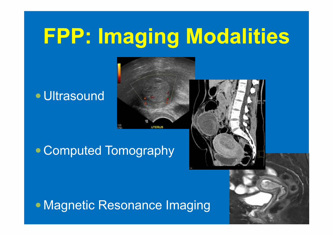
FPP: FPP: ImagingImaging ModalitiesModalities
�Ultrasound
�Computed Tomography
�Magnetic Resonance Imaging
AcuteAcute LowerLower AbdominalAbdominal andand
PelvicPelvic PainPain: :
��UUS S andand CTCT are most commonly used
��MMRIRI beneficial for a subset of pts for whom
CT is not warranted (pregnant pts, younger
pts)
ChronicChronic PelvicPelvic PainPain: :
��UUS S andand MRIMRI are most commonly used
�US (TV US) used initially�US (TV US) used initially
�MRI often used for definitive dx
UltrasoundUltrasound
the first imaging modality to study women w lower
abdominal and pelvic pain
advantages: non-invasive, low cost, no ionizing advantages: non-invasive, low cost, no ionizing radiation, availability, real-time, dynamic exam
US enables final dxs in many clinical conditions and further imaging is indicated when US fxs are inconclusive
UltrasoundUltrasound
� transabdominal US
� transvaginal US
�B-mode, color & power Doppler
�native harmonic and compound imaging�native harmonic and compound imaging
� contrast-enhanced-US
� 3 D Ultrasound
� hysterosonography and hystosalpingo-contrast-sonography (hy-co-sy)
� elastography
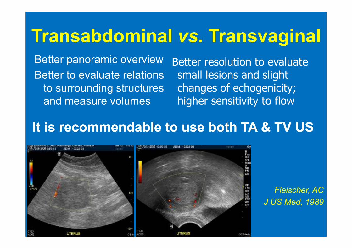
TransabdominalTransabdominal vs.vs. TransvaginalTransvaginalBetter panoramic overview
Better to evaluate relations
to surrounding structures
and measure volumes
Better resolution to evaluate small lesions and slight changes of echogenicity; higher sensitivity to flow
It is recommendable to use both TA It is recommendable to use both TA && TV USTV USIt is recommendable to use both TA It is recommendable to use both TA && TV USTV US
Fleischer, AC
J US Med, 1989
transvaginaltransvaginal USUS alone sufficient to visualise all fxs in
>83% pts
both TV and TA USboth TV and TA US w empty bladder needed for 15%
of pts
only 1.5% of pts required full bladder to visualise
normal ovaries → filling of the bladder for pelvic
sonogram is seldom required
Benacerraf B, et al. J Ultrasound Med 2003
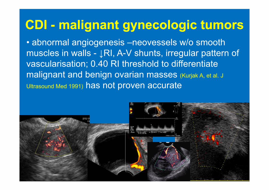
• abnormal angiogenesis –neovessels w/o smooth
muscles in walls - ↓RI, A-V shunts, irregular pattern of
vascularisation; 0.40 RI threshold to differentiate
malignant and benign ovarian masses (Kurjak A, et al. J
Ultrasound Med 1991) has not proven accurate
CDI CDI -- malignantmalignant gynecologicgynecologic tumorstumors
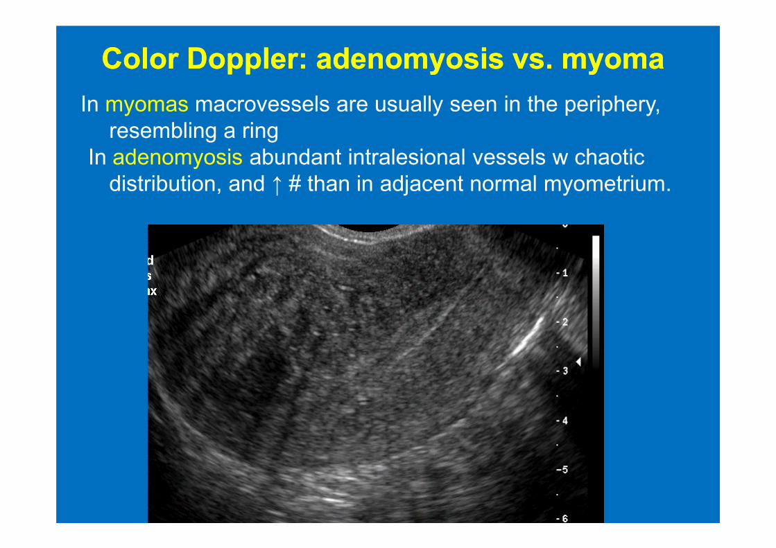
In myomas macrovessels are usually seen in the periphery,
resembling a ring
In adenomyosis abundant intralesional vessels w chaotic
distribution, and ↑ # than in adjacent normal myometrium.
ColorColor Doppler: Doppler: adenomyosisadenomyosis vs. vs. myomamyoma
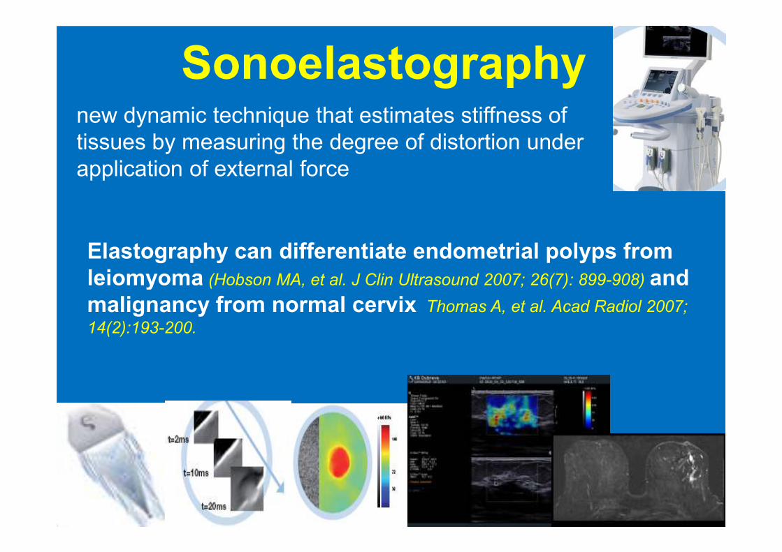
Sonoelastographynew dynamic technique that estimates stiffness of
tissues by measuring the degree of distortion under
application of external force
Elastography can differentiate endometrial polyps from
leiomyoma (Hobson MA, et al. J Clin Ultrasound 2007; 26(7): 899-908) and leiomyoma (Hobson MA, et al. J Clin Ultrasound 2007; 26(7): 899-908) and
malignancy from normal cervix Thomas A, et al. Acad Radiol 2007;
14(2):193-200.
SignificanceSignificance of of normalnormal US US fxfx??
�86 pts w PP and normal US reevaluated after
6-21 mo: 86% w acute/subacute pain & 50%
w chronic pain → resolution of symptoms
� further imaging in 9 pts → only 4 had clin� further imaging in 9 pts → only 4 had clin
sign disease (2 endometriosis, 1 pelvic
adhesions, 1 adenomyosis).
�high NPV (92%) for normal TV US fxs
Harris RD, et al. Clinical outcome in female pts w pelvic pain and
normal pelvic US fxs .Radiology 2000;216:440-3
ComputedComputed TomographyTomography
� not the first-line choice in dxs female pelvic diseases
� pelvis often included as a part of abdominal CT study -
familiarity w appearance of pelvic pathology necessary
� advantage: availability in emergency
� disadvantages: lack of precise definition of pelvic
structures, ionizing radiation (especially problem in
young women and possibly pregnant women)
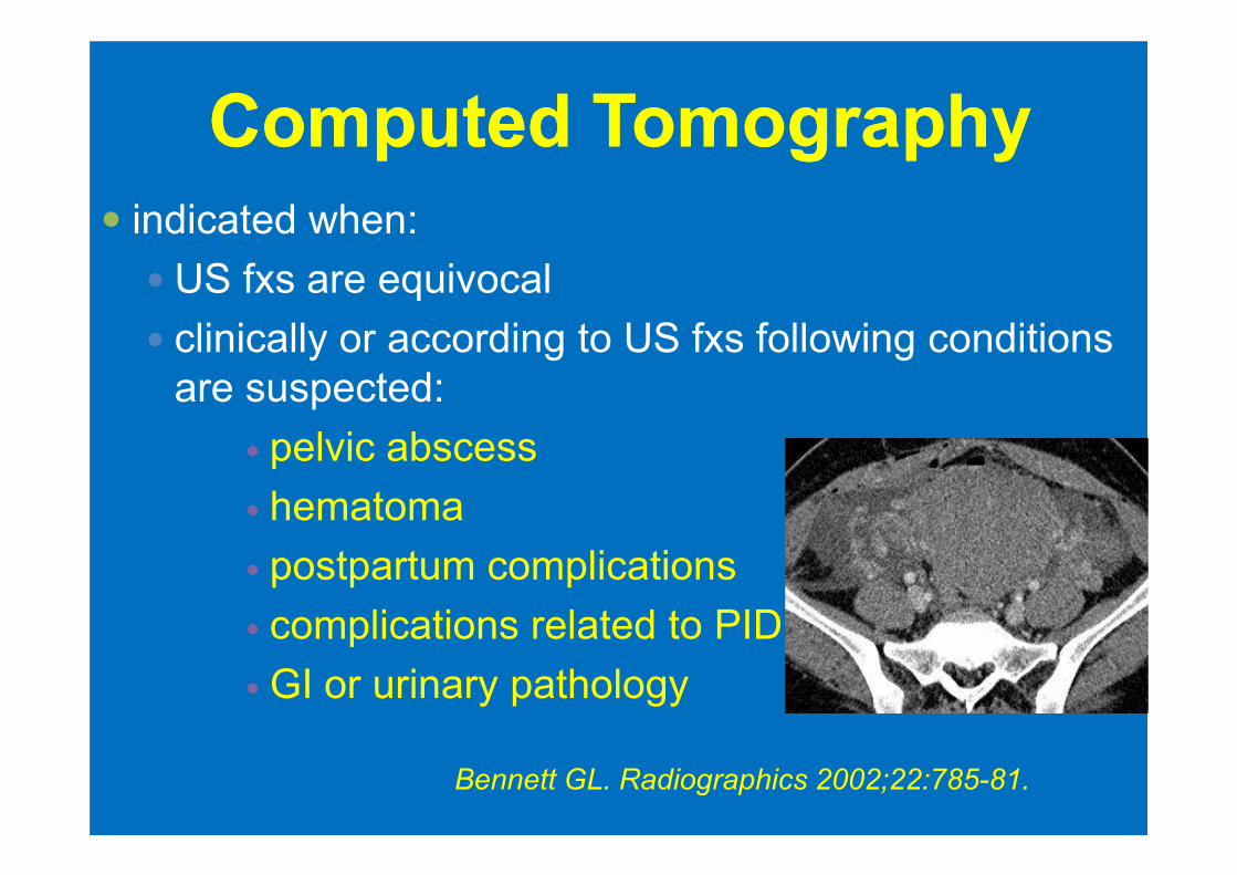
ComputedComputed TomographyTomography
� indicated when:
� US fxs are equivocal
� clinically or according to US fxs following conditions
are suspected:
� pelvic abscess � pelvic abscess
� hematoma
� postpartum complications
� complications related to PID
� GI or urinary pathology
Bennett GL. Radiographics 2002;22:785-81.
ComputedComputed TomographyTomography
� CT often performed in pts referred for pain beyond the pelvis or in pts who present after hours – ↑frequency of use of CT to evaluate pts w acute PP
� PP may exist in the absence of gynecologic cause; CT will depict non-gynecologic disease if the initial imaging protocols are not tailored too narrowly within the pelvis
Potter AW. Radiographics 2008;28:1645-59
CT CT -- acquisitionacquisition parametersparameters::
� (16-MDCT)
� patient position: supine, with elevated arms; scan
range: diaphragm (iliac crest) to pubic symphisis; tube
voltage: 120KVp,140 - 220mAs
� slice collimation: 16x1.5mm, pitch 1.3-1.5, recon.
kernel B20/ B30f, recon. increment 3-5mm
� unenhanced scans in pelvic disease useful only for
detection of acute hemorrhage
CT CT -- acquisitionacquisition parametersparameters::
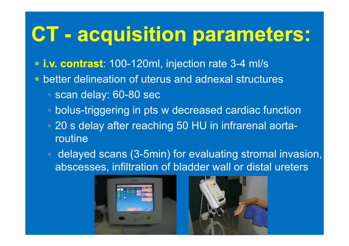
�� ii..v. contrastv. contrast: 100-120ml, injection rate 3-4 ml/s
� better delineation of uterus and adnexal structures
� scan delay: 60-80 sec
� bolus-triggering in pts w decreased cardiac function
� 20 s delay after reaching 50 HU in infrarenal aorta-� 20 s delay after reaching 50 HU in infrarenal aorta-
routine
� delayed scans (3-5min) for evaluating stromal invasion,
abscesses, infiltration of bladder wall or distal ureters
CT CT -- acquisitionacquisition parametersparameters::
�� oral administrationoral administration of 1000-1500 ml of iodinated
water-soluble 2% c.m., or barium-sulfate suspension
- taken continuously over at least 45 min– thus
normal bowel loops can be distinguished fromnormal bowel loops can be distinguished from
abnormal bowel and adnexal structures
� rectal administration of contrast not routinely
performed but may help ddx adnexal process from
rectosigmoid colon disorders
MRI MRI protocolprotocol
�has to be tailored according to the clinical question
�slice orientation and thickness +++�slice orientation and thickness +++
� i.v. contrast
�always / when possible correlate fxs of TV / TA GYN US

Current gynecological Current gynecological
indications for pelvic MRIindications for pelvic MRI
� local staging of biopsy proven cervical or
endometrial ca
�characterization of ovarian masses that are �characterization of ovarian masses that are
suspicios or indeterminate at US
�polyfibromatosis / large uteri before
conservative treatment of the uterus
�chronic PP: endometriosis, adenomyosis
Kinkel K, Eur Radiology 2006
Standard protocol: female pelvis MRIStandard protocol: female pelvis MRI
� 3-6 h prior fasting
� no bladder voiding 1-2 h prior to MRI
� pt interview:
� contraindications to MRI
� symptoms (pain – when, where, how often, how � symptoms (pain – when, where, how often, how
intense?; bleeding)
� date of last menstruation / menopause, medication
(contraceptives?), surgical history
� i.m. injection of an antiperistaltic agent (if no
glaucoma): 20mg butyl-scopalamine (Buscopan®)
Kinkel, K. ECR 2006
Rectal or vaginal Rectal or vaginal contrastcontrast
� vaginal contrast: US gel
� better diagnosis of posterior fornix invasion in cervical
cancer (Van Hoe, Radiology 1999)
� rectal contrast: 200 ml US gel / water enema
� diagnosis of rectal wall invasion (Bazot, Radiology 2004)
� Endometriosis, gynaecological cancer invasion
� pelvic floor analysis
Standard Standard sequencesequence choiceschoices
� three T2 w FSE sequences (12min) – anatomical info
� TR/TE 4000/90 msec, 203x512,
� FOV 22-25cm
� 3 nex, 4 mm, 25 slices� 3 nex, 4 mm, 25 slices
� sagittal, short and long uterine axis (oblique)
� one to three T1 w FSE sequences (TR/TE 500/14
msec, 204x512, 4mm) (15min) – biochemical info
� native: always
� fat suppressed: if lesion hyperintense on native T1
� i.v. contrast-enhanced and fat suppression: list
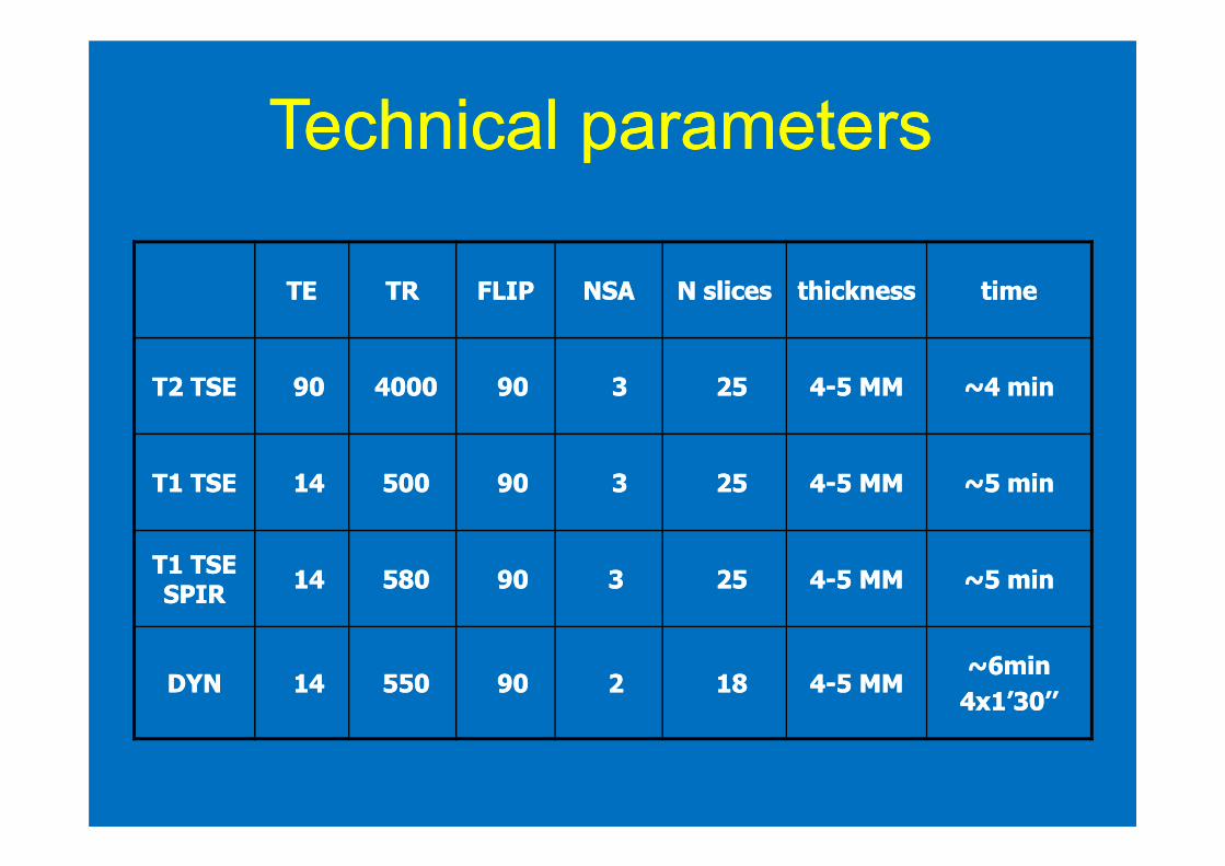
TETE TRTR FLIPFLIP NSANSA N slicesN slices thicknessthickness timetime
T2 TSET2 TSE 9090 40004000 9090 33 2525 44--5 MM5 MM ~4 min~4 min
TechnicalTechnical parametersparameters
T1 TSET1 TSE 1414 500500 9090 33 2525 44--5 MM5 MM ~5 min~5 min
T1 TSE T1 TSE
SPIRSPIR1414 580580 9090 33 2525 44--5 MM5 MM ~5 min~5 min
DYNDYN 1414 550550 9090 22 1818 44--5 MM5 MM~6min~6min
4x1’30’’4x1’30’’
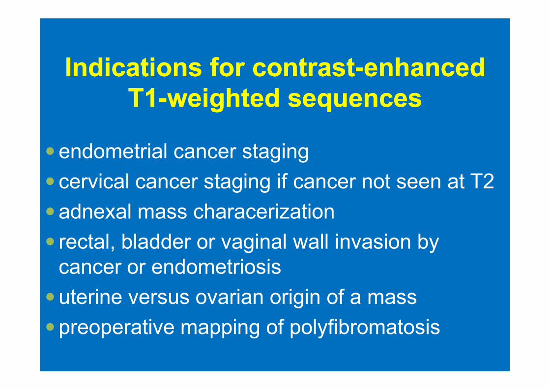
IndicationIndicationss for for contrastcontrast--enhancedenhanced
T1T1--weightedweighted sequencessequences
�endometrial cancer staging
�cervical cancer staging if cancer not seen at T2
�adnexal mass characerization
� rectal, bladder or vaginal wall invasion by
cancer or endometriosis
�uterine versus ovarian origin of a mass
�preoperative mapping of polyfibromatosis
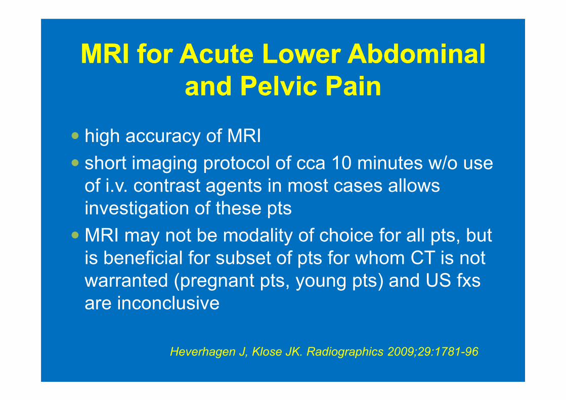
MRI for Acute Lower Abdominal MRI for Acute Lower Abdominal
and Pelvic Painand Pelvic Pain
� high accuracy of MRI
� short imaging protocol of cca 10 minutes w/o use
of i.v. contrast agents in most cases allows
investigation of these ptsinvestigation of these pts
� MRI may not be modality of choice for all pts, but
is beneficial for subset of pts for whom CT is not
warranted (pregnant pts, young pts) and US fxs
are inconclusive
Heverhagen J, Klose JK. Radiographics 2009;29:1781-96
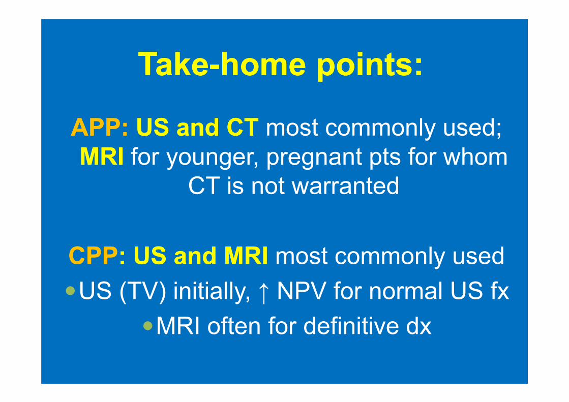
TakeTake--home home pointspoints: :
APP:APP: UUS S andand CTCT most commonly used;
MMRIRI for younger, pregnant pts for whom
CT is not warranted
CPPCPP: : UUS S andand MRIMRI most commonly used
�US (TV) initially, ↑ NPV for normal US fx
�MRI often for definitive dx
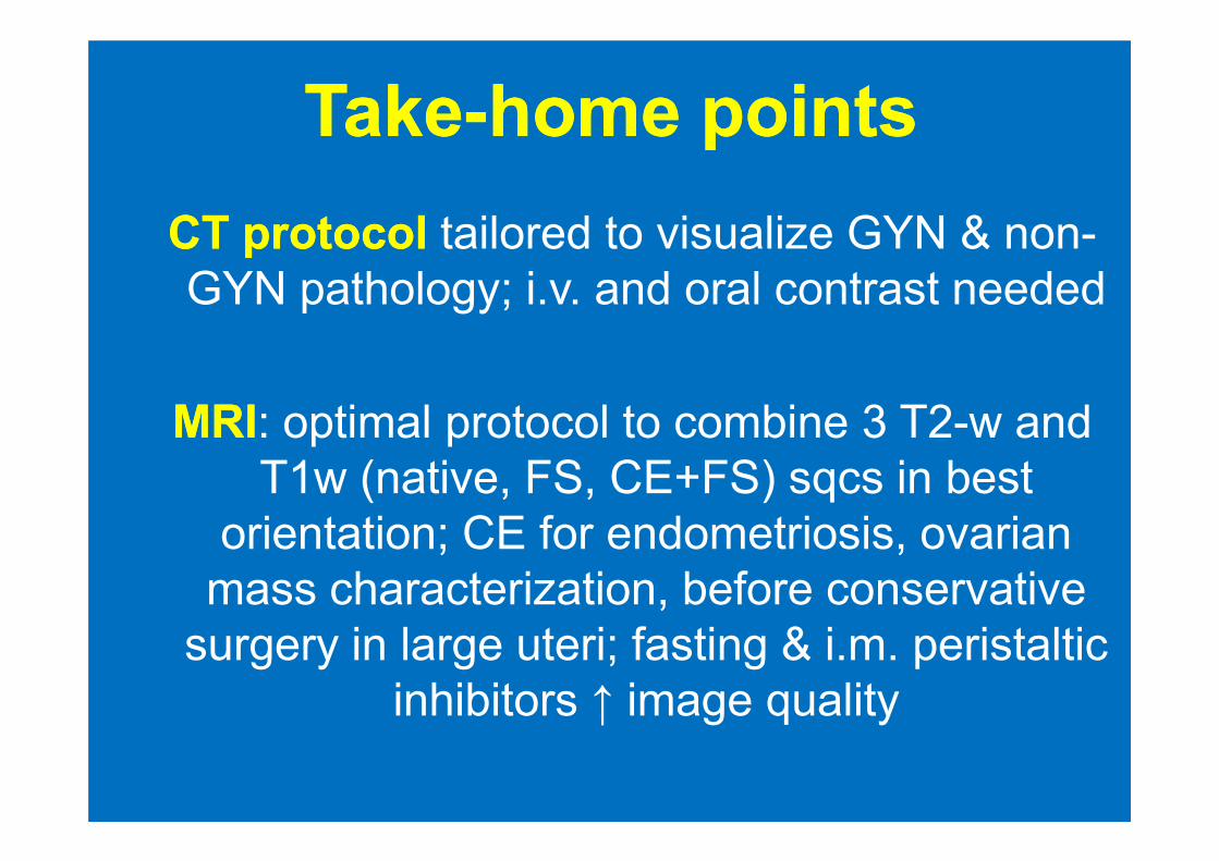
TakeTake--home home pointspoints
CT CT protocolprotocol tailored to visualize GYN & non-
GYN pathology; i.v. and oral contrast needed
MRIMRI: optimal protocol to combine 3 T2-w and MRIMRI: optimal protocol to combine 3 T2-w and
T1w (native, FS, CE+FS) sqcs in best
orientation; CE for endometriosis, ovarian
mass characterization, before conservative
surgery in large uteri; fasting & i.m. peristaltic
inhibitors ↑ image quality
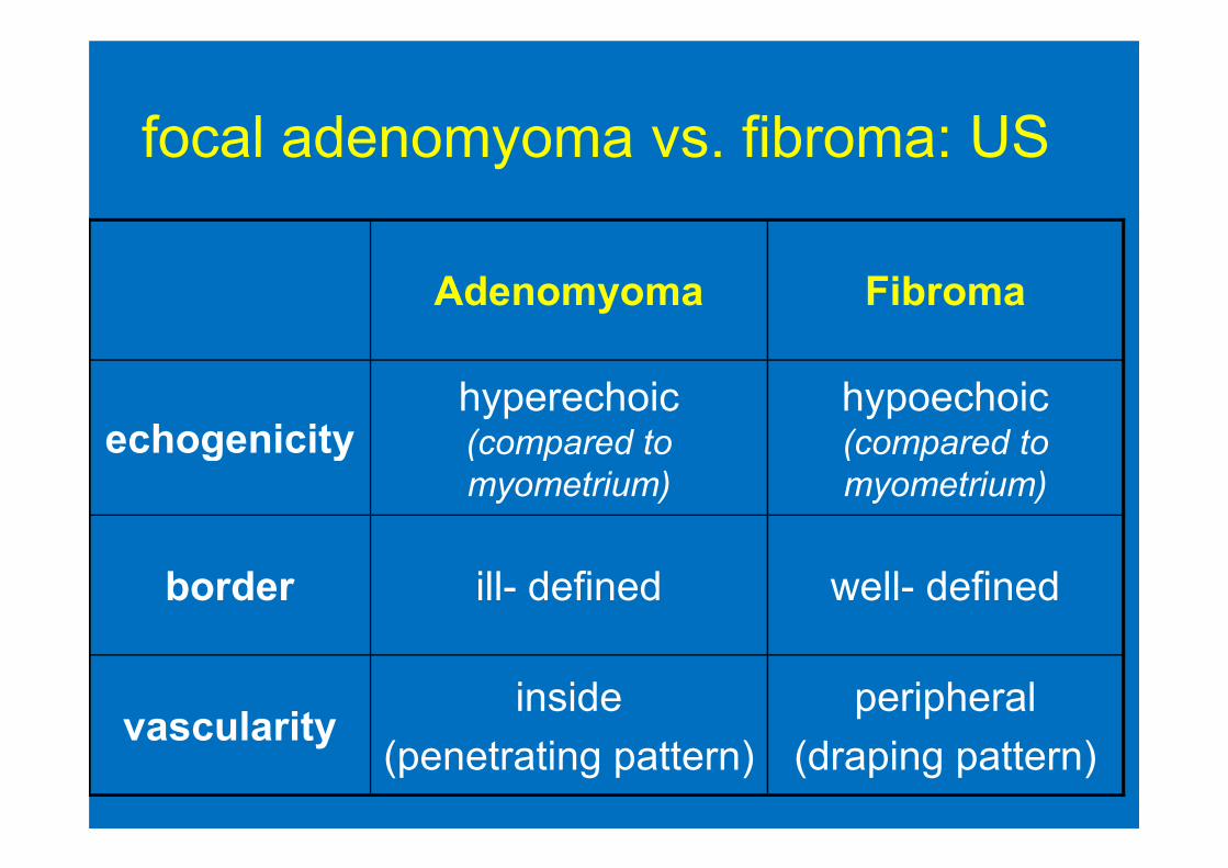
focal adenomyoma vs. fibroma: US
Adenomyoma Fibroma
echogenicityhyperechoic(compared to
hypoechoic(compared to echogenicity (compared to
myometrium)
(compared to
myometrium)
border ill- defined well- defined
vascularityinside
(penetrating pattern)
peripheral
(draping pattern)
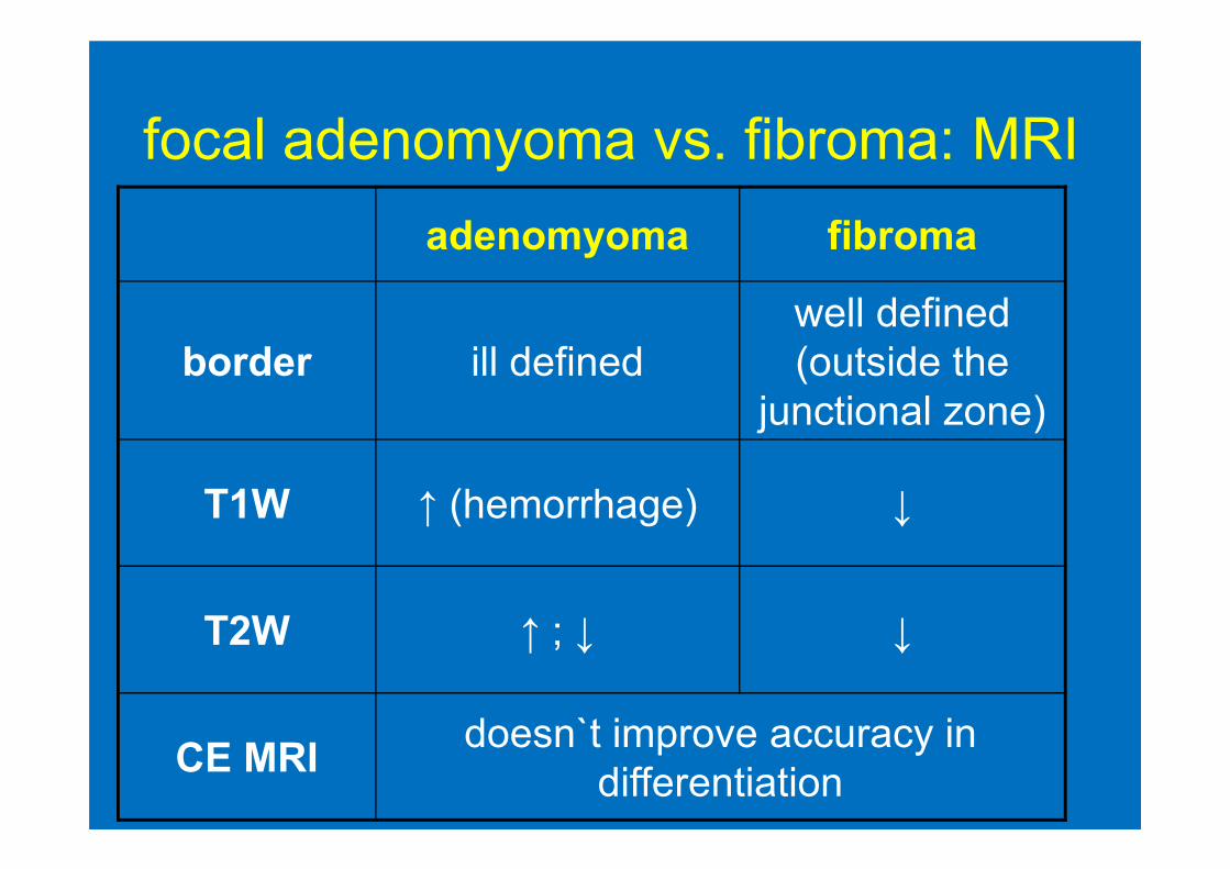
focal adenomyoma vs. fibroma: MRI
adenomyoma fibroma
border ill defined
well defined
(outside the
junctional zone)
T1W ↑ (hemorrhage) ↓
T2W ↑ ; ↓ ↓
CE MRIdoesn`t improve accuracy in
differentiation