HOMMAGE AU PÈRE JEAN DUCRUET NAHR-EL …newhealthconcept.net/uploads/pdf/MedEmergency8.pdf · Le...
56
September 2011-N°8 Endorsed by Trimestriel HOMMAGE AU PÈRE JEAN DUCRUET CARDIAC INJURY IN TRAUMATIC SUBARACHNOID HEMORRHAGE ORGAN AND TISSUE DONATION IN LEBANON QUALITY OF CHEST COMPRESSIONS SPASO TECHNIQUE FOR REDUCING SHOULDER DISLOCATIONS BLUNT THORACIC TRAUMA ISSN 2222-9442 NAHR-EL-BARED MEDICAL SUPPORT MISSION PRISE EN CHARGE DES BLESSÉS GRAVES PRISE EN CHARGE DE L’ENFANT BRÛLÉ . CANCELLED ASSISTANCE CARDIORESPIRATOIRE MOBILE EPENCHEMENTS PLEURAUX : FAUT-IL TOUT DRAINER ? PÉDAGOGIE DE L’ECG N° 8
Transcript of HOMMAGE AU PÈRE JEAN DUCRUET NAHR-EL …newhealthconcept.net/uploads/pdf/MedEmergency8.pdf · Le...

September 2011-N°8Endorsed by
Trim
estri
el
HOMMAGE AU PÈRE JEAN DUCRUET
CARDIAC INJURY IN TRAUMATIC SUBARACHNOID HEMORRHAGE
ORGAN AND TISSUE DONATION IN LEBANON
QUALITY OF CHEST COMPRESSIONS
SPASO TECHNIQUE FOR REDUCING SHOULDER DISLOCATIONS
BLUNT THORACIC TRAUMA
ISSN 2222-9442
NAHR-EL-BARED MEDICAL SUPPORT MISSION
PRISE EN CHARGE DES BLESSÉS GRAVES
PRISE EN CHARGE DE L’ENFANT BRÛLÉ . CANCELLED
ASSISTANCE CARDIORESPIRATOIRE MOBILE
EPENCHEMENTS PLEURAUX : FAUT-IL TOUT DRAINER ?
PÉDAGOGIE DE L’ECG N° 8

1MED EMERGENCY - 2011 No8
MED Emergency PublicationBy New Health Concept
P.O.Box 90.815 Jdeideh - LebanonTel: 00961.1.888921Fax: 00.961.1.888922
Email:[email protected]: www.newhealthconcept.net
Editorial BoardEditor in Chief
Dr. Nagi SOUAIBYManaging Editor
Chantal Saadeh Khalil,Georges Khalil,
Georgina MaaloufDany Matar
Members Jean Claude DESLANDES (France)
Chokri HAMOUDA (Tunisia)AAbdo KHOURY,
Jean Yves Le Coz (France) Afif MUFARRIJ,
Jean-Cyrille PITTELOUD (Switzerland), Alissar RADY.
AUTHORS OF THIS ISSUERIM LAKHDAR, ANTOINE STEPHAN,ANDREAS BOHN, JENARO A. FER-
NANDEZ-VALENCIA, LEANNE PEREZ,KHALIL HELOU, FRANÇOIS BANDALY,
OLIVIER GALL, HICHEM CHENAI-Ç
TIA, PIERRE MICHELET, YANNICK GOTTWALLES.
SCIENTIFIC COMMITTEEPierre ABI HANNA, Georges ABISAAD, Georges ABIZEID, Bahig
ARBID, Rabih ASMAR, Chahine ASSI,Omar AYACH, Melhem AZZI, Charles
BADDOURA, Nasri DIAB, Jean Luc FORTIN (France), Aziz GEAHCHAN,
Bernard GERBAKA, Regis GUARIGUES(France), Berthe HACHEM, Shady HAYEK, Mohamad ACHLAF, Jamil
HALABI, Khalil HELOU, James MOISES(USA), Gladys MOURO, Ahmad
OSMAN (Egypt), Joseph OTAYEK,Maurice KHOURY, MauriceYY HADDAD,
Wassim RAFFOUL (Switzerland), Sami RICHA,Claire GHAFARI ZABLIT
IN PARTNERSHIP WITH
P R É F A C E
Professeur Pierre JOLYPrésident de l’Académie nationale de médecine
When there is a will, there is a way ..
Le Liban et la France sont riches de cultures qui se sont mutuellement valorisées de longue date.
Les Sciences et l’Art de la Médecine n’y ont pas échappé. Différents représentants de l’Aca-démie nationale de médecine parmi les plus prestigieux de France ont porté témoignage à Beyrouth à plusieurs reprises.
Le Docteur SOUAIBY et ses collègues dans leur magnifique publication, que je lis avec autant de joie que de considération, illustrent parfaitement le dynamisme et la qualité de la Médecine Libanaise. Ces travaux de très grande qualité consacrés au malade dans des conditions souvent dramatiques montrent le grand apport de la Médecine Libanaise et des médecins libanais à la Médecine en générale.
Mon propos n’a rien de formel mais je ne résiste pas au grand honneur de rendre hom-mage à la Médecine Libanaise et au rayonnement de celle-ci grâce à des publications aussi remarquables que MED Emergency / Urgence, la Revue Méditerranéenne de Médecined’Urgence.
Dans le cas particulier l’estime ne peut que rejoindre la profonde inclination que j’ai pour le Liban !
Lebanon and France enjoy a wealth of cultures that have mutually enriched over the years. Science and the Art of Medicine are definitely part of this. Various prestigious representatives of the National Academy of Medicine of France have born witness to this on many occasions in Beirut. .
Doctor SOUAIBY and his colleagues illustrate in this magnificent publication, that I always read happily and respectfully, the dynamism and the high quality of Medicine in Lebanon. Those high level works that focus on the patient during very often dramatic moments reflect the great contribution of Lebanese Medicine and Lebanese doctors to Medicine in general.
My statement is far from being a formal one, but I insisted on paying tribute to Lebanese medicine and its radiance thanks to remarkable publications such as MED Emergency / Urgence, the Mediterranean Journal of Emergency Medicine.
I would like here to end by saying that my deepest esteem go hand in hand with the profound liking that I have for Lebanon!

2 MED EMERGENCY - 2011 No8
H O M A G E

3MED EMERGENCY - 2011 No8
H O M M A G E
S U M M A R Y
Hommage au Père Jean Ducruet 3Cardiac injury in traumatic subarachnoid hemorrhage 5Organ and tissue donation in Lebanon 9Quality of chest compressions 11Spaso technique for reducing shoulder dislocations 17Blunt thoracic trauma 21
SWOT analysis of Nahr-El-Bared medical support mission 27Prise en charge des blessés graves 31Prise en charge de l’enfant brûlé cancelled 35Assistance cardiorespiratoire mobile 41Épenchements pleuraux : faut-il tout drainer ? 45Pédagogie de L’ECG N° 8 51
L’accès adéquat aux soins de santé est un droit pour tout homme. Il ne s’agit pas du droit d’être en bonne santé mais le droit de disposer des moyens qui favorisent la promotion, le maintien ou le recouvrement de cet état…Ce droit s’étend aux facteurs déterminants de la santé tels que l’accès à l’eau potable, à une alimentation suffisante, à des conditions hygiéniques de travail, à un environnement sain ainsi qu’à l’éducation et l’information sanitaires… Jean DUCRUET s.j.
Le Père Jean Ducruet, un grand jésuite français devenu «un grand libanais»,
Notre publication qui symbolise l’union entre les deux rives de la Méditerranée et plus particuliè-rement l’amitié franco-libanaise se réjouit de rendre un hommage à cet homme qui a, toute sa vie durant, incarné cette union et cette amitié Le Père Ducruet, natif de Bourg en Bresse en France, est entré dans la Compagnie de Jésus en France en 1942 En 1960, il entame sa mission au Liban où il va consacrer toute sa vie à l’Université Saint-Joseph (USJ) D’abord comme professeur, puis comme Chancelier des Facultés de droit, de sciences économiques et de gestion (1963-1975), puis comme recteur pendant vingt ans (1975-1995) Il préside en même temps (1984-2001) aux destinées de l’Hôtel-Dieu de France avant de prendre la responsabilité du centre d’éthique de l’USJ Il nous a quittés le 13 Mars 2010 à l’âge de 88 ans
Tout a été dit sur ce grand homme :«… Un géant, Un grand bâtisseur aussi bien des pierres brutes que des pierres vivantes, Un homme d’audace, d’action, talentueux, libre et courageux. L’homme des temps difficiles possédant une té-nacité étonnante et un certain sens de l’humour surtout dans les moments graves. Un économiste, visionnaire et résistant. Lucide et déterminé, mais aussi un humaniste, homme de dialogue et de tolérance et surtout un chrétien authentique et discret... »Le père Ducruet était un exemple de courage, de droiture et de modestie La rue qui porte son nom à Beyrouth, inaugurée récemment le 3 Juin 2011, symbolise parfaitement sa personnalité Juxta-posant le rectorat de l’USJ le séparant du nouveau campus de l’innovation, la rue Jean Ducruet est petite mais droite et perpendiculaire à la fameuse « ligne de démarcation », la rue de Damas chère au Père Ducruet qu’il a toujours voulu considérer comme une ligne d’union Reconnu par les plus hautes autorités du pays, il est souvent consulté par les dirigeants politiques Grace à l’ action scientifique et engagée qu’il a menée dans le domaine de la bioéthique, il se voit consacré en 2001 par le Premier ministre comme vice-président du comité consultatif national libanais d’éthique et des sciences de la vie Son regard sur le système de santé en général et sur le système de soins médicaux en particulier était empreint d’humanisme et de dignité envers la personne humaine comme en témoigne l’extrait ci-dessus tiré de son ouvrage « Le service de la santé au Liban » Jacques Chirac dira de lui en le décorant de la croix de commandeur de la Légion d’honneur le 17 juin 1997: « le P. Ducruet ? Un grand serviteur de la pensée et de la culture française dans un Liban qui est devenu naturellement sa seconde patrie. »
Références Le Service de la santé au Liban : ouvrage du Père Ducruet aux éditions de l’USJ Le site de l’USJ : www.usj.edu.lb Le site de la compagnie de Jésus : www.jesuites.com Le quotidien l’Orient Le Jour.
Université Saint-Joseph fondée en 1875 par les Pères Jésuites : Premier établissement d’enseignement universitaire catholique et francophone de la région
Le Père Jean Ducruet


5MED EMERGENCY - 2011 No8
Rim Lakhdar
Article history / info:Received: Sept 14, 2010Reviewed: Oct 30, 2010Received in revised form: July 13, 2011Accepted: August 23, 2011
C A R D I O L O G Y
Rim Lakhdhar*- Nader Baffoun- Kamel Baccar - Chokri Kaddour
IntroductionSubarachnoid hemorrhage (SAH) is a complication of head trauma inducing frequently cerebral vasospasm and even cerebral infarct. Various electrocardiographic abnormalities have been noted in patients with head trauma complicated by subarachnoid hemorrhage (1,2,3,4,5) . They are considered to be secondary to the massive catecholamine discharge in systemic circulation (6,7,8,9). Serum cardiac troponin I (TnI) is considered a highly sensitive and specific marker of myocardial cell lesion and might be regularly performed in these patients to detect early myo-cardial ischemia.
We carried out a prospective study in 35 patients with traumatic SAH (tSAH) in order to assess the incidence of coronary complications during the first five days after admission and to demonstrate the utility of troponin I blood assay in the diagnosis of coronary abnormalities.
Patients and methods:Patients: Our prospective study included 35 patients out of 125 patients with tSAH diagnosed inthe emergency unit in Rabat’s Hospital over a period of 15 months : from January 2008 to March 2009. Patients with any cause of cardiovascular injury were excluded: history of cardiovascular injury; thoracic trauma and vascular-related neurological coma. The selected patients were hos-pitalized in the intensive care and anesthesia department of the National Institute of Neurology.
Methods: The following scoring Systems were used: Acute Physiology and Chronic Health Evaluation II (APACHE II), Injury Severity Score (ISS), the scannographic grade of Fisher and the Glasgow Coma Scale (GCS) after correction of hemodynamic and respirator troubles. A conti-nuous electrocardioscope and a daily electrocardiogram were performed.
A brain CT scan was performed on admission, 48 hours after hospitalization and in case of neurological aggravation. All patients had tracheal intubation and mechanical ventilation with sedative drugs (midazolam and fentanyl) in order to avoid secondary brain insults and to optimize cerebral oxygenation by maintaining mean arterial pressure >90 mmHg, Pa02= 100 mmHg, PaC02 = 30-35 mmHg, a normal osmolarity, and avoiding hyperthermia, hyperglycemia and hypogly-cemia..Serum cardiac troponin Ic, creatine kinase (CK) and its MB fraction (CK-MB) levels were determined on hospital admission and then on the third and the fifth days of hospitalization. This strategy was due to economic reasons and mainly because all blood samples were analyzed in a laboratory out of our hospital.
The statistical analysis was based on the non-parametric variance test of Kruskal-Wallis to com-pare the means; on the chi 2 and Fisher tests to compare percentage, with a significant result at 0.05 percentile and on the Odds ratio non-parametric factors for death. Association between 2 quantitative variables has been analyzed by Pearson coefficient of correlation.
Cardiac injury in traumatic subarachnoid hemorrhage: Prospective study in 35 patientsAtteinte cardiaque au cours de l’hémorragie sous arach-noïdienne post traumatique : Etude prospective de 35 cas
AbstractVarious electrocardiographic abnormali-ties have been noted since 1954 in pa-tients with head trauma complicated by subarachnoid hemorrhage (SAH). Howe-ver, very few studies were interested in these post traumatic SAH (t-SAH) ECG modifications.
Aim of the study: We carried out a pros-pective study in patients with t-SAH in order to assess the incidence of ECG abnormalities during the first five days after admission and the predictive value of these cardiac complications on the mortality in t-SAH.
Patients: Our prospective study included 35 patients out of 125 with traumatic SAH diagnosed in the emergency unit in Rab-ta’s hospital (2001-2009). Patients with cardio vascular history, thoracic trauma, non neurological coma and vascular-re-lated neurological coma were excluded.
Methods: An electrocardiogram monito-ring was performed. A brain CT scan was performed in admission, 48 h after and in case of neurological aggravation. Serum cardiac troponin IC levels were determi-ned on hospital admission and then on the third and fifth days of hospitalization. The statistical analysis was based on the non-parametric variance test of Kruskal-Wallis to compare the means; on the chi 2 and Fisher tests to compare percentage, with a significant result at 0.05 percentile and on the Odds ratio non-parametric
6MED EMERGENCY - 2011 No8
C A R D I O L O G Y
ResultsThe mean age of the 35 patients was 39 ± 17 years. Five patients were treated for hypertension, 3patients for diabetes mellitus, 2 patients had a chronic obstructive pneumopathy, and one patienthad liver cirrhosis. Twenty-three patients had an isolated cranial trauma; the others had associatedtrauma such as facial trauma (4 patients), limb trauma (7 patients) and cervical trauma (1 patient).The mean delay to admission in the intensive care unit was 14 ± 4 hours. Fourteen patientsrequired surgery for different indications: long bone fracture (8 patients), extra-dural hematoma(4 patients), compressive sub-dural hematoma (1 patient) and cerebral contusion (1 patient).
The means ISS, APACHE II, and Glasgow scores were respectively 27 ± 14, 12 ± 6 and 6 ± 3. Thirty patients had a GCS at admission < 8. Twenty patients developed cardiac arrhythmia during thefirst five-day : 8 with tachycardia, 4 with bradycardia, 3 with premature ventricular complexes, 2 with premature atrial complexes and 3 with left or right bundle branch blocks. Seventeen patientsdeveloped cardiac ischemia with ST-segment elevation (1 patient), ST-segment depression (3patients), and T-wave changes (20 patients). The majority of electrocardiographic abnormalitiesappeared on the third day and their mean duration was 4 ± 1 days. Six patients died before thefifth day after admission. Three of them presented ST-segment depression or T-wave changes.
Blood Tn Ic level was increased in 12 patients. CK-MB was elevated in 23 patients and CPK wasincreased in 31 patients. Four patients died before Tn Ic blood level was measured on the thirdday. Three of them had a normal TnIc blood level on the first day and the other one had TnIcincreased at that time. Two patients died before TnIc was determined on the fifth day: one of them had an increased TnIc level on the first day then it get normal on the 3rd day, the secondpatient had a normal TnIc level on the first day then an increased level on the following days.Serum Tn Ic level showed a peak on the 3rd day then it decreased. Both groups of patients withand without increased serum TnIc level were comparable concerning mean ISS (respectively 26 vs 26) and mean APACHE II scores (respectively 12 ± 8 vs 12 ± 7).
There was no correlation between the increase of troponin I level and the severity of initial neurological state in patients assessed by the Glasgow score but this increase was correlated tothe scanographic grade of Fisher with a p value of 0.01.
A significant correlation was found between T-wave changes and the increase of serum TnIclevel, but no correlation was observed between the latter and ST-segment shifts (p=0.5).
Table N°1: Predictive factors of early mortality
Probability of death if factor present
Probabi l i t y of death if factor absent
OR (odds ratio) p
Troponine IC 66.7 21.7 7.2 0.02T wave 56.3 21.1 4.8 0.03ISS 100 26.7 No definite 0.03Fisher 48 54.5 7.7 14.4 0.009Glasgow 43 0 No definite 0.1
In this study, five factors were found as predictive of mortality in the univariate Statistical analysis:troponin, T-wave changes, ISS, the scanographic grade of Fisher at the 48th hour of hospitalizationand the Glasgow Coma Scale (Table I). Patients with increased troponin had a likelihood rate of death of 66.7%, while the latter is of 21% in patients with normal range of Tn Ic. Relative risk of death was 7.2. It became 4.8 higher in patients with T-wave changes.
DiscussionAccording to this study, the prevalence of electrocardiographic abnormalities during SAH-t was57%). It seems to be a common complication seen in case of SAH-t and was reported 50 years
factors for death. Association between 2 quantitative variables was analyzed by Pearson coefficient of correlation. The search engines which have been used were: Pub-Med, Cochrane, and Scopus.
Results: the mean age of the 35 patients was 39+/- 17 years. The sex ratio was 4 in favor of men. The prevalence of elec-trocardiographic changes was 57% (20 patients). Serum Troponin I level showed a peak on the 3rd day then decreased. The majority of electrical abnormalities occurred during the third day after admis-sion and are associated to a markedly in-creased Troponin I plasma level and to the highest rate of mortality. Statistical analysis showed a significant correlation between T wave changes and the increase of serum Tn IC level (p= 0; 0002). The relative risk of mortality was higher than 7.2 times in cases with increase serum TnIc level.
Conclusion: Our study demonstrates that the prevalence of ECG changes were about 57% in patients with t SAH and the major predictive factors of mortality were the increase of serum TnIC and T wave changes.
Key Words: HAS –t, Troponin Ic, ECG, Mortality.

7MED EMERGENCY - 2011 No8
C A R D I O L O G Y
ago. Electrocardiographic abnormalities during SAH have been previously reported by
Burch’s in 1954 (1) who frequently found T-wave inversion and QT segment enlargement in patients with SAH due to aneurysm rupture. Despite the large number of studies interested in myocardial lesions after SAH secondary to aneurysm rupture (2, 3,4,5,6), few clinical studies focused on this specific affection in traumatic SAH.
Physiopathological mechanisms responsible of these electrocardiographic abnormalities remain far from being well-known. Many hypotheses were discussed: coronary organic lesions, massive catecholamine discharge, tachycardia and hypertension secondary to cerebral hemorrhage and coronary vasospasm. The catecholamine discharge hypothesis is the most supported one (7, 8, 9).The incidence of electrocardiographic changes associated to SAH varied in literature. It was of 98% in two prospective studies: The first was carried out by Mayer and al (10) whose study included 57 patients daily monitored by a 12 leads ECG during the first three days after admission. The second was carried out by Browers and al (3) who studied 61 patients with traumatic SAH daily monitored by a 12 leads ECG throughout the 12 first days. They found that the majority of electrocardiographic changes occurred during
the first three days. In our study, all patients had at least one ECG during the first five days of hospitalization. The incidence of electrocardiographic changes was about 57 % and most of them appeared during the third day. Repolarization troubles, especially the T-wave inversion were re-ported to be the most common abnormalities (1,10,11,12,13). These electrical abnormalities were frequently associated to myocardial lesions and to increased serum TnIc level which is specific to the myocardium and may confirm the diagnosis of myocardial lesion especially in trauma patients with lesions of skeletal muscles (6, 14). Elevated levels of troponin have been reported in patients with acute ischemic stroke. Parekh and al (15) had daily measured serum TnIc level in 39 patients with subarachnoid hemorrhage during seven consecutive days. They found that the peak of serum TnIc level occurred on day 1 for 6 patients and on day 2 for one patient. The others had normal range of troponin during the 7 days of the study. In the current study, serum Tn Ic level was quantitatively analyzed on day 1, day 3 and day 5 in all our patients. Both peaks of serum TnIc level and repolarization abnormalities were noted during the third day.
Masaki (16) prospectively evaluated one hundred three patients with SAH in order to determine the relations of TnI to clinical severity, systolic and diastolic cardiac function, pulmonary conges-tion, and length of intensive care unit stay. Highly positive cTnI wit SAH was associated with clinical neurologic severity, systolic and diastolic cardiac dysfunction, pulmonary congestion, and longer intensive care unit stay. Even mild increases in cTnI were associated with diastolic dysfunction and pulmonary congestion.
The severity of neurological status was found to be correlated to a higher incidence of myocardial troubles as diagnosed by biological parameters or echocardiograms (4, 13,15, 16). We found in the current study that the increase of serum TnIc level was independent from physiologic scores and Glasgow coma scale, but it has a statistical significant correlation with the Fisher scanographic scale at the 48th hour (p=0.01) as it was reported by Parekh and al (15).
ConclusionOur study demonstrates first of all that the incidence of electrocardiographic changes was about 57 % in patients with traumatic subarachnoid hemorrhage. Secondly, both peaks of blood serum TnIc level and main repolarization abnormalities were detected on the third day and finally that the major predictive factors of mortality were the increase of serum TnIc level and T-wave changes, with a relative risk of mortality higher than 7.2 times in cases with increased serum Tn Ic level suggesting that daily assaying of this myocardial enzyme must be systematically performed in all patients with tSAH during at least the first seven days.
RésuméL’association d’anomalies électro car-diographiques (ECG), en par ticulier les troubles de la repolarisation, à une hémorragie sous arachnoïdienne post traumatique (HSA) est décrite par plu-sieurs auteurs depuis plus de 50 ans. En l’occurrence très peu d’études se sont intéressées à ces anomalies cardiaques au cours de l’HSA post traumatique (HSA-t).
But de l’étude : Le but de notre travail est de relever l’incidence des complications coronariennes au cours de l’HSA-t et d’ étudier la valeur prédictive de mortalité des anomalies ECG au cours de l’HSA-t
Patients et Méthodes : Cette étude pros-pective menée entre 2001 et 2009 au service de réanimation de l’institut natio-nal de Neurologie de Tunis concerne 35 patients consécutifs traumatisés crâniens avec HSA-t à l’admission. Un électrocar-diogramme a été pratiqué quotidienne-ment pendant les six premiers jours ainsi qu’un dosage de la Troponine Ic au 1er jour post traumatisme puis au 3ème et au 5ème jour. L’analyse statistique était basée sur le test de variance non paramé-trique de Kruskal-Wallis pour comparer les moyennes; et les tests chi 2 et Fisher pour comparer les pourcentages, avec un

MED EMERGENCY - 2011 No88
C A R D I O L O G Y
Todays newsThe Los Angeles Times (9/1, Stein) «Booster Shots» blog reports, «During cardiac arrest time is of the essence, but a longer period of cardiopul-monary resuscitation may be no better than a shorter one,» according to a study published in the New England Journal of Medicine. Investiga-tors «compared outcomes of 9,933 cardiac arrest patients, about half of whom had 30 to 60 seconds of initial CPR from paramedics, or three minutes of the procedure, before heart rhythms were analyzed.» The researchers found that «in both groups, 5.9% of patients survived and were discharged from the hospital in acceptable health.»The Forbes (9/1, Husten) «Cardiobrief» blog reports that in a separate study published in the NEJM, «8718 patients were randomized to treatment with an active or sham impedance threshold device (ITD) intended to improve venous return and cardiac output during CPR. There was nosignificant difference between the groups in the percentage of subjects who survived to hospital discharge with satisfactory function.»MedPage Today (9/1, Gever) reports, «Both trials were designed and conducted by the Resuscitation Outcomes Consortium, a network of treatment centers and emergency response agencies in 10 cities in the US and Canada. Many patients were included in both studies.» Alsocovering the story were AFP (9/1) and HealthDay (9/1, Gardner).
ACEP Sept 1, 2011
LONGER CPR MAY NOT BENEFIT CARDIAC ARREST PATIENTS.
Conflict of interest statement: There is no conflict of interest to declare
REFERENCES
1/Burch GE, Meyers R, Abildskov JA. A new electrocardiographic pattern observed in cerebrovascular accidents. Circulation1954;9:719-723.2/ Cropp GJ, Maiming GW. Electrocardiographic changes simulating myocardial ischemia and infarc-tion associated with spontaneous intracranial hemorrhage.Circulation1960;22:25-38.3/ Brouwers PJ. Westenberg HG, Van Gijn J. Noradrenaline concentrations and electrocardiographic ab-normalities after aneurysmal subarachnoid haemorrhage. J Neurol Neurosurg Psychiatry. 1995;58:614-7.4/ Davies K.R, Gelb A.W, Manninen P.H, Boughner D.R, Bisnaire D. Cardiac function in aneurysmal subarachnoid hemorrhage: A study of electrocardiographic and echocardiographic abnormalities. Br J Anaesth 1991; 67:58-63.5/ Davies Th P, Alexander J, Lesch M. Electrocardiographic changes associated with acute cerebro-vascular disease: a clinical review. Prog Cardiovasc Dis 1993; 36: 245-260.6/ Dominguez H, Torp-Pedersen C. Subarachnoid hemorrhage with transient myocardial injury and normal coronary arteries. Scandinavian Cardiovascular Journal.1999;33: 245-247.7/ Doshi R, Neil-Dwyer G. A clinicopathological study of patients following a subarachnoid hemorrhage J Neurosurg 1980; 52: 295-301. 8/ Elrifai AM, Bailes JE, Shih SR, Brillman J. Characterization of the cardiac effects of acute subarach-noid hemorrhage. Sroke 1996; 27: 737-741.9/Yuki K, Kodama, Onda J, Emoto K. Coronary vasospasm following subarachnoid hemorrhage as a cause of stunned myocardium: A case report. J Neurosurg. 1991;75:308-311.10/ Mayer SA, Li Mandri G, Sherman D, Lennihan L, Fink ME, Solomon RA. Electrocardiographic mar-kers of abnormal left ventricular wall motion in acute subarachnoid hemorrhage. J Neurosurg.1995; 83 : 889-896.11/ Melin J, Fogelholm R. Electrocardiographic findings in subarachnoid hemorrhage. Acta Medica . 1995; 23: 1007-101712/ Fabinyi G, Hunt D, McKinley L. Myocardial creatine kinase isoenzyme in serum after subarachnoid hemorrhage. J Neurol Neurosurg Psychiatry. 1977; 40: 818-820.13/ Zaroff JG, Rordorf GA, Newell JB, Ogilvy CS. Cardiac outcome in patients with subarachnoid he-morrhage and electrocardiographic abnormalities. Neurosurg 1999; 44: 34-39.14/Sommargen CE. Electrocardiographic abnormalities in patients with subarachnoid hemorrhage. AJCC 2002; 11: 48-56.15/ Parekh N, Venkates B, Cross D, Leditschke A, Atherton J, Miles W. Cardiac troponin 1 predicts myocardial dysfunction in aneurysmal subarachnoid hemorrhage. JACC 2000; 36: 1328-1335.16/ Masaki T and al. Relation of Elevation in Cardiac Troponin I to Clinical Severity, cardiac Dysfunction, and Pulmonary Congestion in Patients With Subarachnoid Hemorrhage. Am J Cardiol 2008; 102:1545–155
Rim Lakhdhar*- Nader Baffoun- Kamel Baccar - Chokri KaddourIntensive care and anesthesia department, National institute of Neurology Tunis
*Cardiology department. La Rabta Hospital. Tunis TunisiaContact : [email protected]
Mots clés: HSA-t, Troponines Ic, ECG, mortalité
seuil de significativité 0.05. Des liaisonsentre 2 variables quantitatives ont étéétudiées par le coefficient de corrélationde Pearson.Ensuite nous avons calculé l’Odds ratiopour l étude de la mortalité (risque sup-plémentaire couru par les malades expo-sés aux facteurs par rapport aux maladesnon exposés). Les moteurs de recherchebibliographique utilisés étaient: Pub-Med,Cochrane et Scopus.
Résultats : L’âge des patients est de 39+/-17 ans (14-70).Le sexe ratio est égale à 4en faveur des hommes. On constate desanomalies de la repolarisation à type demodifications de l’onde T et du segmentST chez 20 patients (57%). Les anoma-lies de l’onde T sont les plus fréquenteset observées chez 13 patients. La ma-jorité de ces anomalies surviennent au3ème jour. La Troponine Ic est élevéechez 12 patients soit 34%. Le maximumd’élévation a eu lieu au 3ème jour. L’élé-vation de la Troponine Ic est associée àun mauvais grade scannographique deFISHER (p=0.01) et aux anomalies del’onde T(p=0.002). L’élévation de la Tro-ponine Ic et les anomalies de l’onde Tressortent comme facteurs indépendantsde mortalité d’après une analyse bi variée.
Conclusion : Selon la présente étude, lesauteurs déduisent que l’incidence des mo-difications ECG n’est pas rare au cours del’HSA-t. Les troubles de la repolarisationreprésentent les anomalies les plus fré-quentes et sont corrélées à la troponineIC quis’ élèvent avec un pic à la 72éme heure.D’autre part, ils constituent des facteursprédictifs indépendants de mortalité aucours de l’HSA-t.
9MED EMERGENCY - 2011 No8
Everybody agrees that organ transplan-tation is the best treatment of terminalorgan failure. Everybody knows that organ shortage is universal. Awarenessis the key issue to promote donation.Our experience, in Lebanon, taught us that sensitizing the health professionals is THE essential initial step.
Abstract
Dr Antoine Stephan
Antoine Stephan
Why is organ and tissue donation still timid in Lebanon? What are the obstacles?
There can be no transplantation without organs.The shortage of organs is universal.For the time being, and probably for many years to come, the only accessible source of organs and tissues remains and will remain the human donor.Since many organs (e.g. the heart) and many tissues (e.g. the cornea) can only be obtained from deceased donors; it becomes essential to promote deceased organ donation.The essential prerequisites before initiating any deceased organ donation program are: 1. A law that allows deceased donation 2. The approval of the religious authorities 3. A central organization to collect information, to propagate knowledge, regulate do-nation, allocate organs and analyze results.The success of the deceased organ procurement program requires, then, the full support of the health authorities, the hospital administrations, their physicians, and their health person-nel. Throwing the blame on society is, a too easy way out, for those who do not really want to cooperate.Organ procurement should be regarded as a full-fledged medical specialty, no less important than gynecology, pediatrics or anesthesia.It has been shown that hospitals, that have geared their organigrams to make of organ procu-rement one of their essential divisions, have succeeded in obtaining excellent donation rates.8Organ procurement is made up of a series of challenges that constitute a continuous chain. Every link of this chain is essential to the success of the program.Last year, we concentrated our efforts on the first link of this chain - donor detection. It was, for all practical purposes, almost inexistent. Nobody thought we could succeed! Yet, with the support of an experienced Spanish team (DTI), the backing of the Spanish government (AECID) and the Lebanese M.O.H, but first and foremost the motivation of our organ donation coordinators we have raised our organ donation rate from a mere 28 potential deceased donors per year to 1469 donors for the first year (2010).We have now to pursue our efforts to succeed with the other vital links of the chain. The links we are going to address this year concern primarily the medical community. They are: donor maintenance, early and timely brain death diagnosis and declaration, organ maintenance and evaluation.Improving public awareness is of course essential and should go hand in hand, but it will be ren-dered more effective when the medical profession becomes fully convinced and supportive.5,6,7We have to get rid of our egocentrism and understand that the best way we can actually serve our own interests is through improving our society.We have to realize that every time; we say no to donation we are depriving at least five individuals from a life-enhancing organ or tissue. Our “NO” has to be the result of a profound reflection. We have to weigh carefully the pros and cons of our decision.To be able to reason clearly, we have first; to get rid of several deeply rooted misconceptions:
1. Religion is not an obstacle. Actually, most religious faiths encourage donation.2. There is no age limit to donation:-You can be a potential donor at any age. It is the condition of the donated organs and the status of the donor that will decide of the final destination of your gift of life.3. Disfiguration of the donor is inexistent. All the generous families that have already donated will vow to this.4. The sworn duty of the medical profession is to save lives. It is only after all the efforts to rescue the potential donor fail, and that death occurs, and is irrevocable, that we can
O R G A N D O N A T I O N
Article history / info:Received: June 6, 2011Reviewed: July 14, 2011Received in revised form: August 17, 2011Accepted: August 23, 2011
Key Words Awareness, Organ shortage, Organ do-nation.

speak about donation.5 .It is essential to realize that, with the evolution of reanimation, these days; patients do not die from cardiopulmonary arrest. Death is rather equivalent to the irreversible cessation of brain function. (This is in fact the real cause of death in patients with car-diopulmonary arrest). 1,2,46. Obviously, when death has occurred, we can no longer speak of pain. 7. For the believer, should a miracle occur, the absence of one or more organs will not prevent it!8. There are specific criteria for the diagnosis of brain death. NOOTDT makes sure that these criteria are fulfilled before brain death is declared.1,4
Transplantation is an important component of the modern therapeutic armamentarium. Ob-viously, without organs there can be no transplantation. The organs have to originate from society and they will go back ultimately to society.Donating organs is, thus, the duty of each and every member of this society.
Providing organs is actually the best service we can render to ourselves.NOOTDT, and the people it relies on, are mere facilitators of this vital process.
In conclusion, providing organs relies primarily on the generosity of the public. This apparently just requires correcting some deep rooted misconceptions.It is then essential for the medical and paramedical professionals to show the required modesty to accept to learn and join actively in this vital process.
O R G A N D O N A T I O N
Conflict of interest statement: There is no conflict of interest to declare
MED EMERGENCY - 2011 No810
REFERENCES
1. - Ashton et al. (2002). Effect of rescuer fatigue, one performance of continuous external chest compressions over 3 minutes. Resuscitation 55: 151-155.2. - Berg RA, Sanders AB, Kern KB et al. (2001). Adverse hemodynamic effects of chest compressions for rescue Interrupting breathing cardio-pulmonary resuscitation for ventricular DURING fibrillation cardiac arrest.Circulation 104: 2465-2470.3. - Bohn A, Gude P (2008). Feedback cardiopulmonary resuscitation DURING. Curr Opin Anaesthesiol 21: 200-203.4. - Böttiger BW, Arntz HR, Wenzel V; TROICA Trial Investigators, European Resuscitation Council Study Group (2009). Thrombolysis DURINGresuscitation for out-of-hospital cardiac arrest. New Engl J Med. 359: 2651-62.5. - Christenson J, Berg R, Resuscitation Outcomes Consortium Investigators.(2009) Chest compression fraction survival in patients with DE-TERMINED out-of-hospital ventricular fibrillation. Circulation 120:1241-7.6. - Deakin CD, Nolan JP (2005). European Resuscitation Council Guidelines for Resuscitation 2005. Section 3. Electrical therapies: automatedexternal defibrillators, defibrillation, cardioversion and pacing. Resuscitation 67 [Suppl 1]: S25-S37.7. - Kramer-Johansen J et al. (2006). Quality of out-of-hospital cardiopulmonary resuscitation with real time automated feedback: A prospective interventional study. Resuscitation 71: 283-292.8. - Kramer-Johansen J, Edelson DP, Abella BS et al. (2007). Pauses in chest compression and Inappropriate shocks: a comparison of manual and semi-automatic defibrillation attempts. Resuscitation 73: 212-220.9. - Lloyd MS, Heeke B, Walter PF et al. (2008). Hands-on defibrillation: an analysis of electrical current flow-through direct contact with rescuersin biphasic external defibrillation DURING patients. Circulation 117: 2510-2514.10. - Nolan JP, Deakin CD, Soar J et al. (2005). European Resuscitation Council Guidelines for resuscitation. Section 4. Adult advanced life support.Resuscitation 67 [Suppl 1]: S39-S86.11. - Rittenberg JC, Guimond G, Platt TE, et al. (2006). Quality of BLS resuscitation Decrease with Increasing Complexity. Resuscitation 68: 365-369.12. - Lukas R, C Sengelhoff, Döpke S, Harding G, Mertens P, Osada N, Van Aken H, Weber TP, Bohn A. (2010). Thoraxkompressionsqualität - Feedback Hilft-Technology? Anaesthesist 59 (2): 135-9.13. - Wenzel V, Krismer AC, Arntz HR, Sitter H, Stadlbauer KH, Lindner KH (2004). European Resuscitation Council Cardiopulmonary Resus-citation vasopressor DURING Study Group. A comparison of vasopressin and epinephrine for out-of-hospital cardiopulmonary resuscitation.N Engl J Med 350: 105-113.14. - Wenzel V, Russo S, Arntz HR (2006). European Resuscitation Council [The new 2005 resuscitation guidelines of the European Resuscitation Council: comments and supplements] Anaesthesist 55: 958-966, 968-972, 974-979.15. - Wik L et al. (2003). Delaying defibrillation to Give cardiopulmonary resuscitation basic to patients with out-of-hospital ventricular fibrilla-tion: a randomized trial. JAMA 289: 1389-1395.16. - Wik L et al. (2005). Quality of cardiopulmonary resuscitation DURING out-of-hospital cardiac arrest. JAMA 293: 299-304.
Antoine Stephan, MDVice-President, NOOTDT-Lb
Clinical professor of nephrology, LAU
11MED EMERGENCY - 2011 No8
Dr Andreas Bohn
Andreas Bohn
Quality of chest compressionsCan the computerized feedback be useful in cardiopulmonary resuscitation?The guidelines of the European Resuscitation Council (ERC) noted specifically the importance of basic maneuvers as part of cardiopulmo-nary resuscitation [2,10,14]. However, there are few studies on the quality of chest compressions [3.12]. In his landmark article, Professor Lars Wik and his colleagues have demonstrated that for almost half (48%) of the time, no chest compression was performed during the re-suscitation, i.e. during which time cardiac output was zero.
C . P . R .
Article history / info:Received: Jan 10, 2011Reviewed: June 4, 2011Received in revised form: Aug 28, 2011Accepted: Sept 5, 2011
Overall, only 28% of chest compressions met the criteria for the ERC [16]. The introduction of a system that gives feedback during resuscitation, on the frequency and depth of chest compressions allowed, in the study of Kramer-Johansen, reaching 55% correct compressions, although the frequency of compressions did not comply with the recommendations [7].
Our study focused on resuscitation with a feedback system in an emergency service of a large German city (Münster, in the Land of Nordrhein-West-falien) of 283 000 inhabitants in order to de-termine whether a special system of feedback allows further improve of the quality of chest compressions than the data published so far. These results were compared with ideal stan-dards defined in the recommendations of the ERC 2005 [10.14].
Materials and methodsRescue Service firefighters in the city of Münster per-form on average 150 to 180 CPR-attempts annually.
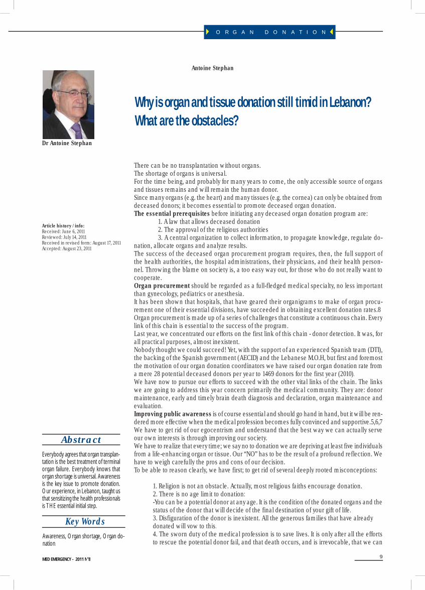
All resuscitations were led by an emergency physi-cian. During the period from April to October 2007, 78 patients were resuscitated with the help of a feedback system. All patients over 18 years requiring resuscitation were included in this study. In 18 of these 78 patients, data were not usable. In 16 cases, technical problems
AED with built-in
accelerometer
such as a defective electrode or a problem in the data storage system were involved. In two cases, the system was activated too late. All these cases were excluded from the study. All cases were documented in the regis-ter of resuscitation of the German Society of Anesthesia and Intensive Care (DGAI).
The study was approved by the ethics committee of the Westphalian Wilhelms University of Münster (File:
2006-571-SF).
The feedback system consisted of an au-tomated external defibrillator (AED) with a built-in accelerometer to measure the chest compressions. This system gives during resuscitation visual feed-back about the deep compressions and gives an audible signal corresponding to a frequency of 100/minute. 16 semi-automatic defibrillators AED Pro ® type
of firm Zoll Medical, Chelmsford, MA, United States equipped with CPR-D Padz ® were used for this study. The sensor is placed on the compression point in the middle of the sternum. The analysis time and the charging of the defibrillator are specific to the device and are therefore not included in the data analysis. The data were analyzed with RescueNet Code Review 4.10 (Zoll Medical Corporation, Chelmsford, MA, United States), Microsoft Office Excel (Microsoft Corporation, Redmond, WA) and SPSSversion 13.0.1 (SPSS Inc., Chicago, Illinois, United States, 2004). Based on the work of Wik and colleagues [15,16] our algorithm provides an intensive initial period of cardiopulmonary resuscitation during 3 minutes. Every 3 minutes, the de-fibrillator gives a signal interruption of chest compres-sions [6,8]. During this pause, ECG analysis takes place followed by defibrillation if needed. All paramedics and doctors were previously familiar with the system. The first 3 cycles of each resuscitation (3 minutes each time followed by an ECG analysis) have been analyzed. The data analyzed included the frequency and depth of chest compressions and the time of circulatory arrest in each cycle. Statistical analysis was performed using SPSS version 13.0.1 (SPSS Inc., Chicago, Illinois, 2004). The frequency of chest compressions, depth and time Demonstration of a resuscitation
12 MED EMERGENCY - 2011 No8
C . P . R .
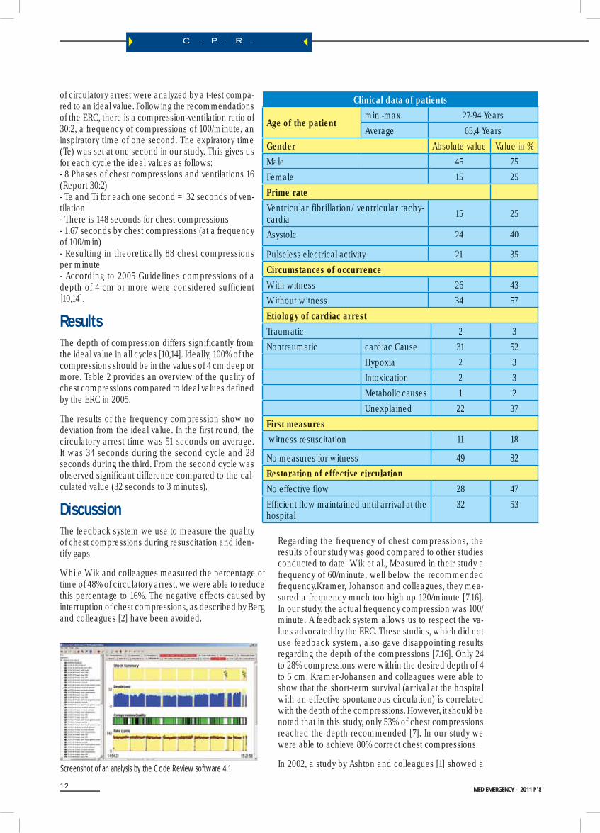
Clinical data of patients
Age of the patientmin.-max. 27-94 Years Average 65,4 Years
Gender Absolute value Value in %Male 45 75Female 15 25Prime rateVentricular fibrillation/ ventricular tachy-cardia 15 25
Asystole 24 40
Pulseless electrical activity 21 35Circumstances of occurrenceWith witness 26 43Without witness 34 57Etiology of cardiac arrestTraumatic 2 3Nontraumatic cardiac Cause 31 52
Hypoxia 2 3Intoxication 2 3Metabolic causes 1 2Unexplained 22 37
First measures witness resuscitation 11 18
No measures for witness 49 82Restoration of effective circulationNo effective flow 28 47Efficient flow maintained until arrival at the hospital
32 53
of circulatory arrest were analyzed by a t-test compa-red to an ideal value. Following the recommendationsof the ERC, there is a compression-ventilation ratio of 30:2, a frequency of compressions of 100/minute, aninspiratory time of one second. The expiratory time(Te) was set at one second in our study. This gives us for each cycle the ideal values as follows:- 8 Phases of chest compressions and ventilations 16(Report 30:2)- Te and Ti for each one second = 32 seconds of ven-tilation- There is 148 seconds for chest compressions- 1.67 seconds by chest compressions (at a frequency of 100/min)- Resulting in theoretically 88 chest compressionsper minute- According to 2005 Guidelines compressions of adepth of 4 cm or more were considered sufficient [10,14].
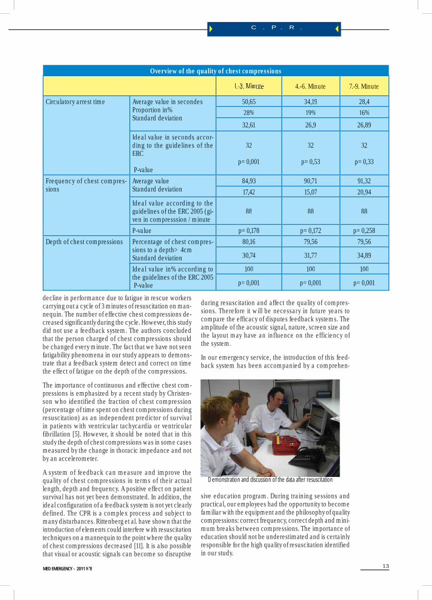
ResultsThe depth of compression differs significantly from the ideal value in all cycles [10,14]. Ideally, 100% of the compressions should be in the values of 4 cm deep or more. Table 2 provides an overview of the quality of chest compressions compared to ideal values defined by the ERC in 2005.
The results of the frequency compression show no deviation from the ideal value. In the first round, the circulatory arrest time was 51 seconds on average.It was 34 seconds during the second cycle and 28 seconds during the third. From the second cycle was observed significant difference compared to the cal-culated value (32 seconds to 3 minutes).
DiscussionThe feedback system we use to measure the quality of chest compressions during resuscitation and iden-tify gaps.
While Wik and colleagues measured the percentage of time of 48% of circulatory arrest, we were able to reducethis percentage to 16%. The negative effects caused by interruption of chest compressions, as described by Bergand colleagues [2] have been avoided.
Screenshot of an analysis by the Code Review software 4.1
Regarding the frequency of chest compressions, theresults of our study was good compared to other studiesconducted to date. Wik et al., Measured in their study afrequency of 60/minute, well below the recommendedfrequency.Kramer, Johanson and colleagues, they mea-sured a frequency much too high up 120/minute [7.16].In our study, the actual frequency compression was 100/minute. A feedback system allows us to respect the va-lues advocated by the ERC. These studies, which did notuse feedback system, also gave disappointing resultsregarding the depth of the compressions [7.16]. Only 24to 28% compressions were within the desired depth of 4to 5 cm. Kramer-Johansen and colleagues were able toshow that the short-term survival (arrival at the hospitalwith an effective spontaneous circulation) is correlatedwith the depth of the compressions. However, it should benoted that in this study, only 53% of chest compressionsreached the depth recommended [7]. In our study wewere able to achieve 80% correct chest compressions.
In 2002, a study by Ashton and colleagues [1] showed a
13MED EMERGENCY - 2011 No8
C . P . R .
Overview of the quality of chest compressions
4.-6. Minute 7.-9. Minute
Circulatory arrest time Average value in secondesProportion in% Standard deviation
50,65 34,19 28,428% 19% 16%
32,61 26,9 26,89
Ideal value in seconds accor-ding to the guidelines of the ERC
P-value
32
p=0,001
32
p=0,53
32
p=0,33
Frequency of chest compres-sions
Average valueStandard deviation
84,93 90,71 91,3217,42 15,07 20,94
Ideal value according to the guidelines of the ERC 2005 (gi-ven in compresssion / minute
88 88 88
P-value p=0,178 p=0,172 p=0,258Depth of chest compressions Percentage of chest compres-
sions to a depth> 4cm Standard deviation
80,16 79,56 79,56
30,74 31,77 34,89
Ideal value in% according to the guidelines of the ERC 2005P-value
100 100 100
p=0,001 p=0,001 p=0,001
decline in performance due to fatigue in rescue workers carrying out a cycle of 3 minutes of resuscitation on man-nequin. The number of effective chest compressions de-creased significantly during the cycle. However, this study did not use a feedback system. The authors concluded that the person charged of chest compressions shouldbe changed every minute. The fact that we have not seen fatigability phenomena in our study appears to demons-trate that a feedback system detect and correct on time the effect of fatigue on the depth of the compressions.
The importance of continuous and effective chest com-pressions is emphasized by a recent study by Christen-son who identified the fraction of chest compression (percentage of time spent on chest compressions during resuscitation) as an independent predictor of survival in patients with ventricular tachycardia or ventricular fibrillation [5]. However, it should be noted that in this study the depth of chest compressions was in some cases measured by the change in thoracic impedance and not by an accelerometer.
A system of feedback can measure and improve the quality of chest compressions in terms of their actual length, depth and frequency. A positive effect on patient survival has not yet been demonstrated. In addition, the ideal configuration of a feedback system is not yet clearly defined. The CPR is a complex process and subject to many disturbances. Rittenberg et al. have shown that the introduction of elements could interfere with resuscitation techniques on a mannequin to the point where the quality of chest compressions decreased [11]. It is also possible that visual or acoustic signals can become so disruptive
during resuscitation and affect the quality of compres-sions. Therefore it will be necessary in future years tocompare the efficacy of disputes feedback systems. Theamplitude of the acoustic signal, nature, screen size andthe layout may have an influence on the efficiency of the system.
In our emergency service, the introduction of this feed-back system has been accompanied by a comprehen-
sive education program. During training sessions andpractical, our employees had the opportunity to becomefamiliar with the equipment and the philosophy of quality compressions: correct frequency, correct depth and mini-mum breaks between compressions. The importance of education should not be underestimated and is certainly responsible for the high quality of resuscitation identifiedin our study.
Demonstration and discussion of the data after resuscitation

14 MED EMERGENCY - 2011 No8
C . P . R .
Conflict of interest statement:There is no conflict of interest to declare
PerspectivesThe quality of chest compressions has so far not been studied in large scale, partly due to the lack of an ade-quate measurement technology [4.13]. The concept of feedback presented here should be a starting point for a more efficient design of future preclinical studies of resuscitation (e.g. on the effectiveness of certain drugs). The bias introduced by a poor quality chest compres-sions, or by long pauses between compressions could well be identified prior to false results. This is particularly important for multicenter studies.
The department managers can use these feedback systems to evaluate the quality of work in the field. As part of debriefing, this data can be used to improve the performance of individual players. Other technological advances to come,as the analysis or defibrillation without interruption of chest compressions in the future will decrease the time of circulatory arrest [9].
ConclusionThe use of technology feedback (with metronome givingan audible signal at a frequency of visual feedback 100/minute and a depth of compressions) has made possiblea correct depth of chest compressions in this study, andled to an improvement in the quality of compressionsfrom previous studies.
Transparent presentation of the quality of chest compres-sions allows more efficient studies. The effective use of technology still requires a feedback training of doctorsand paramedics particularly careful.
AcknowledgmentsThe authors thank the leadership of the fire service of Münster, and Messrs. B. Fritzen and W. Reckert for their effective assistance in this study. This work would nothave been possible without the collaboration of L. Decker,F. Duesmann, P. Mombaur and D. Schwichtenhövel (Fireservice in the city of Münster). Our gratitude also goes tothe fire service ambulances in the city of Münster as wellas emergency physicians.
REFERENCES
1. - Ashton et al. (2002). Effect of rescuer fatigue, one performance of continuous external chest compressions over 3 minutes. Resuscitation 55: 151-155.2. - Berg RA, Sanders AB, Kern KB et al. (2001). Adverse hemodynamic effects of chest compressions for rescue Interrupting breathing cardio-pulmonary resuscitation for ventricular DURING fibrillation cardiac arrest.Circulation 104: 2465-2470.3. - Bohn A, Gude P (2008). Feedback cardiopulmonary resuscitation DURING. Curr Opin Anaesthesiol 21: 200-203.4. - Böttiger BW, Arntz HR, Wenzel V; TROICA Trial Investigators, European Resuscitation Council Study Group (2009). Thrombolysis DURINGresuscitation for out-of-hospital cardiac arrest. New Engl J Med. 359: 2651-62.5. - Christenson J, Berg R, Resuscitation Outcomes Consortium Investigators.(2009) Chest compression fraction survival in patients with DE-TERMINED out-of-hospital ventricular fibrillation. Circulation 120:1241-7.6. - Deakin CD, Nolan JP (2005). European Resuscitation Council Guidelines for Resuscitation 2005. Section 3. Electrical therapies: automatedexternal defibrillators, defibrillation, cardioversion and pacing. Resuscitation 67 [Suppl 1]: S25-S37.
Andreas BOHN 1, 2, Christiane RESING1, Thomas P. WEBER3R , Ulf HARDING1,Roman P. LUKAS1
1. Department of Anesthesiology and Intensive Care, University Hospital Münster, York-Ring 25, 48159 Münster - Germany
2. Medical Director of Emergency Service, Fire service in the city of Münster – Germany
3. Department of Anesthesiology, Hospital of the Catholic University of Bochum, Germany
Email: [email protected]
The editorial board
What we knew? The quality of chest compressions depends on the survival of the victim. The depth of the depression must be 4 cm, and pacearound 100/minute. The latest international recommendations (2010) confirm this.
What brings this article? A feedback device on the gestures performed facilitates the performance and observance of the recommendations. Thissystem could also be used for future studies of resuscitation, particularly on the provision of certain drugs.
15MED EMERGENCY - 2011 No8
C . P . R .
7. - Kramer-Johansen J et al. (2006). Quality of out-of-hospital cardiopulmonary resuscitation with real time automated feedback: A prospectiveinterventional study. Resuscitation 71: 283-292.8. - Kramer-Johansen J, Edelson DP, Abella BS et al. (2007). Pauses in chest compression and Inappropriate shocks: a comparison of manualand semi-automatic defibrillation attempts. Resuscitation 73: 212-220.9. - Lloyd MS, Heeke B, Walter PF et al. (2008). Hands-on defibrillation: an analysis of electrical current flow-through direct contact with rescuersin biphasic external defibrillation DURING patients. Circulation 117: 2510-2514.10. - Nolan JP, Deakin CD, Soar J et al. (2005). European Resuscitation Council Guidelines for resuscitation. Section 4. Adult advanced lifesupport.Resuscitation 67 [Suppl 1]: S39-S86.11. - Rittenberg JC, Guimond G, Platt TE, et al. (2006). Quality of BLS resuscitation Decrease with Increasing Complexity. Resuscitation 68: 365-369.12. - Lukas R, C Sengelhoff, Döpke S, Harding G, Mertens P, Osada N, Van Aken H, Weber TP, Bohn A. (2010). Thoraxkompressionsqualität -Feedback Hilft-Technology? Anaesthesist 59 (2): 135-9.13. - Wenzel V, Krismer AC, Arntz HR, Sitter H, Stadlbauer KH, Lindner KH (2004). European Resuscitation Council Cardiopulmonary Resus-citation vasopressor DURING Study Group. A comparison of vasopressin and epinephrine for out-of-hospital cardiopulmonary resuscitation.N Engl J Med 350: 105-113.14. - Wenzel V, Russo S, Arntz HR (2006). European Resuscitation Council [The new 2005 resuscitation guidelines of the European ResuscitationCouncil: comments and supplements] Anaesthesist 55: 958-966, 968-972, 974-979.15. - Wik L et al. (2003). Delaying defibrillation to Give cardiopulmonary resuscitation basic to patients with out-of-hospital ventricular fibrilla-tion: a randomized trial. JAMA 289: 1389-1395.16. - Wik L et al. (2005). Quality of cardiopulmonary resuscitation DURING out-of-hospital cardiac arrest. JAMA 293: 299-304.
Congress

SAVING LIVES IS OUR AIM
E RE RE RE RR C CCC C OC OOC OO N GN GNNN R ER ER ER EE S SS SS SS S
JJJJJJoooin us: iiiinfffoooo@@@@@@lllllleeeeeebbbbbbrrrrrrcccccc...ooooorgggggg
FoFoFooooooooollllowowininininng g g g onononono t t t ttheheheheeee 1 1110t0t00th h hh on Council Congress in Porto 2010EuEuE roropepepepeanana R RRRResesesesususususcicicic tatatataatitititionooon C CCCouoouo ncn il CCongrgress s in P Poroo toto 2 20101010 and the aandnd theheee ScientiScScientntti c Sc c SySySySyyympmpmpmppososososiuiuiuium m mmm inininininnnnn C C C C CCCCCCCCololoo ogogggggnenenenene 2 2 2 20000000099, , ,,ththhe e ERERERE C C CC inininini vivivv tetetetes s yoyoyoyou u uu tototoo iiiitsts s symymyyy popop sis umum o o n 141411 -1-1-1-15 55 5 OcOcOcOctotobebebeber r r 20201111 iin n VaVaaalllll etettatatata, , , MaMaMaMaMM ltlttl a.a.a.a.
PrPrPrPrelelimimmininnnararary y y y yyy yyy Sccieeeentntntntiiii c c cc PrPrPrPrrogogogograraaammmmmmmmm e:e:e: https://congress2011.erc.edu
MeMeMeMeegagagagalilil thths,s, mmededievaval l ll dudududungn eoeoeoonssnss aandndnnnnnnnn CC Calalalalypypypypsosooso’s’sss C CCCCavavvve e ee - - - ththththt e eee MaMaMaMaMaM ltltltltl esesesese e e ee IsIsIslalalal ndndndnddds s ss arararaaaaare eee mymymym ththhhicicicic. .ThThe e naarrowow m meaeandndererere ining g gg ststrereetets s ofofofofo t theheheheiririri vvilililillalal gees ss araraaa e e crccrowowwdededed d dddd wiwiwiwiththhthth ccccatatatathehehehedrdrdrdrrralalals s ananand d papalalacecec s.s.s..
more about Malta: www.visitmalta.com
onoonononoooo CCCCououououncncn ilil CCCCConongrgrggrgrg esesese ss inn PPPPorororortototot 222201010101000 aaaandndnddnd ttthehehehe ScScScScieiei ntntntn iiii cc SSSS
What is resuscitation 2011?

17MED EMERGENCY - 2011 No8
Article history / info:Received: Jan 10, 2011Reviewed: May30, 2011Received in revised form: Aug 12, 2011Accepted: Aug 23, 2011
IntroductionShoulder dislocations account for almost 50% of all joint dislocations, which are most com-monly anterior (90-98%) and occur due totrauma. Different techniques are commonly used to reduce the dislocation [1]. A new method, the Spaso technique, has been ad-vocated to be a simple and safe procedureto reduce shoulder dislocations and is explai-ned with detail in recent reviews on shoulder dislocation reduction techniques [2,3]. Howe-ver, since the first serie by Miljesic and Kelly [4], the results on the use of this techniquehave been published only in one retrospec-tive serie [5], two prospective series in spa-nish lenguaje journals [6,7] one prospectivestudy [8], and few case reports [9,10].
The literature lacks of prospective studiesevaluating the Spaso technique. In the pre-sent article, we present our results with the ini-tial experience using the Spaso technique in an orthopaedic trauma emergency department of a level-1 hospital [11], and review the literature.
MethodsAll patients who underwent reduction of an anterior shoulder dislocation using the
Spaso technique at Hospital Clínic of Barce-lona Orthopaedic Emergency Department, between January 2007 and May 2007 were the subjects of this study. In this period the Spaso technique was applied in 36 anterior shoulder dislocations.
The inclusion criteria were (1) anterior shoulder dislocation, (2) presentation within 24 hours after the dislocation, (3) no associated frac-tures of the shoulder detectable on routine radiographic examination, except for a greater tuberosity isolated fracture.
Two of the patients were excluded due to a presentation later than 24 hours after the dis-location, leaving a total of 34 dislocations in 33 patients. They occurred in 22 male and 12 female shoulders. The patients ages ranged from twenty-one to eighty years (average, 51 years).
After routine radiographic examination (ante-roposterior, and scapular Y views), the shoul-
der dislocation was reduced using the Spaso technique as follows: the patient was placed in the supine position; the affected arm was
grasped around the wrist or distal forearm and gent-ly lifted vertically, applying gentletraction. While maintaining ver-tical traction, the sh o u l d e r w a sslightly externally rotated (Fig.1) [4].The manoeuvre was applied du-ring a maximum of 3 minutes.
The possibility of using sedation or regional anaesthesia depended on the clinician crite-ria. Exhaustive post-reduction clinical exam was done in order to check any neurological or vascular dysfunction in relation with the reduction.
The demographic data, aetiology, sedation used, history of previous shoulder disloca-tions, successful of the attempt, the degree of the clinician (orthopaedic faculty or resident), complications or any comment from the emer-gencies staff were collected.
Results The results are summarized in the table 1. They were 21 right anterior shoulder dislocations and 13 left, being the casual fall the most fre-quent aetiology (61%). In most of the cases (65%) an oral sedative treatment was admi-nistered previously to the manoeuvre (22/34). A total of 21 patients received oral Diazepam medication, using dose varied from 5 to 10 mgr. sublingual. In two cases this medication was supplemented with NSAIDs (desketoprofen 25 mg). In one case, regional anesthesia using interescalenic block was performed.
The success rate in the present serie using the Spaso technique has been of 67.6% (23 of 34 dislocations). If just those patients with previous shoulder dislocations were taken into account the success rate increased to 83%.
T R A U M A
AbstractThe Spaso technique has been recently described as a new, simple and effectivemanoeuvre for reducing anterior shoul-der dislocation. However there are justfew series in the literature. The aim of thepresent article is to review the results ob-tained with this procedure, and to reportour inicial experience.
Key Words shoulder, dislocation, Spaso.
Figure 1: vertical traction, external FFFFFiiFFFFii 1 iiii ll ii l rotationiiiii
The Spaso technique for reducing shoulder dislocations
Jenaro A. Fernández-Valencia, J. CUÑÉ, E. MuÑoz, Ll. Font .
Jenaro A. Fernández-Valencia

18MED EMERGENCY - 2011 No8
Conflict of interest statement:There is no conflict of interest to declare
From the 34 anterior shoulder dislocations 15 attempts were done by residents. Residents obtained a better success rate than the ortho-paedic faculty: 73% and 63% respectively.
The Spaso technique failed in 11 shoulders, which were reduced using other manoeuvre. Namely, 7 shoulders with Milch method, 3 shoulders with Hippocratic
method and 1 shoulder with Kocher method. After the reduction, no complications were registered. However, in two cases (case 8 and 34) a back discomfort was reported by the clinician after applying the manoeuvre. No other comments were collected.
Discussion The success rate using different reduction ma-noeuvres ranges from 50 to 94%. The Spaso technique is subject of an increasing interest in the literature. Canillas del Rey et al, from Spain, published in 2007 a success rate of 91,3% over a total of 21 shoulder dislocations [7]. On his part, Ugras AA et al, from Turkey, have recently re-ported a sucess rate of 75% of a total of 52 cases [8]. Summarizing the series dealing with the Spaso technique, the success rate ranges from 75% [8] to 91,4% [6]. Our initial experience showed rather worse results (67,6%) [11]. It is possibly related to the learning courve. In this way, Ugras et al showed an increase of success from 75% to 87,5% excluding their initial 20 cases. On the other hand, we evaluated the relationship of the phisician experience and, interestingly, young residents obtained better results, indicating the facility to be learnt.
The originality of the technique has been ques-tioned [6]. Janecky et al reported a forward elevation technique with satisfactory results in a report of 50 cases in 1982 [12].
Waldron reported satisfactory results with the flexion of the elbow and traction gasping the distal humerus with a forward elevation in 1991 [13]. The Spaso manoeuvre, published in 1998, could be considered a variation of the two previous described techniques. It can be also considered the mirror technique of the Stimson method, described the 1900, which is performed prone using the gravity-depen-dent forward flexion at the shoulder of the patient [14]. In any case, the positioning of the muscles, along with the axis of the applied traction, is a rationale to expect easy reduc-tions and few associated complications such
as fractures or neuroapraxia.
Described in previous reports, it has been sta-ted that “a clunk is heard or felt when
reduction is done” [5]. Despite not registeredin our study, we agree with Trueba et al [6]that the reduction may be quite subtle, withoutthe classic “click”, and may be missed by theoperator and the patient.
Some aspects about our serie have to be com-mented. On one hand, the possible discomfortexperienced by the clinician has not been eva-luated previously. It has been reported relatedto reduction of dislocations of the hip, especial-ly when the reduction is performed by a singleperson [15]. In our study, a transient back painwas experienced in 2 orthopaedic faculties,despite the limited time given for the technique(3 minutes). In both cases the patients wereheavily built and the faculties did not recall any previous spinal disorder. We consider that thiscould be an inconvenient for the general useof this technique.
Our serie aimed the free inclusion of patientsby the clinician on emergency, but not all theclinicians collaborated. The exact number of patients that presented the inclusion criteriabut were not included, could not be determi-ned. This could be a limitation due to possibleselection bias. However, all of the cliniciansthat collaborated in the study performedalways this technique during the study period.Another limitation of the serie was the arbitrary use of sedation. Although it is recognized a fa-vourable effect to success with reduction [16],some authors consider that it is not neededif the reduction technique is relatively gentleand painless [17]. We consider that the phar-macologic treatment has to be tailored to thepatients, and some patients do not need it if they are able to relax and calm.
ConclusionIn our experiece, the Spaso technique hasshown lower, although acceptable, successrates than previously reported. In two cases,the manoeuvre was associated to back dis-comfort of the clinician. No complicationswere observed. We consider the Spaso tech-nique as a safe and effective method for redu-cing anterior shoulder dislocation but further studies are needed to compare these resultswith the traditionally performed techniques.
T R A U M A
Jenaro A. Fernández-Valencia,J. Cune, E. Munoz, Ll. Font
Email : [email protected] Orthopaedic Surgeon.
Department of Orthopaedic Surgery.Hospital Clínic. University
of Barcelona. Spain

19MED EMERGENCY - 2011 No8
T R A U M A
Table I. Detailed summary of the cases.
REFERENCES
[1] Riebel GD, McCabe JB. Anterior shoulder dislocation: a review of reduction techniques. Am J Emerg Med 1991;9:180-8. [2] Cunningham NJ. Techniques for reduction of anteroinferior shoulder dislocation. Emerg Med Australas 2005;17(5-6):463–71. [3] Ufberg JW, Vilke GM, Chan TC, et al. Anterior shoulder dislocations: beyond traction-countertraction. J Emerg Med 2004;27:301-6. [4] Miljesic S, Kelly A. Reduction of anterior dislocation of the shoulder: the Spaso technique. Emergency Medicine 1998;10:173-5. [5] Yuen MC, Yap PG, Chan YT, et al. An easy method to reduce anterior shoulder dislocation: the Spaso technique. Emerg Med J 2001;18:370-2.[6] Trueba C, Pozzo A, Gil F, et al. Flexión y rotación externa para reducir la luxación anterior del hombro: Técnica de Spaso. Acta Orto pédica Mexicana 2004; 18(1):18-20. [7] Canillas del Rey FM, Carballo F, Nieto D, González-Criado F. La técnica de Spaso para reducción de la luxación anterior de hombro. Revista Española de Cirugía Osteoarticular 2007;(42): 61-6.[8] Ugras AA, Mahirogullari M, Kural C, Erturk AH, Cakmak S. Reduction of anterior shoulder dislocations by Spaso technique: clinical results. J Emerg Med. 2008 May;34(4):383-7.[9] Yuen MC, Tung WK. The use of the Spaso technique in a patient with bilateral dislocations of shoulder. Am J Emerg Med 2001;19:64-6.[10] Singh S, Kumar S. Bilateral anterior shoulder dislocation: A case report. Eur J Emerg Med 2005;12:33-5. [11] Fernández-Valencia JA, Cuñe J, Casulleres JM, Carreño A, Prat S. The Spaso technique: a prospective study of 34 dislocations. Am J Emerg Med 2009: 27, 466–469.[12] Janecki CJ, Shahcheragh GH. The forward elevation maneuver for reduction of anterior dislocations of the shoulder. Clin Orthop Relat Res 1982;164:177-80.[13] Waldron VD, Hazel D. Tips of the trade #37. Technique for reduction of shoulder dislocation. Orthop Rev 1991;20:563-6.[14] Stimson LA. An easy method of reducing dislocations of the shoulder and hip. Med Rec 1900;57:356-7.[15] Schafer SJ, Anglen JO. The East Baltimore Lift: A simple and effective method for reduction of posterior hip dislocations. J Orthop Trauma 1999;13:56-7.[16] Dunn MJG, Mitchell R, Souza CD, et al. Evaluation of propofol and remifentanil for intravenous sedation for reducing shoulder disloca tions in the emergency department. Emerg Med J 2006;23:57-8.[17] O’Connor DR, Schwarze D, Fragomen AT, et al. Painless reduction of acute anterior shoulder dislocations without anesthesia. Or thopedics 2006;29(6):528-32.
Age, sex, sidePrevious
dislocation (number)
Treatment Associated great tuberosity fracture Clinician degree Successcase
23456789101112131415161718 19202122232425262728293031323334
72,M,R79,M,R38,M,L61,F,R22,M,L38,M,R80,F,R51,F,L32,M,L68,F,R72,F,R63,F,L75,F,R29,M,R63,F,R23,M,R28,M,R27,M,L
27,M,L *26,M,R54,F,R62,F,R75,M,L77,M,L78,F,L58,M,R42,M,L36,M,L21,M,R24,M,L52,M,R68,F,R51,M,R57,M,R
NONO
YES(3)NO
YES(5)NONONO
YES(5)NONONONO
YES(1)NONO
YES(4)YES(8)YES(9)
NOYES(3)
NONONONO
YES(2)NO
YES(5)NO
YES(1)NONONO
YES(1)
DiazepamNONONO
Diazepam+NAIDSInterescalenic A.
DiazepamDiazepam
NONO
DiazepamNO
Diazepam+NAIDSDiazepam
NODiazepamDiazepamDiazepamDiazepamDiazepamDiazepamDiazepamDiazepamDiazepamDiazepam
NODiazepamDiazepam
NONONONO
DiazepamDiazepam
YESNONONONONONONONONONONOYESNONONONONONONONONONONOYESNONONONONONONONONO
OFOFR
OFR
OFOFR
OFOFRRRR
OFRRRR
OFOFOFOFOFOFR
OFR
OFOFR
OFOFR
NOYESYESYESYESNONOYESYESYESNOYESYESYESNONOYESYESYESYESYESYESYESYESNONOYESNOYESYESYESNONOYES

20 MED EMERGENCY - 2011 No8
21MED EMERGENCY - 2011 No8
Article history / info:Received: Jan 21, 2011Reviewed: May 13, 2011Received in revised form: June 6, 2011Accepted: July 10, 2011
T R A U M A
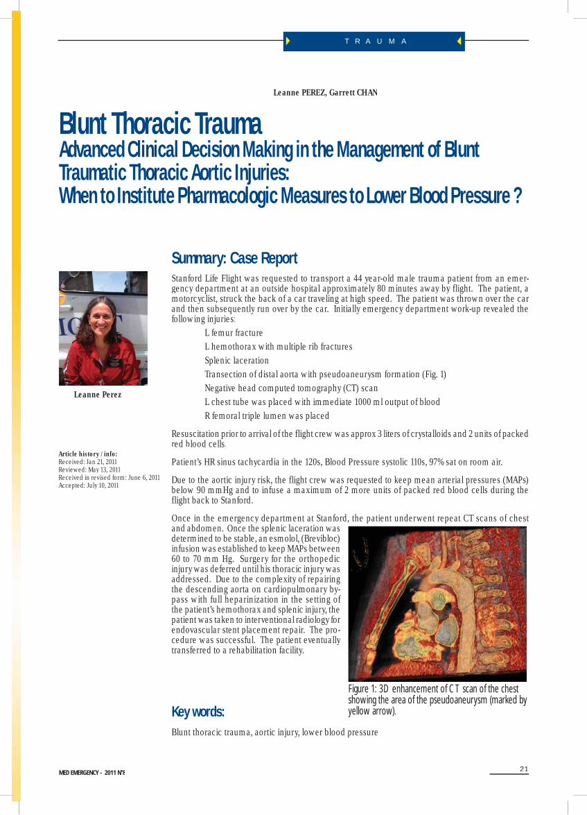
Summary: Case Report Stanford Life Flight was requested to transport a 44 year-old male trauma patient from an emer-gency department at an outside hospital approximately 80 minutes away by flight. The patient, amotorcyclist, struck the back of a car traveling at high speed. The patient was thrown over the car and then subsequently run over by the car. Initially emergency department work-up revealed thefollowing injuries: L femur fracture L hemothorax with multiple rib fractures Splenic laceration Transection of distal aorta with pseudoaneurysm formation (Fig. 1) Negative head computed tomography (CT) scan L chest tube was placed with immediate 1000 ml output of blood R femoral triple lumen was placed
Resuscitation prior to arrival of the flight crew was approx 3 liters of crystalloids and 2 units of packedred blood cells.
Patient’s HR sinus tachycardia in the 120s, Blood Pressure systolic 110s, 97% sat on room air.
Due to the aortic injury risk, the flight crew was requested to keep mean arterial pressures (MAPs)below 90 mmHg and to infuse a maximum of 2 more units of packed red blood cells during theflight back to Stanford.
Once in the emergency department at Stanford, the patient underwent repeat CT scans of chestand abdomen. Once the splenic laceration was determined to be stable, an esmolol, (Brevibloc) infusion was established to keep MAPs between 60 to 70 mm Hg. Surgery for the orthopedic injury was deferred until his thoracic injury was addressed. Due to the complexity of repairing the descending aorta on cardiopulmonary by-pass with full heparinization in the setting of the patient’s hemothorax and splenic injury, the patient was taken to interventional radiology for endovascular stent placement repair. The pro-cedure was successful. The patient eventually transferred to a rehabilitation facility.
Key words: Blunt thoracic trauma, aortic injury, lower blood pressure
Blunt Thoracic TraumaAdvanced Clinical Decision Making in the Management of Blunt Traumatic Thoracic Aortic Injuries: When to Institute Pharmacologic Measures to Lower Blood Pressure ?
Leanne PEREZ, Garrett CHAN
Leanne Perez
Figure 1: 3D enhancement of CT scan of the chestshowing the area of the pseudoaneurysm (marked by yellow arrow).

T R A U M A
MED EMERGENCY - 2011 No822
IntroductionThe true incidence of blunt traumatic aortic injury in the United States is unknown, but has been estimated to be 7,500 to 8,000 cases each year.1,2 Based on several autopsy reports, aortic ruptureoccurs in 12% to 23% of deaths from blunt trauma.3-7 It is the second most common cause of deathin blunt trauma patients second only to head injury.8 Anywhere from 70 to 90% of patients will die on scene or while enroute to the hospital from a complete laceration of the aortic wall.3,7-9 Survivorsthat reach the emergency department usually can be classified into two groups. The hemodyna-mically unstable group has a 2% chance of survival and most of these deaths occur due to a rapid progression of a partial aortic tear to a state of free intrapleural rupture before surgical repair can be achieved.3,10 The hemodynamically stable group can have up to a 90% survival rate with promptdiagnosis, medical management and operative repair. However the risk of a less significant aortic tear progressing to a catastrophic rupture increases over time.8,9 11-14 One of the key elements in early medical management is to institute pharmacologic strategies to lower mean arterial pressures (MAPs) thus blunting and/or preventing this progression to a catastrophic rupture.13, 15-19 Once the diagnosisof BTAI is made, the medical management strategies may be counterintuitive in the trauma setting, because of the initial priority of fluid resuscitation to the lowering of MAPs through pharmacologic interventions.
Aortic Structure and FunctionAn understanding of the basic anatomy of the aorta and the physiology of blood flow are impor-tant concepts to understand in order to appreciate the theoretic premise behind the importance of blood pressure control in this patient popula-tion. The aorta is the largest blood vessel in the human body. It distributes blood from the heart to the circulatory system. It is an elastic artery and is composed of three layers called tunicas. The tunica intima is a thin layer of cells lining the lumen of the vessel, which puts it in direct contact with blood as it flows through the vessel. The middle layer or tunica media is the thickest and most unique part of the aorta. It provides elasticity and tensile strength to the aortic wall. The elastic nature of the aorta is important in maintaining an even blood pressure and in keeping blood flowing smoothly through the vascular system. During systole, the aorta stretches (distends and thins) to accommodate blood ejected by the left ventricle. The stretched aorta has “stored” or potential energy that is released (elastic recoil) during diastole to maintain driving pressure. This pressure wave keeps blood flowing continuously through the circulation.20The outermost layer of the aorta is the tunica adventitia. This layer limits elasticity of the media layer and is the major force of resistance in the aortic wall.21
The degree of aortic tear is a continuum from suboptimal hemorrhage to total aortic rupture.22Theseverity and progression of the aortic tear is undoubtedly the key factor in the likelihood of rupture. Those with full-thickness tears contained only by the mediastinal pleura and false aneurysms which are confined by a thin layer of adventitia and the mediastinal pleura (Fig. 2) likely represent the patients presenting in extremis or those who develop spontaneous rupture.8
Mechanism of injuryThe highest incidence of BTAI (72 to 87%) occurs with victims of motor vehicle collisions with frontal and lateral impacts occurring with approximately equal frequency, followed by falls and pedestrian/vehicular incidents.2,4,5,8 23-27 There is an enormous transfer of energy involved in BTAI. The area of the aorta most commonly injured is in the region of the isthmus. Despite extensive investigation,
f
y
Figure 2: Different Aortic Lesions
23MED EMERGENCY - 2011 No8
T R A U M A
analysis and debate, no consensus or unified agreement as to the exact mechanism of injury in BTAI has emerged.2 Four common theories regarding how BTAIs occur have been postulated in theliterature. First, the predominant postulated theory is that the aorta is at greatest risk from shearing forces (torque and compression) from sudden deceleration. Aortic injury occurs when the rate of deceleration on the relatively mobile aortic arch, differs from that of the relatively fixed descending aorta.22,29-32 A second theory is combined shearing and bending stress where the aorta is injuredfrom deceleration forces and bending stress occurring as the aorta is flexed over the left pulmonary artery and left main stem bronchus.22,29-32 A third theory is a sudden increases in intra-luminal aorticpressure at the moment of impact only can produce the energy to tear the aortic wall.22,33 Lastly, amore recent theory of the forces producing injury at the aortic isthmus is the osseous pinch. The hypothesis is that anterior thoracic osseous structures (manubrium, clavicle and first rib) rotate posteriorly and inferiorly about the axes of the posterior rib attachments. When the force is large enough, these anterior bony structures impact the vertebral column and the portion of the aorta overlying the spine (the isthmus and proximal descending aorta) is pinched between the bones.34,
35 Clinically BTAI involves the ascending aorta about 5% of the time, yet they yield higher mortality rates because of complications of associated injuries such as aortic value rupture, coronary artery laceration and hemopericardium with cardiac tamponade.9
Physical FindingsClinical signs of traumatic aortic injury are rarely present and can be physiologically silent until rup-ture. The diagnosis is based on a high index of suspicion with the mechanism of injury and results of imaging studies.36 Fewer than 50% of BTAI patients manifest in with specific signs or symptoms.28
Less than half of patients have visible signs of chest injury.15,37,38 Clinical signs and symptoms areoften unreliable and variable making it difficult to diagnosis.39 A widened mediastinum has been the most frequently cited chest radiograph (CXR) finding that triggers additional work-up for BTAI.25 However CXR findings have been interpreted as normal at the time of initial evaluation in 2% to 40% of patients with traumatic aortic injury, therefore patients with significant deceleration or acceleration mechanisms with a negative CXR should undergo further screening tests, such as CT angiography of the chest or if available helical or spiral CT.2,19,23,38,40-51
Initial ManagementThe initial management of a multiple-trauma patient is no different with or without suspected aortic disruption. The patient’s airway, breathing, and circulation are addressed first. Primary and secondary surveys are completed. Appropriate venous access is obtained concomitant with initial laboratory and radiographic studies. The priority in the management of hemodynamically unstable patients with potential aortic injury is to rapidly identify and control on-going hemorrhage from other sites and to avoid over-resuscitation. (This cannot be overemphasized; excessive resuscitation resulting in unnecessary blood pressure rise can cause extension of the aortic tear, which can lead to a fatal rupture.) Sites of concealed hemorrhage are identified with chest and pelvic radiographs and fo-cused assessment with sonography for trauma ultrasound (FAST) and/or diagnostic peritoneal lavage with follow-up appropriate CT scans.36 Most patients with aortic disruption also have one or morefractures. These should be stabilized but not definitively treated prior to excluding the diagnosis of aortic rupture or treating an aortic rupture.28
Once the diagnosis of BTAI is made, prompt repair of the blunt TBAI is preferred. However imme-diate repair may not be possible in all patients. If the patient has more immediately life threatening injuries that require intervention such as emergent laparotomy or craniotomy, or if the patient is a poor operative candidate due to age or comorbidities, the aortic repair may be delayed. If a hospital does not have the capability to manage patients with BTAI, outcomes are improved with transfer to a facility that manages these patients routinely. Pharmacologic control of blood pressure with beta-blockers and/or nitroprusside is extremely important when there is a delay to surgery or the patient has been deemed a non-operative candidate.13, 15-19 This is a level II recommendation, according tothe Eastern Association for the Surgery of Trauma.52

24MED EMERGENCY - 2011 No8
T R A U M A
Theory behind medical managementOnce initial stabilization is completed and any acute active bleeding is controlled, BTAI shouldbe treated using a medical management approach. Medical management revolves aroundpreventing progression of any aortic tear to a catastrophic rupture that leads to exsanguinationsand death. The goal is to minimize or blunt the “pounding-type” stress placed on the weake-ned aortic wall during systolic blood flow. Specifically attenuating the rapid rise of the aorticpressure wave during systole(dP/dT). Just as in the pharmacologic treatment of aortic dissection,dP/dT is best controlled by decreasing the force of left ventricular ejection with beta-blockagetherapy (negative inotrope). The mean aortic pressure then is controlled with a systemic arterialvasodilator.13 The goal with therapy is to lower MAPs to the lowest possible level while maintai-ning perfusion to kidneys and brain, usually MAPs between 60 to 80 mmHg. If contraindicatedMAPs should be at least controlled to prevent excessive elevation. Medical treatment shouldalways begin with beta-blockage therapy unless contraindicated, adding vasodilators as nee-ded. Maintain caution when using a vasodilator alone, for this may actually increase the dP/dTby decreasing aortic wall impedance and potentially worsening the aortic lesion. Arterial lineplacement should be considered in patients that require pharmacologic intervention to manageMAPs. If there is a disparity in blood pressure between limbs, the arterial line should be placedin the limb with the higher value. Appropriate narcotic administration for pain managementis an important adjunct but should not be the first line drug for MAP control. The decreases inheart rate and blood pressure seen with opioid use can be very beneficial in many ways for thepatient with BTAI; however, opioid use alone does not blunt the cardiac forces needed to preventprogression of the aortic tear.
Pharmacologic AgentsIdeal characteristics in pharmacologic agents used for the management of blood pressure inpatients with BTAI during the transport phase include rapid onset of action, minimal dosageadjustments and titratable to desired blood pressure.53 Beta-blocker agents are the cornerstonesfor blood pressure management in patients with BTAI. Agents that directly block the inotropicand chronotropic effects of beta1 receptors site (cardioselective beta blockers) act by loweringventricular ejection dynamics (dP/dT), thus decreasing the stress on the aortic wall.19Preferredbeta-blocker agents include esmolol and labetalol (Trandate).
Esmolol is an ultra-short acting intravenous cardioselective beta antagonist (blocking agent). Ithas an extremely short half-life (mean: 9 minutes; range 4 to 16 minutes). A quick onset of action(60 seconds) and full recovery from beta blockade is observed 18 to 30 minutes after terminationof the infusion. A loading dosed of 0.5 to 1mg/kg may be intiated, followed by an infusion at 50 to300 mcg/kg/min. Esmolol is easily titrated to response and adverse effects such as hypotensionand/or bradycardia are rapidly managed by termination of the infusion.
Labetalol is a combined alpha and beta-blocker (1:7 ratio). The onset of action occurs within2 to 5 minutes with peak effect in 5 to 15 minutes. Duration of action is 2 to 4 hours. Labetalolmay be administered as 20 mg intravenous boluses (up to 80 mg) or as a continuous infusionstarting at 1-2 mg/min to a total of 1 to 2 mg/kg.54
When beta blockade therapy is contraindicated, a vasodilator may be used. Great caution is needed when using a vasodilator only. Vasodilators cause a decrease in aortic wall impedance,thus increasing dP/dT. Nitroprusside is the most common vasodilator used to manage bloodpressure in patients with BTAI. Nitroprusside is a combined arterial and venous vasodilator. It isvery potent, acting within seconds with duration of action of 1 to 2 minutes. It is administratedas an infusion starting at 0.3 to 0.5 mcg/kg/min and titrated to desired effect. It may cause a reflex tachycardia, which may necessitate addition of a beta-blocker agent that was originally deferred due to a bradycardic heart rate. Other disadvantages of nitroprusside include decreasecerebral blood, flow and coronary steal.

25MED EMERGENCY - 2011 No8
T R A U M A
Conflict of interest statement:There is no conflict of interest to declare
Leanne PEREZ, Garrett CHANStanford Hospital & Clinics
“Stanford Life Flight”300 Pasteur Drive HG-010 - Stanford CA 94305 5246
Email: [email protected]
REFERENCES
1. Jackson DH. Of TRAs and ROCS. Chest 1984;85:585-7.2. Mattox KL. Fact and fiction about management of aortic transection. Ann Thorac Surg. 1989;48:1-2.3. Parmley L, Mattingly T, Manion W. Nonpenetrating traumatic injury of the aorta. Circulation 1958;17:1086-1101.4. Greendyke RM. Traumatic rupture of aorta; special reference to automobile accidents. JAMA 1966;195:527-530.5. Feczko JD, Lynch L, Pless JE, et al. An autopsy case review of 142 nonpenetrating (blunt) injuries of the aorta. J Trauma 1992;33:846-9.6. Dischinger P, Cowley R, Shankar B. The incidence of ruptured aorta among vehicular fatalities. Proc Assoc Adv Automot med Conf 1988;32:15.7. Smith RS, Chan FC. Traumatic rupture of the aorta: still a lethal injury. Am J Surg 1986;152:660-3.8. Fabian TC, Richardson JD, Croce MA, et al. Prospective study of blunt aortic injury: Multicenter Trial of the American association for the surgery of Trauma. J Trauma 1997;42:374-80.9. Groskin SA. Selected topics in chest trauma. Radiology 1992;183:605-17.10. Attar S, Cardarelli MG, Downing SW, et al. Traumatic aortic rupture: recent outcome with regard to neurologic deficit. Ann Thorar Surg 1996;7:959-64.11. Kim FJ, Moore EE, Moore FA, Read RA, Burch JM. Trauma surgeons can render definitive surgical care for major thoracic injuries. J Trauma 1994;36:871-5.12. Von Oppell UO, Dunne TT, DeGroot MK, Zilla P. Traumatic aortic rupture: twenty-year meta-analysis of mortality and risk of paraplegia. Ann Thorac Surg 1994;58:585-93.13. Warren RL, Akins CW, Conn AKT, et al. Acute traumatic disruption of the thoracic aorta: emergency department management. Ann Emerg Med 1992;21: 391-6.14. Mirvis SE, Shanmuganathan K, Buell J, Rodriguez A. Use of spiral computed tomography for the assessment of blunt trauma patients with potential aortic injury. J Trauma 1998;45:922-30.15. Kish MM, Behrendt DM, Orringer MB, et al. The treatment of acute traumatic rupture of the aorta: A 10-year experience. Ann Surg 1976;184:308-16.16. Akins CW, Buckley MJ, Daggett W, et al. Acute traumatic disruption of the thoracic aorta: A ten-year experience. Ann Thorac 1981;31:305-9.17. Fisher RG, Oria RA, Mattox KL, et al. Conservative management of aortic lacerations due to blunt trauma. J Trauma 1990;30:1562-6.18. Pezzella AT, Todd EP, Dillon ML, et al. Early diagnosis and individualized treatment of blunt thoracic aortic injury. Am Surg 1978;44:699-703.19. Fabian TC, Davis KA, Gavant ML, et al. Prospective study of blunt aortic injury: Helical CT is diagnostic and antihypertensive therapy reduces rupture. Ann Surg 1998;227:666-677.20. Banasik J. Cardiac function and alterations in cardiac function. In: Copstead, L Banasik, J eds. Pathophysiology. St. Louis: Elsevier Saunders, 2003:386,437.
ConclusionClinicians should have a high index of suspicion for TBAI in patients who sustain a frontal or lateral motor vehicle crash, fall, or pedestrian versus automobile mechanism of injury. Early iden-tification and surgical repair of TBAI can potentially increase the chance of survival. However, not all hospitals have qualified staff or available facilities to repair TBAI. Uncontrolled changes in blood pressure can cause further damage to the aorta. Judicious use of beta-blocker agents and/or vasodilator agents are indicated in the management of TBAI until definitive repair can be accomplished:Published in « Air Medir cal Journal ii » Vol. 27.N°3 ; 105-143.°

26MED EMERGENCY - 2011 No8
T R A U M A
21. Blood vessels. Available at: http://greenfield.fortunecity.com/rattler/46/comparison_of_types_of_vessels.htm. Accessed 2006 Jan 7.23. Creasy JD, Chiles C, Routh WD, Dyer RB. Overview of traumatic injury of the thoracic aorta. Radiographics 1997;17:27-45.24. Jenson BT. Fourteen years survival with an untreated traumatic rupture of the thoracic aorta. Am J Forensic Med Pathol 1988;9:58-9.25. Burkhart HM, Gomez GA, Jacobson LE, Pless JE, Broadie TA. Fatal blunt aortic injuries: A review of 242 autopsy cases.26. Katyal D, McLellan BA, Brenneman FD, et al. Lateral impact motor vehicle collisions: Significant cause of blunt traumatic rupture of the thoracic aorta. J Trauma 1997;42:769-72.27. Ben-Menachem Y. Rupture of the thoracic aorta by broadside impacts in road traffic and other collisions: Further angiographic obser vations and preliminary autopsy findings. J Trauma 1993;35:363-7.28. Brundage SI, Harrugg R, Jurkovich GJ, Maier RV. The epidemiology of thoracic aortic injuries in pedestrians. J Trauma 1998;45:1010-14.29. Gleason TG, Bavaria JE. Trauma to great vessels. In: Cohn LH, Edmunds LH Jr, eds. Cardiac Surgery in the Adult. New York: McGraw- Hill, 2003:1229-50.30. Driscoll PA, Hyde JA, Curzon I, Derbyshire S, Graham TR, Nicolson DA. Traumatic disruption of the thoracic aorta: A rational ap proach to imaging. Injury 1996;27:670-85.31. Beel T, Harwood AL. Traumatic rupture of the thoracic aorta. Ann Emerg Med 1980;9:483-6.32. Lundevall J. The mechanism of traumatic rupture of the aorta. Acta Pathol Microbiol Scand 1964;62:34-46.33. Sevitt S. The mechanism of traumatic rupture of the thoracic aorta. Br J Surg 1977;64:166-73.34. Smith MD, Cassidy JM, Southers S, Morris EJ, Sapin PM, Johnson SB, Kearney PA. Transesophageal echocardiography in the diagnosis of traumatic rupture of the aorta. N Engl J Med 199;332:356-62.35. Esterra A, Mattox KL, Wall MJ. Thoracic aortic injury. Semin Vasc Surg 2000;13:345-52.36. Crass JR, Cohen AM, Motta AO, Tomashefski JF Jr, Wiesen EJ. A proposed new mechanism of traumatic aortic rupture: The osseous pinch. Radiology 1990;176:645-49.37. Chest Trauma: Traumatic aortic injury. Available at: www.trauma.org/thoracic/CHESTaorta.html. Accessed at 2005 Dec 6. 38. Lee J, Harris JH, Duke SH, Williams JS. Noncorrelation between thoracic skeletal injuries and acute traumatic aortic tear. J Trauma 1997;43:400-04.39. Kram HB, Appel PL, Wohlmuth DA, Shoemaker WC. Diagnosis of traumatic thoracic aortic rupture: A 10-year retrospective analysis. Ann Thorac Surg 1989;47:282-6.40. Ting JYS. Blunt traumatic aortic injury: A review of initial diagnostic modalities and a proposed diagnostic algorithm. European J Trauma 2003;3:1-11.41. Gavant ML. Helical CT grading of traumatic aortic injuries; Impact on clinical guidelines for medical and surgical management. Radiol Clin North Am 1999;37:553-74.42. Demetriades D, Gomez H, Velmahos GC, et al. Routine helical computed tomographic evaluation of the mediastinum in high-risk blunt trauma patients. Arch Surg 1998;133:1084-88.43. Cook Ad, Klein JS, Rogers FB et al. Chest radiographs of limited utility in the diagnosis of blunt traumatic aortic laceration. J Trauma 2001;50:843-7.44. Burney RE, Gundry SR, MacKenzie JR et al. Chest roentgenograms in diagnosis of traumatic rupture of the aorta; Obserber variation in interpretation. Chest 1984;85:605-9.45. Cigarroa JE, Isselbacher EM, DeSanctis RW, Eagle KA. Diagnostic imaging in the evaluation of suspected Aortic dissection: Old stan dards and new directions. N Engl J med 1993;328:35-43.46. Gundry SR, Burney RE, MacKenzie JR et al. Assessment of mediastinal widening associated with traumatic rupture of the aorta. J Trauma 1983;23:293-9.47. Heystraten FM, Rosenbusch G, Kingman LM et al. Chest radiography in acute traumatic rupture of the thoracic aorta. Acta Radiol 1988;29:411-17.48. Parker MS, Matheson TL, Rao AV et al. Making the transition; The role of helical CT in the evaluation of potentially acute thoracic aor tic injuries. Am J Roentgenol 2001;176:1267-72.49. Raptopoulos V, Sheiman RG, Phillips DA et al. Traumatic aortic tear: Screening with chest CT. Radiology 1992;182:667-73.50. Woodring JH. The normal mediastinum in blunt traumatic rupture of the thoracic aorta and brachiocephalic arteries. J Emerg Med 1990;8:467-76.51. Marsh DG and Sturm JT. Traumatic aortic rupture: Roentgenographic indications for angiography. Ann Thorac Surg 1976;21:337-40.52. Vignon P, Lagrange P, Boncoeur MP et al. Rountine transesophageal echocardiography for the diagnosis of aortic disruption in trauma patients without enlarged mediastinum. J Trauma 1996;40:422-7.53. Nagy K, Fabian T, Rodman G, Fulda G, Rodriguez A, Mirvis S. Guidelines for the diagnosis and management of blunt aortic injury: An EAST practice management guidelines workshop. J Trauma 2000;48:1128-43.54. Papadakos PJ. Hypertensive and hypotensive crisis. Lecture notes from course syllabus of 45th Annual symposium on critical care, trauma and emergency medicine, 2007:699.55. Opie LH. Drugs for the heart. Philadelphia:W.B. Saunders Company,1995:199.

27MED EMERGENCY - 2011 No8
Article history / info:Received: May 10, 2011Reviewed: July 30, 2011Received in revised form: Aug 12, 2011Accepted: Aug 23, 2011
S W O T
SWOT analysis of Nahr El-Bared medical support mission
Khalil Helou
Between the 20th of May and 2nd of Septem-ber 2007, the Lebanese Army had to face oneof the most difficult missions that a regular army could ever face. Fierce fighting took placebetween the Lebanese Army Forces (LAF) and a terrorist group known as “Fath el Islam”. Itwas a successful mission despite the humblemilitary means and the lack of experience insuch situations. Having triumphed at the endof the conflict, the LAF had to draw the lessonsof this experience. A SWOT analysis of themedical support mission was conducted witha view to improving the preparedness of theArmy to face similar situations especially thatthe risk of facing such situations in the future is not negligible.
IntroductionBackground: the scene of the conflict is notmore than 2 square kilometers. Nahr el-Bared a Palestinian refugee camp in Lebanon is lo-cated 16 km to the North of Tripoli the secondlargest city of Lebanon itself at 100 km to the North of Beirut. The LAFs deployed for themission regroups 4000 soldiers split between the 5th infantry brigade, the airborne regiment,the rangers regiment, the Navy SEAL regiment,the engineering regiment , the “Tactical Strike Force”, Air Force, Navy and Medical Services.The Fath el Islam and allies regroup around1000 fighters entrenched inside the camp.
Chronology of operations: On Sunday 20thof May 2007, a surprise attack was launched
on the 5th Infantry Brigade that resulted in the killing of 24 Lebanese soldiers and the occu-pation of some significant positions outside the camp. The LAF retaliated and reoccupied the lost positions and warned the enemy to hand over the killers. The enemy refused and announced its will to fight until the end beyond any logic. The inevitable battle went through 6 different phases and lasted 105 days. Victory was announced on 2nd of September 2007 at 16:00hrs.The human lives loss amongst the fighters is illustrated in tables 1.
Medical and paramedical support was mainly provided by the Lebanese Army Medi-cal Services, the Lebanese Red Cross, the Le-banese local Civil Defense corps, the hospitals of the area mainly Al Yousef medical Center and Kheir hospital and some other local NGOs
AbstractIn the Nahr-El-Bared campaign the Le-banese Armed Forces (LAF) conductedand won a fierce battle against a groupof very well armed and highly motivatedterrorists that were entrenched insidethe Palestinian Camp in North-Lebanon.This battle cost several dozens of humanlives and saw hundreds of casualties. TheLAF Medical Services performed wellwhile providing medical support to thetroops engaged in the battle and evacua-ting the casualties. At the end of the cam-paign these Medical Services underwenta debriefing and evaluation following theSWOT model (analysis of Strengths,Weaknesses, Opportunities and Threats)in order to improve future missions.Despite noticeable strengths and a lot of opportunities, the LAF Medical Servicessuffered some severe weaknesses andsome Threats, but in the end they drew the lessons and got better prepared for future eventual similar missions.
Key Words SWOT analysis, combat medics, Nahr-El-Bared
Killed from Fatah-Al-Islam
350 approximately
Wounded from Fatah-Al-Islam Not known
Martyrs LAF Grade Officers NCO Privates Conscripts TotalNumber 14 34 65 55 168
Wounded (LAF) : 2200
Table 1
Gl Khalil Helou

28MED EMERGENCY - 2011 No8
S W O T
SWOT analysis of the medical services mission of the Lebanese Army.
Strengths, Weaknesses, Opportunities and Threats have been analyzed at various levels of interven-tion and chain of command.
S: Strengths
At the level of fighting units, the LAF was able to count on the Navy SEAL and that had the advan-tage of the availability of 10 “medics” or high level “fight first aiders”.
At the level of the morale, we noticed a signi-ficant level of motivation amongst paramedics and medical teams present on the combat scene.
At the level of competence and ef-ficiency, the paramedical and me-dical field personnel as well as theCommand and Control personnelof the medical sanitary evacuationoperations went through a preciousexperience (MEDEVAC). This has re-flected positively on certain logisticaspects mainly the availability anddispatching of blood donors.
At the level of land sanitary eva-cuation, the LAF have shown rapidity
and efficiency. Except for some tactical delay, no technical delay was noted. There was a certain harmony between the various levels of the eva-cuation chain, thanks to personal relations and direct contacts between the officers of this chain. This helped save precious time.
Civilian hospitals also played a role and contri-buted to increasing the chances of the injured by mobilizing ahead of time the medical and para-medical personnel of these hospitals. The orientation of the wounded according to their case and the decision making process by the me-dical and paramedical personnel was quick and right.
At the level of the aerial sanitation evacuation, we can say that transport by helicopter was inevi-table during this mission as unavoidable transport means in the field of emergency medicine: 160 missions of aerial evacuation were undertaken including 23 night missions. Aerial and land coordi-nation as well as quick intervention by helicopters
were excel-l e n t a n dhelped gain p r e c i o u stime by in-creasing the chances of the woun-ded to the maximum. Rotation of helicoptersto the University Hospital Centers of the ca-pital also helped maintain available beds in the hospitals of the North. This, in turn, had a positive impact on maintaining the morale and the trust.
At the level of consultations for normal pa-thologies, the four military dispensaries avai-lable (Op. Theater - Halba – Tripoli – Kheir) responded efficiently to the demand and li-mited the absences and the withdrawal of soldiers for medical reasons. No SIMULATIONS or FLEEING was reported.
W : Weaknesses
At the level of higher ranks, the absence of a national central military unit specialized in medical support was noticeable. The structure deployed in Nahr el-Bared was not adapted: tactical decisions made by the command were not communicated to the medical personnel that were «managing on their own” to follow the course of the operations. In addition, the toll of human loss was often communicated twice or even in three reports and this led to a great deal of confusion.
The absence of an Emergency Contingency Plan for such situations especially with regards to rapid deployment of a support structure of a combat life-saving unit as well as the mobiliza-tion and deployment of a personnel specialized in advanced combat medics. There were some loopholes noted in the quick deployment of previously stored life saving-first aid equipment and the maintenance of the competence level of paramedics by providing them with CLS and CMAST trainings. The various units engaged, had to rely on their experience gathered from regular practice which has compensated to a certain extent these loopholes while still gi-ving way to some improvisation that is always needed in such situations. Furthermore, the information system that is supposed to be part and parcel of the ECP was not always able to provide accurate data collected from civil hos-pitals (capacity, competence, level ….)
At the level of combat units, most of the units lack « Combat Medics » training with the ex-ception of the naval commando regiment. Very few combat aidmen were present in the majority of combat units that took part to the mission and this had negative repercussions on the operations.
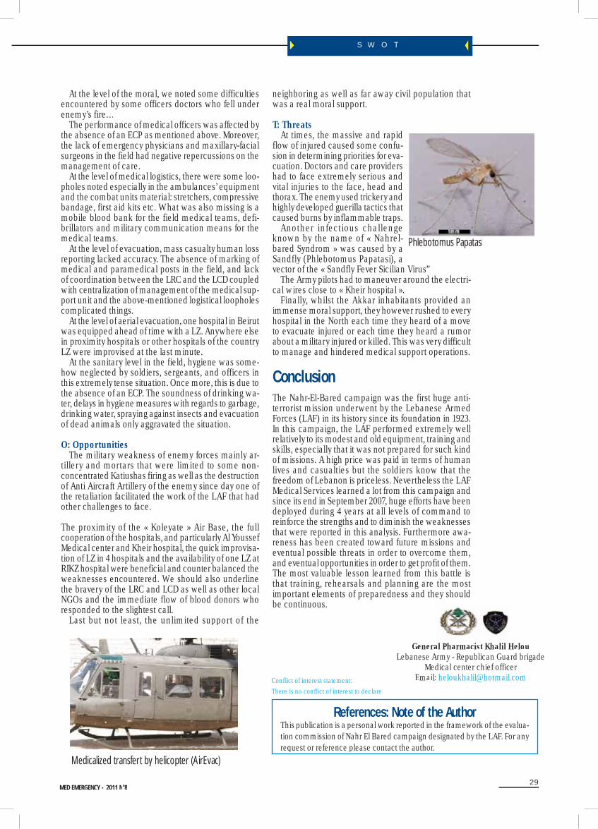
29MED EMERGENCY - 2011 No8
S W O T
At the level of the moral, we noted some difficulties encountered by some officers doctors who fell under enemy’s fire…
The performance of medical officers was affected by the absence of an ECP as mentioned above. Moreover, the lack of emergency physicians and maxillary-facial surgeons in the field had negative repercussions on the management of care.
At the level of medical logistics, there were some loo-pholes noted especially in the ambulances’ equipment and the combat units material: stretchers, compressive bandage, first aid kits etc. What was also missing is a mobile blood bank for the field medical teams, defi-brillators and military communication means for the medical teams.
At the level of evacuation, mass casualty human lossreporting lacked accuracy. The absence of marking of medical and paramedical posts in the field, and lack of coordination between the LRC and the LCD coupled with centralization of management of the medical sup-port unit and the above-mentioned logistical loopholescomplicated things.
At the level of aerial evacuation, one hospital in Beirut was equipped ahead of time with a LZ. Anywhere else in proximity hospitals or other hospitals of the country LZ were improvised at the last minute.
At the sanitary level in the field, hygiene was some-how neglected by soldiers, sergeants, and officers in this extremely tense situation. Once more, this is due to the absence of an ECP. The soundness of drinking wa-ter, delays in hygiene measures with regards to garbage, drinking water, spraying against insects and evacuation of dead animals only aggravated the situation.
O: Opportunities The military weakness of enemy forces mainly ar-
tillery and mortars that were limited to some non-concentrated Katiushas firing as well as the destructionof Anti Aircraft Artillery of the enemy since day one of the retaliation facilitated the work of the LAF that had other challenges to face.
The proximity of the « Koleyate » Air Base, the fullcooperation of the hospitals, and particularly Al Youssef Medical center and Kheir hospital, the quick improvisa-tion of LZ in 4 hospitals and the availability of one LZ at RIKZ hospital were beneficial and counter balanced the weaknesses encountered. We should also underline the bravery of the LRC and LCD as well as other local NGOs and the immediate flow of blood donors who responded to the slightest call.
Last but not least, the unlimited support of the
neighboring as well as far away civil population that was a real moral support.
T: Threats At times, the massive and rapid
flow of injured caused some confu-sion in determining priorities for eva-cuation. Doctors and care providers had to face extremely serious and vital injuries to the face, head and thorax. The enemy used trickery and highly developed guerilla tactics that caused burns by inflammable traps.
Another infectious challenge known by the name of « Nahrel-bared Syndrom » was caused by a Sandfly (Phlebotomus Papatasi), a vector of the « Sandfly Fever Sicilian Virus”
The Army pilots had to maneuver around the electri-cal wires close to « Kheir hospital ».
Finally, whilst the Akkar inhabitants provided an immense moral support, they however rushed to every hospital in the North each time they heard of a move to evacuate injured or each time they heard a rumor about a military injured or killed. This was very difficult to manage and hindered medical support operations.
ConclusionThe Nahr-El-Bared campaign was the first huge anti-terrorist mission underwent by the Lebanese Armed Forces (LAF) in its history since its foundation in 1923. In this campaign, the LAF performed extremely well relatively to its modest and old equipment, training and skills, especially that it was not prepared for such kind of missions. A high price was paid in terms of human lives and casualties but the soldiers know that the freedom of Lebanon is priceless. Nevertheless the LAF Medical Services learned a lot from this campaign and since its end in September 2007, huge efforts have been deployed during 4 years at all levels of command to reinforce the strengths and to diminish the weaknesses that were reported in this analysis. Furthermore awa-reness has been created toward future missions and eventual possible threats in order to overcome them, and eventual opportunities in order to get profit of them. The most valuable lesson learned from this battle is that training, rehearsals and planning are the most important elements of preparedness and they should be continuous.
General Pharmacist Khalil HelouLebanese Army - Republican Guard brigade
Medical center chief officerEmail: [email protected]
References: Note of the AuthorThis publication is a personal work reported in the framework of the evalua-tion commission of Nahr El Bared campaign designated by the LAF. For any request or reference please contact the author.
Medicalized transfert by helicopter (AirEvac)
Phlebotomus Papatasi
Conflict of interest statement:There is no conflict of interest to declare

M É D E C I N E D E C A T A S T R O P H E
*HORS TVA
LE VVOYAGE EN PREMIEREE CCCLLLAASSE
Sed El Bauchrieh: 01 684 684 - Ain El Mreisseh: 01 360 779Agents agrées:City CarBeirut01 803313/4
Bauchrieh CarCenter, Beirut01 880213
Elie TabetJounieh09 918402
Bejco s.a.r.l.Jamhour05 768800
Highway Auto,KhaldehHighway05 800149
Pascal 2 SARLZalka, Seaside03 191900
North MotorsTripoli06 411293/4
Fouad SrourZahleh08 800403
Lana MotorsSaida07 727220
Youssef TradeCompanyTyr & Nabatieh07 351313

31MED EMERGENCY - 2011 No8
François BANDALY, Antoine ZOGHBI, Joseph KHAIRALLAH, Laura BOU ABDO, Chadi SABBAGH, Gabriel ASMAR.
Prise en charge des blessés gravesEnseignements concernant la prise en charge des blessés graves atteints lors des combats du camp de Nahr El Bared en 2007.
Entre le 20 mai et le 2 septembre 2007, des affrontements armés ont opposé dans le camp de Nahr el Bared, prèsde Tripoli (Nord Liban), à environ 110 km de Beyrouth, des insurgés à l’armée libanaise. Après trois mois et demi de combats, l’armée libanaise a réussi à prendre le contrôle du camp au prix de 2600 blessés, 427 morts, dont 163 militaires. Au total, 850 évacuations sanitaires ont été effectuées, dont 100 héliportées. Le nombre exact d’insurgés tués ou blessés n’a pu être précisé en raison des conditions particulières de la survenue de ces évènements.
INTRODUCTIONBasé sur l’étude rétrospective de 109 dossiers de soldats de l’armée libanaise admis pour blessures graves dans deux hôpitaux beyroutins (Hôtel-Dieu de France et hôpital Rizk), ce premier article n’a pour seul objectif que de présenter la chaîne médicale, du relevage des blessés jusqu’à leur admission dans les blocs opératoires de ces deux établisse-ments, et de décrire succinctement les principales lésions rencontrées en rapport avec les particularités des combats en cause. La reprise de ces 109 dossiers dans un travail à effectuer permettra de développer et d’affiner les données présentées ici.
MATÉRIEL ET MÉTHODE Les deux hôpitaux retenus l’ont été en raison du nombreconséquent de blessés reçus. Les patients admis à l’Hôtel-Dieu de France et à l’hôpital Rizk venaient pour 44% d’un centre de triage (échelon II) et pour 32% d’un autre hôpital situé à 30 kilomètres au nord du camp (échelon III). L’ex-ploitation de ces 109 dossiers a nécessité le classement de27 items répartis en 8 rubriques. N’ont été retenus que lesblessés dont l’Injury Severity Score (ISS) était supérieur ou égal à 19 (1, 2, 3). Les soldats porteurs de lésions immédiate-ment létales ne sont pas concernés puisque non admis dans les deux hôpitaux sujets de l’étude. De même, les brûlés nesont pas inclus puisqu’ils ont été transférés directement vers un établissement disposant d’un service spécialisé. L’analysedes données a été effectuée par l’étude dela moyenne pour les variables quantitatives et des proportions pour les variables quali-tatives.
CHAINE MÉDICALEDESCRIPTION : On peut la structurer en quatre échelons de soins : Echelon I (role I OTAN). Au niveau de l’unité decombat élémentaire, les blessés sont pris en charge par leurs camarades ou des sauveteurs formés au secourisme. Les soins effectués à ce niveau ont consisté en la restauration de la liberté des voies aériennes,la tentative d’arrêt des hémorragies extériorisées, les gestesde réanimation élémentaires d’un arrêt cardio-respiratoire.Cet échelon de premier niveau comprend également des postes de premiers secours mobiles, médicalisés avec des techniciens paramédicaux et/ou un médecin, se déplaçant
avec les unités. Ils utilisent des tentes et des véhicules blindésde transport de personnel. Les soins réalisés à ce niveau n’ont pu être plus évolués (pose de voie veineuse,intubation...) en raison de l’exposition des combattants et des secouristes auxsnipers. Deux membres de la croix rouge libanaise ont ététués dans ces circonstances.Echelon II (role II, III OTAN). A ce niveau, outre un triage affiné et une prise en charge réanimatoire complète, on a pu disposer d’un laboratoire et d’une imagerie, procéder à une transfusion sanguine et effectuer des interventions chirurgicales de sauvetage gé-néral ou fonctionnel ainsi qu’une hospitalisation temporaire. La sophistication de la prise en charge a été rendue possible parce que cette fonction a été dévolue, pour des raisons géographiques, à un hôpital bien équipé situé à 5 kilomètres au sud du camp en direction de Beyrouth.Echelon III et IV (role III et IV OTAN). Il s’agitd’hôpitaux disposant d’un plateau technique pluslarge (échelon III) ou complet (échelon IV) avecdes services spécialisés (neurochirurgie, chirurgie vasculaire et thoracique, etc…) comme les deux hôpitauxconcernés par cette étude (Hôtel-Dieu de France et hôpital Rizk à Beyrouth).
PRISE EN CHARGE :Si le relevage des blessés sous le feu n’a pas permis d’effec-tuer à l’échelon I une médicalisation de type SMUR, tous les blessés arrivés vivants à l’échelon II avaient bénéficié des
gestes élémentaires de survie (désobstruction des voies aériennes supérieures, pansements compressifs, garrots…) ce qui traduit la bonne formation des secouristes et de l’ensemble des personnels de l’échelon I qui ont appliqué l’enseignement qui leur avait été prodigué. Leshémorragies artérielles compressibles sont l’archétype des morts évitables si une action réflexe est entreprise par le blessé lui-même ou par un camarade de combat, dans les toutespremières minutes (Platinum Ten Minutes). Si20% des décès sont instantanés et inévitables, 15 à 20% des décès précoces sont encore évi-
tables (4). La pose de garrots artériels est certainement le geste qui sauve le plus de vies et le bénéfice attendu sur la réduction de mortalité est supérieur au risque de lésions ischémiques (4).100% des blessés de notre étude ont été perfusés à l’éche-lon II et ce dans un délai inférieur à une heure après leur atteinte. Pour les 35% qui ont été intubés, la survie a été de
« quatreéchelons
de soins »
François BANDALY
C O N F L I T S A R M É S
Article history / info:Received: Feb 9, 2011Reviewed: Apr 22, 2011Received in revised form:June 1, 2011Accepted: June 23, 2011

32MED EMERGENCY - 2011 No8
90%. A ce niveau de soins, 12% des blessés ont bénéficiéd’un bilan sanguin et d’une imagerie et 20% d’un bilan san-guin ou d’une imagerie. Toujours à l’échelon II, 26% ont étéopérés, principalement pour assurer l’hémostase, avant leur transfert dans les hôpitaux de structure. Ainsi, une chirurgie ultra-précoce améliore la survie dans le cas des hémorra-gies non contrôlables par un garrot et non compressibles (4). Nau et al. (5) dans une étude menée, de 2002 à 2004, sur 3 théâtres d’opérations extérieures (Kosovo, Afghanistan, Côte d’Ivoire) notent que, parmi les blessés, 19% étaient des urgences absolues dont 83% de celles-ci étaient liées à un choc hémorragique, 25% à une détresse ventilatoire et 5% à un coma. Pour les décédés, 92% l’ont été avant triage.On peut donc souligner que, malgré l’impossibilité de prise en charge de type SMUR sur les lieux du combat, le trans-fert immédiat accompagné de gestes de secourisme et la prise en charge médicalisée rapide à l’échelon II a permis d’être dans le délai de la « Golden Hour » et a été tout à fait bénéfique aux blessés. En fonction des capacités des échelons présentés précédemment, on comprend que des patients graves stabilisés à l’échelon II doivent être transférés directement à l’échelon IV (hôpitaux de structure) en court-circuitant l’échelon III qui ne peut apporter obligatoirementla réponse technique adaptée. Ainsi, entre l’échelon II etl’échelon IV (Hôtel-Dieu de France et Hôpital Rizk), pour les 73% de blessés dont le délai de transfert est connu, 3% ont été transférés dans un délai inférieur à une demi-heure (calculé entre leur admission à l’échelon II et leur arrivée à l’échelon IV), 36% dans un délai inférieur à une heure et 63% ont été transférés dans un délai inférieur ou égal à une heure et demie. Le délai moyen de transfert était ainsi de 1h45 si l’on écarte les blessés transférés quelques jours après leurs
blessures ou qui avaient subi une intervention de sauvetageà l’échelon II (Tableau I : Délai du transfert). Aucun décèsn’a été noté durant le transfert entre l’échelon II et l’échelonIV. Les seules complications notées ont été des défaillances hémodynamiques pour 4% des blessés qui ont pu être cor-rigées. Il faut noter 4 blessés morts rapidement après leur arrivée à l’échelon IV.On peut en conclure que, à l’échelon II, la mise en condition préalable au transfert avait été adaptée et soignée (aucuneextubation durant le transfert) et la gravité des patients cor-rectement évaluée (triage). 75% des blessés graves ont béné-ficié d’un transfert héliporté. Tous les drainages thoraciques(11% des cas) ont été mis en place exclusivement dans lesdeux hôpitaux sujets de l’étude où on notait sur l’ensembledes blessés que 56% n’avaient pas reçu d’antibiothérapie avant leur admission et 30% de sérothérapie anti-tétanique.Par contre, 49% étaient déjà porteurs d’une sonde urinaire.Enfin, 83% des admis ont été opérés à l’Hôtel-Dieu de Franceou à l’hôpital Risk dont 75% dans l’heure qui a suivi leur arri-vée ce qui traduit la qualité du tri, le bien-fondé des décisionsde transfert et la bonne transmission des bilans lésionnelsentre l’échelon II et l’échelon IV.
ÉPIDÉMIOLOGIE, LOCALISATION ET ÉTIOLOGIE DES BLESSURES ÉPIDÉMIOLOGIE :La moyenne d’âge des blessés était de 29 ans avec un écart type de 7 ans. 90% avaient un âge inférieur ou égal à 40 ans et 25% inférieur ou égal à 24 ans. Au total, 51 des 109 blessés ont reçu 355 culots globulaires, la moyenne étant de 7 culots globulaires, la médiane étant à 4 culots globulaires avec un minimum d’un culot globulaire et un maximum de 30 culots globulaires. De plus, 136 poches de plasma frais congelé (PFC) ont été utilisées avec unemoyenne de 7 PFC, la médiane étant de 2 PFC, le minimum de 1 PFC et le maximum de 21 PFC. Ces chiffres sont élevés (6)
et peuvent être comparés avec les données de la littérature (5)
ce qui témoigne de la sévérité des atteintes initiales et de la disponibilité des produits sanguins. 36% des blessés étaient de groupe O+ et 27% de groupe A+AA .
LOCALISATION DES BLESSURES :A l’analyse des données, 51% des soldats avaient un seul organe atteint, 31% deux organes atteints et 18% trois organes ou plus. La répartition lésionnelle ne suit pas une distributionaléatoire, mais est le résultat des effets de la visée ennemie, des effets de protection balistique et du type d’arme (7). Letableau II (Localisations des atteintes) donne les proportionssur ces 109 dossiers des différents organes atteints.
Même si l’association tête-cou représente le pourcen-tage le plus élevé des lésions, les membres sont les plus fréquemment atteints (26 % des cas) ce qui est déjàrelevé dans la littérature (4,8) où l’on note que, actuel-lement, les membres et l’extrémité céphalique sont les plus touchés (7,9).
Notons que notre travail ne prend en compte que les soldatsarrivés vivants dans les hôpitaux d’infrastructures ce qui biaise les résultats par élimination des 1/5 des blessés quimeurent quoi que l’on fasse (4) en sachant que 90% des morts au combat décèdent au cours de la première heure (Golden Hour), les exsanguinations par délabrement des membresétant la première cause de décès (4,10).
Délai du transfert
Tableau I : Le délai de transfert est le laps de temps mesuré entre l’arrivée à l’échelon II et l’arrivée à l’échelon IV pour les 73% de blessés dont le délai de transfert est connu.
Une vue du camp deNahr el Bared durant les événements.
C O N F L I T S A R M É S
From 20 May to 2 September 2007 an armed confron-tat ion occurred between the Le-b a n e s e a r m e d forces and rebels in Nahr El Bared camp near Tripoli (Nor th of Leba-non).The fighting led to 2,600 injured, 427 dead (among which 163 service men), 850 medical eva-cuations (MEDE-VAC) 100 of which by helicopter. In a retrospec-tive study on 109 severely injured servicemen of the Lebanese armed forces admitted to two hospitals in
Abstract
33MED EMERGENCY - 2011 No8
Notons également que le port des effets de protection indi-viduels a réduit le pourcentage des lésions cérébrales (12%dans notre étude) et du thorax (6% dans notre étude), parti-culièrement les plaies pénétrantes le plus souvent létales, auprofit des lésions des membres qui représentent désormais60 à 70% des lésions et qui sont en majorité des causes de décès évitables (4,8). Cette évolution est soulignée par Donatet al (7) à partir des études qu’ils citent. Nau (5) précise que les gilets pare-balles ont montré leur efficacité en transformantradicalement les occurrences des blessures mortelles et desblessures graves. Pour lui, les conflits moder-nes induisent une augmentation relative du nombre de blessures des extrémités et de la tête en raison du port de gilets balistiques qui protègent efficacement le thorax et l’abdomen(12% de lésions abdominales et 5% de lésionspelviennes chez nos blessés).Notons enfin que, étant donné le type de com-bat, il existait, outre de nombreux snipers, de multiples engins piégés (mines, engins explo-sifs improvisés également appelés IED) qui ont provoqué des lésions des membres inférieurs. Notre étude n’a malheureusement pas différen-cié les atteintes des membres supérieurs de celles des mem-bres inférieurs. Donat et al (7) notent que la recrudescencedes lésions de polycriblage des membres est associée à unfort contingent de syndromes compartimentaux, de plaiessouillées et d’amputations.Les atteintes ORL, du massif facial, et ophtalmique sont associées chez 5% de nos blessés à une atteinte rachidienne. 11% des blessés souffraient d’une atteinte du massif facial(massif osseux et parties molles à l’exclusion des lésions ophtalmiques et ORL). Ces dernières étaient respectivementde 13% (chiffre élevé peut être en rapport avec l’absence du port de lunettes de protection) et de 9%.L’association des lésions a été discriminée chez les polytrau-matisés. Parmi ceux porteurs d’une atteinte cérébrale, 39%ont au moins une atteinte ophtalmique et/ou ORL et/ou dumassif facial associé. Dans les atteintes du massif facial, 62% avaient en plus une atteinte ophtalmique et/ou ORL.Parmi les atteintes abdominales, 17% avaient en plus des atteintes pelviennes, 17% des atteintes thoraciques, 21% des atteintes rachidiennes. Pour les atteintes des membres, 58%avaient au moins une autre partie du corps touchée. Pour les atteintes rachidiennes (6%), tous les blessés avaient unelésion associée d’un autre système. Tous les polytraumati-sés étaient porteurs d’une lésion rachidienne. Ces chiffres sont à comparer avec les conclusions de Donat et al (7) qui notent que les agents lésionnels responsables des blessures
ont changé, leur capacité létale également. Les IED fontdésormais plus de victimes que les fusils d’assaut, le « poly-blessé - polycriblé - amputé » a pris la place du blessé « brûlé– blasté ». La répartition des lésions s’est également modifiéeavec la généralisation des effets de protection.
ÉTIOLOGIE DES BLESSURES :Au total, 70% des lésions étaient liées aux balles pénétranteset 30% de cause explosive. Dans les grands conflits du 20ème
siècle, (deuxième guerre mondiale, Corée, Vietnam), 90% des blessures de guerre étaient liées à des lésions pénétran-tes (7, 9, 11). Lors de la première partie (invasion blindée) de laguerre en Irak, les lésions pénétrantes représentent, selon les études, entre 43% et 77% des lésions et celles par explosion
entre 13% et 48% (7). A l’inverse, lors de la phase d’occupation de l’Irak, 70% des blessés amé-ricains sont victimes de lésions par explosion(7, 12). Dans la cohorte limitée de nos blessésde Nahr el Bared, la mortalité était de 64% decause balistique et de 36% liée à l’effet blast (tableau III : Cause de la mortalité) avec unemortalité globale de 10% des blessés reçusà l’échelon IV avoisinant les données de la littérature que ce soit dans les conflits plus an-ciens comme le Vietnam (7) ou récents commel’Irak et l’Afghanistan (13). Cette répartition de lamortalité peut être expliquée par la présence
massive des francs tireurs (snipers), spécificité du combatactuel en milieu urbain et par l’effet protecteur, contre lesconséquences mineures des explosions, du casque et du giletbalistique qui comporte une composante pare-éclats (5) quiont réduit le pourcentage des lésions de la tête et du tronc le plus souvent létales au profit des lésions des membres (4,8). Inversement, on peut imaginer que les blessures liées auxexplosions dépassant les capacités d’arrêt de la composante pare-éclats du gilet furent rapidement létales. Les atteintes cérébrales avec ou sans casque n’ont pu malheureusementêtre différenciées.Au niveau thoracique, dans cette étude, les lésions qualifiées« par blast » ont pu être fusionnées avec celles liées aux« effets arrières » dont l’origine est la transmission par le gilet balistique de l’impact du projectile à la paroi thoracique avecdes blessures locales de celle-ci, blessures hémorragiquespulmonaires voire cardiaques ou abdominales (5). La reprise plus discriminante des dossiers devrait permettre d’affiner ces explications.
Enfin, 15% des blessés ont présenté des complications psychiatriques (PTSD = Post-Traumatic Stress Disorder)pour lesquelles une étude bi-variée a montré que ces désordres touchaient préférentiellement les soldats at-teints au rachis ou à l’abdomen et étaient également en rapport avec la durée de séjour à l’hôpital.
Tableau II.
L li ti d tt i tLocalisations des atteintes
« les membres sont préférentiellement
atteints »
Tableau III.
Cause de la mortalitéCause de la mortalité
C O N F L I T S A R M É S
Beirut (Hotel-Dieude Fr ance andHospital Rizk), theauthors firstly des-cribed the first aidsystem and the me-dical care, secondly studied the locationand the etiology of the injuries.They point out thatlimbs are the mostfrequently woun-ded, as describedin the literature.As in the fatalities,the wounds aremainly due to pe-netrating bullets ra-ther than explosivecauses, consistentw i th the seve -rity of sniper bulletwounds. To conclude, theauthors underlinethe importance of a structured andcoordinated medi-cal system; a pre-established codeof practice; rapidmedical evacua-tion by helicopter;the essential issueof using pre-hos-pital medical carewith eff icient welltra ined f ir s t a idp e r sonne l ; t heimperative supply of individual pro-tective equipmentsuch as bulletproof vests, helmets, andprotective glasses.

34 MED EMERGENCY - 2011 No8
CONCLUSIONSSachant que la répartition des blessés est souvent trimodale :ceux qui meurent quoi que l’on fasse (1 sur 5), une majorité de blessés sans gravité (3 sur 5) et ceux en détresse vitale (1 sur 5) qui survivent grâce à une prise en charge agressive (4), on ne peut qu’insister sur l’importance d’une chaîne médicale correctement structurée de-puis le relevage jusqu’à l’accueil dans les hôpi-taux d’infrastructure associée à une doctrine d’emploi pré-établie.On a pu noter une coordination satisfaisante entre les différents niveaux (avant, zone de tri et de soins d’urgences, services d’urgences des hôpitaux d’infrastructure et blocs opératoires), une très bonne régulation et transmission des bilans lésionnels à l’origine des transferts ainsi qu’un accueil aux urgences hospitalières de bonne qualité.L’utilisation intensive de l’hélicoptère, facilitée par la faible étendue du pays, la météorologie et l’absence de menacesol - air sérieuse, même si elle ne s’est pas produite dans les premières phases des combats, a permis des évacuations rapides vers les hôpitaux de l’échelon IV.L’ensemble de ces éléments explique une mortalité de ces blessés graves de 10%, avoisinant celle relevée sur les dif-férents théâtres récents, alors que la mortalité globale des soldats atteints à Narh el Bared était de 25%, chiffre proche des données de la littérature (7). Plus globalement, la rapidité et la qualité des prises en charge,du tri à l’échelon II et des transferts, paradigme d’un servicede santé d’une armée moderne expliquent ces chiffres.
Il faut rappeler que, pour la prise en charge des blesséslors des conflits actuels, au regard du nombre de décèsprécoces encore évitables, les enjeux essentiels se situentd’abord à l’échelon préhospitalier (4).
Davantage de gestes d’urgence enseignés aux opérateurs du sauvetage au combat et des évacuations primaires mé-dicalisées permettent d’amener les blessés à la chirurgie en moins de deux heures avec un meilleur pronostic (4). Le portd’équipements principaux individuels de protection (gilets
balistiques adaptés et modernes, casques)contribue également à ce résultat comme leconfirment les atteintes préférentielles des membres non encore correctement protégés et la diminution des lésions thoraciques etde la tête, point relevé dans la littérature (4,
8). L’importance proportionnelle des décès, tous organes confondus, par atteinte balisti-que (64%) traduit la gravité persistante de ce mode vulnérant d’autant plus que la présence de francs tireurs, particulièrement actifs à Nahr el Bared, est une spécificité du combat actuel en milieu urbain.
Il est important d’insister sur la synergie entre les différentsintervenants qui doit, comme toujours, pouvoir être amélio-rée. Par ailleurs, il serait souhaitable que cette expérience permette le développement des transferts héliportés médi-calisés, en temps de paix, sur tout le territoire libanais.
Ces réflexions devraient aboutir, d’une manière générale, à ce que l’étonnement vienne non pas de l’imprévu, mais du fait que les choses arrivent comme on les a conçues (Raphaël Enthoven, 14)4 .
François BANDALY (1), Antoine ZOGHBI (2), Joseph KHAIRALLAH (2), Laura BOU ABDO (2), Chadi SABBAGH (2), Gabriel ASMAR (1)
(1) Pôle Urgences - SMUR - UHTCD, Centre Hospitalier - Vierzon - France(2) Service des Urgences, Hôtel-Dieu de France - Beyrouth - Liban
Courriel : [email protected]
Cet article est dédié à Madame Béatrice DESBEAUX-SALVIAT. Remerciements à Madame le Docteur Carole ORIOL pour son amitié et ses connaissances en langue anglaise.
RÉFÉRENCES BIBLIOGRAPHIQUES :1. - Jarry J - Prise en charge des traumatismes civils au trauma center de Johannesburg. Urgence Pratique 2009 ; 94 31 - 35.2. - Baker SP, O’Neill B, Haddon W Jr, Long WB - The injury severity score : a method for describing patients with multiple injuries and evalu-ating emergency care. J Trauma. 1974 Mar ; 14 (3) 187 - 96.3. - Stein B - Polytraumatisme - www.anesthesie-esch.lu/download/polytraumatisme% 20 2005. pdf.4. - Martinez JY, Puidupin M, Precloux P, Klack F, Turc J, Escarment J - Les enjeux de la prise en charge des blessés des conflits actuels. Urgences 2009 ; 65 677 - 88.5. - Nau A, Prat N, Fusai T, Sarron JC, Pleytel E. - Intérêts et limites des effets de protection. Urgences 2009 ; 64 667 - 75.6. - Bernard B, Jacques H - La chirurgie en temps de guerre. L’université de tous les savoirs 2007 ; 638.7. - Donat N, Pasquier P, Clapson P, Perez JP, Debien B - Epidémiologie des blessures de guerre. Urgences 2009 ; 63 653 - 66.8. - Carey ME - Analysis of wounds incurred by US Army Seventh Corps personnel treated in corps hospitals during Operation Desert Storm. Februay 20 to march 10, 1991. J Trauma March 1996 ; 40 suppl (3) S165 - S69.9. - Bellamy RF - Combat Trauma Overview. In : Bellamy RF, editor. - Textbook of Military Medecine, Washington DC : Borden Institute ; 1995.10. - Champion HR, Bellamy RF, Roberts CP et al. - A profile of combat injury. - J Trauma May 2003 ; 54 (5 supply) S13 - S19.11. - Sauaia A, Moore FA, Moore EE et al. - Epidemiology of trauma deaths : a reassesment. J Trauma Feb 1995 ; 38 (2) 185 - 93.12. - Peake JB - Beyond the purple heart - Continuity of care for the wounded in Iraq. N England J Med 2005 jan 20 ; 352 (3) : 219 - 22. 13. - Gawande A - Casualties of war-military care for the wounded from Iraq and Afghanistan. N England J Med 2004 ; 351 : 2471 - 5.14.- Enthoven R - Un jeu d’enfant-la philosophie-Paris, Fayard 2007.
« 64% de décès, de cause balistique »
C O N F L I T S A R M É S
Key words War wounded, ME-DEVAC Medical sys-tem, armed conflict,military emergency medicine
Conflict of interest statement:There is no conflict of interest to declare

40 MED EMERGENCY - 2011 No8
The 10th Annual Congress of
The Lebanese Society of Family Medicine
Date: October 14 - 16, 2011
Venue: Bristol Hotel, Beirut – Lebanon
www.lsfm.net
www.infomedweb.com
The 10th Annual Congress of
The Lebanese Society of Family Medicine
Date: October 14 - 16, 2011
Venue: Bristol Hotel, Beirut – Lebanon
www.lsfm.net
www.infomedweb.com
10th Pan Arab Congress of of Endocrinology
& Diabetes
14th Congress of The Lebanese Society of
Endocrinology, Diabetes & Lipids
6 - 9 Oct 2011Phoenicia Intercontinental Hotel - Beirut – Lebanon
www.paced2011.com
European Society of intensive care Medicine24th Annual CongressDate: October 1-5, 2011ICC - Berlin - Germanywww.esicm.org
10th Pan Arab Congress of Endocrinology & Diabetes& 14th Congress of the Lebanese Society of Endocrinology, Diabetes, Lipids.Date: October 6 - 9, 2011Venue: Phoenicia Intercontinental Hotel, Beirut-Lebanonwww.paced2011.com
18th Congress Union of Arab Pediatric SocietiesDate: October 6 - 9, 2011Venue: Bait Al Tabib - Beirut-Lebanonwww.papna.org - www.lpswebsite.org
The 10th Annual Congress of the Lebanese Society of Family MedicineDate: October 14 - 16, 2011Venue: Bristol Hotel, Beirut – Lebanonwww.infomedweb.com
Salon Infirmier 2011Paris - Porte de Versailles12, 13 et 14 octobre 2011www.salon-infirmier.com
Burning Issues on Psychiatry Date: October 27 - 28, 2011
Place: Phoenicia Intercontinental Hotel, Beirut-Lebanon.www.bipsych.org
28ème Congrès scientifique de Médecine d’Urgence26 au 28 octobre 2011Centre Mont-Royal - Montréalwww.amuq.qc.ca ou www.asmuq.org
CEM/IFEM Symposium: Quality and Safety in Emergency CareNovember 15-16, 2011British Museum - London United Kingdomwww.collemergencymed.ac.uk
HPSN Europe 2011Nov 26, 27 Mainz, GermanySimulation & Education conferenceWeb site : hpsn.com
4è Congrès International des SAMU et Urgences Outre-Mer5è Journées Européennes de Médecine d’Urgences Maritimes27 et 28 mars 2012 - Saint Martin30 mars 2012 Saint - Barthélémywww.samu.urgences.outremer.orgAcute Cardiac Care 2012
Acute Cardiac Care 201220 - 23 october 2012Istanbul, Turkeywww.escardio.org
Congresses and conferences
41MED EMERGENCY - 2011 No8
R É A N I M A T I O N
Résumé
Mots clés« suppléance
cardio-respiratoire ou circulatoire à disposition des
SAMU »
Hichem CHENAITIA, Géraldine KARSENTY, Fayez JEBRIL, Jacques BESSEREAU, Richard TOESCA, Emgan QUERELLOU, Laurent JACQUIN, Marion DOUPLAT, Jean-Pierre AUFFRAY, Frédéric COLLART, Pierre MICHELET, Vlad GARIBOLDI.
Assistance cardiorespiratoire mobile :Nouvelle filière en médecine d’urgenceLa mutualisation des moyens et des compétences hyperspécialisées au sein de centres de référence a entraînél’augmentation des demandes de Transferts Inter Hospitaliers (TIH) de patients présentant des défaillances aiguës cardiocirculatoires et/ou respiratoires sévères. Ces transferts de patients à haut risque sont parfois incompatibles avec les moyens classiques de la médecine préhospitalière. Devant ce constat, une réflexion s’est amorcée entre les Services d’Aide Médicale d’Urgence (SAMU) et ces centres de référence dans le but de développer des moyens adaptés pour sécuriser le transfert de ces patients.
L’Oxygénation Membranaire Extra Corporelle (ECMO) est une technique d’assistance cardiores-piratoire dérivée des techniques de Circulation Extra Corporelle (CEC) 1 . Les progrès technologiques et laminiaturisation des appareils ont permis de déve-lopper un système d’ECMO mobile et de le mettre en place dans un délai bref au chevet du patient où qu’il soit. Les Unités Mobile d’Hospitalisation (UMH)des SAMU permettent après stabilisation du patient de réaliser un transfert sécurisé vers un centre spé-cialisé, rompu au management des CEC. Deux nouveaux moyens ont donc été récemment développés sur ce principe : l’Unité Mobile d’Assistance Respiratoire (UMAR) avec une ECMO veino-vei-neuse qui permet une suppléance respiratoire exclusive, et l’Unité Mo-bile d’Assistance Circulatoire (UMAC)avec une ECMO artério-veineuse quipermet une suppléance cardio-circu-latoire ou cardiorespiratoire.L’Assistance Publique des Hôpitauxde Marseille (APHM) et le SAMU zo-nale sud-est ont mis en place ces uni-tés mobilisables en urgence 24h/24et 7 jours/7. Cette zone regroupe la région Pro-vence Alpes Côtes d’Azur (31 400 km2 et 4 891 000 habitants), le Languedoc Roussillon (27 376 km2
et 2 548 000 habitants) et la Corse (8 722 km2 et294 118 habitants), la superficie globale est donc de 67 498 km2, avec une population de 7,7 millions d’habitants ((( 12% de la population française). Ces unités spécialisées peuvent implanter une assis-tance cardiorespiratoire en moins d’une heure dans un rayon de 200 km.
MÉTHODESNous avons réalisé une étude observationnelleprospective des TIH nécessitant la mobilisation de l’UMAC ou de l’UMAR. Nous réalisons le recueil des données cliniques et épidémiologiques des patients, l’étiologie de la décompensation cardio-logique et/ou respiratoire, les complications au cours du transport, les distances parcourues, les délais d’arrivées et les durées des transports, et la mortalité de ces patients à 1 mois.
FONCTIONNEMENT :Le service demandeur appelle le centre 15 duSAMU 13. Après création d’un dossier de régu-lation, le permanencier auxiliaire de régulationmédicale transfère l’appel au médecin régulateur.Selon un protocole de régulation, une « conférence à 3 » (médecin demandeur, médecin régulateur, chirurgien cardiaque) est organisé avec le serviced’Unité de Réanimation Cardio-Circulatoire (URCC)du centre hospitalier universitaire (CHU) de la Ti-mone (Marseille, France). L’indication de l’assis-tance cardiorespiratoire est alors discutée, afin
de ne pas mobiliser une équipe pour un patient au-delà de toute ressource thérapeutique ou, au contraire, pour un patient encore transportable sans assistance. Le SAMU 13 gère complè-tement la logistique du transport, qu’il soit terrestre ou aérien.
ÉQUIPE :Elle est composée d’un chirurgien car-diovasculaire, d’un interne de chirur-gie, pour aider à la mise en place descanules, d’un perfusionniste, capable
de monter la pompe et de purger le circuit en moinsde 10 minutes, et d’un anesthésiste, formé au ma-nagement des patients sous CEC.
MATÉRIEL :L’objectif est d’être totalement autonome en ter-mes de matériel (lourd ou consommable) afin depouvoir implanter l’assistance dans n’importe quel service (urgence, réanimation ou soins intensifs, bloc opératoire, salle de cathétérisme). Il est composé : d’une pompe centrifuge électrique à débit continu avec une manivelle de secours, d’une membrane d’oxygénation et de son support, d’un circuit pré-hépariné, de canules longues veineuses et artérielles pré-hépariné, d’un cathéter et de sa ligne de reper-fusion fémorale, de kits de ponction percutané, d’un soluté de Ringer lactate, de clamps à tuyaux stériles et non stériles, de lames de bistouri, d’aiguilles de suture droites, d’une bouteille d’oxygène. Un sacest dédié à ces transports, et un chariot à roulettes permet de supporter et de faciliter le transport de l’ensemble du dispositif de l’ECMO mobile.
Hichem CHENAITIA
Introduction: Extra Corporeal Membrane IOOxygenation (ECMO) is a technique of OCCardiorespiratory Life Support. 2 new Cmmodels have been recently implemen-mtted: Mobile Unit for Respiratory Support t((MURS) with vein to vein ECMO, for respi-(rratory support exclusively and Mobile Unit rffor Cardiocirculatory Support (MUCS), fwwith a vein to artery Extra Corporeal Life wSSupport (ECLS).SMMethods: We conducted a prospective Moobservational study of all Intra Hospital ottransfers with either MURS or MUCS. tWWe collected clinical & epidemiological Wddata of all these patients, etiology of the drrespiratory or cardiocirculatory failure, rccomplications during the transport, dis-cttance of transport, and patients’ survival taat one month. aRResults: 11 patients needed MURS; trans-Rpport distance ranged from 4 to 202 km. pAAll patients had severe ARDS after a H1N1 Ainfection. Survival at one month was of 445%. 32 patients needed MUCS. No 4
Abstract
A ti l hi t / i fArticle history / info:Article history / info:Received: June 13, 2011Reviewed: July 26, 2011Received in revised form: Aug 5, 2011Accepted: Aug 17, 2011

42 MED EMERGENCY - 2011 No8
Résumé (suite)
« survie de 60% des chocs
cardiogéniques réfractaires »
Elle est percutanée par méthode de Seldinger enutilisant les 2 Scarpa, et peut se faire sous anes-thésie locale ou générale. Le but est la rapiditéde mise en place et la diminution du risque desaignement autour des canules, ce risque étant augmenté par la mobilisation du patient lors dutransport. En cas d’ECMO artério-veineuse, un ca-théter artériel de reperfusion du membre inférieur est systématiquement inséré dans l’artère fémorale superficielle, après stabilisation du patient et avant le transport, pour diminuer le risque d’ischémie du membre inférieur.Indications en médecine d’urgence :- Arrêt cardiorespiratoire réfractaire sous certainesconditions (voir figure 1) [2].- Choc cardiogénique réfractaire aux thérapeuti-ques usuelles, quelques soit la cause (Infarctus du myocarde ; Myocardite fulminante ; Intoxication aux cardiotropes ; Embolie pulmonaire grave ;Choc septique).- Syndrome de Détresse Respiratoire Aigue (SDRA)sévère (Rapport PaO2O /FiO22 2O < 70 depuis 2h au moinset avec une pression télé expiratoire positive (PEEP) > 10 cmH2HH O ; ou PaO2O /FiO22 2O entre 70 et 100 maisavec une pression de plateau > 35 cmH2HH O ; ou la présence d’une acidose respiratoire sévère (pH < 7,15) malgré une fréquence respiratoire à 35/min). - Syndrome de Défaillance Multi-Vis-cérale (SDMV).- Prélèvement Multi-Organe (PMO), avec pour but la sauvegarde des or-ganes internes.
TRANSPORT :Il peut être terrestre ou héliporté, en collaboration avec le SAMU 13 et la sécurité civile, privilégiant à chaque fois la rapidité à l’aller et la stabilité au retour. La mobilisation du patient doit être limitée au maximum, car il existe un risque de déplacement voire d’ablation des canules. Le patient est donc maintenu en décu-bitus dorsal strict. Le patient doit être parfaitement stabilisé avant le départ, car la possibilité de gestes
techniques ou chirurgicaux est limitée pendant le transport.
SURVEILLANCE ET COMPLICATIONS :La surveillance médico-chirurgicale ne peut s’en-visager que dans des services rompus au mana-gement des CEC, et dans des centres habilités à latransplantation. Elle a pour but de veiller au bon fonctionnement du dispositif, au dépistage précocede complication. Une surveillance multimodale (clinique, radiologique, échocardiographique, gazo-métrique et de la compliance pulmonaire) permet d’évaluer le moment opportun pour débuter des épreuves de sevrage par paliers du système.Les complications sont nombreuses :- Hémorragiques : au point d’insertion des canu-les par déchirure des vaisseaux fémoraux et/ou favorisés par le traitement anticoagulant ou anti-agrégant.- Thromboemboliques : embolie cérébrale ou pul-monaire, jusqu’à la thrombose partielle ou com-plète des cavités cardiaques.- Ischémiques du membre inférieur : dans 10 à 20% des cas d’ECMO artério-veineuse malgré une ligne de reperfusion artérielle.- Infectieuses : cellulite au point de ponction ou infections nosocomiales en réanimation.- Hématologiques : thrombopénies, hémolyse intra-
vasculaire- Dysfonction mécanique du systèmeou « caillotage » de la membrane oude la pompe Devenir :- Maintien en place jusqu’à une déci-sion d’arrêt de l’assistance ou « Bridge to décision » : l’assistance est mainte-nue, en attendant d’évaluer les lésions et les séquelles du patients, ainsi que la recherche d’antécédent contre in-diquant le maintien de l’assistance, pour éviter tout acharnement théra-
peutique. Si la décision de limitation des thérapeu-tiques actives est prise, on arrête l’assistance ou on propose un PMO à la famille.- Maintien en place jusqu’à la récupération ou « Bridge to recovery » : situation où le patient récupère une fonction cardiaque ou respiratoire suffisante pour envisager le sevrage progressif de l’assistance. - « Bridge to Bridge » : l’évolution ne permettant pas le sevrage de l’assistance, on réalise le rem-placement de l’ECMO mobile par un système d’as-sistance circulatoire de longue durée.- Maintien en place jusqu’à la transplantation ou « Bridge to transplantation » : pas de récupération des défaillances d’organe mais possibilité de trans-plantation d’organe.
RÉSULTATSUMAC :De janvier 2007 à avril 2009, 32 patients ont pu béné-ficiés de l’UMAC. L’âge moyen est de 41 ans 13-65ans , le sex-ratio M/F est de 1,13 (15 femmes et 17 hommes). La distance parcourue va de 4 à 225 km. Nous n’avons noté aucune complication pendant
Principe ECMO. Source Maquet S.A.
R É A N I M A T I O N
complications during the transport time.Cardiogenic shock was due to myocarditis(28.1%), Acute Cardiac failure of unknown origin (21.9%), Drug poisoning with car-diotropes (18.8%), post Myocardial Infarc-tion (12.5%) & post cardiotomy (12.5%),and refractory Cardiac Arrests (6.2%).Survival rate at one month was of 60%regardless of etiology. Discussion: We found that these MobileUnits have impacted the regular activity of our Out-of-Hospital Mobile Units,enabling transfers which were impossiblebefore the implementation of such spe-cialized units. Information of Emergency Medicine & Intensive Care physicians,Cardiologists and Surgeons is essential for early screening of patients whom wouldbenefit from such transfers.
Mobile Unit for Cardio circulatory Sup-port, Mobile Unit for Respiratory Sup-port, Extra Corporeal Membrane Oxyge-nation (ECMO), Service d’Aide Médicaled’Urgence (SAMU).
Key words
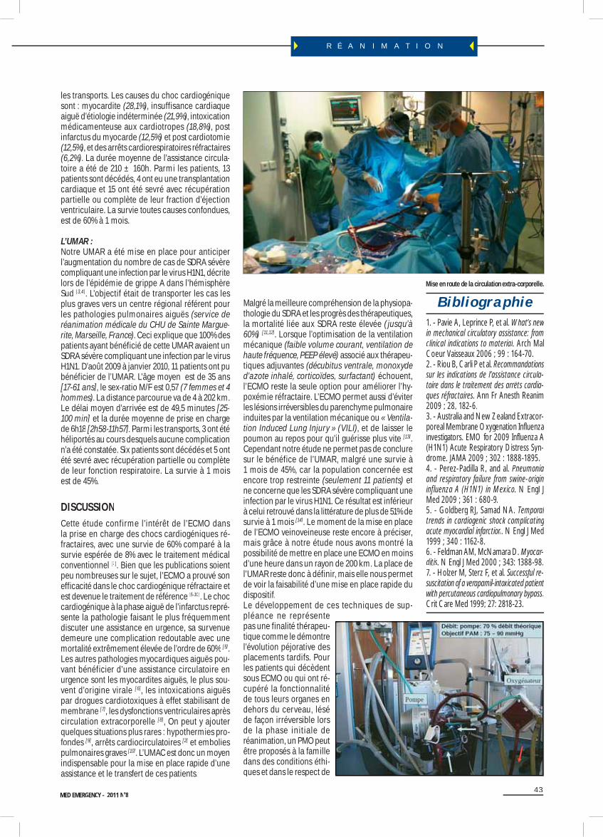
43MED EMERGENCY - 2011 No8
Bibliographie
les transports. Les causes du choc cardiogénique sont : myocardite (28,1%), insuffisance cardiaqueaiguë d’étiologie indéterminée (21,9%), intoxication médicamenteuse aux cardiotropes (18,8%), postinfarctus du myocarde (12,5%) et post cardiotomie(12,5%), et des arrêts cardiorespiratoires réfractaires (6,2%). La durée moyenne de l’assistance circula-toire a été de 210 ± 160h. Parmi les patients, 13 patients sont décédés, 4 ont eu une transplantation cardiaque et 15 ont été sevré avec récupération partielle ou complète de leur fraction d’éjection ventriculaire. La survie toutes causes confondues,est de 60% à 1 mois.
L’UMAR :Notre UMAR a été mise en place pour anticiper l’augmentation du nombre de cas de SDRA sévère compliquant une infection par le virus H1N1, décritelors de l’épidémie de grippe A dans l’hémisphère Sud 3,4 . L’objectif était de transporter les cas les plus graves vers un centre régional référent pour les pathologies pulmonaires aiguës (service deréanimation médicale du CHU de Sainte Margue-rite, Marseille, France). Ceci explique que 100% des patients ayant bénéficié de cette UMAR avaient unSDRA sévère compliquant une infection par le virusH1N1. D’août 2009 à janvier 2010, 11 patients ont pubénéficier de l’UMAR. L’âge moyen est de 35 ans [17-61 ans], le sex-ratio M/F est 0,57 (7 femmes et 4hommes). La distance parcourue va de 4 à 202 km. Le délai moyen d’arrivée est de 49,5 minutes [25-100 min] et la durée moyenne de prise en chargede 6h18 [2h58-11h57]. Parmi les transports, 3 ont été héliportés au cours desquels aucune complication n’a été constatée. Six patients sont décédés et 5 ontété sevré avec récupération partielle ou complète de leur fonction respiratoire. La survie à 1 moisest de 45%.
DISCUSSIONCette étude confirme l’intérêt de l’ECMO dans la prise en charge des chocs cardiogéniques ré-fractaires, avec une survie de 60% comparé à la survie espérée de 8% avec le traitement médicalconventionnel 1 . Bien que les publications soientpeu nombreuses sur le sujet, l’ECMO a prouvé sonefficacité dans le choc cardiogénique réfractaire et est devenue le traitement de référence 5-10 . Le choccardiogénique à la phase aiguë de l’infarctus repré-sente la pathologie faisant le plus fréquemment discuter une assistance en urgence, sa survenuedemeure une complication redoutable avec une mortalité extrêmement élevée de l’ordre de 60% [5]. Les autres pathologies myocardiques aiguës pou-vant bénéficier d’une assistance circulatoire en urgence sont les myocardites aiguës, le plus sou-vent d’origine virale [6], les intoxications aiguëspar drogues cardiotoxiques à effet stabilisant de membrane [7], les dysfonctions ventriculaires aprèscirculation extracorporelle [8], On peut y ajouter quelques situations plus rares : hypothermies pro-fondes [9], arrêts cardiocirculatoires [2] et embolies pulmonaires graves [10]. L’UMAC est donc un moyen indispensable pour la mise en place rapide d’uneassistance et le transfert de ces patients.
Malgré la meilleure compréhension de la physiopa-thologie du SDRA et les progrès des thérapeutiques,la mortalité liée aux SDRA reste élevée (jusqu’à60%) [11,12]. Lorsque l’optimisation de la ventilation mécanique (faible volume courant, ventilation de haute fréquence, PEEP élevé) associé aux thérapeu-tiques adjuvantes (décubitus ventrale, monoxyde d’azote inhalé, corticoïdes, surfactant) échouent, l’ECMO reste la seule option pour améliorer l’hy-poxémie réfractaire. L’ECMO permet aussi d’éviter les lésions irréversibles du parenchyme pulmonaire induites par la ventilation mécanique ou « Ventila-tion Induced Lung Injury » (VILI), et de laisser lepoumon au repos pour qu’il guérisse plus vite [13]. Cependant notre étude ne permet pas de conclure sur le bénéfice de l’UMAR, malgré une survie à 1 mois de 45%, car la population concernée est encore trop restreinte (seulement 11 patients) et ne concerne que les SDRA sévère compliquant une infection par le virus H1N1. Ce résultat est inférieur à celui retrouvé dans la littérature de plus de 51% de survie à 1 mois [14]. Le moment de la mise en placede l’ECMO veinoveineuse reste encore à préciser,mais grâce à notre étude nous avons montré lapossibilité de mettre en place une ECMO en moins d’une heure dans un rayon de 200 km. La place del’UMAR reste donc à définir, mais elle nous permet de voir la faisabilité d’une mise en place rapide du dispositif.Le développement de ces techniques de sup-pléance ne représentepas une finalité thérapeu-tique comme le démontre l’évolution péjorative des placements tardifs. Pour les patients qui décèdentsous ECMO ou qui ont ré-cupéré la fonctionnalitéde tous leurs organes endehors du cerveau, léséde façon irréversible lors de la phase initiale deréanimation, un PMO peutêtre proposés à la famille dans des conditions éthi-ques et dans le respect de
1. - Pavie A, Leprince P, et al. What’s new in mechanical circulatory assistance: fromclinical indications to material. Arch MalCoeur Vaisseaux 2006 ; 99 : 164-70.2. - Riou B, Carli P et al. Recommandations sur les indications de l’assistance circula-toire dans le traitement des arrêts cardia-ques réfractaires. Ann Fr Anesth Reanim2009 ; 28, 182-6.3. - Australia and New Zealand Extracor-poreal Membrane Oxygenation Influenza investigators. EMO for 2009 Influenza A (H1N1) Acute Respiratory Distress Syn-drome. JAMA 2009 ; 302 : 1888-1895.4. - Perez-Padilla R, and al. Pneumoniaand respiratory failure from swine-origin inf luenza A (H1N1) in Mexico. N Engl J Med 2009 ; 361 : 680-9.5. - Goldberg RJ, Samad NA. Temporal trends in cardiogenic shock complicating acute myocardial infarction. N Engl J Med1999 ; 340 : 1162-8.6. - Feldman AM, McNamara D. Myocar-ditis. N Engl J Med 2000 ; 343: 1388-98.7. - Holzer M, Sterz F, et al. Successful re-suscitation of a verapamil-intoxicated patientwith percutaneous cardiopulmonary bypass.Crit Care Med 1999; 27: 2818-23.
Mise en route de la circulation extra-corporelle.
R É A N I M A T I O N

44MED EMERGENCY - 2011 No8
Bibliographie (suite)
la législation française. Cette attitude est justifiée par la pénurie de greffons en France.Le coût financier est estimé à 100 000 euros pour la pompe et 20 000 euros à chaque déplacementde l’équipe, il est pris en charge par l’APHM, avec l’aide une subvention de 250 000 euros du conseilrégional de la région PACA. Il reste inférieur par année de vie gagnée à ceux générés par une trans-plantation hépatique, une greffe de moelle osseuse,une transplantation cardiaque ou rénale [13].
CONCLUSIONOn constate que ces unités mobiles d’assistance cardiorespiratoire ont eu un impact important sur l’activité du SAMU 13, en permettant le transfert de patients considérés jusque là trop instables. L’éva-
luation du gain sur la morbiditéet la mortalité nécessite reste à préciser, mais semble être favo-rable en vue de nos résultats, ilssemblent être d’autant plus favo-rable que l’ECMO est mis en placeprécocement.Un travail d’information desmédecins urgentistes, des car-diologues, des chirurgiens et des réanimateurs est donc nécessaireafin de reconnaître précocement les patients pouvant bénéficier de l’ECMO mobile, avant l’installa-tion d’un SDMV ou de séquellesneurologiques trop importantes,voire d’un ACR.
Cette organisation demande un investissementfinancier, logistique et humain très important, qu’il faudra évaluer avant la création de telles unités au sein d’équipe multidisciplinaire, avec les SAMU au centre du dispositif.
Hichem CHENAITIA(1), Géraldine KARSENTY(1), Fayez JEBRIL(1), Jacques BESSEREAU(1),
Richard TOESCA(1), Emgan QUERELLOU(1),Laurent JACQUIN(1), Marion DOUPLAT(1),
Jean-Pierre AUFFRAY(1,2), Frédéric COLLART(3),Pierre MICHELET(2), Vlad GARIBOLDI(3).
1. Pôle RUSH, SAMU 13, CHU La Timone, Marseille, France2. Pôle RUSH, Réanimation des urgences, CHU St Marguerite, Marseille.3. Unité de réanimation cardio-circulatoire, CHU La Timone, Marseille.
Correspondance : Chenaitia Hichem. Pôle RUSH, SAMU 13.E-mail : [email protected]
8. - Williams MR, Oz MC. Indications and patient selection for mechanicalventricular assistance. Ann Thorac Surg2001 ; 71(Suppl. 3) : S86-91.9. - Vretenar DF, et al. Cardiopulmonarybypass resuscitation for accidental hypo-thermia. Ann Thorac Surg 1994 ; 58 : 895-8.10. - Schwarz B et al. Experience with per-cutaneous venoarterial cardiopulmonary bypass for emergency circulatory support. Crit Care Med 2003 ; 31 :758-64.11. - Hemmila MR et al. ExtracorporealLife Support for Severe Acute Respiratory Distress Syndrome in Adults. Ann Surg2004 ; 240 : 595-605.12. - Peek GJ et al. Efficacy and economic assessment of conventional ventilatory sup-port versus extracorporeal membrane oxy-genation for severe adult respiratory failure(CESAR): a multicentre randomised con-trolled trial. Lancet 2009 ; 374 : 1351-63.13. - Manzona C et al. Extracorporealmembrane oxygenation in the critically illadults. Réanimation 2009 ; 18 : 439-44.14. - Kolla S et al. Extracorporeal life sup-port for 100 adult patients with severe respiratory failure. Ann Surg 1997 ; 226 :544-64.
Revue de presseAssistance circulatoire extracorporelle pour assurer la sur-vie d’une intoxication grave à la flécaïne et au bétaxolol. Benoit Vivien et al. Assistance Publique des Hôpitaux de Paris.Une femme de 40 ans est prise en charge par les services pré-hospitaliers pour une intoxicationthéoriquement létale par 12 g (!) de flécaïne et 400 mg de bétaxo-lol. A l’arrivée aux urgences, elle présente un score de Glasgow à 6(E1,V1,M4), une PA à 70/50 mmHg,un pouls à 32 bpm, et une SpO2
indétectable sous O2 15 L/min. A l’ECG, on peut noter des QRS élargis (0,32 sec.). Après intubationtrachéale, elle fait deux arrêts car-diaques nécessitant 8 et 12 minu-tes de réanimation intensive avecMCE, épinéphrine (8 mg), gluca-gon (9 mg) et Bicarbonate 8,4%
(250 mL). La patiente reste en étatde choc cardiogénique réfractaireà l’épinéphrine en continu. Il estdécidé de tenter une assistancecirculatoire (Rotaflow®, Maquet), dispositif pouvant délivrer un dé-bit cardiaque à 3 L/min environ.Avant la mise en place du dispo-sitif, la patiente fit un autre arrêtcardiaque, récupéré, et était sousdobutamine (20 g/kg/min). Sous assistance circulatoire, le chocs’est progressivement amendé, etl’ECG amélioré, mais l’EEG restaitisoélectrique. Au quatrième jour,après accord de la famille, il fut dé-cidé de prélever ses organes pour transplantation. Quarante-cinqmois plus tard, les organes pré-levés (foi et reins) étaient toujours fonctionnels chez les receveurs.Pour les auteurs, l’assistance cir-culatoire, si elle n’a pu assurer lasurvie de la patiente a permis lavitalité d’organes « transplanta-bles ».
Annals of Emergency MedicineVol. 56.N°4 ; 409-412.
Prise en charge pharmacologi-que (antiagrégants, anticoagu-lants) du syndrome coronarien aigu (SCA).J.-M. Juliard. CHU Bichat.Au cours des SCA, le rôle de l’agrégation plaquettaire se déve-loppant au contact d’une plaquevulnérable rompue est le stimulus principal favorisant la formation d’un thrombus fibrinocruoriqueocclusif au cours du SCA ST+ f (in-farctus du myocarde), parfois non occlusif avec embolies distalesdans le lit coronaire au cours duSCA ST- (angor instable). Aspirine et clopidogrel sont incon-tournables en phase aiguë.Les indications des anti-GPIIb/IIIa se sont restreintes au profit des doses de charge de clopidogrel au cours des SCA ST-, ces molécules étant plus volontiers introduites au cours de l’angioplastie à hautrisque thrombotique. Au cours des SCA ST+, les anti-GP IIb/IIIa associés à l’héparine sont en com-pétition avec la bivaluridine seule,
dont l’usage en préhospitalier n’a pas encore été validé. En ce qui concerne les anticoagu-lants, l’héparine non fractionnée(HNF) a été remplacée par les hé-parines de bas poids moléculaire, puis par le fondaparinux au coursdes SCA ST-. En association avec la fibrinolyse au cours des SCA ST+, l’énoxaparine a été validée, avec un recours possible à l’HNF pour les sujets âgés à fonction rénale normale. En cas d’angioplastie pri-maire, l’énoxaparine est en cours d’évaluation en comparaison avec l’HNF. Le développement de nouvelles molécules antiplaquettaires et anticoagulantes sera source de prochaines modifications théra-peutiques. Il faut cependant, rester vigilant, et veiller au rapport béné-fice/risque, l’efficacité antithrom-botique d’un médicament allant souvent de pair avec son potentiel hémorragique.
Réanimation (2010) 19, 95-102.
LUCAS2_French_final_v2.indd 1 9/3/2009 2:15:11 PM
R É A N I M A T I O N
Conflict of interest statement:There is no conflict of interest to declare
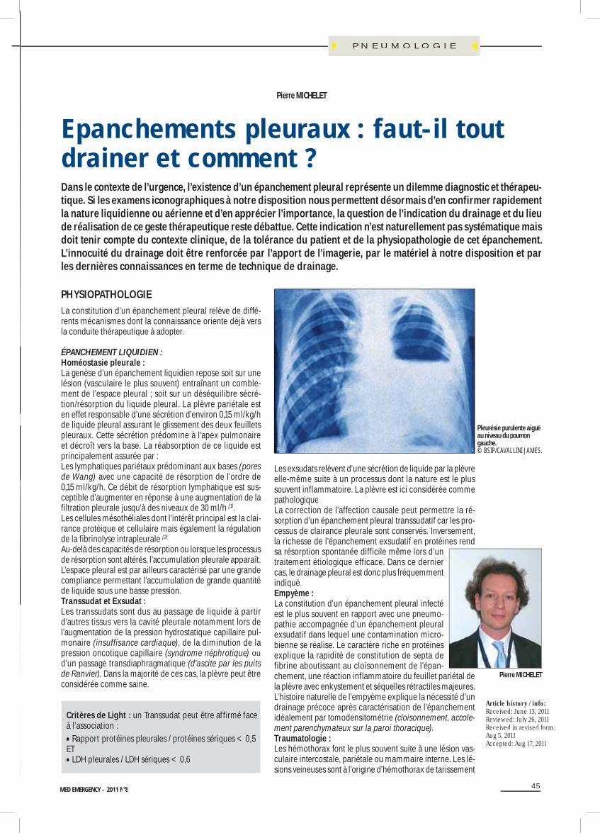
45MED EMERGENCY - 2011 No8
Pierre MICHELET
Epanchements pleuraux : faut-il tout drainer et comment ?Dans le contexte de l’urgence, l’existence d’un épanchement pleural représente un dilemme diagnostic et thérapeu-tique. Si les examens iconographiques à notre disposition nous permettent désormais d’en confirmer rapidementla nature liquidienne ou aérienne et d’en apprécier l’importance, la question de l’indication du drainage et du lieu de réalisation de ce geste thérapeutique reste débattue. Cette indication n’est naturellement pas systématique mais doit tenir compte du contexte clinique, de la tolérance du patient et de la physiopathologie de cet épanchement. L’innocuité du drainage doit être renforcée par l’apport de l’imagerie, par le matériel à notre disposition et par les dernières connaissances en terme de technique de drainage.
PHYSIOPATHOLOGIE La constitution d’un épanchement pleural relève de diffé-rents mécanismes dont la connaissance oriente déjà vers la conduite thérapeutique à adopter.
ÉPANCHEMENT LIQUIDIEN :Homéostasie pleurale :La genèse d’un épanchement liquidien repose soit sur unelésion (vasculaire le plus souvent) entraînant un comble-ment de l’espace pleural ; soit sur un déséquilibre sécré-tion/résorption du liquide pleural. La plèvre pariétale est en effet responsable d’une sécrétion d’environ 0,15 ml/kg/hde liquide pleural assurant le glissement des deux feuillets pleuraux. Cette sécrétion prédomine à l’apex pulmonaireet décroît vers la base. La réabsorption de ce liquide estprincipalement assurée par :Les lymphatiques pariétaux prédominant aux bases (poresde Wang) avec une capacité de résorption de l’ordre de 0,15 ml/kg/h. Ce débit de résorption lymphatique est sus-ceptible d’augmenter en réponse à une augmentation de la filtration pleurale jusqu’à des niveaux de 30 ml/h [1]. Les cellules mésothéliales dont l’intérêt principal est la clai-rance protéique et cellulaire mais également la régulationde la fibrinolyse intrapleurale [2].Au-delà des capacités de résorption ou lorsque les processusde résorption sont altérés, l’accumulation pleurale apparaît.L’espace pleural est par ailleurs caractérisé par une grande compliance permettant l’accumulation de grande quantité de liquide sous une basse pression.Transsudat et Exsudat :Les transsudats sont dus au passage de liquide à partir d’autres tissus vers la cavité pleurale notamment lors de l’augmentation de la pression hydrostatique capillaire pul-monaire (insuffisance cardiaque), de la diminution de la pression oncotique capillaire (syndrome néphrotique) oud’un passage transdiaphragmatique (d’ascite par les puitsde Ranvier). Dans la majorité de ces cas, la plèvre peut êtreconsidérée comme saine.
Critères de Light : un Transsudat peut être affirmé face à l’association :
Rapport protéines pleurales / protéines sériques < 0,5 ET
LDH pleurales / LDH sériques < 0,6
Les exsudats relèvent d’une sécrétion de liquide par la plèvre elle-même suite à un processus dont la nature est le plus souvent inflammatoire. La plèvre est ici considérée comme pathologiqueLa correction de l’affection causale peut permettre la ré-sorption d’un épanchement pleural transsudatif car les pro-cessus de clairance pleurale sont conservés. Inversement, la richesse de l’épanchement exsudatif en protéines rend sa résorption spontanée difficile même lors d’un traitement étiologique efficace. Dans ce dernier cas, le drainage pleural est donc plus fréquemment indiqué.Empyème :La constitution d’un épanchement pleural infectéest le plus souvent en rapport avec une pneumo-pathie accompagnée d’un épanchement pleural exsudatif dans lequel une contamination micro-bienne se réalise. Le caractère riche en protéines explique la rapidité de constitution de septa defibrine aboutissant au cloisonnement de l’épan-chement, une réaction inflammatoire du feuillet pariétal de la plèvre avec enkystement et séquelles rétractiles majeures.L’histoire naturelle de l’empyème explique la nécessité d’un drainage précoce après caractérisation de l’épanchement idéalement par tomodensitométrie (cloisonnement, accole-ment parenchymateux sur la paroi thoracique).Traumatologie :Les hémothorax font le plus souvent suite à une lésion vas-culaire intercostale, pariétale ou mammaire interne. Les lé-sions veineuses sont à l’origine d’hémothorax de tarissement
Pierre MICHELET
Pleurésie purulente aiguëau niveau du poumongauche.© BSIP/CAVALLINI JAMES.
P N E U M O L O G I E
LUCAS2_French_final_v2.indd 1 9/3/2009 2:15:11 PM
Article history / info:Received: June 13, 2011Reviewed: July 26, 2011Received in revised form: Aug 5, 2011Accepted: Aug 17, 2011
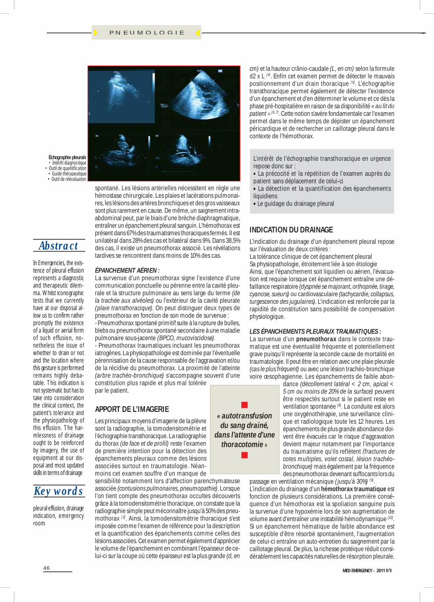
46 MED EMERGENCY - 2011 No8
« autotransfusion du sang drainé,
dans l’attente d’unethoracotomie »
spontané. Les lésions artérielles nécessitent en règle une hémostase chirurgicale. Les plaies et lacérations pulmonai-res, les lésions des artères bronchiques et des gros vaisseaux sont plus rarement en cause. De même, un saignement intra-abdominal peut, par le biais d’une brèche diaphragmatique, entraîner un épanchement pleural sanguin. L’hémothorax est présent dans 67% des traumatismes thoraciques fermés. Il est unilatéral dans 28% des cas et bilatéral dans 9%. Dans 38,5% des cas, il existe un pneumothorax associé. Les révélations tardives se rencontrent dans moins de 10% des cas.
ÉPANCHEMENT AÉRIEN :La survenue d’un pneumothorax signe l’existence d’une communication ponctuelle ou pérenne entre la cavité pleu-rale et la structure pulmonaire au sens large du terme (de la trachée aux alvéoles) ou l’extérieur de la cavité pleurale (plaie transthoracique). On peut distinguer deux types depneumothorax en fonction de son mode de survenue : - Pneumothorax spontané primitif suite à la rupture de bulles, blebs ou pneumothorax spontané secondaire à une maladie pulmonaire sous-jacente (BPCO, mucoviscidose).- Pneumothorax traumatiques incluant les pneumothorax iatrogènes. La physiopathologie est dominée par l’éventuelle pérennisation de la cause responsable de l’aggravation et/oude la récidive du pneumothorax. La proximité de l’atteinte (arbre trachéo-bronchique) s’accompagne souvent d’une constitution plus rapide et plus mal tolérée par le patient.
APPORT DE L’IMAGERIELes principaux moyens d’imagerie de la plèvre sont la radiographie, la tomodensitométrie et l’échographie transthoracique. La radiographie du thorax (de face et de profil) reste l’examende première intention pour la détection des épanchements pleuraux comme des lésions associées surtout en traumatologie. Néan-moins cet examen souffre d’un manque de sensibilité notamment lors d’affection parenchymateuse associée (contusions pulmonaires, pneumopathie). Lorsque l’on tient compte des pneumothorax occultes découvertsgrâce à la tomodensitométrie thoracique, on constate que laradiographie simple peut méconnaître jusqu’à 50% des pneu-mothorax [3]. Ainsi, la tomodensitométrie thoracique s’est imposée comme l’examen de référence pour la descriptionet la quantification des épanchements comme celles deslésions associées. Cet examen permet également d’apprécier le volume de l’épanchement en combinant l’épaisseur de ce-lui-ci sur la coupe où cette épaisseur est la plus grande (d, en
cm) et la hauteur crânio-caudale (L, en cm) selon la formule d2 x L [4]. Enfin cet examen permet de détecter le mauvais positionnement d’un drain thoracique [5]. L’échographie transthoracique permet également de détecter l’existenced’un épanchement et d’en déterminer le volume et ce dès laphase pré-hospitalière en raison de sa disponibilité « au lit dupatient » [6, 7]. Cette notion s’avère fondamentale car l’examenpermet dans le même temps de dépister un épanchementpéricardique et de rechercher un caillotage pleural dans lecontexte de l’hémothorax.
L’intérêt de l’échographie transthoracique en urgence repose donc sur : La précocité et la répétition de l’examen auprès du
patient sans déplacement de celui-ci La détection et la quantification des épanchements
liquidiens Le guidage du drainage pleural
INDICATION DU DRAINAGE L’indication du drainage d’un épanchement pleural repose sur l’évaluation de deux critères :La tolérance clinique de cet épanchement pleuralSa physiopathologie, étroitement liée à son étiologieAinsi, que l’épanchement soit liquidien ou aérien, l’évacua-tion est requise lorsque cet épanchement entraîne une dé-faillance respiratoire (dyspnée se majorant, orthopnée, tirage,cyanose, sueurs) ou cardiovasculaire (tachycardie, collapsus,turgescence des jugulaires). L’indication est renforcée par larapidité de constitution sans possibilité de compensation physiologique.
LES ÉPANCHEMENTS PLEURAUX TRAUMATIQUES :La survenue d’un pneumothorax dans le contexte trau-matique est une éventualité fréquente et potentiellement grave puisqu’il représente la seconde cause de mortalité en traumatologie. Il peut être en relation avec une plaie pleurale (cas le plus fréquent) ou avec une lésion trachéo-bronchique voire œsophagienne. Les épanchements de faible abon-
dance (décollement latéral < 2 cm, apical < 5 cm ou moins de 20% de la surface) peuventêtre respectés surtout si le patient reste en ventilation spontanée [8]. La conduite est alors une oxygénothérapie, une surveillance clini-que et radiologique toute les 12 heures. Les épanchements de plus grande abondance doi-vent être évacués car le risque d’aggravation devient majeur notamment par l’importance du traumatisme qu’ils reflètent (fractures decotes multiples, volet costal, lésion trachéo-bronchique) mais également par la fréquence des pneumothorax devenant suffocants lors du
passage en ventilation mécanique (jusqu’à 30%) [9].L’indication du drainage d’un hémothorax traumatique est fonction de plusieurs considérations. La première consé-quence d’un hémothorax est la spoliation sanguine puis la survenue d’une hypoxémie lors de son augmentation de volume avant d’entraîner une instabilité hémodynamique [10]. Si un épanchement hématique de faible abondance est susceptible d’être résorbé spontanément, l’augmentation de celui-ci entraîne un auto-entretien du saignement par lacaillotage pleural. De plus, la richesse protéique réduit consi-dérablement les capacités naturelles de résorption pleurale.
Échographie pleurale• Intérêt diagnostique
• Outil de quantification• Guide thérapeutique• Outil de réévaluation
P N E U M O L O G I E
AbstractIn Emergencies, the exis-tence of pleural effusion represents a diagnostic and therapeutic dilem-ma. Whilst iconographic tests that we currently have at our disposal al-low us to confirm rather promptly the existence of a liquid or aerial form of such ef fusion, no-netheless the issue of whether to drain or not and the location where this gesture is performed remains highly deba-table. This indication is not systematic but has to take into consideration the clinical context, the patient’s tolerance and the physiopathology of this effusion. The har-mlessness of drainage ought to be reinforced by imagery, the use of equipment at our dis-posal and most updated skills in terms of drainage.
Key wordspleural effusion, drainage indication, emergency room.
y

47MED EMERGENCY - 2011 No8
Enfin, le risque de surinfection de cette collection hématiqueou de séquelles rétractiles pleurales sont bien réel [11]. Il existe par ailleurs différent arguments expérimentaux et cliniquesrapportant le risque de tamponnade cardiaque suite à unhémothorax soit par l’importance de celui-ci soit par l’exis-tence d’un caillotage en regard des cavités cardiaques etcomprimant celles-ci [12, 13]. Si l’indication du drainage estretenue et que l’état clinique du patient le permet, celui-cidoit être réalisé dans les meilleures conditions de sécurité.Une radiographie du thorax ainsi qu’un scanner permettrons ainsi de repérer la position de la coupole diaphragmatique et d’écarter une lésion de cette dernière avec risque de migration des structures digestives (faux positif). En tenant compte de ces différents aspects, il peut être proposé dif-férents scénarii :
Un hémothorax de faible abondance (estimé à 200-300 cc)sans instabilité respiratoire ou cardio-vasculaire peut être respecté avec une surveillance radiologique (échographique)et clinique.
Un hémothorax de plus grande abondance mais bien toléré cliniquement doit pouvoir être drainé en milieu spé-cialisé après un bilan iconographique et dans les meilleurs conditions d’asepsie. L’indication du drainage repose ici sur l’impossibilité d’une résorption spontanée, l’altération del’oxygénation secondaire au troubles ventilatoires induits, les conséquences infectieuses ou fonctionnelles futures (séquel-les rétractiles) [14]. Le drainage permet en outre de surveiller ledébit hémorragique et de poser éventuellement l’indicationd’une thoracotomie secondaire. Une thoracotomie ou vidéo-thoracoscopie peut être indiquée lors d’un drainage > 1 500 ml d’emblée, d’un débit de drainage > 250-300 ml/h pendant2 à 4 heures, un drainage quotidien > 1 500 ml. Face à un hémothorax massif avec instabilité hémodyna-
mique majeure, l’indication du drainage pleural est parfois discuté car susceptible d’entraîner une exsanguination. Rete-nons qu’outre le risque très hypothétique de levé du contrôle du saignement par l’hémothorax lui même, il est bien établiqu’un hémothorax massif peut provoquer une tamponnadecardiaque et que c’est le drainage en urgence qui permettra la correction de ces troubles en association avec une expan-sion volémique massive [15, 16]. Face à la poursuite d’un saigne-ment massif après drainage, l’urgence est à la thoracotomie d’hémostase et diagnostique. Une autotransfusion du sang drainé, soit par un système non spécifique (recueil du sang dans une poche à urine stérile) [17], soit, de préférence, par un système prêt à l’emploi (inclus dans le kit de drainage ouCell Saver™ ; Haemonetics) doit être réalisé dans l’attente de la thoracotomie si celle-ci ne peut être réalisée rapidement (en préhospitalier notamment). Face à un hémothorax traumatique associé à une rupture
isthmique de l’aorte (lésion des vaisseaux médiastinaux la plus fréquente), l’indication du drainage est également dis-cutée selon le principe que l’hémothorax prévient la rupturecomplète de l’aorte ou réduit le débit du saignement. Si l’état hémodynamique du patient permet l’attente de la solution chirurgicale (pose d’endoprothèse ou chirurgie ouverte), il est en effet logique de retarder le drainage. Lorsque lepatient est en détresse circulatoire deux cas de figure sont possibles. Soit la rupture vasculaire est complète, l’hémotho-rax est alors le reflet de la gravité extrême de la lésion et lagrande compliance de la cavité pleurale ne serait générer des pressions suffisantes pour stopper un saignement arté-riel [10]. Dans ce cas, la thoracotomie de sauvetage est la seule solution thérapeutique et la mortalité reste très élevée. Soit enfin et c’est le cas le plus fréquent, la rupture est incomplète, ne concernant pas toutes les tuniques vasculaires (intima et
média), et c’est le risque de tamponnade et de pérennisation de l’hémorragie qui induit un risque vital. Nous avons ainsi pu rapporter l’intérêt du drainage thoracique auprès de pa-tients en détresse circulatoire terminale (dont un patient en arrêt cardio-circulatoire) et présentant une lésion isthmiquechez lesquels le drainage thoracique à permis une restau-ration de l’hémodynamique sans aggravation de la lésionvasculaire qui fut contrôlée par pose d’une endoprothèse aortique [18]. Cette réflexion a par ailleurs été rapportée par d’autres équipes confirmant que les hémothorax associés ne sont quasiment jamais causé par la rupture isthmique mais par les lésions pulmonaires ou costales associées [19, 20].
ÉPANCHEMENTS PLEURAUX LIQUIDIENS NON TRAUMA-TIQUES :Les causes d’épanchements pleuraux de nature « médicale »sont nombreuses (remplissage excessif, causes cardiaques,atélectasies, hypoalbuminémie, hépathopathie...). L’indi-cation du drainage repose sur le retentissement clinique(respiratoire) de celui-ci ou sur la nécessité diagnostique.En effet, la résorption est souvent obtenue par le traitement étiologique. De plus, il ne faut jamais méconnaître le risque iatrogène et les conséquences algiques (parfois handica-pantes) de la procédure de drainage surtout si un dispositif est laissé en place. Pour un épanchement pleural satellited’une pneumopathie, la démarche thérapeutique reposesur l’importance de l’épanchement, sa nature et la durée de l’évolution. Ainsi un épanchement pleural modéré, dont laponction révèle un liquide non purulent peut faire surseoir au drainage (ponction évacuatrice uniquement) pour un traite-ment antibiotique et kinésithérapeutique d’épreuve. Inverse-ment, tout épanchement de moyenne à grande abondance,purulent, non résolutif sous un traitement antibiotique bien conduit ou entraînant une défaillance respiratoire nécessiteun drainage complet par un drain thoracique laissé en placeet ce sans délai [21].
PNEUMOTHORAX SPONTANÉS :La réflexion concernant le pneumothorax porte sur les ca-pacités de résorption spontanée du pneumothorax ainsi quele risque de récidive ou d’aggravation. On distingue ainsi lespetits pneumothorax (décollement < 2 cm latéral, 5 cm à l’apex) dont la résorption spontanée est probable (2 à 5%par jour) si la cause est contrôlée, des pneumothorax deplus grande taille plus souvent symptomatiques requérant une évacuation et un contrôle à 24 heures. La récidive ou la survenue dans la cadre d’une maladie pulmonaire han-dicapante (mucoviscidose) peut motiver le drainage voire le talcage pleural.
Les différents principes• Le drainage
chirurgical :- Incision cutanée, la
dissection souscutanée et musculaire à
l’index ou la pincecourbe de Kelly
- Guidage du drain au doigt
- Plus traumatisant maisplus sûr
• Le drainage avectrocart court type
Monod- Effondrement de la
plèvre pariétale etl’orientation du drain.
- Incision plus réduite, procédure accélérée.
- Plus de malpositions.
P N E U M O L O G I E

48 MED EMERGENCY - 2011 No8
TECHNIQUE DE DRAINAGEMATÉRIEL :Le drainage thoracique, lorsque sont indication est retenue, peut être réalisée avec différentes tailles de drain en fonction de la nature de l’épanchement. Les épanchements aériens sont susceptibles d’être évacué pas des drains de petitestailles type Pleurocath® (8 French) [22] ou des drains de 16 à 20 French. Toute suspicion d’épanchement liquidien doitpouvoir bénéficier d’un drainage par un drain dont la taille sera de 24 à 32 French afin de limiter le risque d’obstructiondu drain [23].
TECHNIQUE :• Le site de drainage le plus souvent rapporté est par lavoie latérale, dans le triangle de sécurité bordé en avant par le bord latéral du muscle grand pectoral et en arrière par le bord latéral du muscle grand dorsal, en haut par le 3° EIC et en bas par le 5° EIC. La voie antérieure (peu recommandée par les équipes anglo-saxonnes) entre le 2° et 3° EIC sur laligne médioclaviculaire est essentiellement utilisée pour le drainage des pneumothorax.
La majorité des épanchements drainés entre le 3° et le 5° EIC, patient en décubitus dorsal, le sont avec un abord pleural en dehors de la zone de collection liquidienne. L’abord se fait donc feuillet pariétal contre feuillet viscéral (voir schéma).
• Il existe cinq principes possibles pour le drainage :- Le drainage chirurgical qui consiste en une incision cuta-lnée, la dissection sous cutanée et musculaire à l’index ou lapince courbe de Kelly, le franchissement de la plèvre parié-tale au doigt puis en l’introduction prudente du drain guidépar le doigt ou la pince dans la direction dictée par la nature de l’épanchement à la surface du parenchyme pulmonairelimitant ainsi le risque de traumatisme pulmonaire [24].
- Le drainage avec un trocart court type Monod qui permettl’effondrement de la plèvre pariétale et l’orientation du drain. Si l’incision peut être plus réduite et la procédure accélérée,il semble que cette technique soit grevée d’un manque d’an-gulation avec de plus fréquentes malpositions [5].
- Le drainage avec trocart long avec utilisation d’un drainthoracique borgne à bout mousse rigidifié par un guide mé-tallique type Joly ou Mallinckrodt. La phase de passage de la plèvre pariétale doit être très prudente en raison du risque de lésion parenchymateuse. La phase de montée du drain (sur son guide orienté) est en revanche simplifiée.
- Le drainage à travers une aiguille type « Pleurocath ». La phase de passage des plans cutanés et musculaires est facilitée par le caractère tranchant de l’aiguille dans laquelleest placé le drain. Le risque de ponction du parenchyme estpar contre important. Ce risque pourrait être fortement réduitpar l’utilisation de nouveau dispositif à embout mousse etrétractable sur lequel est monté le drain de 8F type « Argyle Turkel ».
- Le drainage selon la technique de « Seldinger » avec une ponction à l’aiguille de l’espace interpleural dans lequel estmonté le guide. Sur ce dernier seront introduits un dilatateur puis un drain multiperforé de 9 à 14F. La ponction initiale àl’aiguille présente un risque de ponction parenchymateuseréservant souvent cette technique aux ponctions écho-gui-dées. L’utilisation d’aiguilles moins traumatiques de type aiguille pour ponction péridurale peut réduire ce risque etpermettre une orientation optimisée du drainage.
Seuls les épanchements engageant le pronostic vitalimmédiat sont drainés en urgence sans bilan radiologique préalable. Le premier geste est alors la mise en place d’une aiguille afin de décompresser la cavité pleurale (fréquence des pneumothorax suffocant) et de repérerl’épanchement. Ce geste sera suivi d’un véritable drainage de la cavité.
• En contexte traumatologique, l’absence de localisationdes coupoles diaphragmatiques (possible surélévation ouissue d’organes intestinaux) motive une ponction « haute »soit antérieure au niveau du deuxième espace intercostalsoit latérale au niveau 3°-4° EIC (Schéma 1). La nécessité d’ungeste rapide fait souvent préférer le drainage par trocart courtou long, une dissection au doigt n’étant pas recommandéeen raison du risque de lésion par esquille osseuse. Lors d’untraumatisme thoracique pénétrant, il n’est pas recommandé d’introduire le drain par l’orifice même lorsque la plaie estsoufflante. En effet, l’introduction du drain dans le trajetlésionnel est susceptible d’aggraver les lésions.
• L’aspiration : suite à la pose du drain, un système aspiratif est le plus souvent adapté afin de maintenir la dépression
La majorité des épanche-ments drainés entre le 3°et le 5° EIC, patient en dé-cubitus dorsal, le sont avec un abord pleural en dehors de la zone de collection li-quidienne. L’abord se fait donc feuillet pariétal contrefeuillet viscéral avec un ris-que de traumatisme paren-chymateux.
Exemple d’un hémothorax de 800 ccenviron avec drainage au 4° ou 5° EIC, patient en décubitus dorsal A ; patient en position demi-assise B.
P N E U M O L O G I E
A
B

49MED EMERGENCY - 2011 No8
RÉFÉRENCES BIBLIOGRAPHIQUES : 1. - Miserocchi G. Physiology and pathophysiology of pleural fluid turnover. Eur Respir J. 1997 ; 10 : 219-25.rr2. - Zocchi L. Physiology and pathophysiology of pleural fluid turnover. Eur Respir J. 2002 ; 20 : 1545-58.rr3. - Ball CG., Hameed SM. , Evans D. et al. Occult pneumothorax in the mechanically ventilated trauma patient. Can J Surg 2003 ; 46 : 373-9.4. - Mergo PJ., Helmberger T., Didovic J. et al. New formula for quantification of pleural effusions from computed tomography. J ThoracImaging 1999; 14 : 122-5.5. - Remérand F., Luce V., et al. Incidence of chest tube malposition in the critically ill: a prospective computed tomography study. Anesthe-siology. 2007 Jun ; 106(6) : 1112-9.6. - Rozycki GS., Pennington SD., Feiciano DV. Surgeon-performed ultrasound in the critical care setting: its use as an extension of the physical examination to detect pleural effusion. J Trauma 2001 ; 50 : 636-42.7. - Roch A., Bojan M., Michelet P. et al. Usefulness of ultrasonography in predicting pleural effusions > 500 mL in patients receiving mechani-cal ventilation. Chest 2005 Jan ; 127(1) : 224-32.8. - Hill SL, Edmisten T, Holtzman G, et al. The occult pneumothorax : an increasing diagnostic entity in trauma. Am Surg 1999 ; 65 : 254-8.9. - Enderson BL, Abdalla R, Frame SB, et al. Tube thoracostomy for occult pneumothorax : a prospective randomized study of its use. J Trauma 1993 ; 35 : 726-30.10. - Nishida O., Arellano R., Cheng D. et al. Gas exchange and hemodynamics in experimental pleural effusion. Crit Care Med. 1999 ; 27 :583-87.11. - Eddy AC, Luna GK, Copass M. Empyema thoracis in patients undergoing emergent closed tube thoracostomy for thoracic trauma. Am J Surg 1989 ; 157 : 494-7.12. - Alam HB. Can pleural effusions cause cardiac tamponade. Chest 1999 ; 116 : 1820-22.13. - Vaska K, Wann LS, Sagar K et al. Pleural effusion as a cause of right ventricular diastolic collapse. Circulation 1992; 86 : 609-17.14. - Watkins JA, Spain DA, et al. Empyema and restrictive pleural processes after blunt trauma : an under-recognized cause of respiratory failure. Am Surg 2000 ; 66 : 210-14.15. - Kopterides P, Lignos M, Papanikolaou S et al. Pleural effusion causing cardiac tamponade : report of two cases and review of the litera-ture. Heart Lung 2006 ; 35 : 66-7. 16. - Kaplan LM, Epstein SK, Schwartz SL et al. Clinical, echocardiographic and hemodynamic evidence of cardiac tamponade caused by large pleural effusion. Am J Respir Crit Care Med 1995 ; 151 : 904-8.17. - Barriot P, Riou B, Viars P. Prehospital autotransfusion in life-threatening hemothorax. Chest 1988 ; 93 : 522-6.18. - Michelet P, Roch A, Amabile P et al. Traitement endovasculaire des ruptures isthmiques de l’Aorte chez le polytraumatisé : avancées dans la prise en charge. Ann Fr Anesth Réa 2005 ; 24 : 355-360.19. - Steingruber IE., et al. Placement of endovascular stent-grafts for emergency repair of acute traumatic aortic rupture: a single-centreexperience. Eur Radiol 2007 Jul ; 17(7) : 1727-37.20. - Richeus L., Dambrin C., Marcheix B., Towards a new management of acute traumatic aortic ruptures. J Radiol 2004 ; 85 : 101-6.21. - Sahn SA. Diagnosis and management of parapneumonic effusions and empyema. Clin Inf Dis 2007 ; 45 : 1480-6.22. - Reinhold C, Bret PM. Treatment of pleural effusions and pneumothorax with catheters placed percutaneously under imaging guidance. Am J Roentgenol 1989 ; 152 : 1189.23. - Hyde J., et al. Reducing morbidity from chest drains. BMJ 1997 Mar 29 ; 314(7085) : 914-5.24. - Laws D, Duffy J. BTS guidelines for the insertion of a chest drain. Thorax 2003 ; 58 : ii53-59.25. - Ali J, Qi W. Effectiveness of chest tube clamping in massive hemothorax. J Trauma 1995 ; 38 : 59-63.26. - Maxwell RA, Campbell DJ, Fabian TC, et al. Use of presumptive antibiotics following tube thoracostomy for traumatic hemopneumothoraxin the prevention of empyema and pneumonia - a multi-center trial. J Trauma 2004 ; 57 : 742-8.
et la vacuité pleurale. La perméabilité du drain doit êtrerégulièrement vérifiée. Ainsi, une attention toute particulière doit être portée aux dispositifs d’aspiration dont les débits peuvent être considérablement réduit par la longueur des dispositifs et l’interposition de raccord. La persistance d’unsaignement actif signe le plus souvent une lésion chirurgi-cale. La première mesure est alors de supprimer l’aspiration en mettant le bocal du drain en siphonage simple. En effet, le clampage du drain ne modifierait pas le débit hémorra-gique [25] mais serait susceptible de rendre l’hémothorax compressif majorant ainsi la défaillance hémodynamique et respiratoire.
La prescription systématique d’une antibiothérapieaprès drainage thoracique est controversée. Dans une étude randomisée récente portant sur 224 patients, une antibiothé-rapie après drainage d’hémopneumothorax ne semble pas limiter l’incidence des empyèmes et des pneumonies [26]. Inversement , il semble que les drainages réalisé lors de plaies thoraciques pénétrante puissent bénéficier d’un trai-tement antibiotique.
CONCLUSIONLe drainage thoracique n’est pas un geste dénué de toute complication et ne pourrait-être systématique face à la présence d’un épanchement pleural. Néanmoins, la des-cription de quelques cas cliniques grevés d’effets adverses « retentissants » au sein de la littérature dans le contextetraumatologique ne doit pas conduire le praticien à adopter une attitude d’éviction toute aussi systématique. En effet, les conséquences respiratoires et hémodynamiques du non drai-nage sont désormais reconnues et scientifiquement établies. Le développement des moyens d’imagerie disponibles « au lit du patient » associé à l’amélioration des techniques de drai-nage nous permettent désormais d’adopter une démarcheréfléchie et sécuritaire vis-à-vis de ce geste technique.
Pierre MicheletRéanimation des Urgences
Hôpital Sainte Marguerite - Place Aristide Boyer. 13009. Marseille
P N E U M O L O G I E
Conflict of interest statement:There is no conflict of interest to declare
A B
• Basic and Advanced Life Support• Emergency Meddiiciine Techhniiques• Disaster Management• EEmergency DDepartment MManagement• Road Safety (new)• Intensive Care Techniques (new)• Combat Life Saver: CLS (new)• Combat Medic Advanced Skill Training: CMAST (new)• Deminingg Medics Advanced Life Supppport (new)
When experience and professionalism meet quality
All our courses are accredited by the Lebanese Ministry of Education
For more information contact New Health Concept SALFanar – Lebanon
Tel: +961-1-888921 / Fax: +961-1-888922Email: [email protected] / website: www.newhealthconcept.net
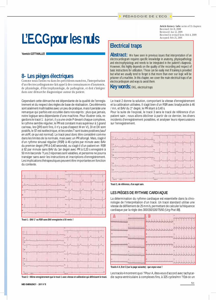
51MED EMERGENCY - 2011 No8
L’E.C.G pour les nulsYannick GOTTWALLES
8 - Les pièges électriques
Cependant cette démarche est dépendante de la qualité de l’enregis-trement et du respect des règles de base de réalisation. Ces éléments sont aisément maîtrisables avec un peu de pratique, mais il persiste une remarque qui parfois est occultée dans nos esprits : plus que jamais, notre logique sera dépendante d’une machine. Pour illustrer cela, re-gardons le tracé 1 : à priori, il y a une onde P devant chaque complexe, le rythme semble régulier, le PR est constant mais supérieur à 1 grand carreau, les QRS sont fins, il n’y a pas d’aspect M en V1, DI et DII sont positifs, le ST est isoélectrique, et les ondes T sont toutes positives (sauf en aVR, ce qui est normal). Le tracé peut donc être considéré commedans les limites de la normale, mais avec un PR allongé. Mais, s’agit-il d’un rythme sinusal régulier (RSR) à 46 cycles par minute avec BAV du premier degré (PR à 0,40 seconde), ou s’agit-il d’un patient en RSR à 92 par minute sans BAV du 1er degré avec PR à 0,20 s enregistré à50 mm/seconde ? Les 2 réponses sont valables, et personne ne pourratransiger sans avoir les instructions et inscriptions d’enregistrement.Les implications thérapeutiques peuvent être importantes en fonction du contexte.
Le tracé 2 donne la solution, comportant la vitesse d’enregistrementet la calibration utilisées. Il s’agit bien d’un RSR avec bradycardie à 46/ mn, et BAV du 1er degré, le PR étant à 0,40 s.r
Pour la suite de l’exposé, le tracé 3 sera le tracé de référence d’unpatient sain ; nous allons décliner à partir de ce dernier, les diversincidents d’enregistrement possibles, et analyser leurs répercussions sur l’enregistrement.
Tracé 1 : BAV 1° ou RSR sans BAV enregistrée à 50 mm/s ?
Tracé 2 : Même enregistrement que le tracé 1 avec vitesse et calibration qui définissent le tracé.
Tracé 3, de référence, d’un sujet sain.
LES PIÈGES DE RYTHME CARDIAQUELa détermination du rythme cardiaque est essentielle dans la chro-nologie de l’interprétation d’un tracé. Un tracé standard utilise unevitesse de défilement de 25 mm/s, permettant de calculer la fréquence cardiaque par la règle des 300/150/100/75/60 (Urg Prat 88).
Les tracés 4 montrent quoi ? Pour A, êtes-vous d’accord avec tachycar-die supra-ventriculaire à complexes fins, à 325 cycles/mn ? Est-ce un
Tracés 4 A, B et C (sur la page suivante) : que voyez-vous ?
AA BB
PÉDAGOGIE DE L’ECG
8- Les pièges électriquesComme nous l’avons vu dans les précédents numéros, l’interprétation d’un électrocardiogramme fait appel à des connaissances d’anatomie, de physiologie, d’électrophysiologie, de pathogénie, et doit s’intégrer
g q pdans une démarche diagnostique autour du patient.
Electrical trapsAbstract: We have seen in previous issues that interpretation of anelectrocardiogram requires specific knowledge in anatomy, physiopathology and electrophysiology and needs to be integrated in the patient’s diagnosis. However, this highly depends on the quality of the recording and respect of basic instructions for utilization. Those can be easily met if training is provided but what we usually tend to forget is that more than ever our logic will be prisoner of a machine. In this chapter, we cover the main electrical traps of an electrocardiogram and ways to avoid them.
Key words: EKG, electrical traps
Article history / info: series of 11 chaptersReceived: Oct 8, 2008Reviewed: Jan 12, 2009Received in revised form: Feb 4, 2009Accepted: Feb 25, 2009

52 MED EMERGENCY - 2011 No8
accès de maladie de Bouveret ? Pour le tracé B, tachycardie sinusale à 162 cycles/mn ? Pour le tracé C, tachycardie sinusale à 130 cycles/mn ? Toutes ces réponses sont justes et fausses en même temps car aucune référence à la vitesse d’enregistrement n’est notée. En fait, il s’agit dumême tracé enregistré en A à une vitesse de 5 mm/s, en B à 10 mm/s, et en C à 12,5 mm/s. La fréquence est toujours la même à 65 cycles/mn, ce n’est que le défilement qui varie. Bien entendu, la prise en charge d’un accès de Bouveret ou d’une tachycardie sinusale, ou d’un RSR n’est pas du tout identique.
LES PIÈGES DE L’AMPLITUDE DU TRACÉUn enregistrement standard s’effectue à 10 mm pour 1 mV, avec une calibration notée en début de tracé. Si cette calibration fait défaut ou n’est pas réglée sur cette valeur, des perturbations peuvent survenir,soit à type d’écrasement des complexes ou d’étirement de ces derniers. Le tracé 5, enregistré avec une amplitude à 2,5 mm pour 1 mV, révèle des complexes microvoltés évoquant un épanchement péricardique voire une tamponnade. Apparaissent des dérivations quasiment plates, notamment en périphérie (DIII ou aVF). Idem pour le tracé 6 sous unecalibration de 5 mm / mV.
CC
Tracé 5 : enregistrement avec une amplitude de 2,5 mm / mV.
À l’inverse, les amplitudes supérieures à 10 mm / mV donneront des tracés avec chevauchement des QRS, surtout en précordial, et feront évoquer à tort des hypertrophies ventriculaires, voire des sus-déca-lages débutants chez les patients jeunes avec un certain éréthisme cardiaque (tracé 7).
Tracé 6 : enregistrement avec une amplitude de 5 mm / mV.
LES PIÈGES AVEC ASSOCIATIONS VITESSEET AMPLITUDECes deux erreurs de calibration peuvent bien entendu se potentialiser,comme cela est visible dans les tracés suivants (tracé 8 A et B). Le tracé A est enregistré à une vitesse de 5 mm/s, avec une amplitude calibréeà 20 mm / mV. Il en résulte un aspect de tachycardie rapide, à com-plexes très fins, à plus de 300 par minute, évoquant une tachycardie supra-ventriculaire très rapide. Un tel tracé est possible sous certaines conditions (sujet très jeune, nouveau-né). Le tracé B semble plus plau-sible et pourrait correspondre à un adolescent après un effort.
Tracé 7 : amplitude de 20 mm.
Tracé 8 A : vitesse de 5 mm/s et amplitude de 20 mm / mV.
Tracé 8 B : vitesse de 10 mm/s et 20 mm / mV.
AA
BB
PÉDAGOGIE DE L’ECG

53MED EMERGENCY - 2011 No8
C
A
B
Parfois, l’aspect du tracé peut être moins choquant, et ce n’est que lafréquence observée qui étonnera. Le tracé 9, avec double vitesse et double amplitude, enregistre des complexes QRS d’aspect normal,mais avec un possible BAV du 1er degré, et une bradycardie à 33 cycles / mn. La repolarisation paraît suspendue notamment en inféro-latéral.
LES PIÈGES DE POSITION DES ÉLECTRODESLes inversions d’électrodes sont fréquentes et régulières, mais sontaisément décelables en respectant, et par conséquent en recherchant, quelques règles de base que l’on peut appeler les règles de territoire.Les complexes doivent être concordant entre eux, notamment dans les territoires coronaires explorés.
L’interventriculaire antérieure qui irrigue la paroi antérieure du ven-tricule gauche, correspond au territoire précordial : de V1 à V5 (voire V6) il doit y avoir une progression régulière de R, et en parallèle maisun peu retardée, une diminution de la profondeur de S. Un rabotagede R en antérieur en possible en cas de séquelles d’infarctus, mais la logique persiste et n’est que retardée avec un démarrage de R en V3ou V4 (Urg Prat 88).
En clair,• R en V1 est inférieur à R en V2 qui est inférieur à R en V3, qui est inférieur à R en V4, qui est inférieur à R en V5, qui est inférieur àR en V5, et• S en V1 est plus profonde que S en V2, qui est plus profonde que S en V3, qui est plus profonde que S en V4, qui est plus profonde que S en V5, qui est plus profonde que S en V6 • o u p l u s s c h é m a t i q u e [R 1< R 2< R 3< R 4 < R 5] e t[S1>S2>S3>S4>S5>S6].
Tracé 9 : vitesse de 50 mm/s et amplitude de 20 mm / mV.
Le tracé 10 ne respecte pas cette logique : R1<R2<R3<R4=R5>R6 et S1>S2>>S3<S4<S5>S6. Ces anomalies sont dues à une inversion de V3 et de V5, qui perturbent la logique de progression de R et de régression de S.
Tracé 10 : inversion de V3 et de V5.
Le tracé 11 révèle une malposition d’une électrode un peu plus difficileà visualiser mais très fréquente, à savoir R2>R3. Si une droite verticale, parallèle à la colonne vertébrale est tracée dans l’espace, la bonne po-sition des électrodes se vérifie par une progression de V1 à V6 (schémaUrg Prat 86). Les droites passant par V2 et V3 sont très proches l’une del’autre, mais V3 reste entre V2 et V4. Il ne s’agit pas réellement d’une inversion d’électrodes, mais V3 est placée entre V1 et V2, le plus souvent du fait de l’anatomie de la personne (sein, prothèse mammaire, …). À l’extrême, une onde Q isolée en V3 est possible.
Dans les dérivations périphériques, DI et aVL (territoire latéral haut, irrigué par l’artère circonflexe), les complexes QRS doivent se ressem-bler de prêt, même aspect. DI positif et aVL négatif (ou l’inverse) n’est pas possible physiologiquement.
Toujours en périphérie, DII, DIII et aVF (territoire inférieur, généralement irrigué pas la coronaire droite). Les tracés 12 à 14 révèlent des inversions d’électrodes par non respect des couleurs RNVJ.
Une inversion entre le Vert et le Noir (membres inférieurs droit et gauche) ne donne quasiment pas de modifications au tracé (tracé 12). Le tracé est dans les limites de la normale.
Tracé 11 : malposition de V3.
PÉDAGOGIE DE L’ECG

54 MED EMERGENCY - 2011 No8
Une inversion des électrodes rouge et noire (membres supérieur et inférieur droits) est plus flagrante avec un enregistrement quasi-inexistant sur DII, DIII, aVF, et microvolté en DI-aVL, comme le prouve le tracé 13.
toutes des causes potentielles d’artéfacts inscrits sur le tracé. Ils sont en général aisément reconnaissables par des parasites de la ligne de base,plus ou moins amples, mais avec persistance d’un tracé ECG normal surajouté à ces artéfacts. La mise en place d’eau comme conducteur entre les électrodes et la peau, l’utilisation d’électrodes pré gélifiées, l’utilisation de filtres, le fait de demander au patient de se relaxer, de ne plus parler, … sont tous des éléments permettant de minimiser ces interférences. Le tracé 15 est caricatural. Il s’agit d’un tracé fait à titre systématique, préopératoire, chez un patient de 70 ans présentant une fracture du col du fémur. Un diagnostic de flutter ventriculaire a été évoqué. Mailles larges, régulières à 300 par minute en DI. Est-cepossible ? Non, et pour de multiples raisons. La première est qu’un flutter ventriculaire est rarissime, et en général dégénère en quelques secondes en fibrillation ventriculaire. Devant tout ECG d’interprétation difficile, il faut revenir aux bases. Voit-on des ondes P ? Le rythme est-il régulier ? Les QRS sont-ils fins ou larges ? Ces 3 questions vous donne la réponse.
Tracé 12 : inversion V et N.
L’inversion la plus aisée à reconnaître est celle reproduite sur le tracé 14, avec inversion des électrodes rouge et jaune (membres supérieurs droit et gauche), puisque apparaît alors une discordance entre DII – négatif -, DIII - positif -, mais surtout aVR se positive.
Tracé 13 : inversion R et N.
LES PIÈGES LIÉS AUX APPAREILS ENREGISTREURSDeux principaux types sont à reconnaître dans cette catégorie, à savoir les artéfacts et les erreurs d’interprétation (pour les appareils dotés d’un module d’aide à l’interprétation).
Les artéfacts peuvent avoir des causes multiples. Contractions muscu-laires, comitialité, maladie de Parkinson, maladie musculaire, hoquet, interférence électrique, prise de mise à terre défectueuse, type de courant, artéfact des 60 Hz, … et la liste est loin d’être exhaustive, sont
Tracé 14 : inversion R et J.
Voit-on des ondes P ? A priori non, du moins pas au premier abord.Le rythme est-il régulier ? En précordial à priori oui.Les QRS sont-ils fins ou larges ? En précordial, ils sont fins, avec une fréquence à 74 cycles / mn.Complexes fins, réguliers à 74/mn, c’est trop rapide pour une origineventriculaire (Urg Prat 91). Cela ne peut donc être que supra-ventricu-laire (oreillette ou nœud). Si cela est supra-ventriculaire, les mailles de la ligne de base sont trop grandes pour être une fibrillation, et bien plus organisée. Est-ce un flutter ou une tachycardie atriale ? Si oui, les rap-ports entre ces ondes et les QRS en précordial doivent être identiques d’un complexe à l’autre. Or si sur le 2e QRS en V2, cette onde est proche du QRS, elle l’est moins pour le 3e QRS en encore plus distante pour le 4e complexe, donc cela ne colle pas. Un rythme QRS régulier avec une activité supra-ventriculaire irrégulière ne peut pas exister. Notre réflexion est par conséquent fausse. Reprenons alors notre pre-mière question : Voit-on des ondes P ? Les ondes P sont classiquement mieux visibles dans les dérivations périphériques, et le seul endroit qui se rapproche le plus d’une trace lisible est DII. Les QRS sont réguliers en précordial, ils doivent l’être en périphérie. Les complexes QRS sont donc les ondes les plus amples que l’on devine en DII, entrecoupées de 2 ondes pointues plus petites, qui ont aussi l’air régulières mais à une autre fréquence (environ 160/mn). Si les QRS sont réguliers à 74/mn, ilfaut trouver une onde P avant. En regardant de prêt, le 3e QRS en DII est précédé d’une onde P tout à fait nette, avec un PR à 0,16 s. Cette onde P permet en fait de démasquer les autres ondes P qui précèdent les QRS en DII, et qui s’ajoutent aux artéfacts autres. Trouver une onde P sur un rythme régulier signifie que le rythme est sinusal. Il s’agit effectivement d’un tracé en rythme sinusal régulier à 74 / mn, avec
Tracé 15 : artéfacts électriques avec complexes QRS réguliers visibles.
PÉDAGOGIE DE L’ECG

55MED EMERGENCY - 2011 No8
QRS fins, et si l’on examine les ondes en regard des QRS détectés en DII, on remarque sur les autres dérivations en périphérie les QRS qui s’ajoutent aux ondulations de base. L’appareil a été éteint, réinitialisé, les électrodes restées en place chezce patient, le secteur débranché, l’appareil utilisé sur batteries et unnouvel enregistrement s’est avéré strictement normal. Ces ondulations de base étaient dues à un onduleur.
Faut-il croire les modules d’aide à l’interprétation des enregistreurs ?La réponse pour moi est simple : autant on peut se fier aux mesures de durée effectuées par l’appareil, autant il ne faut pas tenir comptede son interprétation. Le tracé 16 en est encore une illustration pure etsimple. La qualité des données empêche l’interprétation !!!En médecine d’urgence, il est rare d’obtenir un tracé avec si peud’artéfact.
Interprétation selon « l’ECG pour les nuls » Traduction classiqueOndes P visibles en DII, toutes suivies d’un QRS et , Q chaque QRS précédé d’une onde P Rythme sinusal régulierchaque QRS précédé d’une onde P Rythme sinusal régulierOndes P non visibles dans les autres dérivations, mais présence d’une saccade à 280/mnsemblant régulière, mais rythme sinusal régulier enregistré concomitamment sur d’autres dérivations concordantes, il s’agit donc d’artéfactsgFréquence régulière à 50 cycles/mn à 50 cycles/mnFréquence régulière à 50 cycles/mn à 50 cycles/mnPR constant à 0,20 s Pas de BAV du premier degré ou limitep gQRS fins, sans oreille de lapin en V1 ou V6 Pas de bloc de brancheQRS fins, sans oreille de lapin en V1 ou V6 Pas de bloc de brancheT positive dans toutes les dérivations sauf aVR et V1 Pas d’ischémieT positive dans toutes les dérivations sauf aVR et V1 Pas d’ischémieConclusion : rythme sinusal régulier à 50 cycles / mn, sans troubles conductifs, sans troubles de la repolarisation, mais avec des artéfacts diffus, non visibles isolément en DII. Il s’agit d’un patient ayant une maladie de Parkinson de l’hémicorps gauche
CE QUE L’HOMME PEUT FAIRELa présence d’une onde Q isolée, peu profonde en DIII peut êtrephysiologique, et l’inspiration profonde du sujet la fera disparaître. Letracé 17 est une caricature de ce que peut faire l’homme simplementen simulant une manœuvre vagale. Observez bien DIII. Le rythmeest sinusal, régulier à 72 / mn. On demande de faire une inspiration profonde en bloquant sa respiration, dès le 7e complexe, le QRS sepositive, le rythme provient du sinus coronaire (onde P négative en DIII)et se ralentit, on observe même un sus-décalage de ST en DII-DIII-aVFqui apparaît mais reste ascendant. La situation revient à la normale après reprise de la respiration.
Tracé 16 : interprétation non possible ???
Tracé 17 : même sujet toujours, avec inspiration profonde.
CE QU’IL FAUT RETENIR• L’interprétation du tracé est dépendante de la qualité du tracé• La standardisation du tracé est de 25 mm/s pour la vitesse, avecune amplitude de 10 mm/mV• Les inversions d’électrodes peuvent simuler des ondes Q• Il faut être critique, surtout lorsque l’interprétation nous semblecomplexe, et revenir à des questions simples• Il faut parfois voir plus loin que ce que nos yeux veulent nous fairevoir au premier abord• Un tracé peut en cacher un autre
Exercice avec interprétation selon « L’ECG pour les nuls »
PÉDAGOGIE DE L’ECGConflict of interest statement:There is no conflict of interest to declare

56 MED EMERGENCY - 2011 No8
DIRECTOR OF PUBLICATIONDr. Nagi SOUAIBY
EDITORIAL BOARDJean Claude DESLANDES (France)
Chokri HAMOUDA (TUNISIA)Abdo KHOURY, Jean Yves LE COZ (France),
Afif MUFARRIJJean-Cyrille PITTELOUD (Switzerland),
Alissar RADY
COVER PICTURES CT Thorax: BTAI
AED accelerometer Pleural Effusion
ECMO
PRINTING AND LAYOUT
WIDE EXPERTISEUNILEB BLDG 1ST FLOOR
MAR ANTONIOS STR. JDEIDEH, LEBANON
TELEFAX: +961-1-888545
Quarterly Journal ISSN No 2222-9442Printed in Lebanon
All rights reserved. Please note Med Emergency PUublication copyright in all reprints.
The Advertising Organizations:
Fattal LifeScan - cover page 2; Sanofi Aventis - inside page 2; Storz - page 4; LRC and ERC - page 16; Patient Health care – page 20 ; Bassoul & Hneiné - page 30; Congresses and conferences – page 40; NSEC – page 50; ACAL - cover page 3; IHF Dubai - cover page 4.
MEMBERSHIP YEAR ($USD)
8 ISSUES/2 YEARS($USD)
Individual 80 140
Student 60 100
Institution 100 180
Outside Lebanon* Add +20% Add +20%Name : . . . . . . . . . . . . . . . . . . . . . . . . . . .Surname : . . . . . . . . . . . . . . . . . . . . . . . .. . . . . . . . . . . . . . . . . . . . . . . . . . . . . . . . .Address : . . . . . . . . . . . . . . . . . . . . . . . . . . . . . . . . . . . . . . . . . . . . . . . . . . . . . . . . . . . . . . . . . . . . . . . . . . . . . . . .. . . . . . . . . . . . . . . . . . . . . . . . . . . . . . . . . . . . . . . . . . . . . . . . . . . . . . . . . . . . . . . . . . . . . . . . . . . . . . . . . . . . . . . . P.O. Box: : . . . . . . . . . . . . . . . . . . . . . . . . .City : . . . . . . . . . . . . . . . . . . . . . . . . . . . . . . . . . . . . . . . . . . . . . . . . . . Country : . . . . . . . . . . . . . . . . . . . . . . . . . Email : . . . . . . . . . . . . . . . . . . . . . . . . . . . . . . . . . . . . . . . . . . . . . . . .
Telephone: --------------------------
Profession: -------------------------- Affiliation: -------------------------------------------
Bank Check (Cheque Bancaire)Please send to: MED EMERGENCY PUBLICATIONS - New Health Concept, Samra Center, Block C 4th floor Fanar, Jdeidet El Metn P.O. Box 90.815.
Kindly fill and return to: MED EMERGENCY PublicationsP.O. Box 90.815, Jdeideh- Lebanon, Tel: +961-1-888921;Fax: +961-1-888922
* +$10 USD to send outside Lebanon
www.nootdt.org

57MED EMERGENCY - 2011 No8

58 MED EMERGENCY - 2011 No8


INDEX® Conferences & Exhibitions Organisation Est.
8 - 10November 2011
ATLANTIS
It's All About Hospitals !
Early Registration Rates Now!
H.H. Sheikh Hamdan Bin Rashid Al Maktoum
H. H. Sheikh Mohammed Bin Rashid Al Maktoum
World class exhibitsawait all visitors


![Homology of gaussian groups - Centre Mersenne · 493 1.1. Gaussian and locally Gaussian monoids. Our notations follow those of [42] on the one hand, and those of [25] and [23] on](https://static.fdocuments.fr/doc/165x107/5fd4001a720ab320977220ad/homology-of-gaussian-groups-centre-mersenne-493-11-gaussian-and-locally-gaussian.jpg)