Genetic diversity of Anaplasma species major surface ... · Genetic diversity of Anaplasma species...
17
Genetic diversity of Anaplasma species major surface proteins and implications for anaplasmosis serodiagnosis and vaccine development Jose ´ de la Fuente 1,2 *, Ala Lew 3 , Hans Lutz 4 , Marina L. Meli 4 , Regina Hofmann-Lehmann 4 , Varda Shkap 5 , Thea Molad 5 , Atilio J. Mangold 6 , Consuelo Almaza ´n 1 , Victoria Naranjo 2 , Christian Gorta ´zar 2 , Alessandra Torina 7 , Santo Caracappa 7 , Ana L. Garcı ´a-Pe ´rez 8 , Marta Barral 8 , Beatriz Oporto 8 , Luigi Ceci 9 , Grazia Carelli 9 , Edmour F. Blouin 1 and Katherine M. Kocan 1 1 Department of Veterinary Pathobiology, Center for Veterinary Health Sciences, Oklahoma State University, Stillwater, OK 74078- 2007, USA 2 Instituto de Investigacio ´n en Recursos Cinege ´ticos IREC (CSIC-UCLM-JCCM), Ronda de Toledo s/n, 13005 Ciudad Real, Spain 3 Department of Primary Industries and Fisheries, Locked Mail Bag No. 4, Moorooka, 4105 QLD, Australia 4 Clinical Laboratory, Vetsuisse Faculty, University of Zurich, CH-8057 Zurich, Switzerland 5 Division of Parasitology, Kimron Veterinary Institute, P.O. Box 12, Bet Dagan 50250, Israel 6 Instituto Nacional de Tecnologı ´a Agropecuaria, Estacio ´n Experimental Agropecuaria Rafaela, CC 22, CP2300 Rafaela, Santa Fe, Argentina 7 Istituto Zooprofilattico Sperimentale della Sicilia, Via G. Marinuzzi n 3, 90129 Palermo, Italy 8 Departamento de Sanidad Animal, Instituto Vasco de Investigacio ´n y Desarrollo Agrario (NEIKER), Berreaga 1, 48160 Derio (Bizkaia), Spain 9 Dipartimento di Sanita ` e Benessere degli Animali, Sezione di Clinica Medica, Facolta ` di Medicina Veterinaria, Universita ` degli studi di Bari, Strada per Casamassima km 3, Valenzano (Ba), Italy Received 5 January 2005; Accepted 14 March 2005 Abstract The genus Anaplasma (Rickettsiales: Anaplasmataceae) includes several pathogens of veterinary and human medical importance. An understanding of the diversity of Anaplasma major surface proteins (MSPs), including those MSPs that modulate infection, development of persistent infections, and transmission of pathogens by ticks, is derived in part, by characterization and phylogenetic analyses of geographic strains. Information concerning the genetic diversity of Anaplasma spp. MSPs will likely influence the development of serodiagnostic assays and vaccine strategies for the control of anaplasmosis. Keywords: Anaplasma, major surface proteins, diagnosis, vaccination Introduction The genus Anaplasma (Rickettsiales: Anaplasmataceae) contains obligate intracellular organisms found exclu- sively within membrane-bound vacuoles in the cytoplasm of both vertebrate and invertebrate host cells (Dumler et al., 2001). The genus Anaplasma includes pathogens of ruminants, A. marginale, A. centrale (A. marginale ssp. centrale), A. bovis (formerly Ehrlichia bovis), and A. ovis. Also included in this genus is A. phagocytophilum (previously recognized as E. equi, E. phagocytophila, and the human granulocytic ehrlichiosis (HGE) agent), which infects a wide range of hosts including humans, rodents, birds, dogs and cattle, and A. platys (formerly E. platys) which infects dogs. Aegyptianella, which is infective for birds, was originally retained as a genus incertae sedis due to lack of sequence information, but was recently confirmed to be closely related to Anaplasma spp., based *Corresponding author: E-mail: [email protected] and [email protected] * c CAB International 2005 Animal Health Research Reviews 6(1); 75–89 ISSN 1466-2523 DOI: 10.1079/AHR2005104
Transcript of Genetic diversity of Anaplasma species major surface ... · Genetic diversity of Anaplasma species...
Genetic diversity of Anaplasma species majorsurface proteins and implications foranaplasmosis serodiagnosis andvaccine development
Jose de la Fuente1,2*, Ala Lew3, Hans Lutz4, Marina L. Meli4,Regina Hofmann-Lehmann4, Varda Shkap5, Thea Molad5, Atilio J. Mangold6,Consuelo Almazan1, Victoria Naranjo2, Christian Gortazar2, Alessandra Torina7,Santo Caracappa7, Ana L. Garcıa-Perez8, Marta Barral8, Beatriz Oporto8,Luigi Ceci9, Grazia Carelli9, Edmour F. Blouin1 and Katherine M. Kocan1
1Department of Veterinary Pathobiology, Center for Veterinary Health Sciences, Oklahoma State
University, Stillwater, OK 74078-2007, USA 2Instituto de Investigacion en Recursos Cinegeticos
IREC (CSIC-UCLM-JCCM), Ronda de Toledo s/n, 13005 Ciudad Real, Spain 3Department of Primary
Industries and Fisheries, Locked Mail Bag No. 4, Moorooka, 4105 QLD, Australia 4Clinical
Laboratory, Vetsuisse Faculty, University of Zurich, CH-8057 Zurich, Switzerland 5Division of
Parasitology, Kimron Veterinary Institute, P.O. Box 12, Bet Dagan 50250, Israel 6Instituto Nacional
de Tecnologıa Agropecuaria, Estacion Experimental Agropecuaria Rafaela, CC 22, CP2300 Rafaela,
Santa Fe, Argentina 7Istituto Zooprofilattico Sperimentale della Sicilia, Via G. Marinuzzi n�3, 90129
Palermo, Italy 8Departamento de Sanidad Animal, Instituto Vasco de Investigacion y Desarrollo
Agrario (NEIKER), Berreaga 1, 48160 Derio (Bizkaia), Spain 9Dipartimento di Sanita e Benessere
degli Animali, Sezione di Clinica Medica, Facolta di Medicina Veterinaria, Universita degli studi di
Bari, Strada per Casamassima km 3, Valenzano (Ba), Italy
Received 5 January 2005; Accepted 14 March 2005
AbstractThe genus Anaplasma (Rickettsiales: Anaplasmataceae) includes several pathogens of
veterinary and human medical importance. An understanding of the diversity of Anaplasma
major surface proteins (MSPs), including those MSPs that modulate infection, development
of persistent infections, and transmission of pathogens by ticks, is derived in part, by
characterization and phylogenetic analyses of geographic strains. Information concerning the
genetic diversity of Anaplasma spp. MSPs will likely influence the development of
serodiagnostic assays and vaccine strategies for the control of anaplasmosis.
Keywords: Anaplasma, major surface proteins, diagnosis, vaccination
Introduction
The genus Anaplasma (Rickettsiales: Anaplasmataceae)
contains obligate intracellular organisms found exclu-
sively within membrane-bound vacuoles in the cytoplasm
of both vertebrate and invertebrate host cells (Dumler
et al., 2001). The genus Anaplasma includes pathogens of
ruminants, A. marginale, A. centrale (A. marginale ssp.
centrale), A. bovis (formerly Ehrlichia bovis), and A. ovis.
Also included in this genus is A. phagocytophilum
(previously recognized as E. equi, E. phagocytophila, and
the human granulocytic ehrlichiosis (HGE) agent), which
infects a wide range of hosts including humans, rodents,
birds, dogs and cattle, and A. platys (formerly E. platys)
which infects dogs. Aegyptianella, which is infective for
birds, was originally retained as a genus incertae sedis due
to lack of sequence information, but was recently
confirmed to be closely related to Anaplasma spp., based*Corresponding author: E-mail: [email protected] [email protected]
*c CAB International 2005 Animal Health Research Reviews 6(1); 75–89ISSN 1466-2523 DOI: 10.1079/AHR2005104
on 16 S rRNA and groEL gene sequences of Ae. pullorum
(Rikihisa et al., 2003). However, Aegyptianella has yet to
be formally renamed as a member of the Anaplasma
genus.
The type species for the genus Anaplasma, A. margi-
nale, is an obligate intraerythrocytic pathogen that causes
bovine anaplasmosis (reviewed by Kocan et al., 2003).
Anaplasmosis is endemic in tropical and sub-tropical
regions of the world and the disease causes considerable
economic losses globally to both dairy and beef industries
worldwide (reviewed by Kocan et al., 2003). Biological
transmission of A. marginale is effected by ticks and
approximately 20 species of ticks have been incriminated
as vectors worldwide, while mechanical transmission
occurs when infected blood is transferred to susceptible
cattle by biting flies or blood-contaminated fomites
(reviewed by Kocan et al., 2004). Many geographic
strains of A. marginale have been identified that differ
in biology, morphology, protein sequences, antigenic
characteristics, and infectivity for ticks (reviewed by de la
Fuente et al., 2001a; Kocan et al., 2003, 2004). Bovine
anaplasmosis often results in the development of mild to
severe anemia and icterus without hemoglobinemia and
hemoglobinuria. Clinical symptoms may include fever,
weight loss, abortion, lethargy, icterus, and often death in
animals over 2-year old (reviewed by Kocan et al., 2003,
2004). Cattle that survive acute infection develop persis-
tent infections characterized by cyclic low-level rickett-
semia (Kieser et al., 1990). Persistently infected or carrier
cattle serve as reservoirs of A. marginale and provide a
source of infective blood for both mechanical transmis-
sion and biological transmission by ticks.
Anaplasma centrale is less pathogenic for cattle than
A. marginale, is only occasionally associated with clinical
disease, and is presently used as a live vaccine in Israel,
Australia, Africa, and South America (reviewed by Bock
et al., 2003). A. ovis, a pathogen of wild and domestic
sheep, does not establish persistent infection or cause
disease in cattle (reviewed by Kocan et al., 2003).
Anaplasma phagocytophilum causes human granulo-
cytic anaplasmosis (HGA), tick-borne fever of ruminants,
and equine and canine granulocytic anaplasmosis
(Dumler et al., 2001). HGA was first described in the
United States in 1994 and subsequently reported in
Europe and South and North America. Since this time,
A. phagocytophilum had become a predominant form of
anaplasmosis and among the most common tick-borne
pathogens in the United States and Europe (Massung and
Slater, 2003; Parola, 2004). Human anaplasmosis is
characterized by fever, headache, myalgia, and malaise,
as well as leukopenia, thrombocytopenia, and elevated
levels of C-reactive protein and liver transaminases, both
of which are evidence of hepatic injury (reviewed by
Carlyon and Fikrig, 2003). Although the disease is usually
self-limiting, severe complications can result, including
prolonged fever, shock, seizures, pneumonitis, acute
renal failure, hemorrhage, rhabdomyolysis, and
opportunistic infections that may result in death
(reviewed by Carlyon and Fikrig, 2003).
Anaplasma phagocytophilum is transmitted by tick
species belonging to the Ixodes persulcatus complex,
including I. scapularis and I. pacificus in the United
States, I. ricinus in Western Europe, and I. persulcatus in
eastern Europe and Asia, but other tick species may
subsequently prove to be vectors (de la Fuente et al.,
2004a). A. phagocytophilum infections are maintained in
nature, at least in part, in small- and medium-sized
mammals including white-footed mice (Peromyscus
leucopus), raccoons (Procyon lotor), and gray squirrels
(Sciurus carolinensis) (Telford et al., 1996; Levin et al.,
2002; Petrovec et al., 2002). Wild rabbits, birds, and
cats have been also implicated in the epidemiology of
A. phagocytophilum (Daniels et al., 2002; Goethert and
Telford, 2003; Lappin et al., 2004). Evidence suggests that
subclinical persistent infections occur in domestic and
wild ruminants, including white-tailed, red, and roe deer
(Dumler et al., 2001; de la Fuente et al., 2004b; Polin et al.,
2004). The clinical and host diversity of A. phagocyto-
philum suggest the presence of genetic differences
among these strains that have not been characterized
and may contribute to the diversity of complex infection–
transmission networks that impact the epidemiology of
the disease.
Recent reports have demonstrated concurrent infec-
tions of Anaplasma spp. in ruminants and ticks (de la
Fuente et al., 2004a; Hofmann-Lehmann et al., 2004; Lin
et al., 2004). The establishment of concurrent Anaplasma
spp. infections in reservoir hosts is likely to increase the
risk of pathogen transmission among wildlife, domestic
animals, and humans, and emphasizes the need for
effective diagnostic assays and vaccines for the control of
anaplasmosis.
Several MSPs have been identified in Anaplasma spp.,
which have been most extensively characterized in
A. marginale (Palmer et al., 1985; reviewed by de la
Fuente et al., 2001a; Kocan et al., 2003, 2004). Some of
these MSPs have been tested for use in serodiagnostic
assays and vaccines. Sequence information that allows
assessment of genetic diversity among Anaplasma strains
have been obtained thus far for MSP1a, MSP2, MSP4, and
MSP5 and therefore this review will focus on genetic
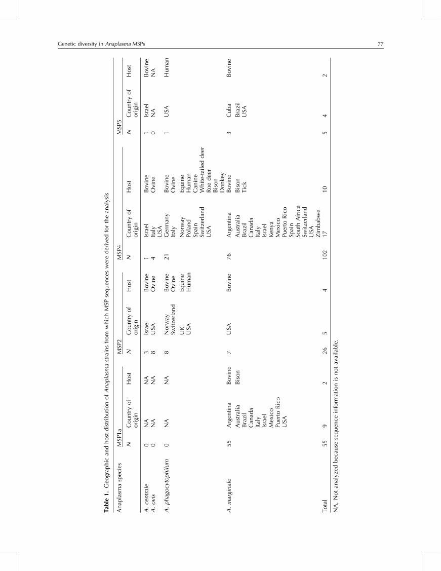
diversity of Anaplasma spp. using these MSPs (Table 1).
The implications of the results of these sequence analyses
for the diagnosis and control of anaplasmosis will be
discussed.
Anaplasma major surface proteins
MSP1a
The gene msp1a, encoding for MSP1a, has only been
identified thus far in A. marginale despite attempts
to clone this gene from A. centrale, A. ovis, and
76 J. de la Fuente et al.
Tab
le1.
Geo
grap
hic
and
host
dis
trib
uti
on
of
Anap
lasm
ast
rain
sfr
om
whic
hM
SPse
quen
ces
wer
eder
ived
for
the
anal
ysis
Anap
lasm
asp
ecie
sM
SP1a
MSP
2M
SP4
MSP
5
NC
ountr
yof
ori
gin
Host
NC
ountr
yof
ori
gin
Host
NC
ountr
yof
ori
gin
Host
NC
ountr
yof
ori
gin
Host
A.
centr
ale
0N
AN
A3
Isra
elB
ovi
ne
1Is
rael
Bovi
ne
1Is
rael
Bovi
ne
A.
ovi
s0
NA
NA
8U
SAO
vine
4It
aly
Ovi
ne
0N
AN
AU
SAA
.phag
ocy
tophil
um
0N
AN
A8
Norw
ayB
ovi
ne
21
Ger
man
yB
ovi
ne
1U
SAH
um
anSw
itze
rlan
dO
vine
Ital
yO
vine
UK
Equin
eN
orw
ayEq
uin
eU
SAH
um
anPola
nd
Hum
anSp
ain
Can
ine
Swit
zerl
and
Whit
e-ta
iled
dee
rU
SAR
oe
dee
rB
ison
Donke
yA
.m
argi
nal
e55
Arg
enti
na
Bovi
ne
7U
SAB
ovi
ne
76
Arg
enti
na
Bovi
ne
3C
uba
Bovi
ne
Aust
rali
aB
ison
Aust
rali
aB
ison
Bra
zil
Bra
zil
Bra
zil
Tic
kU
SAC
anad
aC
anad
aIt
aly
Ital
yIs
rael
Isra
elM
exic
oK
enya
Puer
toR
ico
Mex
ico
USA
Puer
toR
ico
Spai
nSo
uth
Afr
ica
Swit
zerl
and
USA
Zim
bab
we
Tota
l55
92
26
54
102
17
10
54
2
NA
,N
ot
anal
yzed
bec
ause
sequen
cein
form
atio
nis
not
avai
lable
.
Genetic diversity in Anaplasma MSPs 77
A. phagocytophilum (Lew et al., 2002; unpublished
results). MSP1a is one of the six MSPs that have been
identified in A. marginale from bovine erythrocytes and
found to be conserved on A. marginale derived from ticks
and cultured tick cells (reviewed by Kocan et al., 2003,
2004). MSP1a is a part of the MSP1 complex composed of
a heterodimer of two structurally unrelated polypeptides:
MSP1a, which is encoded by a single gene and MSP1b,
which is encoded by at least two genes, msp1b1 and
msp1b2 (Barbet et al., 1987; Viseshakul et al., 2000;
Camacho-Nuez et al., 2000; Bowie et al., 2002). The
MSP1a of A. marginale geographic strains differs in
molecular weight because of a variable number of
tandem 28 or 29 amino acid repeats, and has been used
for strain identification (Allred et al., 1990; de la Fuente
et al., 2001a, 2003a). Within a strain, msp1a does not vary
and has proved to be a stable genetic marker of the
rickettsia through its developmental cycle in cattle and
ticks (Bowie et al., 2002).
MSP1a was shown to be an adhesin for bovine
erythrocytes and both native and cultured tick cells in
microtitre hemagglutination and adhesion assays using
recombinant Escherichia coli expressing MSP1a (McGarey
and Allred, 1994; McGarey et al., 1994; de la Fuente
et al., 2001b), and by light and electron microscopy
(de la Fuente et al., 2001b). MSP1a was also shown to
be involved in the infectivity of A. marginale strains
for Dermacentor spp. (de la Fuente et al., 2001c).
Expression of MSP1a was found to be down-regulated
in tick cells, as compared with organisms derived from
bovine erythrocytes. In addition, both native and recom-
binant MSP1a were found to be glycosylated, a feature
of the repeated N-terminal peptides which appears
to contribute to the adhesive properties and thus
the function of the protein (Garcia-Garcia et al.,
2004a, b, c).
The adhesion domain of MSP1a was identified on the
extracellular N-terminal region of MSP1a that contains the
repeated peptides (de la Fuente et al., 2003b). We
demonstrated by the use of mutant proteins that the
repeated peptides of MSP1a were necessary and sufficient
to mediate adhesion of recombinant MSP1a to tick cells
and bovine erythrocytes, a prerequisite to infection of
host cells (de la Fuente et al., 2003b). The adhesive
capacity of individual peptides for tick cell extract (TCE)
was then evaluated using synthetic peptides representing
the predominant repeated sequences (de la Fuente et al.,
2003b) and was found to be associated with specific
amino acids. Peptides containing acidic amino acids D or
E at position 20 bound to TCE, while peptides with a G as
the 20th amino acid were not adhesive to TCE. Antibodies
produced in rabbits against a synthetic repeated peptide
significantly reduced A. marginale infection of cultured
tick cells, and the neutralization observed was similar to
that obtained using antibodies produced against the
whole MSP1a recombinant protein (de la Fuente et al.,
2003b). Analyses of the tandemly repeated MSP1a
peptides of several geographic strains of A. marginale
revealed a complex relationship between the msp1agenotype and the tick-transmissible phenotype of the
strain and suggested that both the sequence and
conformation of this portion of MSP1a influenced the
adhesive properties of the protein (de la Fuente et al.,
2003b). The results of these studies reinforced the
functional importance of the MSP1a tandem repeats in
adhesion, invasion, and transmission of A. marginale.
A. marginale MSPs are involved in interactions with
both vertebrate and invertebrate hosts (reviewed by de la
Fuente et al., 2001a; Kocan et al., 2003, 2004), and
therefore are likely to evolve more rapidly than other
nuclear genes because they are subjected to selective
pressures exerted by host immune systems. While both
msp1a and msp4 were found to be stable genetic markers
during the multiplication of a given A. marginale strain
(de la Fuente et al., 2001d; Bowie et al., 2002), they
proved to be useful in phylogenetic analysis among
geographic A. marginale strains.
Initially, we chose two A. marginale MSPs, MSP1a and
MSP4, for phylogenetic analysis and these studies
provided evidence that msp1a is under positive selection
pressure (de la Fuente et al., 2003a). Phylogenetic analysis
of A. marginale was first performed on strains from the
USA, using msp1a and msp4 genes and derived protein
sequences (de la Fuente et al., 2001d). Results of these
analyses strongly supported a southeastern clade of
A. marginale comprising Virginia and Florida isolates.
Furthermore, analysis of 16 S rDNA fragment sequences
from the tick vector of A. marginale, D. variabilis, from
various areas of the USA was performed, which suggested
co-evolution of the vector and pathogen (de la Fuente
et al., 2001d).
Subsequent phylogenetic studies of MSP1a DNA and
protein sequences from 20 New World strains of
A. marginale failed to provide phylogeographic resolu-
tion (de la Fuente et al., 2002). Most of the variations in
MSP1a sequences appeared unique for a given strain.
However, similar DNA sequence variation in MSP1a was
detected within strains from Idaho and Florida and from
Idaho and Argentina. These results suggested that MSP1a
sequences may be rapidly evolving and questioned the
use of MSP1a sequences for defining geographic strains of
A. marginale. These findings were then confirmed in
phylogenetic analyses of MSP1a DNA and protein
sequences of 13 strains from Oklahoma, in comparison
with seven Latin American and 13 strains from the USA,
which demonstrated no geographic clustering (de la
Fuente et al., 2003a). From these studies, we concluded
that MSP1a is not useful as a marker for the characteriza-
tion of geographic strains of A. marginale. The genetic
heterogeneity observed among strains of A. marginale
within Oklahoma, and within other endemic regions like
Oregon (Palmer et al., 2001), Minas Gerais in Brazil (de la
Fuente et al., 2004c), Castilla-La Mancha in Spain (de la
Fuente et al., 2004b), Kansas (Palmer et al., 2004) and
78 J. de la Fuente et al.
Sicily in Italy (de la Fuente et al., 2005b) could be
explained by cattle movement and maintenance of
different genotypes by independent transmission events,
due to infection exclusion of A. marginale in cattle and
ticks which results in the establishment of only one
genotype per animal (de la Fuente et al., 2003a).
However, recent results have documented the low
frequency appearance of animals infected with two
A. marginale strains in a cattle herd with high prevalence
of infection (Palmer et al., 2004). The A. marginale msp1agenotypes in animals infected with two strains were not
closely related and may reflect a situation similar to the
A. centrale/A. marginale co-infection reported in vacci-
nated cattle (Shkap et al., 2002b). When cattle movement
imports a new A. marginale genotype, it becomes
established and maintained by mechanical and/or bio-
logical transmission to susceptible cattle. Transmission of
A. marginale strains appears to be stochastic, at least
within a herd with a high prevalence of infection (Palmer
et al., 2004). These results predict that genotypic variation
of A. marginale strains would be minimal in regions with
few cattle/A. marginale introductions, while a highly
heterogeneous population of A. marginale genotypes
would be expected to occur in regions with extensive
cattle movement, such as Oklahoma.
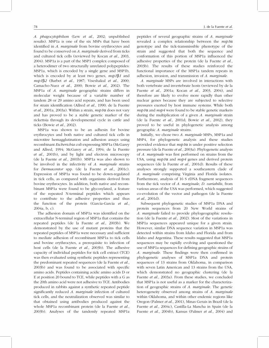
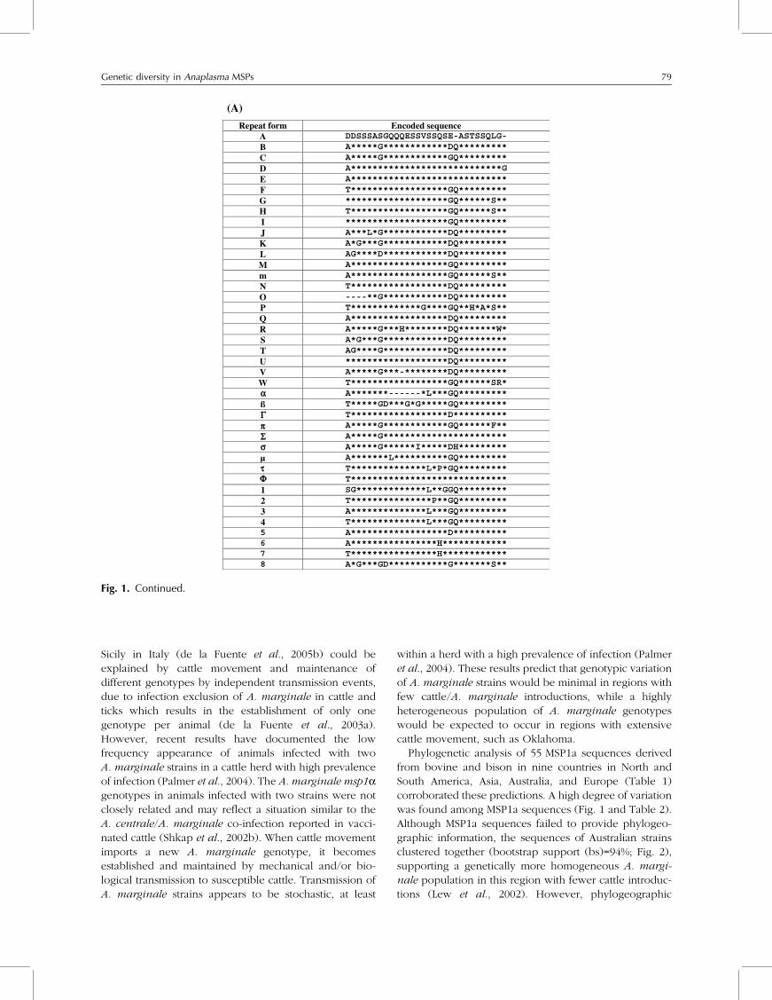
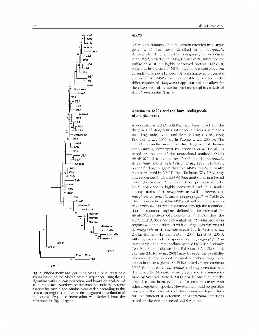
Phylogenetic analysis of 55 MSP1a sequences derived
from bovine and bison in nine countries in North and
South America, Asia, Australia, and Europe (Table 1)
corroborated these predictions. A high degree of variation
was found among MSP1a sequences (Fig. 1 and Table 2).
Although MSP1a sequences failed to provide phylogeo-
graphic information, the sequences of Australian strains
clustered together (bootstrap support (bs)=94%; Fig. 2),
supporting a genetically more homogeneous A. margi-
nale population in this region with fewer cattle introduc-
tions (Lew et al., 2002). However, phylogeographic
(A)
Repeat form Encoded sequence A DDSSSASGQQQESSVSSQSE-ASTSSQLG- B A*****G************DQ********* C A*****G************GQ********* D A****************************G E A***************************** F T******************GQ********* G *******************GQ******S** H T******************GQ******S** I *******************GQ********* J A***L*G************DQ********* K A*G***G************DQ********* L AG****D************DQ********* M A******************GQ********* m A******************GQ******S** N T******************DQ********* O ----**G************DQ********* P T*************G****GQ**H*A*S** Q A******************DQ********* R A*****G***H********DQ*******W* S A*G***G************DQ********* T AG****G************DQ********* U *******************DQ********* V A*****G***-********DQ********* W T******************GQ******SR*
A*******------*L***GQ********* ß ΓπΣσ
τΦ
αT*****GD***G*G*****GQ********* T******************D********** A*****G************GQ******F** A*****G*********************** A*****G******I*****DH*********
µ A*******L**********GQ********* T**************L*P*GQ********* T*****************************
1 SG*************L**GGQ********* 2 T***************P**GQ********* 3 A**************L***GQ********* 4 T**************L***GQ********* 5 A*********
A******************D**********
6 *******H************7 T****************H************8 A*G***GD***********G*******S**
Fig. 1. Continued.
Genetic diversity in Anaplasma MSPs 79
(B) A. marginale isolate Structure of MSP1a tandem repeats No. of repeats
Florida A B B B B B B B 8 Idaho D D D D D E 6 Virginia A B 2 Washington B B B C 4 Wetumka, OK K C H 3 Cushing, OK L C B C 4 Cushing 2,OK S N N F H 5 Glencoe 1, OK S F N F H 5 Glencoe 2, OK B M F H 4 Glencoe 3, OK T B C 3 Stillwater, OK S F F F H 5 Stillwater 2, OK L B C C 4 Stillwater 68, OK S B M F H 5 Oklahoma City, OK U 1 Okmulgee, OK S B V C 4 Stigler, OK T B B C 4 Pawhuska, OK I H 2 New Castle, OK L B C B 4 St. Maries, ID J B B 3 California B B C 3 Okeechobee, FL L B C B C 5 Mississippi D D D D E 5 Missouri B B B B 4 Illinois M N B M H 5 Texas O B M P 4 Texas 198 B B m B m 5 South Dakota A F H 3 Oregon A F H 3 Kansas 3261 B B 2 Kansas 4102 B B B 3 Kansas 2267 B B B B 4 Kansas 0141 B B B B B 5 Kansas 0063 B B B B B B 6 Kansas 5076 D D D D D 5 Kansas 7042 D D E 3 Kansas 4318 D D D D D E 6 Kansas 2070 D D D D D D E 7 Kansas 7030 D D D D D D D D D E 10 Kansas 0050 E M 3 Canadian bison D Q Q R 4 U.S. bison (buffalo) K B M F W 5 Yucatán T C B B C B π 7
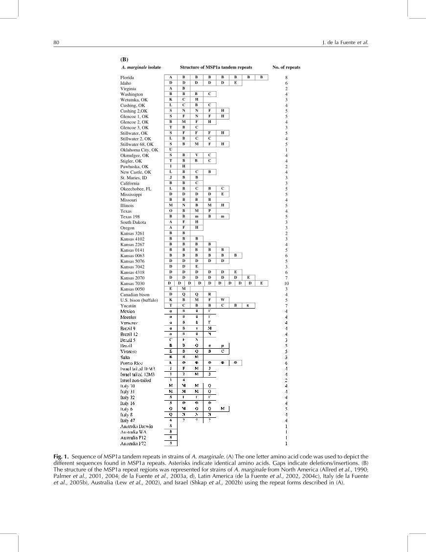
Fig. 1. Sequence of MSP1a tandem repeats in strains of A. marginale. (A) The one letter amino acid code was used to depict thedifferent sequences found in MSP1a repeats. Asterisks indicate identical amino acids. Gaps indicate deletions/insertions. (B)The structure of the MSP1a repeat regions was represented for strains of A. marginale from North America (Allred et al., 1990;Palmer et al., 2001, 2004; de la Fuente et al., 2003a, d), Latin America (de la Fuente et al., 2002, 2004c), Italy (de la Fuenteet al., 2005b), Australia (Lew et al., 2002), and Israel (Shkap et al., 2002b) using the repeat forms described in (A).
80 J. de la Fuente et al.
resolution was not obtained in all the other regions of the
world analyzed, suggesting multiple introductions of A.
marginale strains from different geographic locations.
MSP2
MSP2 is an immunodominant outer membrane protein
with orthologs in all Anaplasma spp. studied thus far
(Palmer et al., 1998; Shkap et al., 2002a; Lin et al., 2004).
In A. marginale, A. centrale, and A. ovis, MSP2 is encoded
by a multigene family (Palmer et al., 1998; Shkap et al.,
2002a), while the corresponding gene in A. phagocyto-
philum is encoded by a single gene, different from the
antigenically-related p44 multigene family (Lin et al.,
2004). Cattle and ticks become persistently infected with
A. marginale and antigenic variation of MSP2 occurs
during these persistent infections (French et al., 1998,
1999; de la Fuente and Kocan, 2001). Multiple antigenic
variants arise as the result of combinatorial gene conver-
sion into a single expression site (Brayton et al., 2002).
This mechanism of antigenic variation has been suggested
to allow A. marginale to evade the bovine immune
response, thus contributing to the maintenance of
persistent infections in cattle (French et al., 1998, 1999).
Male ticks also become persistently infected with
A. marginale (Kocan et al., 1992a, b). Male ticks thus
also serve as a reservoir host and they are capable of
transmitting the pathogen to multiple hosts (reviewed by
Kocan et al., 2003, 2004). MSP2 variants appear on
A. marginale during tick feeding on multiple hosts
suggesting that antigenic variation of MSP2 in D. varia-
bilis is independent of the bovine immune response (de la
Fuente and Kocan, 2001).
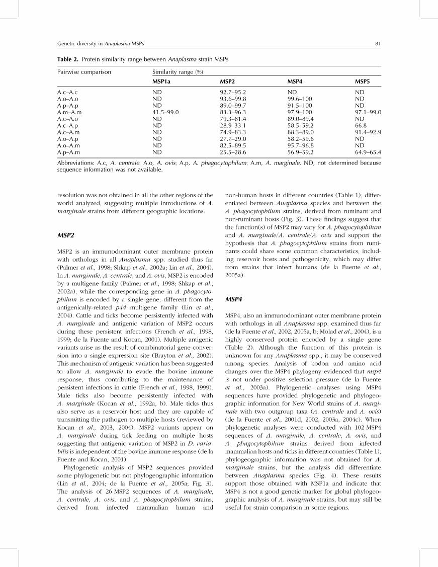
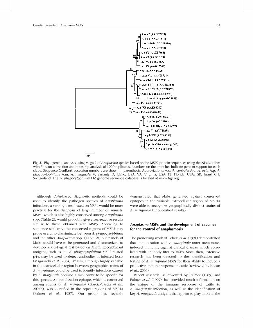
Phylogenetic analysis of MSP2 sequences provided
some phylogenetic but not phylogeographic information
(Lin et al., 2004; de la Fuente et al., 2005a; Fig. 3).
The analysis of 26 MSP2 sequences of A. marginale,
A. centrale, A. ovis, and A. phagocytophilum strains,
derived from infected mammalian human and
non-human hosts in different countries (Table 1), differ-
entiated between Anaplasma species and between the
A. phagocytophilum strains, derived from ruminant and
non-ruminant hosts (Fig. 3). These findings suggest that
the function(s) of MSP2 may vary for A. phagocytophilum
and A. marginale/A. centrale/A. ovis and support the
hypothesis that A. phagocytophilum strains from rumi-
nants could share some common characteristics, includ-
ing reservoir hosts and pathogenicity, which may differ
from strains that infect humans (de la Fuente et al.,
2005a).
MSP4
MSP4, also an immunodominant outer membrane protein
with orthologs in all Anaplasma spp. examined thus far
(de la Fuente et al., 2002, 2005a, b; Molad et al., 2004), is a
highly conserved protein encoded by a single gene
(Table 2). Although the function of this protein is
unknown for any Anaplasma spp., it may be conserved
among species. Analysis of codon and amino acid
changes over the MSP4 phylogeny evidenced that msp4
is not under positive selection pressure (de la Fuente
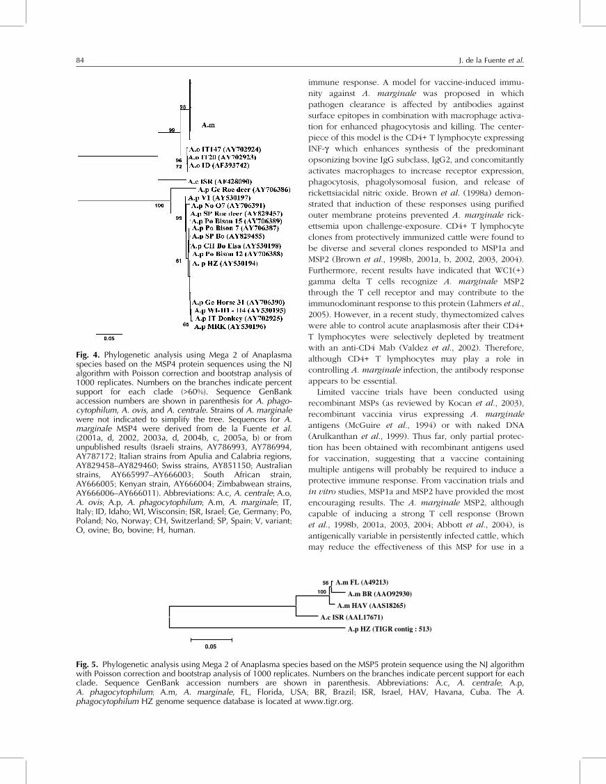
et al., 2003a). Phylogenetic analyses using MSP4
sequences have provided phylogenetic and phylogeo-
graphic information for New World strains of A. margi-
nale with two outgroup taxa (A. centrale and A. ovis)
(de la Fuente et al., 2001d, 2002, 2003a, 2004c). When
phylogenetic analyses were conducted with 102 MSP4
sequences of A. marginale, A. centrale, A. ovis, and
A. phagocytophilum strains derived from infected
mammalian hosts and ticks in different countries (Table 1),
phylogeographic information was not obtained for A.
marginale strains, but the analysis did differentiate
between Anaplasma species (Fig. 4). These results
support those obtained with MSP1a and indicate that
MSP4 is not a good genetic marker for global phylogeo-
graphic analysis of A. marginale strains, but may still be
useful for strain comparison in some regions.
Table 2. Protein similarity range between Anaplasma strain MSPs
Pairwise comparison Similarity range (%)
MSP1a MSP2 MSP4 MSP5
A.c–A.c ND 92.7–95.2 ND NDA.o–A.o ND 93.6–99.8 99.6–100 NDA.p–A.p ND 89.0–99.7 91.5–100 NDA.m–A.m 41.5–99.0 83.3–96.3 97.9–100 97.1–99.0A.c–A.o ND 79.3–81.4 89.0–89.4 NDA.c–A.p ND 28.9–33.1 58.5–59.2 66.8A.c–A.m ND 74.9–83.3 88.3–89.0 91.4–92.9A.o–A.p ND 27.7–29.0 58.2–59.6 NDA.o–A.m ND 82.5–89.5 95.7–96.8 NDA.p–A.m ND 25.5–28.6 56.9–59.2 64.9–65.4
Abbreviations: A.c, A. centrale; A.o, A. ovis; A.p, A. phagocytophilum; A.m, A. marginale, ND, not determined becausesequence information was not available.
Genetic diversity in Anaplasma MSPs 81
MSP5
MSP5 is an immunodominant protein encoded by a single
gene, which has been identified in A. marginale,
A. centrale, A. ovis, and A. phagocytophilum (Visser
et al., 1992; Molad et al., 2004; Dreher et al., submitted for
publication). It is a highly conserved protein (Table 2),
which, as in the case of MSP4, may have a conserved but
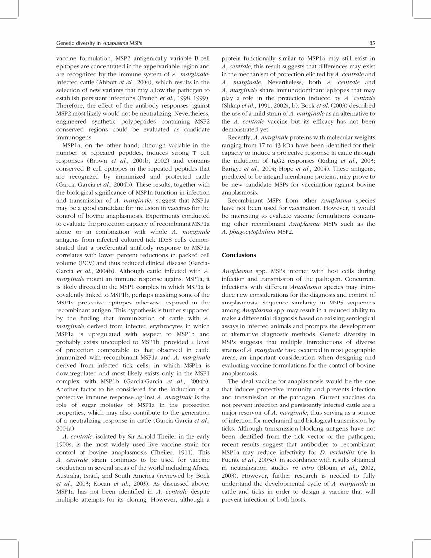
currently unknown function. A preliminary phylogenetic
analysis of five MSP5 sequences (Table 1) resulted in the
differentiation of Anaplasma spp. but did not allow for
the assessment of its use for phylogeographic analysis of
Anaplasma strains (Fig. 5).
Anaplasma MSPs and the immunodiagnosisof anaplasmosis
A competitive ELISA (cELISA) has been used for the
diagnosis of Anaplasma infection in various ruminants
including cattle, ovine, and deer (Ndung’u et al., 1995;
Knowles et al., 1996; de la Fuente et al., 2004b). The
cELISA currently used for the diagnosis of bovine
anaplasmosis, developed by Knowles et al. (1996), is
based on the use of the monoclonal antibody (Mab)
ANAF16C1 that recognizes MSP5 in A. marginale,
A. centrale, and A. ovis (Visser et al., 1992). However,
recent findings suggest that this MSP5 ELISA, currently
commercialized by VMRD, Inc. (Pullman, WA, USA), may
also recognize A. phagocytophilum antibodies in infected
cattle (Dreher et al., submitted for publication). The
MSP5 sequence is highly conserved and thus similar
among strains of A. marginale, as well as between A.
marginale, A. centrale, and A. phagocytophilum (Table 2).
The cross-reactivity of the MSP5 test with multiple species
of Anaplasma has been confirmed through the identifica-
tion of common regions defined to be essential for
ANAF16C1 reactivity (Munodzana et al., 1998). Thus, the
MSP5 cELISA does not differentiate Anaplasma species in
regions where co-infection with A. phagocytophylum and
A. marginale or A. centrale occurs (de la Fuente et al.,
2004a; Hofmann-Lehmann et al., 2004; Lin et al., 2004).
Although a second test specific for A. phagocytophilum
(for example the immunofluorescence HGE IFA Antibody
Test Kit; Fuller Laboratories, Fullerton, CA, USA) or A.
centrale (Molloy et al., 2001) may be used, the possibility
of cross-infection cannot be ruled out when using these
assays in these regions. An ELISA based on recombinant
MSP5 for indirect A. marginale antibody detection was
developed by Morzaria et al. (1999) and is commercia-
lized by Svanova Biotech AB (Uppsala, Sweden) but the
assay has not been evaluated for cross-reactivity with
other Anaplasma species. However, it should be possible
to explore the possibility of developing serological tests
for the differential detection of Anaplasma infections
based on the non-conserved MSP5 regions.
Fig. 2. Phylogenetic analysis using Mega 2 of A. marginalestrains based on the MSP1a protein sequences using the NJalgorithm with Poisson correction and bootstrap analysis of1000 replicates. Numbers on the branches indicate percentsupport for each clade. Strains were coded according to thecountry of origin to emphasize the geographic distribution ofthe strains. Sequence information was derived from thereferences in Fig. 1 legend.
82 J. de la Fuente et al.
Although DNA-based diagnostic methods could be
used to identify the pathogen species of Anaplasma
infections, a serologic test based on MSPs would be more
practical for the diagnosis of large number of animals.
MSP4, which is also highly conserved among Anaplasma
spp. (Table 2), would probably give cross-reactive results
similar to those obtained with MSP5. According to
sequence similarity, the conserved regions of MSP2 may
prove useful to discriminate between A. phagocytophilum
and the other Anaplasma spp. (Table 2), but panels of
Mabs would have to be generated and characterized to
develop a serological test based on MSP2. Recombinant
antigens, such as the A. phagocytophilum MSP2-related
p44, may be used to detect antibodies in infected hosts
(Magnarelli et al., 2004). MSP1a, although highly variable
in the extracellular region between geographic strains of
A. marginale, could be used to identify infections caused
by A. marginale because it may prove to be specific for
this species. A neutralization epitope, which is conserved
among strains of A. marginale (Garcia-Garcia et al.,
2004b), was identified in the repeat regions of MSP1a
(Palmer et al., 1987). Our group has recently
demonstrated that Mabs generated against conserved
epitopes in the variable extracellular region of MSP1a
were able to recognize geographically distinct strains of
A. marginale (unpublished results).
Anaplasma MSPs and the development of vaccinesfor the control of anaplasmosis
The pioneering work of Tebele et al. (1991) demonstrated
that immunization with A. marginale outer membranes
induced immunity against clinical disease which corre-
lated with antibody titer to MSPs. Since then, extensive
research has been devoted to the identification and
testing of A. marginale MSPs for their ability to induce a
protective immune response in cattle (reviewed by Kocan
et al., 2003).
Recent research, as reviewed by Palmer (1989) and
Palmer et al. (1999), has provided much information on
the nature of the immune response of cattle to
A. marginale infection, as well as the identification of
key A. marginale antigens that appear to play a role in the
Fig. 3. Phylogenetic analysis using Mega 2 of Anaplasma species based on the MSP2 protein sequences using the NJ algorithmwith Poisson correction and bootstrap analysis of 1000 replicates. Numbers on the branches indicate percent support for eachclade. Sequence GenBank accession numbers are shown in parenthesis. Abbreviations: A.c, A. centrale; A.o, A. ovis; A.p, A.phagocytophilum; A.m, A. marginale; V, variant; ID, Idaho, USA; VA, Virginia, USA; FL, Florida, USA; ISR, Israel; CH,Switzerland. The A. phagocytophilum HZ genome sequence database is located at www.tigr.org.
Genetic diversity in Anaplasma MSPs 83
immune response. A model for vaccine-induced immu-
nity against A. marginale was proposed in which
pathogen clearance is affected by antibodies against
surface epitopes in combination with macrophage activa-
tion for enhanced phagocytosis and killing. The center-
piece of this model is the CD4+ T lymphocyte expressing
INF-g which enhances synthesis of the predominant
opsonizing bovine IgG subclass, IgG2, and concomitantly
activates macrophages to increase receptor expression,
phagocytosis, phagolysomosal fusion, and release of
rickettsiacidal nitric oxide. Brown et al. (1998a) demon-
strated that induction of these responses using purified
outer membrane proteins prevented A. marginale rick-
ettsemia upon challenge-exposure. CD4+ T lymphocyte
clones from protectively immunized cattle were found to
be diverse and several clones responded to MSP1a and
MSP2 (Brown et al., 1998b, 2001a, b, 2002, 2003, 2004).
Furthermore, recent results have indicated that WC1(+)
gamma delta T cells recognize A. marginale MSP2
through the T cell receptor and may contribute to the
immunodominant response to this protein (Lahmers et al.,
2005). However, in a recent study, thymectomized calves
were able to control acute anaplasmosis after their CD4+
T lymphocytes were selectively depleted by treatment
with an anti-CD4 Mab (Valdez et al., 2002). Therefore,
although CD4+ T lymphocytes may play a role in
controlling A. marginale infection, the antibody response
appears to be essential.
Limited vaccine trials have been conducted using
recombinant MSPs (as reviewed by Kocan et al., 2003),
recombinant vaccinia virus expressing A. marginale
antigens (McGuire et al., 1994) or with naked DNA
(Arulkanthan et al., 1999). Thus far, only partial protec-
tion has been obtained with recombinant antigens used
for vaccination, suggesting that a vaccine containing
multiple antigens will probably be required to induce a
protective immune response. From vaccination trials and
in vitro studies, MSP1a and MSP2 have provided the most
encouraging results. The A. marginale MSP2, although
capable of inducing a strong T cell response (Brown
et al., 1998b, 2001a, 2003, 2004; Abbott et al., 2004), is
antigenically variable in persistently infected cattle, which
may reduce the effectiveness of this MSP for use in a
Fig. 4. Phylogenetic analysis using Mega 2 of Anaplasmaspecies based on the MSP4 protein sequences using the NJalgorithm with Poisson correction and bootstrap analysis of1000 replicates. Numbers on the branches indicate percentsupport for each clade (>60%). Sequence GenBankaccession numbers are shown in parenthesis for A. phago-cytophilum, A. ovis, and A. centrale. Strains of A. marginalewere not indicated to simplify the tree. Sequences for A.marginale MSP4 were derived from de la Fuente et al.(2001a, d, 2002, 2003a, d, 2004b, c, 2005a, b) or fromunpublished results (Israeli strains, AY786993, AY786994,AY787172; Italian strains from Apulia and Calabria regions,AY829458–AY829460; Swiss strains, AY851150; Australianstrains, AY665997–AY666003; South African strain,AY666005; Kenyan strain, AY666004; Zimbabwean strains,AY666006–AY666011). Abbreviations: A.c, A. centrale; A.o,A. ovis; A.p, A. phagocytophilum; A.m, A. marginale; IT,Italy; ID, Idaho; WI, Wisconsin; ISR, Israel; Ge, Germany; Po,Poland; No, Norway; CH, Switzerland; SP, Spain; V, variant;O, ovine; Bo, bovine; H, human.
A.m FL (A49213)
A.m BR (AAO92930)
A.m HAV (AAS18265)
A.c ISR (AAL17671)
A.p HZ (TIGR contig : 513)
100
56
0.05
Fig. 5. Phylogenetic analysis using Mega 2 of Anaplasma species based on the MSP5 protein sequence using the NJ algorithmwith Poisson correction and bootstrap analysis of 1000 replicates. Numbers on the branches indicate percent support for eachclade. Sequence GenBank accession numbers are shown in parenthesis. Abbreviations: A.c, A. centrale; A.p,A. phagocytophilum; A.m, A. marginale, FL, Florida, USA; BR, Brazil; ISR, Israel, HAV, Havana, Cuba. The A.phagocytophilum HZ genome sequence database is located at www.tigr.org.
84 J. de la Fuente et al.
vaccine formulation. MSP2 antigenically variable B-cell
epitopes are concentrated in the hypervariable region and
are recognized by the immune system of A. marginale-
infected cattle (Abbott et al., 2004), which results in the
selection of new variants that may allow the pathogen to
establish persistent infections (French et al., 1998, 1999).
Therefore, the effect of the antibody responses against
MSP2 most likely would not be neutralizing. Nevertheless,
engineered synthetic polypeptides containing MSP2
conserved regions could be evaluated as candidate
immunogens.
MSP1a, on the other hand, although variable in the
number of repeated peptides, induces strong T cell
responses (Brown et al., 2001b, 2002) and contains
conserved B cell epitopes in the repeated peptides that
are recognized by immunized and protected cattle
(Garcia-Garcia et al., 2004b). These results, together with
the biological significance of MSP1a function in infection
and transmission of A. marginale, suggest that MSP1a
may be a good candidate for inclusion in vaccines for the
control of bovine anaplasmosis. Experiments conducted
to evaluate the protection capacity of recombinant MSP1a
alone or in combination with whole A. marginale
antigens from infected cultured tick IDE8 cells demon-
strated that a preferential antibody response to MSP1a
correlates with lower percent reductions in packed cell
volume (PCV) and thus reduced clinical disease (Garcia-
Garcia et al., 2004b). Although cattle infected with A.
marginale mount an immune response against MSP1a, it
is likely directed to the MSP1 complex in which MSP1a is
covalently linked to MSP1b, perhaps masking some of the
MSP1a protective epitopes otherwise exposed in the
recombinant antigen. This hypothesis is further supported
by the finding that immunization of cattle with A.
marginale derived from infected erythrocytes in which
MSP1a is upregulated with respect to MSP1b and
probably exists uncoupled to MSP1b, provided a level
of protection comparable to that observed in cattle
immunized with recombinant MSP1a and A. marginale
derived from infected tick cells, in which MSP1a is
downregulated and most likely exists only in the MSP1
complex with MSP1b (Garcia-Garcia et al., 2004b).
Another factor to be considered for the induction of a
protective immune response against A. marginale is the
role of sugar moieties of MSP1a in the protection
properties, which may also contribute to the generation
of a neutralizing response in cattle (Garcia-Garcia et al.,
2004a).
A. centrale, isolated by Sir Arnold Theiler in the early
1900s, is the most widely used live vaccine strain for
control of bovine anaplasmosis (Theiler, 1911). This
A. centrale strain continues to be used for vaccine
production in several areas of the world including Africa,
Australia, Israel, and South America (reviewed by Bock
et al., 2003; Kocan et al., 2003). As discussed above,
MSP1a has not been identified in A. centrale despite
multiple attempts for its cloning. However, although a
protein functionally similar to MSP1a may still exist in
A. centrale, this result suggests that differences may exist
in the mechanism of protection elicited by A. centrale and
A. marginale. Nevertheless, both A. centrale and
A. marginale share immunodominant epitopes that may
play a role in the protection induced by A. centrale
(Shkap et al., 1991, 2002a, b). Bock et al. (2003) described
the use of a mild strain of A. marginale as an alternative to
the A. centrale vaccine but its efficacy has not been
demonstrated yet.
Recently, A. marginale proteins with molecular weights
ranging from 17 to 43 kDa have been identified for their
capacity to induce a protective response in cattle through
the induction of IgG2 responses (Riding et al., 2003;
Barigye et al., 2004; Hope et al., 2004). These antigens,
predicted to be integral membrane proteins, may prove to
be new candidate MSPs for vaccination against bovine
anaplasmosis.
Recombinant MSPs from other Anaplasma species
have not been used for vaccination. However, it would
be interesting to evaluate vaccine formulations contain-
ing other recombinant Anaplasma MSPs such as the
A. phagocytophilum MSP2.
Conclusions
Anaplasma spp. MSPs interact with host cells during
infection and transmission of the pathogen. Concurrent
infections with different Anaplasma species may intro-
duce new considerations for the diagnosis and control of
anaplasmosis. Sequence similarity in MSP5 sequences
among Anaplasma spp. may result in a reduced ability to
make a differential diagnosis based on existing serological
assays in infected animals and prompts the development
of alternative diagnostic methods. Genetic diversity in
MSPs suggests that multiple introductions of diverse
strains of A. marginale have occurred in most geographic
areas, an important consideration when designing and
evaluating vaccine formulations for the control of bovine
anaplasmosis.
The ideal vaccine for anaplasmosis would be the one
that induces protective immunity and prevents infection
and transmission of the pathogen. Current vaccines do
not prevent infection and persistently infected cattle are a
major reservoir of A. marginale, thus serving as a source
of infection for mechanical and biological transmission by
ticks. Although transmission-blocking antigens have not
been identified from the tick vector or the pathogen,
recent results suggest that antibodies to recombinant
MSP1a may reduce infectivity for D. variabilis (de la
Fuente et al., 2003c), in accordance with results obtained
in neutralization studies in vitro (Blouin et al., 2002,
2003). However, further research is needed to fully
understand the developmental cycle of A. marginale in
cattle and ticks in order to design a vaccine that will
prevent infection of both hosts.
Genetic diversity in Anaplasma MSPs 85
The full genome sequences of A. marginale and
A. phagocytophilum are complete (Brayton et al., 2005)
or close to completion and should be released in early
2005 (A. phagocytophilum and A. marginale genome
sequence databases are located at www.tigr.org and
www.vetmed.wsu.edu/research%5Fvmp/anagenome/,
respectively). This information will prove to be essential
for global comparative studies of Anaplasma strains,
which should add considerable information on the
genetic diversity of Anaplasma MSPs. Genome sequences
can be also used for in silico search and biological
throughput screening of polypeptides with potential
value for diagnosis and vaccine development.
Acknowledgments
This research was supported by the project no. 1669 of
the Oklahoma Agricultural Experiment Station, the
Endowed Chair for Food Animal Research (K.M. Kocan),
the Instituto de Ciencias de la Salud, Spain (ICS-JCCM)
(project no. 03052-00), Australian Centre for International
Agricultural Research (project no. AS2/1999/063), the
Ministry of Health, Italy, the Federal Veterinary Office,
Switzerland and National Institutes of Health Centers for
Biomedical Research Excellence through a subcontract to
J. de la Fuente from the Oklahoma Medical Research
Foundation, and the Oklahoma Center for the Advance-
ment of Science and Technology, Applied Research
Grants, AR00(1)-001 and ARO21-37. Consuelo Almazan
is supported by grants-in-aid from the CONACYT and
Promep (University of Tamaulipas), Mexico and a grant
from Pfizer Animal Health, Inc., Kalamazoo, Michigan,
USA. This work is a contribution to project CICYT
AGL2001-3947, Ministerio de Educacion y Ciencia (Spain),
and to the agreement between Yolanda Fierro and the
University of Castilla–La Mancha. Janet J. Rogers (Core
Sequencing Facility, Department of Biochemistry and
Molecular Biology, Noble Research Center, Oklahoma
State University) is acknowledged for DNA sequencing.
References
Abbott JR, Palmer GH, Howard CJ, Hope JC and Brown WC(2004). Anaplasma marginale major surface protein 2CD4+-T-cell epitopes are evenly distributed in conservedand hypervariable regions (HVR), whereas linear B-cellepitopes are predominantly located in the HVR. Infectionand Immunity 72: 7360–7366.
Allred DR, McGuire TC, Palmer GH, Leib SR, Harkins TM,McElwain TF and Barbet AF (1990). Molecular basis forsurface antigen size polymorphisms and conservation of aneutralization-sensitive epitope in Anaplasma marginale.Proceedings of the National Academy of Sciences of theUnited States of America 87: 3220–3224.
Arulkanthan A, Brown WC, McGuire C and Knowles DP (1999).Biased immunoglobulin G1 isotype responses induced incattle with DNA expressing msp1a of Anaplasma margi-nale. Infection and Immunity 67: 3481–3487.
Barbet AF, Palmer GH, Myler PJ and McGuire TC (1987).Characterization of an immunoprotective protein complexof Anaplasma marginale by cloning and expression of thegene coding for polypeptide Am105L. Infection andImmunity 55: 2428–2435.
Barigye R, Garcia-Ortiz MA, Rojas Ramirez EE and Rodriguez SD(2004). Identification of IgG2-specific antigens in MexicanAnaplasma marginale strains. Annals of the New YorkAcademy of Sciences 1026: 84–94.
Blouin EF, de la Fuente J, Garcia-Garcia JC, Sauer JR, Saliki JTand Kocan KM (2002). Applications of a cell culturesystem for studying the interaction of Anaplasmamarginale with tick cells. Animal Health Research Reviews3: 57–68.
Blouin EF, Saliki JT, de la Fuente J, Garcia-Garcia JC and KocanKM (2003). Antibodies to Anaplasma marginale majorsurface proteins 1a and 1b inhibit infectivity for culturedtick cells. Veterinary Parasitology 111: 247–260.
Bock RE, de Vos AJ, Kingston TG and Carter PD (2003).Assessment of a low virulence Australian isolate ofAnaplasma marginale for pathogenicity, immunogenicityand transmissability by Boophilus microplus. VeterinaryParasitology 118: 121–131.
Bowie JV, de la Fuente J, Kocan KM, Blouin EF and Barbet AF(2002). Conservation of major surface protein 1 genesof the ehrlichial pathogen Anaplasma marginale duringcyclic transmission between ticks and cattle. Gene282: 95–102.
Brayton KA, Palmer GH, Lundgren A, Yi J and Barbet AF (2002).Antigenic variation of Anaplasma marginale msp2 occursby combinatorial gene conversion. Molecular Microbiology43: 1151–1159.
Brayton KA, Kappmeyer LS, Herndon DR, Dark MJ, Tibbals DL,Palmer GH, McGuire TC and Knowles Jr DP (2005).Complete genome sequencing of Anaplasma marginalereveals that the surface is skewed to two superfamilies ofouter membrane proteins. Proceedings of the NationalAcademy of Sciences of the United States of America PNAS102: 844–849.
Brown WC, Shkap V, Zhu D, McGuire TC, Tuo W, McElwain TFand Palmer GH (1998a). CD4+ T-lymphocyte and immuno-globulin G2 responses in calves immunized withAnaplasma marginale outer membranes and protectedagainst homologous challenge. Infection and Immunity66: 5406–5413.
Brown WC, Zhu D, Shkap V, McGuire TC, Blouin ED, Kocan KMand Palmer GH (1998b). The repertoire of Anaplasmamarginale antigens recognized by CD4+ T-lymphocyteclones from protectively immunized cattle is diverse andincludes major surface protein 2 (MSP-2) and MSP-3.Infection and Immunity 66: 5414–5422.
Brown WC, McGuire TC, Zhu D, Lewin HA, Sosnow J andPalmer GH (2001a). Highly conserved regions of theimmunodominant major surface protein 2 of the genogroupII Ehrlichial pathogen Anaplasma marginale are rich innaturally derived CD4+ T lymphocyte epitopes that elicitstrong recall responses. Journal of Immunology166: 1114–1124.
Brown WC, Palmer GH, Lewin HA and McGuire TC (2001b).CD4+ T lymphocytes from calves immunzied withAnaplasma marginale major surface protein 1 (MSP1), aheteromeric complex of MSP1a and MSP1b, preferentiallyrecognize the MSP1a carboxyl terminus that is conservedamong strains. Infection and Immunity 69: 6853–6862.
Brown WC, McGuire TC, Mwangi W, Kegerreis KA, Macmillan H,Lewin HA and Palmer GH (2002). Major histocompatibilitycomplex class II DR-restricted memory CD4(+) T lympho-cytes recognize conserved immunodominant epitopes of
86 J. de la Fuente et al.
Anaplasma marginale major surface protein 1a. Infectionand Immunity 70: 5521–5532.
Brown WC, Brayton KA, Styer CM and Palmer GH (2003). Thehypervariable region of Anaplasma marginale majorsurface protein 2 (MSP2) contains multiple immunodomi-nant CD4+ T lymphocyte epitopes that elicit variant-specificproliferative and IFN-gamma responses in MSP2 vaccinates.Journal of Immunology 170: 3790–3798.
Brown WC, Palmer GH, Brayton KA, Meeus PF, Barbet AF,Kegerreis KA and McGuire TC (2004). CD4+ T lymphocytesfrom Anaplasma marginale major surface protein 2 (MSP2)vaccinees recognize naturally processed epitopesconserved in MSP3. Infection and Immunity72: 3688–3692.
Camacho-Nuez J, De Lourdes Munoz M, Suarez CE, McGuire TC,Brown WC and Palmer GH (2000). Expression of poly-morphic msp1b genes during acute Anaplasma marginalerickettsemia. Infection and Immunity 68: 1946–1952.
Carlyon JA and Fikrig E (2003). Invasion and survival strategiesof Anaplasma phagocytophilum. Cellular Microbiology5: 743–754.
Daniels TJ, Battaly GR, Liveris D, Falco RC and Schwartz I (2002).Avian reservoirs of the agent of human granulocyticehrlichiosis? Emerging Infectious Diseases 8: 1524–1525.
de la Fuente J and Kocan KM (2001). Expression of Anaplasmamarginale major surface protein 2 variants in persistentlyinfected ticks. Infection and Immunity 69: 5151–5156.
de la Fuente J, Garcia-Garcia JC, Blouin EF, Rodrıguez SD, GarcıaMA and Kocan KM (2001a). Evolution and function oftandem repeats in the major surface protein 1a of theehrlichial pathogen Anaplasma marginale. Animal HealthResearch Reviews 2: 163–173.
de la Fuente J, Garcia-Garcia JC, Blouin EF and Kocan KM(2001b). Differential adhesion of major surface proteins 1aand 1b of the ehrlichial cattle pathogen Anaplasmamarginale to bovine erythrocytes and tick cells. Interna-tional Journal of Parasitology 31: 145–153.
de la Fuente J, Garcia-Garcia JC, Blouin EF and Kocan KM(2001c). Major surface protein 1a effects tick infection andtransmission of the ehrlichial pathogen Anaplasma margi-nale. International Journal of Parasitology 31: 1705–1714.
de la Fuente J, Van Den Bussche RA and Kocan KM (2001d).Molecular phylogeny and biogeography of North Americanisolates of Anaplasma marginale (Rickettsiaceae: Ehrli-chieae). Veterinary Parasitology 97: 65–76.
de la Fuente J, Van Den Bussche RA, Garcia-Garcia JC, RodrıguezSD, Garcıa MA, Guglielmone AA, Mangold AJ, Passos LM,Blouin EF and Kocan KM (2002). Phylogeography of NewWorld isolates of Anaplasma marginale (Rickettsiaceae:Ehrlichieae) based on major surface protein sequences.Veterinary Microbiology 88: 275–285.
de la Fuente J, Van Den Bussche RA, Prado T and Kocan KM(2003a). Anaplasma marginale major surface protein 1agenotypes evolved under positive selection pressure butare not a marker for geographic isolates. Journal of ClinicalMicrobiology 41: 1609–1616.
de la Fuente J, Garcia-Garcia JC, Blouin EF and Kocan KM(2003b). Characterization of the functional domain of majorsurface protein 1a involved in adhesion of the rickettsiaAnaplasma marginale to host cells. Veterinary Microbiol-ogy 91: 265–283.
de la Fuente J, Kocan KM, Garcia-Garcia JC, Blouin EF, Halbur Tand Onet V (2003c). Immunization against Anaplasmamarginale major surface protein 1a reduces infectivity forticks. The International Journal of Applied Research inVeterinary Medicine 1: 285–292.
de la Fuente J, Golsteyn Thomas EJ, Van Den Bussche RA,Hamilton RG, Tanaka EE, Druhan SE and Kocan KM
(2003d). Characterization of Anaplasma marginale isolatedfrom North American bison. Applied and EnvironmentalMicrobiology 69: 5001–5005.
de la Fuente J, Naranjo V, Ruiz-Fons F, Vicente J, Estrada-PenaA, Almazan C, Kocan KM, Martın MP and Gortazar C(2004a). Prevalence of tick-borne pathogens in ixodid ticks(Acari: Ixodidae) collected from European wild boar (Susscrofa) and Iberian red deer (Cervus elaphus hispanicus) incentral Spain. European Journal of Wildlife Research50: 187–196.
de la Fuente J, Vicente J, Hofle U, Ruiz-Fons F, Fernandezde Mera IG, Van Den Bussche RA, Kocan KM and GortazarC (2004b). Anaplasma infection in free-ranging Iberian reddeer in the region of Castilla-La Mancha, Spain. VeterinaryMicrobiology 100: 163–173.
de la Fuente J, Passos LMF, Van Den Bussche RA, Ribeiro MFB,Facury-Filho EJ and Kocan KM (2004c). Genetic diversityand molecular phylogeny of Anaplasma marginale isolatesfrom Minas Gerais, Brazil. Veterinary Parasitology121: 307–316.
de la Fuente J, Massung RF, Wong SJ, Chu FK, Lutz H, Meli M,von Loewenich FD, Grzeszczuk A, Torina A, Caracappa S,Mangold AJ, Naranjo V, Stuen S and Kocan KM (2005a).Sequence analysis of the msp4 gene of Anaplasmaphagocytophilum strains. Journal of Clinical Microbiology43: 1309–1317.
de la Fuente J, Torina A, Caracappa S, Tumino G, Furla R,Almazan C and Kocan KM (2005b). Serologic and molecularcharacterization of Anaplasma species infection in farmanimals and ticks from Sicily. Veterinary Parasitology:in press.
Dreher UM, de la Fuente J, Hofmann-Lehmann R, Meli ML,Pusterla N, Kocan KM, Woldehiwet Z, Regula G, StaerkKDC and Lutz H (2005). Serologic cross reactivity betweenAnaplasma marginale and Anaplasma phagocytophilum.Journal of Clinical Microbiology: submitted for publication.
Dumler JS, Barbet AF, Bekker CPJ, Dasch GA, Palmer GH, RaySC, Rikihisa Y and Rurangirwa FR (2001). Reorganization ofthe genera in the families Rickettsiaceae and Anaplasmata-ceae in the order Rickettsiales: unification of some speciesof Ehrlichia with Anaplasma, Cowdria with Ehrlichia andEhrlichia with Neorickettsia, descriptions of six new speciescombinations and designation of Ehrlichia equi and “HGEagent” as subjective synonyms of Ehrlichia phagocytophila.International Journal of Systematic Evolutionary Micro-biology 51: 2145–2165.
French DM, McElwain TF, McGuire TC and Palmer GH (1998).Expression of Anaplasma marginale major surface protein2 variants during persistent cyclic rickettsemia [publishederratum appears in Infection and Immunity (1998)66: 2400]. Infection and Immunity 66: 1200–1207.
French DM, Brown WC and Palmer GH (1999). Emergence ofAnaplasma marginale antigenic variants during persistentrickettsemia. Infection and Immunity 67: 5834–5840.
Garcia-Garcia JC, de la Fuente J, Bell G, Blouin EF and Kocan KM(2004a). Glycosylation of Anaplasma marginale majorsurface protein 1a its putative role in adhesion to tick cells.Infection and Immunity 72: 3022–3030.
Garcia-Garcia JC, de la Fuente J, Kocan KM, Blouin EF, Halbur T,Onet VC and Saliki JT (2004b). Mapping of B-cell epitopesin the N-terminal repeated peptides of Anaplasma margi-nale major surface protein 1a and characterization of thehumoral immune response of cattle immunized withrecombinant and whole organism antigens. VeterinaryImmunology and Immunopathology 98: 137–151.
Garcia-Garcia JC, de la Fuente J, Blouin EF, Halbur T, Onet VC,Saliki JT and Kocan KM (2004c). Differential expression ofthe msp1a gene of Anaplasma marginale occurs in bovine
Genetic diversity in Anaplasma MSPs 87
erythrocytes and tick cells. Veterinary Microbiology98: 261–272.
Goethert HK and Telford III SR (2003). Enzootic transmission ofBabesia divergens among cottontail rabbits on NantucketIsland, Massachusetts. American Journal of Tropical Medi-cine and Hygiene 69: 455–460.
Hofmann-Lehmann R, Meli ML, Dreher UM, Gonczi E, DeplazesP, Braun U, Engels M, Schupbach J, Jorger K, Thoma R,Griot C, Stark K, Willi B, Schmidt J, Kocan KM and Lutz H(2004). Concurrent infections with vector-borne pathogensas etiology of fatal hemolytic anemia in a cattle herdfrom Switzerland. Journal of Clinical Microbiology42: 3775–3780.
Hope M, Riding G, Menzies M and Willadsen P (2004). A novelantigen from Anaplasma marginale: characterization,expression and preliminary evaluation of the recombinantprotein. Vaccine 22: 407–415.
Kieser ST, Eriks IE and Palmer GH (1990). Cyclic rickettsemiaduring persistent Anaplasma marginale infection in cattle.Infection and Immunity 58: 1117–1119.
Knowles D, Torioni de Echaide S, Palmer G, McGuire T, Stiller Dand McElwain T (1996). Antibody against an Anaplasmamarginale MSP5 epitope common to tick and erythrocytestages identifies persistently infected cattle. Journal ofClinical Microbiology 34: 2225–2230.
Kocan KM, Stiller D, Goff WL, Claypool PL, EdwardsW, Ewing SA, McGuire TC, Hair JA and Barron SJ(1992a). Development of Anaplasma marginale in maleDermacentor andersoni transferred from infected tosusceptible cattle. American Journal of Veterinary Research53: 499–507.
Kocan KM, Goff WL, Stiller D, Claypool PL, Edwards W, EwingSA, Hair JA and Barron SJ (1992b). Persistence ofAnaplasma marginale (Rickettsiales: Anaplasmataceae) inmale Dermacentor andersoni (Acari: Ixodidae) transferredsuccessively from infected to susceptible cattle. Journal ofMedical Entomology 29: 657–668.
Kocan KM, de la Fuente J, Guglielmone AA and Melendez RD(2003). Antigens and alternatives for control of Anaplasmamarginale infection in cattle. Clinical Microbiology Reviews16: 698–712.
Kocan K, de la Fuente J, Blouin EF and Garcia-Garcia JC (2004).Anaplasma marginale (Rickettsiales: Anaplasmataceae):recent advances in defining host–pathogen adaptations ofa tick-borne rickettsia. Parasitolology 129: S285–S300.
Lahmers KK, Norimine J, Abrahamsen MS, Palmer GH andBrown WC (2005). The CD4+ T cell immunodominantAnaplasma marginale major surface protein 2 stimulates{gamma}{delta} T cell clones that express unique T cellreceptors. Journal of Leukocyte Biology 77: 1–10.
Lappin MR, Breitschwerdt EB, Jensen WA, Dunnigan B, Rha JY,Williams CR, Brewer M and Fall M (2004). Molecular andserologic evidence of Anaplasma phagocytophilum infec-tion in cats in North America. Journal of the AmericanVeterinary Medical Association 225: 893–896.
Levin ML, Nicholson WL, Massung RF, Sumner JW and Fish D(2002). Comparison of the reservoir competence ofmedium-sized mammals and Peromyscus leucopus forAnaplasma phagocytophilum in Connecticut. Vector BorneZoonotic Diseases 2: 125–136.
Lew AE, Bock RE, Minchin CM and Masaka S (2002). A msp1apolymerase chain reaction assay for specific detection anddifferentiation of Anaplasma marginale isolates. VeterinaryMicrobiology 86: 325–335.
Lin Q, Rikihisa Y, Felek S, Wang X, Massung RF and WoldehiwetZ (2004). Anaplasma phagocytophilum has a functionalmsp2 gene that is distinct from p44. Infection andImmunity 72: 3883–3889.
Magnarelli LA, IJdo JW, Ramakrishnan U, Henderson DW,Stafford III KC and Fikrig E (2004). Use of recombinantantigens of Borrelia burgdorferi and Anaplasma phago-cytophilum in enzyme-linked immunosorbent assays todetect antibodies in white-tailed deer. Journal of WildlifeDiseases 40: 249–258.
Massung RF and Slater KG (2003). Comparison of PCR assays fordetection of the agent of human granulocytic ehrlichiosis,Anaplasma phagocytophilum. Journal of Clinical Micro-biology 41: 717–722.
McGarey DJ and Allred DR (1994). Characterization of hemag-glutinating components on the Anaplasma marginaleinitial body surface and identification of possible adhesins.Infection and Immunity 62: 4587–4593.
McGarey DJ, Barbet AF, Palmer GH, McGuire TC and Allred DR(1994). Putative adhesins of Anaplasma marginale: majorsurface polypeptides 1a and 1b. Infection and Immunity62: 4594–4601.
McGuire TC, Stephens EB, Palmer GH, McElwain TF, Leichten-steiger CA, Leib SR and Barbet AF (1994). Recombinantvaccinia virus expression of Anaplasma marginale surfaceprotein MSP-1a: effect of promoters, leader sequences andGPI anchor sequence on antibody response. Vaccine12: 465–471.
Molad T, Brayton KA, Palmer GH, Michaeli S and Shkap V(2004). Molecular conservation of MSP4 and MSP5 inAnaplasma marginale and A. centrale vaccine strain.Veterinary Microbiology 100: 55–64.
Molloy JB, Bock RE, Templeton JM, Bruyeres AG, Bowles PM,Blight GW and Jorgensen WK (2001). Identification ofantigenic differences that discriminate between cattlevaccinated with Anaplasma centrale and cattle naturallyinfected with Anaplasma marginale. International Journalof Parasitology 31: 179–186.
Morzaria SP, Katende J, Musoke A, Nene V, Skilton R andBishop R (1999). Development of sero-diagnostic andmolecular tools for the control of important tick-borne pathogens of cattle in Africa. Parasitologia 41(Supplement 1): 73–80.
Munodzana D, McElwain TF, Knowles DP and Palmer GH(1998). Conformational dependence of Anaplasma margi-nale major surface protein 5 surface-exposed B-cellepitopes. Infection and Immunity 66: 2619–2624.
Ndung’u LW, Aguirre C, Rurangirwa FR, McElwain TF, McGuireTC, Knowles DP and Palmer GH (1995). Detection ofAnaplasma ovis infection in goats by major surface protein5 competitive inhibition enzyme-linked immunosorbentassay. Journal of Clinical Microbiology 33: 675–679.
Palmer GH (1989). Anaplasma vaccines. In: Wright IG (ed.)Veterinary Protozoan and Hemoparasite Vaccines. BocaRaton, FL: CRC Press, pp. 2–29.
Palmer GH, Kocan KM, Barron SJ, Hair JA, Barbet AF, Davis WCand McGuire TC (1985). Presence of common antigens,including major surface protein epitopes, between thecattle (intraerythrocytic) and tick stages of Anaplasmamarginale. Infection and Immunity 50: 881–886.
Palmer GH, Waghela SD, Barbet AF, Davis WC and McGuire TC(1987). Characterization of a neutralization sensitiveepitope on the AM 105 surface protein of Anaplasmamarginale. Journal of Parasitology 17: 1279–1285.
Palmer GH, Abbott JR, French DM and McElwain TF (1998).Persistence of Anaplasma ovis infection and conservationof the msp-2 and msp-3 multigene families within the genusAnaplasma. Infection and Immunity 66: 6035–6039.
Palmer GH, Rurangirwa FR, Kocan KM and Brown WC (1999).Molecular basis for vaccine development against theehrlichial pathogen Anaplasma marginale. ParasitologyToday 15: 253–300.
88 J. de la Fuente et al.
Palmer GH, Rurangirwa FR and McElwain TF (2001). Straincomposition of the ehrlichia Anaplasma marginale withinpersistently infected cattle, a mammalian reservoir fortick transmission. Journal of Clinical Microbiology39: 631–635.
Palmer GH, Knowles Jr DP, Rodriguez JL, Gnad DP, Hollis LC,Marston T and Brayton KA (2004). Stochastic transmiss-ion of multiple genotypically distinct Anaplasmamarginale strains in a herd with high prevalence ofAnaplasma infection. Journal of Clinical Microbiology42: 5381–5384.
Parola P (2004). Tick-borne rickettsial diseases: emerging risks inEurope. Comparative Immunology, Microbiology andInfectious Diseases 27: 297–304.
Petrovec M, Bidovec A, Sumner JW, Nicholson WL, Childs JE andAvsic Zupanc T (2002). Infection with Anaplasma phago-cytophila in cervids from Slovenia: evidence of twogenotypic lineages. Wiener Klinische Wochenschrift114: 641–647.
Polin H, Hufnagl P, Haunschmid R, Gruber F and Ladurner G(2004). Molecular evidence of Anaplasma phagocytophilumin Ixodes ricinus ticks and wild animals in Austria. Journalof Clinical Microbiology 42: 2285–2286.
Riding G, Hope M, Waltisbuhl D and Willadsen P (2003).Identification of novel protective antigens from Anaplasmamarginale. Vaccine 21: 1874–1883.
Rikihisa Y, Zhang C and Christensen BM (2003). Molecularcharacterization of Aegyptianella pullorum (Rickettsiales,Anaplasmataceae). Journal of Clinical Microbiology41: 5294–5297.
Shkap V, Pipano E, McGuire TC and Palmer GH (1991).Identification of immunodominant polypeptides com-mon between Anaplasma centrale and Anaplasma margi-nale. Veterinary Immunology and Immunopathology29: 31–40.
Shkap V, Molad T, Brayton KA, Brown WC and Palmer GH(2002a). Expression of major surface protein 2 variants withconserved T-cell epitopes in Anaplasma centrale vacci-nates. Infection and Immunity 70: 642–648.
Shkap V, Molad T, Fish L and Palmer GH (2002b). Detection ofthe Anaplasma centrale vaccine strain and specific differ-entiation from Anaplasma marginale in vaccinated andinfected cattle. Parasitology Research 88: 546–552.
Tebele N, McGuire TC and Palmer GH (1991). Induction ofprotective immunity by using Anaplasma marginale initialbody membranes. Infection and Immunity 59: 3199–3204.
Telford III SR, Dawson JE, Katavolos P, Warner CK, Kolbert CPand Persing DH (1996). Perpetuation of the agent of humangranulocytic ehrlichiosis in a deer tick–rodent cycle.Proceedings of the National Academy of Sciences of theUnited States of America 93: 6209–6214.
Theiler A (1911). Further investigations into anaplasmosis ofSouth African cattle. 1st Report of the Director of VeterinaryResearch, Department of Agriculture of the Union of SouthAfrica pp. 7–46.
Valdez RA, McGuire TC, Brown WC, Davis WC, Jordan JM andKnowles DP (2002). Selective in vivo depletion of CD4+ Tlymphocytes with anti-CD4 monoclonal antibody duringacute infection of calves with Anaplasma marginale.Clinical and Diagnostic Laboratory Immunology9: 417–424.
Viseshakul N, Kamper S, Bowie MV and Barbet AF (2000).Sequence and expression analysis of a surface antigen genefamily of the rickettsia Anaplasma marginale. Gene253: 45–53.
Visser ES, McGuire TC, Palmer GH, Davis WC, Shkap V, PipanoE and Knowles Jr DP (1992). The Anaplasma marginalemsp5 gene encodes a 19-kilodalton protein conserved in allrecognized Anaplasma species. Infection and Immunity60: 5139–5144.
Genetic diversity in Anaplasma MSPs 89


Reproduced with permission of the copyright owner. Further reproduction prohibited without permission.