forests New species of Cortinarius sect. Austroamericani
19
Full Terms & Conditions of access and use can be found at http://www.tandfonline.com/action/journalInformation?journalCode=umyc20 Mycologia ISSN: 0027-5514 (Print) 1557-2536 (Online) Journal homepage: http://www.tandfonline.com/loi/umyc20 New species of Cortinarius sect. Austroamericani, sect. nov., from South American Nothofagaceae forests Beatriz San-Fabian, Tuula Niskanen, Kare Liimatainen, Pepijn W. Kooij, Alija B. Mujic, Camille Truong, Ursula Peintner, Philipp Dresch, Eduardo Nouhra, P. Brandon Matheny & Matthew E. Smith To cite this article: Beatriz San-Fabian, Tuula Niskanen, Kare Liimatainen, Pepijn W. Kooij, Alija B. Mujic, Camille Truong, Ursula Peintner, Philipp Dresch, Eduardo Nouhra, P. Brandon Matheny & Matthew E. Smith (2018): New species of Cortinarius sect. Austroamericani, sect. nov., from South American Nothofagaceae forests, Mycologia, DOI: 10.1080/00275514.2018.1515449 To link to this article: https://doi.org/10.1080/00275514.2018.1515449 Published online: 29 Nov 2018. Submit your article to this journal Article views: 61 View Crossmark data
Transcript of forests New species of Cortinarius sect. Austroamericani
Full Terms & Conditions of access and use can be found athttp://www.tandfonline.com/action/journalInformation?journalCode=umyc20
Mycologia
ISSN: 0027-5514 (Print) 1557-2536 (Online) Journal homepage: http://www.tandfonline.com/loi/umyc20
New species of Cortinarius sect. Austroamericani,sect. nov., from South American Nothofagaceaeforests
Beatriz San-Fabian, Tuula Niskanen, Kare Liimatainen, Pepijn W. Kooij, AlijaB. Mujic, Camille Truong, Ursula Peintner, Philipp Dresch, Eduardo Nouhra,P. Brandon Matheny & Matthew E. Smith
To cite this article: Beatriz San-Fabian, Tuula Niskanen, Kare Liimatainen, Pepijn W. Kooij, AlijaB. Mujic, Camille Truong, Ursula Peintner, Philipp Dresch, Eduardo Nouhra, P. Brandon Matheny &Matthew E. Smith (2018): New species of Cortinarius sect. Austroamericani, sect. nov., from SouthAmerican Nothofagaceae forests, Mycologia, DOI: 10.1080/00275514.2018.1515449
To link to this article: https://doi.org/10.1080/00275514.2018.1515449
Published online: 29 Nov 2018.
Submit your article to this journal
Article views: 61
View Crossmark data
New species of Cortinarius sect. Austroamericani, sect. nov., from South AmericanNothofagaceae forestsBeatriz San-Fabian a, Tuula Niskanen a, Kare Liimatainen a, Pepijn W. Kooij a, Alija B. Mujic b,c,Camille Truongb,c, Ursula Peintnerd, Philipp Dreschd, Eduardo Nouhrae, P. Brandon Matheny f,and Matthew E. Smithc
aJodrell Laboratory, Royal Botanic Gardens, Kew, Surrey TW9 3AB, United Kingdom; bInstituto de Biología, Universidad Nacional Autónomade México, Tercer Circuito s/n, Ciudad Universitaria, Delegación Coyoacán, C.P. 04510, Mexico City, Mexico; cDepartment of Plant Pathology,University of Florida, PO Box 110680, Gainesville, Florida 32611; dInstitute of Microbiology, University Innsbruck, Technikerstraße 25, 6020Innsbruck, Austria; eInstituto Multidisciplinario de Biología Vegetal (CONICET), Facultad de Ciencias Exactas, Físicas y Naturales, UniversidadNacional de Córdoba, Argentina; fDepartment of Ecology and Evolutionary Biology, University of Tennessee, 334 Hesler Biology Building,Knoxville, Tennessee 37996
ABSTRACTIn this study, we document and describe the new Cortinarius section Austroamericani. Our resultsreveal high species diversity within this clade, with a total of 12 recognized species. Of these, onlyC. rufus was previously documented. Seven species are described as new based on basidiomatacollections. The four remaining species are only known from environmental sequences. Allexamined species form ectomycorrhizal associations with species of Nothofagaceae and arecurrently only known from Argentinean and Chilean Patagonia. The phylogenetic analysis basedon the nuc rDNA internal transcriber spacer (ITS1-5.8S-ITS2 = ITS) and partial 28S gene (28S)sequences shows that this section is related to other taxa from the Southern Hemisphere. Speciesin this group do not belong to subg. Telamonia, where C. rufus was initially placed. Cortinariusrufus and the newly described C. subrufus form a basal clade within sect. Austroamericani that hasa weakly supported relationship with the core clade. Because the two species are morphologicallysimilar to species from the core clade and share their distribution and Nothofagaceae associations,we include them here as part of sect. Austroamericani sensu lato (s.l.) until more material isavailable to refine the delimitation.
ARTICLE HISTORYReceived 19 January 2018Accepted 21 August 2018
KEYWORDSAgaricales; diversity; DNAbarcoding; ectomycorrhizalfungi; Patagonia; speciesdelimitation; southerntemperate forests; 8 new taxa
INTRODUCTION
The genus Cortinarius (Pers.) Gray is one of the mostspecies-rich ectomycorrhizal (ECM) genera ofAgaricales. Cortinarius species are ecologically impor-tant and critical for nutrient cycling in forests, espe-cially at higher latitudes (Bödeker et al. 2014). They arealso used as indicators of valuable forests (e.g.,Vesterholt 1991). This genus is one of the most widelydistributed ECM genera throughout the Northern andSouthern Hemispheres (Kirk et al. 2008). However,Cortinarius diversity in the Southern Hemisphereremains insufficiently studied compared with the diver-sity in the Northern Hemisphere (Peintner et al. 2004;Garnica et al. 2005). Cortinarius is the most species-rich genus in southern South American Nothofagaceaeforests, with approximately 250 species described fromthe region to date (Garnica et al. 2002, 2003; Romanoand Lechner 2014). The earliest publications of SouthAmerican Cortinarius species are from Spegazzini
(1887a, 1887b), whereas the largest contribution wasmade by Moser and Horak (1975) and Horak (1980)from Andino-Patagonian forests. Singer and Moser(1965), Garrido (1988), Valenzuela and Esteve-Raventós (1994), and Garnica et al. (2002) were alsoinvolved in the discovery and description of Cortinariusspecies from South America. DNA sequencing of ECMroots subsequently revealed that Cortinarius are alsohighly abundant and speciose on the roots ofNothofagaceae host trees (Nouhra et al. 2013).
In southern South America, temperate Nothofagaceaeforests host a high diversity of previously unknownCortinarius species that have not been reported fromother regions (Truong et al. 2017a). This suggests thatthere are probably many species and lineages that areendemic to the region but that these taxa have not beenformally recognized in the past. It is critical to name thesespecies and lineages so that the unique biodiversity of thisregion can be conserved. The extensive explorations and
CONTACT Tuula Niskanen [email protected]
MYCOLOGIAhttps://doi.org/10.1080/00275514.2018.1515449
© 2018 The Mycological Society of America
Published online 29 Nov 2018
mycological collections made by the authors inArgentinean and Chilean Patagonia will contribute to abetter understanding of this hyperdiverse genus globallyand in South America. In addition to potentially endemiclineages, theCortinariusmycobiota of South America alsohas similarities to those of other Austral regions, i.e.,Australia-Tasmania and New Zealand. For example,Peintner et al. (2004) and Garnica et al. (2005) previouslyshowed that the Pseudotriumphantes clade is sharedbetween South America and Oceania (Australia-Tasmania and New Zealand) and is only found in theSouthern Hemisphere. Such “southern Gondwana” con-nections are probably explained by the presence of spe-cific host plants in the Southern Hemisphere that areabsent in other regions and vice versa (Tedersoo et al.2010; Kuhar et al. 2017; Truong et al. 2017b). TheseGondwanan hosts harbor unique ECM associations andsome lineages not found in the Northern Hemisphere(Truong et al. 2017a). Some lineages of Cortinarius haverepresentative species in both the Northern and SouthernHemispheres, including sections Anomali, Delibuti, andObtusi, among others (Garnica et al. 2005). Generally,species from different geographic regions form distinctmonophyletic subgroups within these sections (Garnicaet al. 2005). They are also associated with different hostplants in the Southern Hemisphere (i.e., Nothofagus,Eucalyptus) from those in the Northern Hemisphere(i.e., Fagaceae, Betulacea, Malvaceae, Salicaceae, orPinaceae) (Niskanen et al. 2008).
The structure of ECM fungal communities dependsstrongly on their host associations (Tedersoo et al. 2008,2012). Nouhra et al. (2012) studied forests dominated bythe evergreen N. dombeyi and the deciduous N. pumilio,as well as how the community composition of hypo-geous fungi is affected by whether the host is evergreenor deciduous. They found that the evergreen forests (N.dombeyi), occurring in warmer, wetter, more acidic, andlower altitude areas, showed a greater species richnessand biomass production. A later study by Nouhra et al.(2013), however, did not reveal significant host prefer-ences for different ectomycorrhizal fungi among differ-ent species of Nothofagaceae (N. dombeyi and thedeciduous species Lophozonia obliqua and L. alpina)based on sampling of ectomycorrhizal root tips. Fungalcommunity composition is also influenced by environ-mental factors such as elevation, precipitation, and tem-perature (Nouhra et al. 2012; Tedersoo et al. 2012).
The present study concentrates on PatagonianCortinarius species that are morphologically similar tothe telamonioid clades Obtusi Melot (known from boththe Northern and Southern Hemispheres) andFulvescentes/Laeti Melot (known only from theNorthern Hemisphere). Using both morphological
and molecular data, we studied the species limits andphylogenetic placement of Patagonian specimens col-lected in 2016 and 2017. The purpose of this paper is to(i) determine the number of species that belong to thisgroup based on morphological and molecular data; (ii)describe the previously unknown species as new; (iii)provide the taxonomic placement of the studied speci-mens; and (iv) produce DNA barcodes for all of thesespecies for the RefSeq (Schoch et al. 2014) and UNITEdatabases (Kõljalg et al. 2013).
MATERIALS AND METHODS
Material.—Sixteen collections made Mar–May 2016and 2017 (Southern Hemisphere autumn) fromNothofagaceae forest sites in Patagonia (Argentina andChile) were studied. Dried fungal material is depositedin the Museum Botánico of Córdoba (CORD) or MuseoNacional de Historia Natural de Chile (SGO), and theduplicates in Royal Botanic Gardens, Kew (K). We alsoexamined the type specimen of C. rufus M.M. Moser(IB19630369) preserved in the mycological collection ofthe herbarium Innsbruck (IB). Herbarium acronymsfollow Index Herbariorum (Thiers [continuouslyupdated]). Collectors are represented by their initials:Camille Truong (CT), Matthew E. Smith (MES),Meinhard Moser (MM), and Tuula Niskanen (TN).Collections indicated with MES numbers were made bynumerous collaborators associated with the Universityof Florida and University of Tennessee research team aspart of National Science Foundation grant DEB-1354802(see collection data for more details).
Morphological studies.—Macroscopic descriptionsare based on observations of basidiomata at differentstages of development when possible and made fromnotes and photographs of fresh material. Themicroscopic descriptions are made from exsiccatae.Color codes for exsiccatae were taken from Munsell(2009). Descriptions and measurements of themicromorphology of basidiospores, basidia, lamellaestructures, and pileipellis were made from driedmaterial mounted in Melzer’s reagent (MLZ). Thecolor of the spores was also recorded from material in10% KOH. Twenty basidiospores were measured fromone basidiome of each specimen, from the sporedeposit on the pileipellis after examining the sporemorphology on the lamellae to confirm that thespores belonged to that species. The length and widthwere measured from each spore, and their Q values(basidiospore length/width ratio) were calculated.Mean values are indicated by “av.” When more than
2 SAN-FABIAN ET AL.: SOUTH AMERICAN CORTINARII
one specimen was studied and the averages differ fromone another, the range of averages is given. Two basidiawere measured from each specimen, except in thespecies with only one collection where six basidiawere measured. Pileipellis structure was studied fromboth radial freehand sections and scalps made frommidway to the pileus center.
DNA extraction, PCR amplification, andsequencing.—Our data set included five publishedsequences of the nuc rDNA internal transcriberspacers (ITS1-5.8S-ITS2 = ITS) from Truong et al.(2017a) and six newly produced sequences. The fullmethods for DNA extraction, polymerase chainreaction (PCR) amplification, and sequencing forthese specimens are detailed in the supportinginformation of Truong et al. (2017a).
In our preliminary analysis, 3 of these 11 sequenceswere unique and represented three putative species.From those collections, a different basidiome wassequenced to verify the quality and identity of thesequence. DNA was extracted from dried material(pieces of lamellae) using the Phire Plant Direct PCRkit (F130WH; Thermo Scientific; Waltham,Massachusetts) following the manufacturer’s instruc-tions. Amplification of ITS was conducted with primersITS1F (Gardes and Bruns 1993) and ITS4 (White et al.1990). PCR amplification and sequencing followedLiimatainen et al. (2014).
In addition, eight ITS sequences and eight nuc rDNApartial 28S gene (28S) sequences were produced from sixadditional collections and two previously sequenced col-lections following Ivanova et al. (2006) with the modifica-tions suggested by Dentinger et al. (2010). The ITS regionwas amplified using the primers ITS1F and ITS4, and the28S region using the primers LR0R (Bunyard et al. 1994)and LR5 (Vilgalys and Hester 1990). PCR reactions wereprepared in 10 µL final volume with 5 µL DreamTaqGreen PCR Master Mix (2×) (K1081; ThermoScientific), 2 µL TBT-PAR (5×), 0.2 µL each of forwardand reverse primers, 1.6 µL double-distilled water(ddH2O), and 1 µL of undiluted DNA, and the PCRconditions were 4 min initial denaturing at 94 C, followedby 36 cycles of 30 s at 94 C, 45 s at 53 C, 1.5 min at 72 C,and 10 min final extension at 72 C. PCR products wereverified by electrophoresis on 1% agarose gel and purifiedusing exonuclease I and alkaline phosphatase (ThermoScientific). Purified products were sequenced bidirection-ally using the same PCR primers on an ABI PRISM 3730automated DNA sequencer (Applied Biosystems, FosterCity, California) at Royal Botanic Gardens, Kew.
Data analyses.—A total of 18 ITS sequences and eight28S sequences were newly generated (GenBank numbersare indicated in Taxonomy, after correspondingspecimens) and, together with the five publishedsequences from Truong et al. (2017a), were assembledand edited with Sequencher 4.1 (Gene Codes, AnnArbor, Michigan). The sequences were then comparedwith those in GenBank (https://www.ncbi.nlm.nih.gov/)and UNITE (Abarenkov et al. 2010) databases throughBLAST searches to obtain published sequences that aresimilar or identical to the target sequences in our data set.The sequence of the type specimen of C. rufus onlyincluded the ITS2 region and was excluded from thephylogenetic analysis. We also incorporated in ourphylogenetic analysis sequences of other selectedCortinarius species that represent different lineages ofstipitocarpic Cortinarii, mainly from other traditionalsubgenera except Phlegmacium, to resolve therelationships of our target sequences (stipitocarpic iswhen the stipe lengthens first and then the pileus opens,in contrast to pileocarpic wherein the pileus opens firstand then the stipe lengthens). Sequences were mostlyobtained from Garnica et al. (2005), Harrower et al.(2011), and Stensrud et al. (2014). Sequences fromsection Phlegmacioides were selected as the outgroupbased on Stensrud et al. (2014).
A total of 87 ITS and 70 28S sequences were alignedseparately for both regions using MAFFT 7 (Katoh andStandley 2013) with the G-ING-i algorithm (Katohet al. 2005). The alignments were then manuallyimproved in SeaView (Galtier et al. 1996). Two align-ments of the more variable ITS region were createdwith and without ambiguously aligned regions. Thephylogenetically informative indels in the ITS and 28Sregions were coded as characters (Simmons andOchoterena 2000) with FastGap 1.2 (Borchsenius2009). Alignments were concatenated in Mesquite 3.2(Maddison and Maddison 2017). Finally, three differentdata sets were created for the phylogenetic analyses: (i)ITS and 28S regions with gap coding included; (ii) ITSand 28S regions excluding gap coding; and (iii) ITS and28S regions without gap coding and excluding ambigu-ously aligned regions. Three phylogenetic trees weregenerated using maximum likelihood (ML) analyseswith 1000 bootstrap replicates under theGTRGAMMA model in RAxML 8 (Stamatakis 2014).Analyses including and excluding gap coding showedthat including the gaps improved the phylogeny.Incorrectly aligned regions may have an effect on thephylogeny, but removing ambiguously aligned regionsalso leads to the loss of informative characters.Therefore, to retain as much information as possiblebut also to test the sensitivity of our data set to these
MYCOLOGIA 3
regions, we performed analyses with and without theseambiguously aligned regions.
Genetic differences within and between species werecalculated for paired sequences by dividing the number ofindels and/or substitution found in the ITS1+5.8S+ITS2regions by the length of the shortest sequence in the pair.
RESULTS
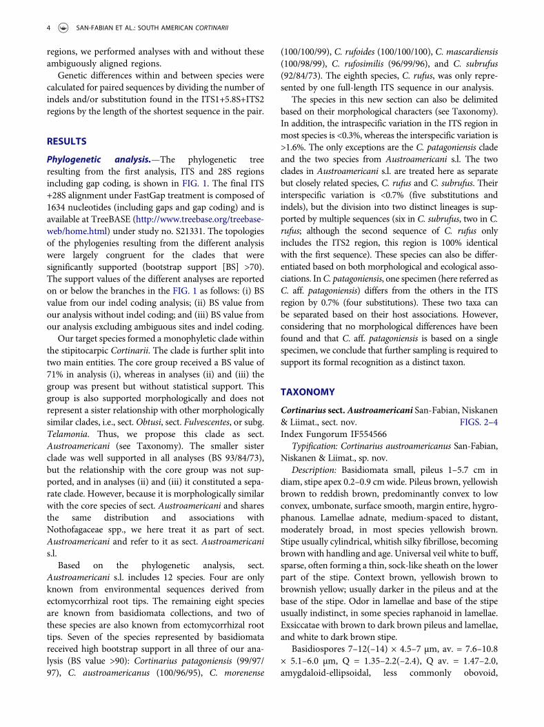
Phylogenetic analysis.—The phylogenetic treeresulting from the first analysis, ITS and 28S regionsincluding gap coding, is shown in FIG. 1. The final ITS+28S alignment under FastGap treatment is composed of1634 nucleotides (including gaps and gap coding) and isavailable at TreeBASE (http://www.treebase.org/treebase-web/home.html) under study no. S21331. The topologiesof the phylogenies resulting from the different analysiswere largely congruent for the clades that weresignificantly supported (bootstrap support [BS] >70).The support values of the different analyses are reportedon or below the branches in the FIG. 1 as follows: (i) BSvalue from our indel coding analysis; (ii) BS value fromour analysis without indel coding; and (iii) BS value fromour analysis excluding ambiguous sites and indel coding.
Our target species formed a monophyletic clade withinthe stipitocarpic Cortinarii. The clade is further split intotwo main entities. The core group received a BS value of71% in analysis (i), whereas in analyses (ii) and (iii) thegroup was present but without statistical support. Thisgroup is also supported morphologically and does notrepresent a sister relationship with other morphologicallysimilar clades, i.e., sect. Obtusi, sect. Fulvescentes, or subg.Telamonia. Thus, we propose this clade as sect.Austroamericani (see Taxonomy). The smaller sisterclade was well supported in all analyses (BS 93/84/73),but the relationship with the core group was not sup-ported, and in analyses (ii) and (iii) it constituted a sepa-rate clade. However, because it is morphologically similarwith the core species of sect. Austroamericani and sharesthe same distribution and associations withNothofagaceae spp., we here treat it as part of sect.Austroamericani and refer to it as sect. Austroamericanis.l.
Based on the phylogenetic analysis, sect.Austroamericani s.l. includes 12 species. Four are onlyknown from environmental sequences derived fromectomycorrhizal root tips. The remaining eight speciesare known from basidiomata collections, and two ofthese species are also known from ectomycorrhizal roottips. Seven of the species represented by basidiomatareceived high bootstrap support in all three of our ana-lysis (BS value >90): Cortinarius patagoniensis (99/97/97), C. austroamericanus (100/96/95), C. morenense
(100/100/99), C. rufoides (100/100/100), C. mascardiensis(100/98/99), C. rufosimilis (96/99/96), and C. subrufus(92/84/73). The eighth species, C. rufus, was only repre-sented by one full-length ITS sequence in our analysis.
The species in this new section can also be delimitedbased on their morphological characters (see Taxonomy).In addition, the intraspecific variation in the ITS region inmost species is <0.3%, whereas the interspecific variation is>1.6%. The only exceptions are the C. patagoniensis cladeand the two species from Austroamericani s.l. The twoclades in Austroamericani s.l. are treated here as separatebut closely related species, C. rufus and C. subrufus. Theirinterspecific variation is <0.7% (five substitutions andindels), but the division into two distinct lineages is sup-ported by multiple sequences (six in C. subrufus, two in C.rufus; although the second sequence of C. rufus onlyincludes the ITS2 region, this region is 100% identicalwith the first sequence). These species can also be differ-entiated based on both morphological and ecological asso-ciations. InC. patagoniensis, one specimen (here referred asC. aff. patagoniensis) differs from the others in the ITSregion by 0.7% (four substitutions). These two taxa canbe separated based on their host associations. However,considering that no morphological differences have beenfound and that C. aff. patagoniensis is based on a singlespecimen, we conclude that further sampling is required tosupport its formal recognition as a distinct taxon.
TAXONOMY
Cortinarius sect.Austroamericani San-Fabian, Niskanen& Liimat., sect. nov. FIGS. 2–4Index Fungorum IF554566
Typification: Cortinarius austroamericanus San-Fabian,Niskanen & Liimat., sp. nov.
Description: Basidiomata small, pileus 1–5.7 cm indiam, stipe apex 0.2–0.9 cm wide. Pileus brown, yellowishbrown to reddish brown, predominantly convex to lowconvex, umbonate, surface smooth, margin entire, hygro-phanous. Lamellae adnate, medium-spaced to distant,moderately broad, in most species yellowish brown.Stipe usually cylindrical, whitish silky fibrillose, becomingbrownwith handling and age. Universal veil white to buff,sparse, often forming a thin, sock-like sheath on the lowerpart of the stipe. Context brown, yellowish brown tobrownish yellow; usually darker in the pileus and at thebase of the stipe. Odor in lamellae and base of the stipeusually indistinct, in some species raphanoid in lamellae.Exsiccatae with brown to dark brown pileus and lamellae,and white to dark brown stipe.
Basidiospores 7–12(–14) × 4.5–7 µm, av. = 7.6–10.8× 5.1–6.0 µm, Q = 1.35–2.2(–2.4), Q av. = 1.47–2.0,amygdaloid-ellipsoidal, less commonly obovoid,
4 SAN-FABIAN ET AL.: SOUTH AMERICAN CORTINARII
C. austroamericanus N. dombeyiMES2028*; ARG, N Patagonia;
C. patagoniensis N. antarcticaMES2022; ARG, N Patagonia;C. patagoniensis MES2048; ARG, N patagonia; N. antarctica
C. patagoniensis N. pumilioaff. CT4610; CHI, S patagonia;
C. rubellus AY669595; EUR sect. Orellani
C. laniger AY669666; EUR subg.Telamonia
-/59/66
99/97/97
100/99/98
83/--/--
C. austroamericanus N. pumilioJX316442; ARG, N Patagonia;C. austroamericanus N. dombeyiMES2028*; ARG, N Patagonia;
sect. Austroamericani
sect. Austroamericani s .lato
71/-/--
C N. pumilio. sp. 1 UDB014287; ARG, N Patagonia;C N. pumilio. sp. 1 UDB014311; ARG, N Patagonia;
C. morenense N. dombeyi, N. pumilioMES1877; ARG, N Patagonia;C. morenense N. antarcticaMES1929; ARG, N Patagonia;C. morenense N. dombeyi, N. pumilioMES1867; ARG, N Patagonia;
C. rufoides MES2007*; ARG, N Patagonia; N. pumilioC. rufoides N. pumilioMES2007*; ARG, N Patagonia;
C. mascardiensis MES2020*; ARG, N Patagonia; N. antarcticaC. mascardiensis N. antarcticaMES2020*; ARG, N Patagonia;C. mascardiensis N. antarcticaMES2020*; ARG, N Patagonia;
C N. pumilio. sp. 2 JX316452; ARG, N Patagonia;C. N. obliquasp. 3 JX316338; ARG, N Patagonia;C N. pumilio. sp. 4 UDB014299; ARG, N Patagonia;
C. rufosimilis MES1864*; ARG, N Patagonia; N. dombeyi, N. pumilioC. rufosimilis N. dombeyi, N. pumilioMES1864*; ARG, N Patagonia;
C. subrufus N. antarcticaTN16-261; ARG, N Patagonia;C. subrufus N. antarcticaCT4647; CHI, S Patagonia;C. subrufus N. antarcticaCT4649; CHI, S Patagonia;C. subrufus N. pumilioUDB014290; ARG, N Patagonia;C. subrufus MES2047; ARG, N Patagonia; N. antarcticaC. subrufus N. antarcticaMES2084; ARG, N Patagonia;C. rufus N. dombeyi, N. pumilioMES1865; ARG, N Patagonia;
C. orixanthus KJ635245; NZLC. sp. UDB014322; ARG
C. sp. KJ635232; NZL
C. pachynemeus AF539727; CHIC. tenellus AF539728; CHI
C. squamiger AF539729; CHIC. acutus FJ157002; NA
C. obtususcf. FJ039610; NAC. cystidiocatenatus AY669651; TAS
C. acutovelatus AY669655; EUR
C. icterinoides KJ635221; NZL
C. sp. UDB014297; ARGC. sp. KY462570; CHI
C. canarius AY669630; TASC. sp. JQ282169; NZLC. cramesinusaff. JX679128; AUS
C. ardesiacus AY669650; TAS
C. uliginosus AY669584; EURC. palustris tubarius AY669581; EUR
C. sanguineus AY669582; EUR
C. malicorius AY669583; EUR
C. sp. KJ635212; NZLC. luteostriatulus AF539707; CHI
C. elaiops KT875181; NZLC. sp. KJ635217; NZL
C. olivaceoniger KJ635240; NZL
C. pseudobulliardioides KX388641; NAC. fulvescens KX388634; EUR
C. tenuifulvescens KX388644; EURC. sp. FJ157124; NA
C. laetus FJ157034; NAC. fillionii FJ717565; NA
C. ochrophyllus FJ039604; NA
C. sejuctus ined. AY669636; TASC. globuliformis AY669602; AUS
C. bulliardii AY669659; EUR
C. fulvoconicus AY669677; CHI
C. armillatus AY669671; EUR
C. splendidus AY669598; AUS
C. clandestinus FJ039583; NAC. parkeri FJ039585; NA
C. sp. FJ039586; NAC. kula AY669643; TAS
C. sclerophyllorum AY669637; TASC. tristis AY669648; CHI
C. caninus AY669646; EURC. anomalus AY669645; EUR
C. delibutus AY669587; EURC. salor AY669592; EUR
C. spilomeus AY669654; EURC. bolaris AY669596; EUR
C. balteatus AY669526; EURC. balteatocumatilis AY174801; EUR
C. violaceus AY669579; EURC. rotundisporus AY669612; AUS
C. spadicellus AY669539; EURC. lavendulensis AY669617; AUS
sect. Obtusi
subg. Dermocybe
sect. Fulvescentes
/Splendidi
subg. Leprocybe
sect. Anomali
sect. Delibuti
sect. Cortinarius
sect. Phlegmacioides
98/84/72
100/96/9551/--/--
100/100/99
93/84/73
0.05
100/100/100
100/100/99
100/98/95
71/-/-- 77/--/--
96/99/96
53/--/--
92/88/83
99/97/92
91/80/76100/100/99
74/68/71100/100/100
95/--/--55/--/--
92/87/96
56/62/53
51/56/54
64/63/--
78/-/--
100/100/99
100/100/100
51/59/65
72/85/--75/-/--
100/99/9999/98/85
55/--/--
97/89/8790/73/69
93/96/94100/100/100
74/--/--
100/84/6993/--/89
93/67/60
-/70/64
100/100/100
100/100/99
100/100/99
77/57/63100/99/93
92/83/82
62/--/83
97/77/69
Figure 1. Maximum likelihood phylogeny based on ITS and 28S (D1–D2) sequences. Topology based on analyses under indel codingtreatment. Three bootstrap values of different analyses are shown as follows: BS value from indel coding analysis/BS value fromanalysis without indel coding/BS value from analysis without indel coding and excluding ambiguous sites. Bootstrap values ≥50%are indicated above branches. “-” indicates <50% bootstrap value; “–” indicates nonexistent topology; “*” indicates sequencesgenerated from different basidiomata of the same collection. Species names of newly described type specimens and sections are inboldface. Bootstrap values of sect. Austroamericani s.s. and s.l. as well as of the species within the section are in boldface. ARG =Argentina; CHI = Chile; NZL = New Zealand; AUS = Australia; TAS = Tasmania; NA = North America; EUR = Europe.
MYCOLOGIA 5
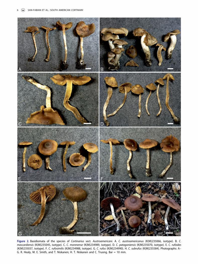
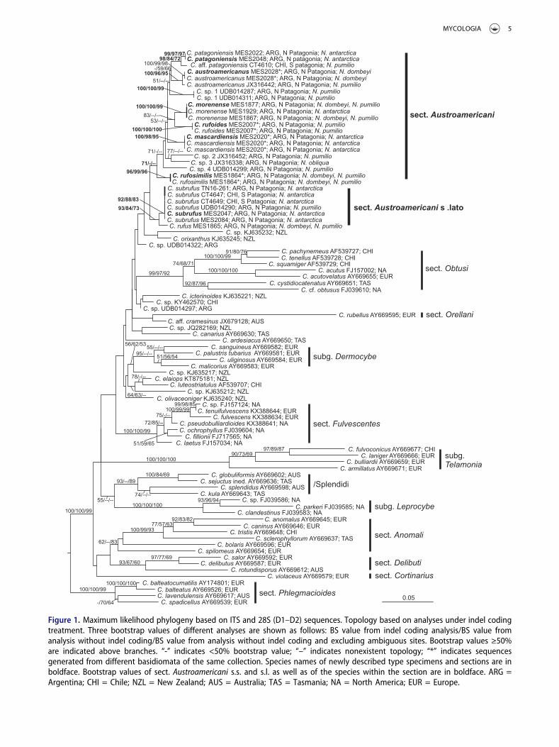
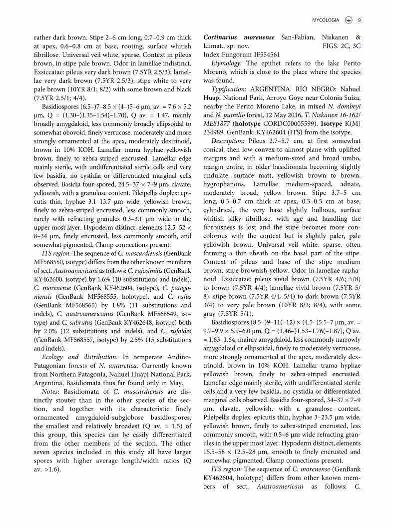
Figure 2. Basidiomata of the species of Cortinarius sect. Austroamericani. A. C. austroamericanus (K(M)235066, isotype). B. C.mascardiensis (K(M)235045, isotype). C. C. morenense (K(M)234989, isotype). D. C. patagoniensis (K(M)235070, isotype). E. C. rufoides(K(M)235037, isotype). F. C. rufosimilis (K(M)234988, isotype). G. C. rufus (K(M)234990). H. C. subrufus (K(M)235584). Photographs: A–G. R. Healy, M. E. Smith, and T. Niskanen; H. T. Niskanen and C. Truong. Bar = 10 mm.
6 SAN-FABIAN ET AL.: SOUTH AMERICAN CORTINARII
broadly ellipsoidal in C. mascardiensis; finely tostrongly ornamented, more strongly ornamented atthe apex, somewhat to strongly dextrinoid. Lamellartrama hyphae yellowish brown, finely to zebra-stripedencrusted in MLZ. Lamellar edge mainly sterile, withundifferentiated sterile cells and a very few basidia, nocystidia or differentiated marginal cells observed.Basidia four-spored, clavate, yellowish, with a granulosecontent. Pileipellis duplex: epicutis thin, hyphae up to23 µm wide, yellowish brown, finely to zebra-stripedencrusted, less commonly smooth, with refracting gran-ules. Hypoderm distinct, smooth to finely encrustedand somewhat pigmented. Clamp connections present.
Ecology and distribution: Associated with evergreen(Nothofagus dombeyi) or deciduous (N. pumilio and N.antarctica) species of Nothofagus. Currently onlyknown from Patagonia, South America.
Notes: The species of Cortinarius sect.Austroamericani are reminiscent of the species of C.sect. Fulvescentes/Laeti and Obtusi. The members ofthese sections, however, do not have refracting granulesin their epicutis. In addition, the members of sectionsFulvescentes and Laeti are currently only known fromthe Northern Hemisphere and have a colored universalveil (yellow, ochraceous, pink to vinaceous) in contrastto the white to buff veil of the species of sect.Austroamericani. The species of sect. Obtusi occurboth in the Northern and Southern Hemispheres but
have a smell of iodoform at the base of the stipe whenslightly dried, and some species have lamellar tramawith ellipsoidal inflated hyphae ending in balloon-shaped cystidia.
Cortinarius austroamericanus San-Fabian, Niskanen &Liimat., sp. nov. FIGS. 2A, 3A, 4Index Fungorum IF554559
Etymology: The epithet refers to the distribution ofthe species in South America.
Typification: ARGENTINA. RIO NEGRO: NahuelHuapi National Park, road to Tronador, after PampaLinda, at the base of the hill, in N. dombeyi forest, 14May 2016, T. Niskanen 16-235/MES2028 (holotypeCORDC00005643). Isotype K(M)235066. GenBank:MF568549 (ITS) and KY462644 (ITS), generated fromtwo different basidiomes of the isotype.
Description: Pileus 2.7–3.6 cm, at first somewhatconical, then low convex to almost plane with a verylow umbo, margin entire, in older basidiomata becom-ing slightly undulating, surface smooth, somewhatwaxy-looking, medium brown, hygrophanous.Lamellae medium-spaced to distant, adnate, moderatelybroad, yellow brown. Stipe 5.9–8.5 cm long, 0.3–0.6 cmthick at apex, 0.5–0.9 cm at base, cylindrical, at the verybase slightly bulbous, surface whitish silky fibrillose,with age and handling the fibrousness is lost and thestipe becomes more concolorous with the context but
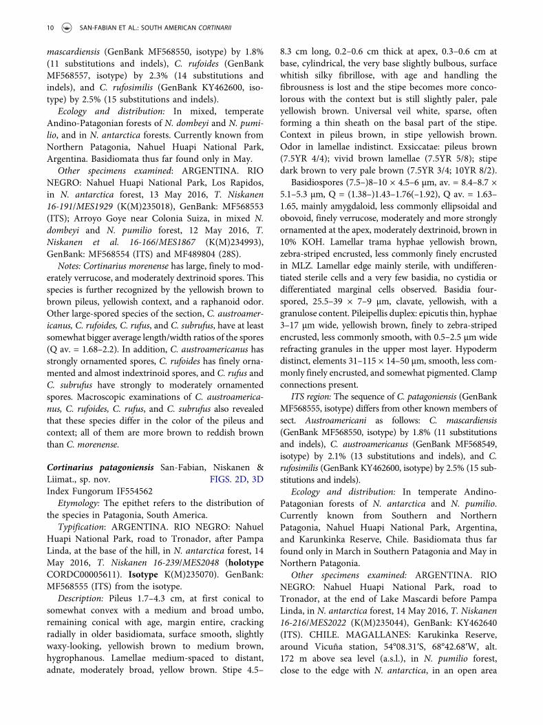
Figure 3. Basidiospores of the species of Cortinarius sect. Austroamericani. A. C. austroamericanus (K(M)235066, isotype). B. C.mascardiensis (K(M)235045, isotype). C. C. morenense (K(M)234989, isotype). D. C. patagoniensis (K(M)235070, isotype). E. C. rufoides (K(M)235037, isotype). F. C. rufosimilis (K(M)234988, isotype). G. C. rufus (K(M)234990). H. C. subrufus (K(M)235069, isotype).Photographs: B. San-Fabian. Bar = 10 µm.
MYCOLOGIA 7
remains slightly paler, pale brown. Universal veil white,sparse, often forming a thin sheath on the basal part ofthe stipe. Context medium brown in pileus and stipe.Odor in lamellae and base of the stipe indistinct.Exsiccatae: pileus dark brown (7.5YR 3/3), lamellaedark brown (7.5YR 3/3), stipe dark brown to white(7.5YR 3/2; 10YR 8/1).
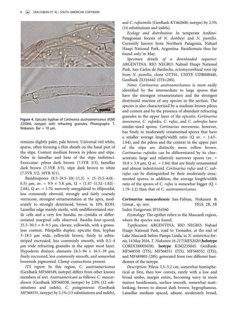
Basidiospores (8.5–)9.5–10(–11.5) × (5–)5.5–6.0(–6.5) µm, av. = 9.9 × 5.8 µm, Q = (1.47–)1.52–1.82(–2.04), Q av. = 1.70, narrowly amygdaloid to ellipsoidal,less commonly obovoid, strongly and fairly coarselyverrucose, strongest ornamentation at the apex, mod-erately to strongly dextrinoid, brown in 10% KOH.Lamellar edge mainly sterile, with undifferentiated ster-ile cells and a very few basidia, no cystidia or differ-entiated marginal cells observed. Basidia four-spored,25.5–30.5 × 8–9.5 µm, clavate, yellowish, with a granu-lose content. Pileipellis duplex: epicutis thin, hyphae3–18.5 µm wide, yellowish brown, finely to zebra-striped encrusted, less commonly smooth, with 0.5–4µm wide refracting granules in the upper most layer.Hypoderm distinct, elements 24.5–94 × 16.5–39 µm,finely encrusted, less commonly smooth, and somewhatbrownish pigmented. Clamp connections present.
ITS region: In this region, C. austroamericanus(GenBank MF568549, isotype) differs from other knownmembers of sect. Austroamericani as follows: C. mascar-diensis (GenBank MF568550, isotype) by 2.0% (12 sub-stitutions and indels), C. patagoniensis (GenBankMF568555, isotype) by 2.1% (13 substitutions and indels),
and C. rufosimilis (GenBank KY462600, isotype) by 2.3%(14 substitutions and indels).
Ecology and distribution: In temperate Andino-Patagonian forests of N. dombeyi and N. pumilio.Currently known from Northern Patagonia, NahuelHuapi National Park, Argentina. Basidiomata thus farfound only in May.
Specimen details of a downloaded sequence:ARGENTINA. RIO NEGRO: Nahuel Huapi NationalPark, San Carlos de Bariloche, ectomycorrhizal root tipfrom N. pumilio, clone OTT01, UNITE UDB008440,GenBank JX316442 (ITS+28S).
Notes: Cortinarius austroamericanus is most easilyidentified by the intermediate to large spores thathave the strongest ornamentation and the strongestdextrinoid reaction of any species in the section. Thespecies is also characterized by a medium brown pileusand context and by the presence of abundant refractinggranules in the upper layer of the epicutis. Cortinariusmorenense, C. rufoides, C. rufus, and C. subrufus havesimilar-sized spores. Cortinarius morenense, however,has finely to moderately ornamented spores that havea smaller average length/width ratio (Q av. = 1.63–1.64), and the pileus and the context in the upper partof the stipe are distinctly more yellow brown.Cortinarius rufoides can be differentiated by its char-acteristic large and relatively narrower spores (av. =10.8 × 5.9 µm, Q av. = 1.84) that are finely ornamentedand almost indextrinoid. Cortinarius rufus and C. sub-rufus can be distinguished by their moderately orna-mented spores; in addition, the average length/widthratio of the spores of C. rufus is somewhat bigger (Q =1.79–2.2) than that of C. austroamericanus.
Cortinarius mascardiensis San-Fabian, Niskanen &Liimat., sp. nov. FIGS. 2B, 3BIndex Fungorum IF554560
Etymology: The epithet refers to the Mascardi region,where the species was found.
Typification: ARGENTINA. RIO NEGRO: NahuelHuapi National Park, road to Tronador, at the end ofLake Mascardi before Pampa Linda, in N. antarctica for-est, 14May 2016, T. Niskanen 16-217/MES2020 (holotypeCORDC00005658). Isotype K(M)235045. GenBank:MF568550 (ITS), MF568551 (ITS), MF568552 (ITS),and MF489803 (28S), generated from two different basi-diomes of the isotype.
Description: Pileus 3.5–5.2 cm, somewhat hemisphe-rical at first, then low convex, rarely with a low andbroad umbo, margin entire, becoming wavy in moremature basidiomata, surface smooth, somewhat matt-looking, brown to almost dark brown, hygrophanous.Lamellae medium spaced, adnate, moderately broad,
Figure 4. Epicutis hyphae of Cortinarius austroamericanus (K(M)235066, isotype) with refracting granules. Photographs: T.Niskanen. Bar = 10 µm.
8 SAN-FABIAN ET AL.: SOUTH AMERICAN CORTINARII
rather dark brown. Stipe 2–6 cm long, 0.7–0.9 cm thickat apex, 0.6–0.8 cm at base, rooting, surface whitishfibrillose. Universal veil white, sparse. Context in pileusbrown, in stipe pale brown. Odor in lamellae indistinct.Exsiccatae: pileus very dark brown (7.5YR 2.5/3); lamel-lae very dark brown (7.5YR 2.5/3); stipe white to verypale brown (10YR 8/1; 8/2) with some brown and black(7.5YR 2.5/1; 4/4).
Basidiospores (6.5–)7–8.5 × (4–)5–6 µm, av. = 7.6 × 5.2µm, Q = (1.30–)1.35–1.54(–1.70), Q av. = 1.47, mainlybroadly amygdaloid, less commonly broadly ellipsoidal tosomewhat obovoid, finely verrucose, moderately and morestrongly ornamented at the apex, moderately dextrinoid,brown in 10% KOH. Lamellar trama hyphae yellowishbrown, finely to zebra-striped encrusted. Lamellar edgemainly sterile, with undifferentiated sterile cells and veryfew basidia, no cystidia or differentiated marginal cellsobserved. Basidia four-spored, 24.5–37 × 7–9 µm, clavate,yellowish, with a granulose content. Pileipellis duplex: epi-cutis thin, hyphae 3.1–13.7 µm wide, yellowish brown,finely to zebra-striped encrusted, less commonly smooth,rarely with refracting granules 0.3–3.1 µm wide in theupper most layer. Hypoderm distinct, elements 12.5–52 ×8–34 µm, finely encrusted, less commonly smooth, andsomewhat pigmented. Clamp connections present.
ITS region: The sequence of C. mascardiensis (GenBankMF568550, isotype) differs from the other knownmembersof sect.Austroamericani as follows:C. rufosimilis (GenBankKY462600, isotype) by 1.6% (10 substitutions and indels),C. morenense (GenBank KY462604, isotype), C. patago-niensis (GenBank MF568555, holotype), and C. rufus(GenBank MF568565) by 1.8% (11 substitutions andindels), C. austroamericanus (GenBank MF568549, iso-type) and C. subrufus (GenBank KY462648, isotype) bothby 2.0% (12 substitutions and indels), and C. rufoides(GenBank MF568557, isotype) by 2.5% (15 substitutionsand indels).
Ecology and distribution: In temperate Andino-Patagonian forests of N. antarctica. Currently knownfrom Northern Patagonia, Nahuel Huapi National Park,Argentina. Basidiomata thus far found only in May.
Notes: Basidiomata of C. mascardiensis are dis-tinctly stouter than in the other species of the sec-tion, and together with its characteristic finelyornamented amygdaloid-subglobose basidiospores,the smallest and relatively broadest (Q av. = 1.5) ofthis group, this species can be easily differentiatedfrom the other members of the section. The otherseven species included in this study all have largerspores with higher average length/width ratios (Qav. >1.6).
Cortinarius morenense San-Fabian, Niskanen &Liimat., sp. nov. FIGS. 2C, 3CIndex Fungorum IF554561
Etymology: The epithet refers to the lake PeritoMoreno, which is close to the place where the specieswas found.
Typification: ARGENTINA. RIO NEGRO: NahuelHuapi National Park, Arroyo Goye near Colonia Suiza,nearby the Perito Moreno Lake, in mixed N. dombeyiand N. pumilio forest, 12 May 2016, T. Niskanen 16-162/MES1877 (holotype CORDC00005599). Isotype K(M)234989. GenBank: KY462604 (ITS) from the isotype.
Description: Pileus 2.7–5.7 cm, at first somewhatconical, then low convex to almost plane with upliftedmargins and with a medium-sized and broad umbo,margin entire, in older basidiomata becoming slightlyundulate, surface matt, yellowish brown to brown,hygrophanous. Lamellae medium-spaced, adnate,moderately broad, yellow brown. Stipe 3.7–5 cmlong, 0.3–0.7 cm thick at apex, 0.3–0.5 cm at base,cylindrical, the very base slightly bulbous, surfacewhitish silky fibrillose, with age and handling thefibrousness is lost and the stipe becomes more con-colorous with the context but is slightly paler, paleyellowish brown. Universal veil white, sparse, oftenforming a thin sheath on the basal part of the stipe.Context of pileus and base of the stipe mediumbrown, stipe brownish yellow. Odor in lamellae rapha-noid. Exsiccatae: pileus vivid brown (7.5YR 4/6; 5/8)to brown (7.5YR 4/4); lamellae vivid brown (7.5YR 5/8); stipe brown (7.5YR 4/4; 5/4) to dark brown (7.5YR3/4) to very pale brown (10YR 8/3; 8/4), with somegray (7.5YR 5/1).
Basidiospores (8.5–)9–11(–12) × (4.5–)5.5–7 µm, av. =9.7–9.9 × 5.9–6.0 µm, Q = (1.46–)1.53–1.76(–1.87), Q av.= 1.63–1.64, mainly amygdaloid, less commonly narrowlyamygdaloid or ellipsoidal, finely to moderately verrucose,more strongly ornamented at the apex, moderately dex-trinoid, brown in 10% KOH. Lamellar trama hyphaeyellowish brown, finely to zebra-striped encrusted.Lamellar edge mainly sterile, with undifferentiated sterilecells and a very few basidia, no cystidia or differentiatedmarginal cells observed. Basidia four-spored, 34–37 × 7–9µm, clavate, yellowish, with a granulose content.Pileipellis duplex: epicutis thin, hyphae 3–23.5 µm wide,yellowish brown, finely to zebra-striped encrusted, lesscommonly smooth, with 0.5–6 µm wide refracting gran-ules in the upper most layer. Hypoderm distinct, elements15.5–58 × 12.5–28 µm, smooth to finely encrusted andsomewhat pigmented. Clamp connections present.
ITS region: The sequence of C. morenense (GenBankKY462604, holotype) differs from other known mem-bers of sect. Austroamericani as follows: C.
MYCOLOGIA 9
mascardiensis (GenBank MF568550, isotype) by 1.8%(11 substitutions and indels), C. rufoides (GenBankMF568557, isotype) by 2.3% (14 substitutions andindels), and C. rufosimilis (GenBank KY462600, iso-type) by 2.5% (15 substitutions and indels).
Ecology and distribution: In mixed, temperateAndino-Patagonian forests of N. dombeyi and N. pumi-lio, and in N. antarctica forests. Currently known fromNorthern Patagonia, Nahuel Huapi National Park,Argentina. Basidiomata thus far found only in May.
Other specimens examined: ARGENTINA. RIONEGRO: Nahuel Huapi National Park, Los Rapidos,in N. antarctica forest, 13 May 2016, T. Niskanen16-191/MES1929 (K(M)235018), GenBank: MF568553(ITS); Arroyo Goye near Colonia Suiza, in mixed N.dombeyi and N. pumilio forest, 12 May 2016, T.Niskanen et al. 16-166/MES1867 (K(M)234993),GenBank: MF568554 (ITS) and MF489804 (28S).
Notes: Cortinarius morenense has large, finely to mod-erately verrucose, and moderately dextrinoid spores. Thisspecies is further recognized by the yellowish brown tobrown pileus, yellowish context, and a raphanoid odor.Other large-spored species of the section, C. austroamer-icanus, C. rufoides, C. rufus, and C. subrufus, have at leastsomewhat bigger average length/width ratios of the spores(Q av. = 1.68–2.2). In addition, C. austroamericanus hasstrongly ornamented spores, C. rufoides has finely orna-mented and almost indextrinoid spores, and C. rufus andC. subrufus have strongly to moderately ornamentedspores. Macroscopic examinations of C. austroamerica-nus, C. rufoides, C. rufus, and C. subrufus also revealedthat these species differ in the color of the pileus andcontext; all of them are more brown to reddish brownthan C. morenense.
Cortinarius patagoniensis San-Fabian, Niskanen &Liimat., sp. nov. FIGS. 2D, 3DIndex Fungorum IF554562
Etymology: The epithet refers to the distribution ofthe species in Patagonia, South America.
Typification: ARGENTINA. RIO NEGRO: NahuelHuapi National Park, road to Tronador, after PampaLinda, at the base of the hill, in N. antarctica forest, 14May 2016, T. Niskanen 16-239/MES2048 (holotypeCORDC00005611). Isotype K(M)235070). GenBank:MF568555 (ITS) from the isotype.
Description: Pileus 1.7–4.3 cm, at first conical tosomewhat convex with a medium and broad umbo,remaining conical with age, margin entire, crackingradially in older basidiomata, surface smooth, slightlywaxy-looking, yellowish brown to medium brown,hygrophanous. Lamellae medium-spaced to distant,adnate, moderately broad, yellow brown. Stipe 4.5–
8.3 cm long, 0.2–0.6 cm thick at apex, 0.3–0.6 cm atbase, cylindrical, the very base slightly bulbous, surfacewhitish silky fibrillose, with age and handling thefibrousness is lost and the stipe becomes more conco-lorous with the context but is still slightly paler, paleyellowish brown. Universal veil white, sparse, oftenforming a thin sheath on the basal part of the stipe.Context in pileus brown, in stipe yellowish brown.Odor in lamellae indistinct. Exsiccatae: pileus brown(7.5YR 4/4); vivid brown lamellae (7.5YR 5/8); stipedark brown to very pale brown (7.5YR 3/4; 10YR 8/2).
Basidiospores (7.5–)8–10 × 4.5–6 µm, av. = 8.4–8.7 ×5.1–5.3 µm, Q = (1.38–)1.43–1.76(–1.92), Q av. = 1.63–1.65, mainly amygdaloid, less commonly ellipsoidal andobovoid, finely verrucose, moderately and more stronglyornamented at the apex, moderately dextrinoid, brown in10% KOH. Lamellar trama hyphae yellowish brown,zebra-striped encrusted, less commonly finely encrustedin MLZ. Lamellar edge mainly sterile, with undifferen-tiated sterile cells and a very few basidia, no cystidia ordifferentiated marginal cells observed. Basidia four-spored, 25.5–39 × 7–9 µm, clavate, yellowish, with agranulose content. Pileipellis duplex: epicutis thin, hyphae3–17 µm wide, yellowish brown, finely to zebra-stripedencrusted, less commonly smooth, with 0.5–2.5 µm widerefracting granules in the upper most layer. Hypodermdistinct, elements 31–115 × 14–50 µm, smooth, less com-monly finely encrusted, and somewhat pigmented. Clampconnections present.
ITS region: The sequence of C. patagoniensis (GenBankMF568555, isotype) differs from other known members ofsect. Austroamericani as follows: C. mascardiensis(GenBank MF568550, isotype) by 1.8% (11 substitutionsand indels), C. austroamericanus (GenBank MF568549,isotype) by 2.1% (13 substitutions and indels), and C.rufosimilis (GenBank KY462600, isotype) by 2.5% (15 sub-stitutions and indels).
Ecology and distribution: In temperate Andino-Patagonian forests of N. antarctica and N. pumilio.Currently known from Southern and NorthernPatagonia, Nahuel Huapi National Park, Argentina,and Karunkinka Reserve, Chile. Basidiomata thus farfound only in March in Southern Patagonia and May inNorthern Patagonia.
Other specimens examined: ARGENTINA. RIONEGRO: Nahuel Huapi National Park, road toTronador, at the end of Lake Mascardi before PampaLinda, in N. antarctica forest, 14 May 2016, T. Niskanen16-216/MES2022 (K(M)235044), GenBank: KY462640(ITS). CHILE. MAGALLANES: Karukinka Reserve,around Vicuña station, 54°08.31′S, 68°42.68′W, alt.172 m above sea level (a.s.l.), in N. pumilio forest,close to the edge with N. antarctica, in an open area
10 SAN-FABIAN ET AL.: SOUTH AMERICAN CORTINARII
grazed by horses with dense understory, 26 Mar 2017,T. Niskanen & C. Truong CT4610, (K(M)235581).GenBank: MF568556 (ITS) and MF489804 (28S).
Notes: This small and slender species looks like thosein sect. Obtusi. This is the only species known fromsect. Austroamericani with a conical yellowish brown tomedium brown pileus. The basidiospores are of inter-mediate size, finely verrucose, and almost indextrinoid.The spores of C. rufosimilis are of similar size andshape but they are moderately ornamented and mod-erately dextrinoid and the pileus is convex and reddishbrown. Based on molecular data, C. patagoniensis iscomposed of two taxa, C. patagoniensis and C. aff.patagoniensis, because our specimen CT4610 differsfrom the type of C. patagoniensis by four substitutions.To date, C. patagoniensis has only been found in forestsof N. antarctica and C. aff. patagoniensis in forests of N.pumilio. However, considering that no morphologicaldifferences have been found, we treat all of the collec-tions mentioned here as C. patagoniensis s.l. Furtherstudies with additional specimens are needed to deter-mine the species limits in this group.
Cortinarius rufoides San-Fabian, Niskanen & Liimat.,sp. nov. FIGS. 2E, 3EIndex Fungorum IF554563.
Etymology: The epithet indicates that the color of thebasidiomata resembles that of C. rufus.
Typification: ARGENTINA. RIO NEGRO: NahuelHuapi National Park, road to Tronador, just before themountain base, in N. pumilio forest, 14 May 2016, T.Niskanen 16-209/MES2007 (holotype CORDC00005552).Isotype K(M)235037. GenBank: MF568557 (ITS) andMF568558 (ITS), generated from two different basidiomesof the isotype.
Description: Pileus 2–2.8 cm, at first somewhat con-ical, then convex with a low and broad umbo, marginentire, surface smooth, slightly waxy-looking, not viscid,medium red brown, center often dark brown, sometimesyellow brown toward the margin, hygrophanous.Lamellae medium-spaced, adnate, moderately broad,yellow brown. Stipe 3.1–4 cm long, 0.3–0.5 cm thick atapex, 0.4–0.5 cm at base, cylindrical, surface whitish silkyfibrillose, with age and handling the fibrousness is lostand the stipe becomes more concolorous with the con-text but is slightly paler, pale yellowish brown. Universalveil white, sparse, often forming a thin sheath on thebasal part of the stipe. Context in pileus reddish brown,in stipe yellowish brown. Odor in lamellae indistinct.Exsiccatae: pileus dark brown to vivid brown (7.5YR 3/4;4/6); lamellae brown (7.5YR 4/4); stipe dark brown tovery dark brown (7.5YR 2.5/2; 3/4).
Basidiospores (8.5–)10–12(–12.5) × (4.5–)5.5–6.5µm, av. = 10.8 × 5.9 µm, Q = (1.72–)1.74–1.94(–2.13),Q av. = 1.84, narrowly amygdaloid-ellipsoidal, lesscommonly obovoid, finely verrucose, moderately orna-mented at the apex, somewhat dextrinoid, brown in10% KOH. Lamellar trama hyphae yellowish brown,zebra-striped encrusted, less commonly finelyencrusted in MLZ. Lamellar edge mainly sterile, withundifferentiated sterile cells and a very few basidia, nocystidia or differentiated marginal cells observed.Basidia four-spored, 30.5–41 × 7–10 µm, clavate, yel-lowish, with a granulose content. Pileipellis duplex:epicutis thin, hyphae 4.5–19 µm wide, yellowishbrown, finely to zebra-striped encrusted, less com-monly smooth, with 0.5–5.5 µm wide refracting gran-ules in the upper most layer. Hypoderm distinct,elements 19–86 × 15.5–47 µm, finely encrusted, lesscommonly smooth, and somewhat pigmented. Clampconnections present.
ITS region: The sequence of C. rufoides (GenBankMF568557, isotype) deviates from the other knownmembers of sect. Austroamericani by at least 2.3% (14substitutions and indels).
Ecology and distribution: In temperate Andino-Patagonian forests of N. pumilio. Currently known fromNorthern Patagonia, Nahuel Huapi National Park,Argentina. Basidiomata thus far found only in May.
Notes: Cortinarius rufoides has among the largest andrelatively narrowest spores of the section, and they arefinely ornamented and almost indextrinoid. The pileus ismedium red brown and often with a dark brown center.The sister species C. morenense has slightly more orna-mented and more dextrinoid spores with a smaller aver-age length/width ratio (Q av. = 1.63–1.64). Cortinariusrufosimilis, C. rufus, and C. subrufus are macroscopicallyrather similar to C. rufoides but often have somewhatdarker, more reddish brown pileus and can be distin-guished from C. rufoides by their moderately verrucoseand moderately dextrinoid spores. The spores of C. rufusand C. subrufus are similar in size to those of C. rufoides,but the spores of C. rufosimilis are smaller and relativelybroader (Q av. = 1.65).
Cortinarius rufosimilis San-Fabian, Niskanen &Liimat., sp. nov. FIGS. 2F, 3FIndex Fungorum IF554564
Etymology: The epithet refers to the similarity in thecolor of the basidiomata of this species to C. rufus.
Typification: ARGENTINA. RIO NEGRO: HuapiNational Park, Arroyo Goye near Colonia Suiza, in mixedN. dombeyi and N. pumilio forest, 12 May 2016, T.Niskanen 16-161/MES1864 (holotype CORDC00005566).Isotype K(M)234988. GenBank: KY462600 (ITS),
MYCOLOGIA 11
MF568559 (ITS), and MF489802 (28S), generated fromtwo different basidiomes of the isotype.
Description: Pileus 2.5–3.2 cm, at first somewhatconical, then convex umbonate with a medium andbroad umbo, margin entire, becoming slightly undu-late in more mature basidiomata, surface smooth,slightly waxy-looking, vivid reddish brown to darkreddish brown often with a darker center, hygro-phanous. Lamellae medium-spaced, adnate, moder-ately broad, yellow brown. Stipe 5–5.4 cm long, 0.3–0.4 cm thick at apex, 0.5–0.7 cm at base, cylindrical,the very base slightly bulbous, surface whitish silkyfibrillose, with age and handling the fibrousness islost and the stipe becomes more concolorous withthe context but is slightly paler, pale brown.Universal veil white to buff, sparse, often forminga thin sheath on the basal part of the stipe. Contextin pileus and in base of the stipe stronger brown, instipe yellowish brown. Odor in lamellae faintlyraphanoid. Exsiccatae: pileus dark brown (7.5YR 3/4); lamellae dark brown (7.5YR 3/4); stipe darkbrown to very pale brown (7.5YR 3/2; 10YR 8/2).
Basidiospores 8–9.5(–10) × 5–6 µm, av. = 8.7 × 5.3µm, Q = 1.49–1.78(–1.83), Q av. = 1.65, mainlyamygdaloid, less commonly ellipsoidal and obovoid,moderately to strongly verrucose, strongest ornamen-ted at the apex, moderately dextrinoid, brown in 10%KOH. Lamellar trama hyphae yellowish brown, finelybut distinctly encrusted. Lamellar edge mainly sterile,with undifferentiated sterile cells and a very few basi-dia, no cystidia or differentiated marginal cellsobserved. Basidia four-spored, 21.5–33 × 7–9 µm,clavate, yellowish, with a granulose content.Pileipellis duplex: epicutis thin, hyphae 3–19 µmwide, yellowish brown, finely to zebra-stripedencrusted, less commonly smooth, with 0.5–4 µmwide refracting granules in the upper most layer.Hypoderm distinct, elements 23.5–83 × 15.5–37 µm,finely encrusted or with more distinctive encrusta-tions, less commonly smooth, and somewhat pigmen-ted. Clamp connections present.
ITS region: The sequence of C. rufosimilis (GenBankKY462600, isotype) differs from other known membersof sect. Austroamericani as follows: C. mascardiensis(GenBank MF568550, isotype) and C. subrufus(GenBank KY462648, isotype) both by 1.6% (10 substitu-tions and indels), C. rufus (GenBank MF568565) by 1.8%(11 substitutions and indels), C. austroamericanus(GenBank MF568549, isotype) by 2.3% (14 substitutionsand indels), and C. morenense (GenBank KY462604, iso-type) and C. patagoniensis (GenBank MF568555, isotype)both by 2.4% (15 substitutions and indels).
Ecology and distribution: In mixed Andino-Patagonian forests of N. dombeyi and N. pumilio.Currently known from Northern Patagonia, NahuelHuapi National Park, Argentina. Basidiomata thus farfound only in May.
Notes: Cortinarius rufosimilis is characterized by avivid reddish brown to dark reddish brown pileus, aslightly raphanoid odor in the lamellae, and intermedi-ate-sized, moderately to strongly verrucose spores. Thespores of C. patagoniensis are of similar size but are finelyornamented and almost indextrinoid. In addition, thepileus is more conical and yellowish brown. Cortinariusrufoides, C. rufus, and C. subrufus could be macroscopi-cally confused with C. rufosimilis because of the similarcolor and shape of the basidiomata. However, theirspores are larger and the average Q value is typicallysomewhat larger (Q av. = 1.68–2.2) than in C. rufosimilis.
Cortinarius sect. Austroamericani s.l.—The twospecies listed below form a well-supported basal cladethat is separated from the core sect. Austroamericani.This clade is treated as Austroamericani s.l. because therelationship with Austroamericani s.s. is not statisticallysupported. Additional taxon sampling and geneticstudies with other loci are needed to resolve thedelimitation of this section.
Cortinarius rufus M.M. Moser, Beihefte zur NovaHedwigia 52:335. 1975. FIGS. 2G, 3G
Typification: ARGENTINA. NEUQUÉN: CerroCortinario, Puerto Manzano, in N. pumilio forest, 18Apr 1963, M. Moser 1963/0369 (holotype IB19630369).GenBank: MF568564 (ITS2).
Description: Pileus up to 2 cm wide, at first some-what conical, then convex umbonate, margin entire,surface smooth to radially fibrillose, reddish brown tosomewhat dark brown, hygrophanous. Lamellae med-ium-spaced to distant, adnate, moderately broad,vividly cinnamon brown. Stipe 2.5–3 cm long, 0.3–0.4 cm thick at apex, up to 0.5 cm at base, cylindrical,the very base slightly bulbous, surface at first whitishsilky fibrillose, with age and handling the fibrousness islost and the stipe becomes more concolorous with thecontext. Universal veil white, sparse, forming a thinsheath on the basal part of the stipe. Context in pileusand stipe dark brown. Odor in lamellae indistinct.Exsiccatae: pileus dark brown (7.5YR 3/3); lamellaedark brown (7.5YR 3/3); stipe dark brown (7.5YR 3/4)to white (10YR 8/1).
Basidiospores (9.5–)10–12(–13.5) × 5–6(–6.5) µm, av. =10.1–11.1 × 5.6 µm, Q = (1.61–)1.7–2.2(–2.4), Q av. = 1.79–2.0, narrowly amygdaloid to ellipsoidal, often with a
12 SAN-FABIAN ET AL.: SOUTH AMERICAN CORTINARII
suprahilar depression, moderately to strongly verrucose,strongest ornamented at the apex, moderately to almoststrongly dextrinoid, brown in 10% KOH. Lamellar tramahyphae yellowish brown, finely to zebra-striped encrusted.Lamellar edge mainly sterile, with undifferentiated sterilecells and a very few basidia, no cystidia or differentiatedmarginal cells observed. Basidia four-spored, 31.5–37 ×8–9 µm, clavate, yellowish, with a granulose content.Pileipellis duplex: epicutis thin, hyphae 4.7–19 µm wide,yellowish brown, finely to zebra-striped encrusted, lesscommonly smooth, with some refracting granules 0.3–1µm wide in the upper most layer. Hypoderm distinct,elements 33–100 × 23–37 µm, smooth to finely encrustedand somewhat pigmented. Clamp connections present.
ITS region: The sequence of C. rufus (GenBankMF568565) deviates from the other known members ofsect.Austroamericani as follows: from the sister species C.subrufus (GenBank KY462648, isotype) by 0.7% (5 sub-stitutions and indels), from C. mascardiensis (GenBankMF568550, isotype) and C. rufosimilis (GenBankKY462600, holotype) both by 1.8% (11 substitutions andindels), and from C. rufoides (GenBank MF568557, iso-type) by 2.5% (15 substitutions and indels).
Ecology and distribution: In temperate Andino-Patagonian forests of N. pumilio and in forest mixedwith N. dombeyi. Currently known from NorthernPatagonia, Puerto Manzano, and Nahuel HuapiNational Park, Argentina.
Specimen examined: ARGENTINA. RIO NEGRO:Nahuel Huapi National Park, Arroyo Goye nearColonia Suiza, in mixed N. dombeyi and N. pumilioforest, 12 May 2016, T. Niskanen et al. 16-163/MES1865 (K(M)234990). GenBank: MF568565 (ITS)and MF489798 (28S).
Notes: Cortinarius rufus is notable for its large, mod-erately to strongly verrucose, and relatively narrowspores (Q av. = 1.8–2.0). This species is also defined bya rather small (up to 2 cm diam), reddish brown tobrown pileus. The sister species C. subrufus is morpho-logically similar, but its spores are somewhat smaller (av.= 9.2–9.7 × 5.4–5.6 µm) and more ellipsoidal (Q av. =1.68–1.70). Cortinarius austroamericanus, C. morenense,and C. rufoides are also large-spored species. However,the spores of C. austroamericanus are broader (Q av. =1.7), C. morenense has broader and more finely orna-mented spores (Q av. = 1.63–1.64), and C. rufoides hasfinely ornamented and almost indextrinoid spores.Cortinarius morenense and C. rufoides can also be differ-entiated by their slightly paler and more yellowish pileiand stipe context, especially in C. morenense where thesefeatures are more obvious.
Our observations of Cortinarius rufus fit withMoser’s original description except that the pileus was
described as radially fibrillose and the spores weredescribed as warty and 10.5–13(–14) × 5.5–7 µm. Wereexamined the spores of the type specimen andobserved the following measurements: (10–)10.5–12(–13.5) × 5–6(–6.5) µm, av. = 11.1 × 5.6 µm, Q = (1.6–)1.8–2.2(–2.4), Q av. = 2.0. Observed values from thetype are close to our measurements from recently col-lected material, (9.5–)10–10.5(–11.5) × 5–6 µm, av. =10.1 × 5.6 µm, Q = (1.6–)1.7–1.9(–2.1), Q av. = 1.8.Furthermore, the shape, ornamentation, and dextri-noidity of the spores in our new collection are alsosimilar to those of the type. The ITS2 sequence fromthe type specimen of C. rufus is identical to thesequence from MES1865. Our conclusion is that thesetwo specimens represent the same species; thus, weprovide an updated description of this species above.
Cortinarius subrufus San-Fabian, Niskanen & Liimat.,sp. nov. FIGS. 2H, 3HIndex Fungorum IF554565.
Etymology: The epithet refers to the affinity to C. rufus.Typification: ARGENTINA. RIO NEGRO: Nahuel
Huapi National Park, road to Tronador, after PampaLinda, at the base of the hill, in N. antarctica forest, 14May 2016, T. Niskanen 16-238/MES2047 (holotypeCORDC00005558). Isotype K(M)235069. GenBank:KY462648 (ITS), generated from the isotype.
Description: Pileus 1.8–3.4 cm, at first somewhatconical, then convex umbonate with a medium tobroad umbo, margin entire, becoming slightly undulatein more mature basidiomata, surface smooth, slightlywaxy-looking, vivid reddish brown to dark reddishbrown, hygrophanous. Lamellae medium-spaced to dis-tant, adnate, moderately broad, yellow brown. Stipe3.7–7.5 cm long, 0.3–0.5 cm thick at apex, 0.5–0.8 cmat base, cylindrical, the very base slightly bulbous, sur-face whitish silky fibrillose, rather stiff, with age andhandling the fibrousness is lost and the stipe becomesmore concolorous with the context but is slightly paler,pale brown. Universal veil white, sparse, forming a thinsheath on the basal part of the stipe. Context in pileusand base of the stipe dark brown, in stipe yellowishbrown to pale brown. Odor in lamellae indistinct.Exsiccatae: pileus dark brown to brown (7.5YR 3/4; 4/4); vivid brown lamellae (7.5YR 4/6); stipe dark brown(7.5YR 3/3) to light gray and white (10YR 7/2; 8/1).
Basidiospores (8.5–)9–11 × 5–6.5 µm, av. = 9.2–9.7 ×5.4–5.6 µm, Q = (1.45–)1.50–1.88(–1.96), Q av. = 1.68–1.70, narrowly amygdaloid to ellipsoidal, less com-monly obovoid; moderately to strongly verrucose,strongest ornamentation at the apex, moderately toalmost strongly dextrinoid, brown in 10% KOH.Lamellar trama hyphae yellowish brown, finely to
MYCOLOGIA 13
zebra-striped encrusted. Lamellar edge mainly sterile,with a very few basidia, no cystidia or differentiatedmarginal cells observed. Basidia four-spored, 20.5–32 ×6.5–9 µm, clavate, yellowish, with a granulose content.Pileipellis duplex: epicutis thin, hyphae 6–20 µm wide,yellowish brown, finely to zebra-striped encrusted, lesscommonly smooth, with 0.5–3 µm wide refractinggranules in the upper most layer. Hypoderm distinct,elements 18.5–89 × 14–42 µm, smooth to finelyencrusted and somewhat pigmented. Clamp connec-tions present.
ITS region: The sequence of C. subrufus (GenBankKY462648, isotype) differs from the other known mem-bers of sect. Austroamericani as follows: from its sisterspecies C. rufus (GenBank MF568565) by 0.7% (5 sub-stitutions and indels), from C. rufosimilis (GenBankKY462600, isotype) by 1.6% (10 substitutions andindels), and from C. mascardiensis (GenBankMF568550, isotype) by 2.0% (12 substitutions andindels).
Ecology and distribution: In temperate Andino-Patagonian forests of N. antarctica and N. pumilio.Currently known from both Southern and NorthernPatagonia, Nahuel Huapi National Park, Argentina,and Karukinka Reserve, Chile. Basidiomata thus farfound only in Mar in Southern Patagonia and May inNorthern Patagonia.
Other specimens examined: ARGENTINA. RIONEGRO: Nahuel Huapi National Park, Lago Hess,in N. antarctica forest, 17 May 2016, T. Niskanen16-259/MES2084 (K(M)235093), GenBank:MF568560 (ITS); loc. cit., 16 May 2016, T. Niskanen16-261 (K(M)235095), GenBank: MF568563 (ITS)and MF489801 (28S). CHILE. MAGALLANES:Karukinka Reserve, along the path to Laguna delCura from Vicuña station, by the stream, 54°08′46.0″S, 68°43′40.2″W, alt. 252 m a.s.l., in N. antarc-tica forest, close by N. pumilio forest, by the creek, 26Mar 2017, T. Niskanen & C. Truong CT4647 (SGO; K(M)235584), GenBank: MF568562 (ITS) andMF489800 (28S); Karukinka Reserve, along the pathto Laguna del Cura from Vicuña station, by thestream, 54°08′46.0″S, 68°43′40.2″W, alt. 252 m a.s.l.,in N. antarctica forest, close by N. pumilio forest, bythe creek, 26 Mar 2017, T. Niskanen & C. TruongCT4649 (SGO; K(M)235583), GenBank: MF568561(ITS) and MF489799 (28S).
Specimen details of a downloaded sequence:ARGENTINA. RIO NEGRO: Nahuel Huapi NationalPark, San Carlos de Bariloche, ectomycorrhizal root tipfrom N. pumilio, UNITE UDB014290 (ITS+28S).
Notes: Cortinarius subrufus is recognized by a vividreddish brown to brown pileus and intermediate-sized
to large-sized, moderately to strongly verrucose spores.Spores of the sister species C. rufus are somewhat larger(av. = 10.1–11.1 × 5.6 µm), and the average length/widthratio of the spores is higher (Q av. = 1.8–2.0). Cortinariusaustroamericanus and C. morenense also have spores ofsimilar sizes. The spores of C. austroamericanus, how-ever, are strongly verrucose, and the spores of C. mor-enense finely ornamented and the average length/widthratio is smaller (Q av. = 1.63–1.64). Cortinarius more-nense can also be differentiated by its notably moreyellowish pileus and context. Based on macroscopiccharacters, C. rufoides and C. rufosimilis could be mis-identified as C. subrufus because all three species havesimilar reddish brown pilei. However, the spores of C.rufoides are larger (av. = 10.8 × 5.9), more finely orna-mented, narrower (Q av. = 1.84), and almost indextri-noid, whereas the spores of C. rufosimilis are smaller (av.= 8.7 × 5.3) and the average length/width ratio of thespores is higher (Q av. = 1.65).
KEY TO THE DESCRIBED SPECIES OFCORTINARIUS SECT. AUSTROAMERICANI
1. Basidiospores on average <9 µm long. ............... 21′. Basidiospores larger, on average >9 µm long ... 42. Basidiospores on average <8 µm long; basidio-
mata comparatively stouter, apex of the stipeoften ≥0.7 cm wide. ................... C. mascardiensis
2′. Basidiospores on average >8 µm long; basidio-mata slender, apex of the stipe often ≤0.6 cmwide .......................................................................... 3
3. Pileus yellow brown; basidiospores finely verru-cose, almost indextrinoid . ......... C. patagoniensis
3′. Pileus reddish brown; basidiospores moderatelyverrucose, moderately dextrinoid... C. rufosimilis
4. Q av. >1.75 . ............................................................ 54′. Q av. <1.75 .............................................................. 65. Pileus medium red brown, center often dark
brown, sometimes yellow brown toward the mar-gin; context in the upper part of the stipe yellowbrown; basidiospores finely verrucose, somewhatdextrinoid................................................. C. rufoides
5′. Pileus reddish brown to almost dark brown; con-text in the upper part of the stipe brown to darkbrown; basidiospores moderately to strongly ver-rucose and dextrinoid................................. C. rufus
6. Q av. <1.65, basidiospores finely to moderatelyverrucose, moderately dextrinoid.. . C. morenense
6′. Q av. >1.65, basidiospores moderately to stronglyverrucose and dextrinoid. ...................................... 7
7. Pileus medium brown; basidiospores on average9.9 × 5.8 µm.............. ............. C. austroamericanus
14 SAN-FABIAN ET AL.: SOUTH AMERICAN CORTINARII
7′. Pileus vivid reddish brown to dark reddish brown;basidiospores on average 9.2–9.7 × 5.4–5.6 µm............................................................................ C. subrufus
DISCUSSION
Species delimitation and phylogenetic relationships.—Twelve species are recognized within the newly describedsect. Austroamericani. All species have an intraspecificvariation of less than 0.3% in the ITS region. Theinterspecific variation is greater than 1.6%, except in thespecies pair C. rufus/C. subrufus where variation betweenthe species is also 0.7%. All species received high support inour phylogenetic analysis, and their delimitation is alsosupported by morphological data. This corresponds wellwith previous studies of Cortinarius that have shown thatan ITS similarity threshold value of 99% is suitable fordistinguishing species in the majority of lineages, althoughsome morphologically distinguishable species may haveeven higher threshold values, probably indicating recentradiation events (Niskanen et al. 2011; Garnica et al.2016). The barcodes of the species will be deposited in theRefSeq database (Schoch et al. 2014) and used to setreference sequences in UNITE (Kõljalg et al. 2013).
The species studied here formed a monophyleticclade consisting of two main entities, sect.Austroamericani s.s., which grouped together in allanalyses, and sect. Austroamericani s.l., which formeda separate clade that is not statistically supported assister to sect. Austroamericani s.s. However, we treatsect. Austroamericani s.l. as part of the sectionAustroamericani because the morphology, distribution,and host plant associations are consistent with sect.Austroamericani s.s. The Austroamericani s.l. clade issister to Austroamericani s.s. in all analyses, suggestingthat C. rufus and the closely related C. subrufus (thetwo species in the Austroamericani s.l. clade) mayrepresent an ancestral clade within the section.
The only previously known species of the section,C. rufus, was originally placed in subg. Telamonia(Moser and Horak 1975). However, based on currentknowledge, subg. Telamonia mainly includes repre-sentatives from the Northern Hemisphere (Peintneret al. 2004; Garnica et al. 2005). Our phylogeneticanalysis revealed that C. rufus does not belong tosubg. Telamonia and suggests that its closest relativesare Nothofagaceae-associated species from SouthAmerica and New Zealand. The current classificationfor South American Cortinarius was proposed byMoser and Horak (1975) and is based on morpholo-gical characters. Recent studies have shown that tra-ditional classifications are at least partly artificial andare in a need of revision based on molecular data
(Peintner et al. 2001, 2004; Garnica et al. 2003, 2005).Our results show that sect. Austroamericani is notclosely related to morphologically similar clades thathave been described previously; we therefore proposeit as a new section. Based on morphological andmolecular features, this section belongs to the largeand well-supported monophyletic lineage of stipito-carpic Cortinarii that includes subgenera Cortinariusand Telamonia, most of subg. Myxacium, and somephlegmacioid species (Stensrud et al. 2014). However,additional relationships within the lineage were notresolved in our current analysis.
The short branch leading to sect. Austroamericani sug-gests that it has undergone a rapid evolutionary diversifica-tion. In Cortinarius, Southern Hemisphere clades generallyhave shorter branch lengths than clades from the NorthernHemisphere ormixed clades that include species from boththe Northern and Southern Hemispheres, i.e., Obtusi,Fulvescentes, and Telamonia (FIG. 1). This is not a featurederived from biased sampling because the same tree topol-ogy was also observed in analyses that included additionalspecies from the Northern Hemisphere (T. Niskanen,unpubl. data). The recent radiation observed in sect.Austroamericani could be explained by an adaptive radia-tion during the process of speciation. We hypothesize thatthe species in sect.Austroamericanimay have diversified ina relatively short time. Colonization of a new areamay haveled to rapid adaptation because of the availability of newniches. This phenomenon of short branches has not beenreported so far in other studies of South American fungi;therefore, further studies of other Cortinarius groups areneeded to verify this trend. Future directions include time-calibrated analyses to date lineage divergence events as wellas phylogenies of Nothofagaceae and other plant associa-tions, in order to understand coevolutionary processes.Moreover, dating the phylogeny could help to reveal howthe occurrence of adaptive radiation is correlated withhistorical events (e.g., movement of landmasses or forestexpansion). In addition, a biogeographic characterizationcould help to resolve the biogeographic history of SouthernHemispheric species, including the direction of coloniza-tion and characterization of ancestral clades.
Morphological characteristics.—We found severalmorphological characters that in combination can beused to recognize the members of sect.Austroamericani. All species form small basidiomataand have a more or less brown and hygrophanouspileus, white to buff universal veil, and brown tobrownish yellow context in the stipe. The pileipellis isduplex, with a thin epicutis and a distinctive hypoderm.Zebra-striped encrusted hyphae are commonly found in
MYCOLOGIA 15
both the lamellae and epicutis, and refracting granulescan be found in the epicutis. The refracting granules inthe epicutis are not a common feature in telamonioidCortinarii and may be a very useful character fordistinguishing the members of this section from othermorphologically similar species. However,morphological convergence exists with other cladessuch as subg. Telamonia, sect. Fulvescentes/Laeti, andsect. Obtusi (Peintner et al. 2004; Garnica et al. 2005;Niskanen 2008). Currently, because we are far fromknowing all species and their characters in Cortinarius,sequence-based identification is generally needed forreliable section-level recognition.
For species-level identification within the section,the most important diagnostic characters are mainlyspore size (length, width), shape (Q value), and orna-mentation. The color and in some cases the shape ofthe pileus and the color of the context in the stipe arealso useful for species identification when used togetherwith additional significant diagnostic features.
Ecology and distribution.—The species of sect.Austroamericani s.l. are found in association withNothofagaceae species, including N. antarctica, N.dombeyi, N. pumilio, and Lophozonia obliqua (Heenanand Smissen 2013). The ectomycorrhizal associationsformed by species in sect. Austroamericani did not showany evident host specificity, as also previously suggested byNouhra et al. (2013). This suggests that species are notstrictly constrained to one host but may grow inassociation with different tree species fromNothofagaceae. However, most of our sampling was inforests in the southern part of Patagonia that weredominated by species of Nothofagus. We only rarelysampled in association with trees in the genusLophozonia, and we were unable to visit endangered treesin the genus Fuscospora. Further sampling with thesephylogenetically divergent hosts in the northern part ofPatagonia may uncover additional new species that couldbelong to sect. Austroamericani.
All studied species of sect. Austroamericani have thusfar been found only from Argentina and Chile, so wesuggest that this section is likely endemic to SouthAmerica. All collecting was done on the drier side ofthe Andes, and it is unknown whether these species alsooccur in more humid coastal areas. Most of the speciesare currently known only from a very limited area, withthe exception of C. patagoniensis and C. subrufus, whichhave been found from both Southern and NorthernPatagonia. The limited number of collections availabledoes not permit us to draw further conclusions about thedistribution of these species so far.
Conclusions.—This study contributes to the knowledgeon Patagonian fungal diversity by formally recognizingthe new section Austroamericani, represented by 12species, of which 7 are described as new in this study.We have included Cortinarius rufus and C. subrufus aspart of sect. Austroamericani s.l., although itsrelationship with the remaining species in the coresect. Austroamericani s.s. is not statistically supportedby the phylogenetic analysis. However, we found thatall the species are morphologically similar, form ECMassociations with the trees from the familyNothofagaceae, and are endemic to southern SouthAmerica. We therefore treat them all as part of thesame section. Our study aims to encourage similarresearch in South America, a region that is largelyunderstudied and with a presumably large fungaldiversity yet to be described.
ACKNOWLEDGMENTS
We thank the Administracion de Parques Nacionales ofArgentina for authorizing our collecting expeditions inParque Nacional Nahuel Huapi and Parque Nacional Laninunder Projecto 2016/720 (to E. Nouhra). The WildlifeConservation Society Chile in Parque Karukinka kindly author-ized our investigation (C.T. and T.N.). We thank the staff at theCORD herbarium for helping to rapidly process loans to facil-itate this work. We acknowledge critical logistical support andphotography help during field collecting by R. Healy, F. Kuhar,G. Furci, N. Fernandez, and D. Pfister. We thank Ester Gaya forinspiring discussions on the evolution on South AmericanCortinarii during the preparation of the manuscript. Lastly,we would like to thank the editor and two anonymousreviewers who helped us to improve the manuscript.
FUNDING
This work was supported by the US National ScienceFoundation grant DEB 1354802 (to M.E.S. and P.B.M.), twoBentham-Moxon Trust grants in 2016 and 2017 for fieldwork (to T.N.), an Advanced Postdoc Mobility Fellowshipfrom the Swiss National Science Foundation (to C.T.), andthe University of Florida Institute for Food and AgriculturalSciences (IFAS) (to M.E.S.).
ORCID
Beatriz San-Fabian http://orcid.org/0000-0001-5797-4584Tuula Niskanen http://orcid.org/0000-0003-1479-5548Kare Liimatainen http://orcid.org/0000-0002-5422-2384Pepijn W. Kooij http://orcid.org/0000-0002-2619-0813Alija B. Mujic http://orcid.org/0000-0002-5810-5521P. Brandon Matheny http://orcid.org/0000-0003-3857-2189
16 SAN-FABIAN ET AL.: SOUTH AMERICAN CORTINARII
LITERATURE CITED
Abarenkov K, Tedersoo L, Nilsson RH, Vellak K, Saar I,Veldre V, Parmasto E, Prous M, Aan A, Ots M, KurinaO, Ostonen I, Jõgeva J, Halapuu S, Põldmaa K, TootsM, Truu J, Larsson K-H, Kõljalg U. 2010. PlutoF—aweb based workbench for ecological and taxonomicresearch, with an online Implementation for fungalITS sequences. Evolutionary Bioinformatics Online6:189–196.
Bödeker ITM, Clemmensen KE, de Boer W, Martin F,Olson A, Lindahl BD. 2014. EctomycorrhizalCortinarius species participate in enzymatic oxidationof humus in northern forest ecosystems. NewPhytologist 203:245–256.
Borchsenius F. 2009. FastGap 1.2. Department ofBiosciences, Aarhus University, Denmark. [cited 2017May 21]. Available from: http://www.aubot.dk/FastGap_home.htm
Bunyard BA, Nicholson MS, Royse DJ. 1994. A systematicassessment of Morchella using RFLP analysis of the 28Sribosomal RNA gene. Mycologia 86:762–772.
Dentinger BTM, Margaritescu S, Moncalvo J-M. 2010. Rapidand reliable high-throughput methods of DNA extractionfor use in barcoding and molecular systematics of mush-rooms. Molecular Ecology Resources 10:628–633,doi:10.1111/j.1755-0998.2009.02825.x
Galtier N, Gouy M, Gautier C. 1996. SEAVIEW andPHYLO_WIN: two graphic tools for sequence alignmentand molecular phylogeny. Computer Applications in theBiosciences 12:543–548.
Gardes M, Bruns TD. 1993. ITS primers with enhanced speci-ficity for basidiomycetes—application to the identification ofmycorrhizae and rusts. Molecular Ecology 2:113–118.
Garnica S, Schön ME, Abarenkov K, Riess K, Liimatainen K,Niskanen T, Dima B, Soop K, Frøslev TG, Jeppesen TS,Peintner U, Kuhnert-Finkernagel R, Brandrud TE, Saar G,Oertel B, Ammirati JF. 2016. Determining threshold valuesfor barcoding fungi: lessons fromCortinarius (Basidiomycota),a highly diverse and widespread ectomycorrhizal genus. FEMSMicrobiology Ecology 92: fiw045.
Garnica S, Weiss M, Oberwinkler F. 2002. New Cortinariusspecies from Nothofagus forests in South Chile. Mycologia94:136–145.
Garnica S, Weiss M, Oberwinkler F. 2003. Morphological andmolecular phylogenetic studies in South AmericanCortinarius species. Mycological Research 107:1143–1156.
Garnica S, Weiss M, Oertel B, Oberwinkler F. 2005. A frame-work for a phylogenetic classification in the genusCortinarius (Basidiomycota, Agaricales) derived from mor-phological and molecular data. Canadian Journal Botany83:1457–1477.
Garrido N. 1988. Agaricales s.l. und ihre Mykorrhizen in denNothofagus-Wäldern Mittelchiles. Bibliotheca Mycologica120: 1–528.
Harrower E, Ammirati JF, Cappuccino AA, Ceska O,Kranabetter JM, Kroeger P, Lim SR, Taylor T, BerbeeML. 2011. Cortinarius species diversity in BritishColumbia and molecular phylogenetic comparison withEuropean specimen sequences. Botany 89:799–810.
Heenan PB, Smissen RD. 2013. Revised circumscription ofNothofagus and recognition of the segregate genera
Fuscospora, Lophozonia, and Trisyngyne (Nothofagaceae).Phytotaxa 146:1–31.
Horak E. 1980. Fungi, Basidiomycetes. Agaricales yGasteromycetes secotioides. Flora Criptogámica de Tierradel Fuego 11(6):1–524.
Ivanova NV, Dewaard JR, Hebert PDN. 2006. An inexpen-sive, automation-friendly protocol for recovering high-quality DNA. Molecular Ecology Notes 6:998–1002.
Katoh K, Kuma K, Toh H, Miyata T. 2005. MAFFT version 5:improvement in accuracy of multiple sequence alignment.Nucleic Acids Research 33:511–518.
Katoh K, Standley DM. 2013. MAFFT Multiple SequenceAlignment Software version 7: improvements in perfor-mance and usability. Molecular Biology and Evolution30:772–780.
Kirk PM, Cannon PF,Minter DW, Stalpers JA. 2008. Dictionaryof the fungi, 10th ed. Wallingford, UK: CSIRO Publishing. p.784.
Kõljalg U, Nilsson RH, Abarenkov K, Tedersoo L, TaylorAFS, Bahram M, Bates ST, Bruns TD, Bengtsson-Palme J,Callaghan TM, Douglas B, Drenkhan T, Eberhardt U,Dueñas M, Grebenc T, Griffith GW, Hartmann M, KirkPM, Kohout P, Larsson E, Lindahl BD, Lücking R, MartínMP, Matheny PB, Nguyen NH, Niskanen T, Oja J, PeayKG, Peintner U, Peterson M, Põldmaa K, Saag L, Saar I,Schüßler A, Scott JA, Senés C, Smith ME, Suija A, TaylorDL, Telleria MT, Weiß M, Larsson K-H. 2013. Towards aunified paradigm for sequence-based identification ofFungi. Molecular Ecology 22:5271–5277.
Kuhar F, Smith ME, Mujic A, Truong C, Nouhra E. 2017. Asystematic overview of Descolea (Agaricales) in theNothofagaceae forests of Patagonia. Fungal Biology121:876–889.
Liimatainen K, Niskanen T, Dima B, Kytövuori I, AmmiratiJF, Frøslev TG. 2014. The largest type study of Agaricalesspecies to date: bringing identification and nomenclatureof Phlegmacium (Cortinarius) into the DNA era. Persoonia33:98–140.
Maddison WP, Maddison DR. 2017. Mesquite: a modular sys-tem for evolutionary analysis. Version 3.2. [cited 2017 May24]. Available from: http://mesquiteproject.org/
Moser M, Horak E. 1975. Cortinarius Fr. und nahe verwandteGattungen in Südamerika. Beihefte zur Nova Hedwigia 52:1–628.
Munsell. 2009. Munsell soil color charts: with genuineMunsell color chips. 2009 rev. ed. Grand Rapids,Michigan: Munsell Color.
Niskanen T. 2008. Cortinarius subgenus Telamonia p.p. inNorth Europe [PhD academic dissertation]. Helsinki,Finland: Yliopistopaino. p. 33.
Niskanen T, Kytövuori I, Bendisken E, Bendisken K,Brandrud TE, Frøslev TG, Høiland K, Jeppesen TS,Liimatainen K, Lindström H. 2008. Cortinarius. In:Knudsen H, Vesterholt J, eds. Funga Nordica: Agaricoid,Boletoid and Cyphelloid genera. Copenhagen, Denmark:Nordsvamp. p. 661–777.
Niskanen T, Kytövuori I, Liimatainen K. 2011. Cortinarius sect.Armillati in northern Europe. Mycologia 103:1080–1101.
Nouhra E, Urcelay C, Longo S, Fontenla S. 2012. Differentialhypogeous sporocarp production from Nothofagus dom-beyi and N. pumilio forests in southern Argentina.Mycologia 104:45–52.
MYCOLOGIA 17
Nouhra E, Urcelay C, Longo S, Tedersoo L. 2013.Ectomycorrhizal fungal communities associated toNothofagus species in Northern Patagonia. Mycorrhiza23:487–496.
Peintner U, Bougher NL, Castellano MA, Moncalvo JM,Moser MM, Trappe JM, Vilgalys R. 2001. Multipleorigins of sequestrate fungi related to Cortinarius(Cortinariaceae). American Journal of Botany88:2168–2719.
Peintner U, Moncalvo JM, Vilgalys R. 2004. Toward a betterunderstanding of the infrageneric relationships inCortinarius (Agaricales, Basidiomycota). Mycologia96:1042–1058.
Romano GM, Lechner BE. 2014. The Cortinariaceae ofArgentina’s Nothofagus forests. Mycotaxon link page 126:247.
Schoch C, Robbertse B, Robert V, Vu D, Cardinali G, Irinyi, L,Meyer W, Nilsson H, Hughes K, Miller AN, Kirk PM,Abarenkov K, Aime MC, Ariyawansa HA, Bidartondo M,Boekhout T, Buyck B, Cai Q, Chen J, Crespo A, Crous PW,Damm U, De Beer ZW, Dentinger BTM, Divakar PK,Dueñas M, Feau N, Fliegerova K, García MA, Ge ZW,Griffith GW, Groenewald JZ, Groenewald M, Grube M,Gryzenhout M, Gueidan C, Guo L, Hambleton S, HamelinR, Hansen K,Hofstetter V, Hong SB, Houbraken J, Hyde KD,Inderbitzin P, Johnston PR, Karunarathna SC, Kõljalg U,Kovács GM, Kraichak E, Krizsan K, Kurtzman CP, LarssonKH, Leavitt S, Letcher PM, Liimatainen K, Liu JK, Lodge J,Luangsa-ard JJ, Lumbsch HT, Maharachchikumbura SSN,Manamgoda D, Martín MP, Minnis AM, Moncalvo JM,Mulè G, Nakasone KK, Niskanen T, Olariaga I, Papp T,Petkovits T, Pino-Bodas R, Powell MJ, Raja HA, RedeckerD, Sarmiento-Ramirez JM, Seifert KA, Shrestha B, StenroosS, Stielow B, Suh SO, Tanaka K, Tedersoo L, Telleria MT,Udayanga D, Untereiner WA, Uribeondo JD, Subbarao KV,Vágvölgyi C, Visagie C, Voigt K,Walker DM,Weir BS,WeißM, Wijayawardene NN, Wingfield MJ, Xu JP, Yang ZL,Zhang N, Zhuang WY, Federhen S. 2014. Finding needlesin haystacks: linking scientific names, reference specimensand molecular data for Fungi. Database: The Journal ofBiological Databases and Curation 2014:bau061.
Simmons MP, Ochoterena H. 2000. Gaps as characters insequence-based phylogenetic analyses. Systematic Biology49:369–381.
Singer R, Moser M. 1965. Forest mycology and forest com-munities in South America. 1. The early fall aspect of themycoflora of the Cordillera Pelada (Chile).Mycopathologia et Mycologia Applicata 26:129–191.
Spegazzini C. 1887a. Fungi patagonici. Boletín de laAcademia Nacional de Ciencias de Córdoba 11:5–64.
Spegazzini C. 1887b. Fungi fuegiani. Boletín de la AcademiaNacional de Ciencias de Córdoba 11:135–308.
Stamatakis A. 2014. RAxML version 8: a tool for phylogeneticanalysis and post-analysis of large phylogenies.Bioinformatics 30:1312–1313.
Stensrud Ø, Orr RJS, Reier-Røberg K, Schumacher T, HøilandK. 2014. Phylogenetic relationships in Cortinarius withfocus on North European species. Karstenia 54:57–71.
Tedersoo L, Bahram M, Toots M, Diédhiou AG, Henkel TW,Kjøller R, Morris MH, Nara K, Nouhra E, Peay KG, PõlmeS, Ryberg M, Smith ME, Kõljalg U. 2012. Towards globalpatterns in the diversity and community structure of ecto-mycorrhizal fungi. Molecular Ecology 21:4160–4170.
Tedersoo L, Jairus T, Horton BM, Abarenkov K, Suvi T, SaarI, Kõljalg U. 2008. Strong host preference of ectomycor-rhizal fungi in a Tasmanian wet sclerophyll forest asrevealed by DNA barcoding and taxon-specific primers.New Phytologist 180:479–490.
Tedersoo L, May TW, Smith ME. 2010. Ectomycorrhizallifestyle in fungi: global diversity, distribution, andevolution of phylogenetic lineages. Mycorrhiza20:217–263.
Thiers B. [continuously updated]. Index Herbariorum: a glo-bal directory of public herbaria and associated staff. NewYork Botanical Garden’s Virtual Herbarium. [cited 2017Jun 25]. Available from: http://sweetgum.nybg.org/ih/
Truong C, Mujic AB, Healy R, Kuhar F, Furci G, Torres D,Niskanen T, Sandoval-Leiva PA, Fernández N, Escobar JM,Moretto A, Palfner G, Pfister D, Nouhra E, Swenie R,Sánchez-García M, Matheny PB, Smith ME. 2017a. Howto know the fungi: combining field inventories and DNA-barcoding to document fungal diversity. New Phytologist214:913–919.
Truong C, Sánchez-Ramírez S, Kuhar F, Kaplan Z, SmithME. 2017b. The Gondwanan connection—southern tem-perate Amanita lineages and the description of the firstsequestrate species from the Americas. Fungal Biology121:638–651.
Valenzuela E, Esteve-Raventós F. 1994. Cortinarius horakii, anew species from Chile. Mycological Research 98:973–938.
Vesterholt J. 1991. Knold-slørhatte (Cortinarius underslaegtPhlegmacium) som indikatorarter for en type vaerdifuldeløvskovslokaliteter. Svampe 24:27–48.
Vilgalys R, Hester M. 1990. Rapid genetic identification andmapping of enzymatically amplified ribosomal DNA fromseveral Cryptococcus species. Journal of Bacteriology172:4238–4246.
White TJ, Bruns TD, Lee SB, Taylor JW. 1990.Amplification and direct sequencing of fungal ribosomalRNA genes for phylogenetics. In: Innis MA, GelfandDH, Sninsky JJ, White TJ, eds. PCR protocols: a guideto methods and applications. New York: AcademicPress. p. 315–322.
18 SAN-FABIAN ET AL.: SOUTH AMERICAN CORTINARII