Fabrication and characterization of etoposide loaded ...
12
International Journal of Drug Delivery 6 (2014) 24-35 http://www.arjournals.org/index.php/ijdd/index Original Research Article Fabrication and characterization of etoposide loaded magnetic polymeric microparticles Vankayalu Devendiran Sundar 1 , Magharla Dasaratha Dhanaraju 1 , Nandhakumar Sathyamoorthy 1 *Corresponding author: Vankayalu Devendiran Sundar 1 GIET School of Pharmacy India Abstract The purpose of this study is to develop a targeted drug carrier system. Magnetic poly ( - caprolactone) (PCL) microspheres were prepared using classical oil-in-water solvent evaporation method by loading magnetite nanoparticles and anticancer drug etoposide. The prepared magnetic microspheres were smooth, free flowing, individual and homogenous in nature. Fourier transformed infrared spectroscopy studies revealed the absence of any potential incompatibility of drug with other excipients. DSC studies were conducted to study the state of etoposide in the formulation. Further the magnetic microspheres were characterized for entrapment efficiency, drug loading, invitro release studies and subjected to particle size analysis and scanning electron microscopy. The magnetite nanoparticles were well dispersed in polymer matrix, which are responsible for magnetic response. The magnetic property of the prepared microspheres was measured by using vibrating sample magnetometer. The amount of magnetite in the formulation was estimated quantitatively by atomic absorption spectroscopy which was about 31.5%. The experimental results proved that the magnetic microspheres exhibited superparamagnetic behavior and the saturation magnetization was 7.26 emu/g. The optimized formulations exhibited a narrow size distribution which were below 10 μm and are evident from SEM analysis. Formulation batches prepared with drug/polymer ratio 1:10 showed a maximum encapsulation efficiency and the invitro release profile in phosphate buffer (pH 7.4) solution showed an extended release of etoposide up to 76.25% at the end of 21 day. Histopathological studies proved that the etoposide loaded magnetic microspheres were nontoxic and safe. Keywords: poly ( -caprolactone), solvent evaporation, superparamagnetic, saturation magnetization Introduction The rationale behind the development of polymeric controlled delivery systems is to make a therapeutic agent do its best when administered into the body. These systems are one of the most attractive areas in drug delivery and drug targeting [1,2]. Despite several advantages offered by controlled drug release, a major problem associated with all these systems so far developed is uniform biodistribution throughout the body, lack of drug specificity, requirement of excess dose to achieve high local concentration, non-specific toxicity and adverse side effects. Hence the development of drug system that deliver the drug molecules to the tumor site, without a concurrent increase in its concentration near healthy cells of the tissues, is one of the most active area of investigation in cancer research [3]. Among all the existing targeting drug delivery systems, a promising one will be the system associated with external or feedback control such as magnetic control [4,5]. The technique is based on the principle that the drug is encapsulated with magnetite nanoparticles, covered by a polymer shell. The existence of magnetic particles in magnetic microspheres will make them specifically transported to the target site under the influence of external magnetic field. On reaching the target organ or tissue, the magnetic vehicles release the drug in a temporal manner. The magnetically targeted therapy can be effective to lower the toxic effects and to enhance the therapeutic effect of drug because of the characteristics of driven magnetic accuracy, targeting and high drug capacity[6-8]. Magnetic particles are usually made of magnetite (Fe 3 O 4 ), maghemite (γ ă Fe 2 O 3 ), cobalt ferrite (Fe 2 CoO 4 ), chromium dioxide (CrO 2 ) etc. Among them magnetite is extensively used, properly characterized in various aspects, whose toxicity has been demonstrated to be low and well tolerated in human body[9,10]. Biocompatible and biodegradable polymers have been widely used for the controlled drug release [11,12]. The polymeric shell can consist of different kinds of polymers such as poly ( -caprolactone) (PCL), polylactides (PLA) and polyglycolide (PGA) are of great interest in design of carrier based drug delivery. The biocompatibility, biodegradability and greater permeability of PCL make it suitable for controlled drug delivery and wide use in medical applications. Its compatibility with wide range of drugs enables uniform drug distribution in the formulation matrix and its long term degradation facilitates drug release up to several months[13,14]. The investigation on PCL formulations revealed ISSN: 0975-0215 This work is licensed under a Creative Commons Attribution 3.0 License .
Transcript of Fabrication and characterization of etoposide loaded ...

International Journal of Drug Delivery 6 (2014) 24-35 http://www.arjournals.org/index.php/ijdd/index
Original Research Article
Fabrication and characterization of etoposide loaded magnetic polymeric
microparticles Vankayalu Devendiran Sundar1, Magharla Dasaratha Dhanaraju1, Nandhakumar Sathyamoorthy1
*Corresponding author: Vankayalu Devendiran Sundar 1GIET School of Pharmacy India
A b s t r a c t The purpose of this study is to develop a targeted drug carrier system. Magnetic poly ( -caprolactone) (PCL) microspheres were prepared using classical oil-in-water solvent evaporation method by loading magnetite nanoparticles and anticancer drug etoposide. The prepared magnetic microspheres were smooth, free flowing, individual and homogenous in nature. Fourier transformed infrared spectroscopy studies revealed the absence of any potential incompatibility of drug with other excipients. DSC studies were conducted to study the state of etoposide in the formulation. Further the magnetic microspheres were characterized for entrapment efficiency, drug loading, invitro release studies and subjected to particle size analysis and scanning electron microscopy. The magnetite nanoparticles were well dispersed in polymer matrix, which are responsible for magnetic response. The magnetic property of the prepared microspheres was measured by using vibrating sample magnetometer. The amount of magnetite in the formulation was estimated quantitatively by atomic absorption spectroscopy which was about 31.5%. The experimental results proved that the magnetic microspheres exhibited superparamagnetic behavior and the saturation magnetization was 7.26 emu/g. The optimized formulations exhibited a narrow size distribution which were below 10 μm and are evident from SEM analysis. Formulation batches prepared with drug/polymer ratio 1:10 showed a maximum encapsulation efficiency and the invitro release profile in phosphate buffer (pH 7.4) solution showed an extended release of etoposide up to 76.25% at the end of 21 day. Histopathological studies proved that the etoposide loaded magnetic microspheres were nontoxic and safe. Keywords: poly ( -caprolactone), solvent evaporation, superparamagnetic, saturation magnetization
Introduction The rationale behind the development of polymeric controlled delivery systems is to make a therapeutic agent do its best when administered into the body. These systems are one of the most attractive areas in drug delivery and drug targeting [1,2]. Despite several advantages offered by controlled drug release, a major problem associated with all these systems so far developed is uniform biodistribution throughout the body, lack of drug specificity, requirement of excess dose to achieve high local concentration, non-specific toxicity and adverse side effects. Hence the development of drug system that deliver the drug molecules to the tumor site, without a concurrent increase in its concentration near healthy cells of the tissues, is one of the most active area of investigation in cancer research [3]. Among all the existing targeting drug delivery systems, a promising one will be the system associated with external or feedback control such as magnetic control [4,5]. The technique is based on the principle that the drug is encapsulated with magnetite nanoparticles, covered by a polymer shell. The existence of magnetic particles in magnetic microspheres will make them specifically transported to the target site under the influence of
external magnetic field. On reaching the target organ or tissue, the magnetic vehicles release the drug in a temporal manner. The magnetically targeted therapy can be effective to lower the toxic effects and to enhance the therapeutic effect of drug because of the characteristics of driven magnetic accuracy, targeting and high drug capacity[6-8]. Magnetic particles are usually made of magnetite (Fe3O4), maghemite (γ Fe2O3), cobalt ferrite (Fe2CoO4), chromium dioxide (CrO2) etc. Among them magnetite is extensively used, properly characterized in various aspects, whose toxicity has been demonstrated to be low and well tolerated in human body[9,10]. Biocompatible and biodegradable polymers have been widely used for the controlled drug release [11,12]. The polymeric shell can consist of different kinds of polymers such as poly ( -caprolactone) (PCL), polylactides (PLA) and polyglycolide (PGA) are of great interest in design of carrier based drug delivery. The biocompatibility, biodegradability and greater permeability of PCL make it suitable for controlled drug delivery and wide use in medical applications. Its compatibility with wide range of drugs enables uniform drug distribution in the formulation matrix and its long term degradation facilitates drug release up to several months[13,14]. The investigation on PCL formulations revealed
ISSN: 0975-0215
This work is licensed under a Creative Commons Attribution 3.0 License.
Sundar et al. International Journal of Drug Delivery 6 (1) 24-35 [2014]
PAGE | 25 |
that the mechanism of drug release is often dominated by diffusion from microsphere matrix, by which the lipophilic drug release can be controlled in vivo from PCL formulations for much longer duration [15,16]. Etoposide is a semisynthetic derivative of podophyllotoxin that exhibits superior antitumor activity. Etoposide inhibits DNA topoisomerase II, thereby inhibiting DNA synthesis at the premiotic stage of cell division. Etoposide is cell cycle dependent and phase specific, affecting mainly the S and G2 phases of cell division and cause cell death. Etoposide is considered as major standard cytotoxic drug for small lung cancer [17]. The chemotherapy regimens that utilize etoposide are more effective when the drug is given over an extended period of time [18, 19]. The present work is aimed to develop etoposide loaded PCL magnetic microspheres that ensure the delivery of concentrated dose of etoposide directly into the tumor, which will eliminate the need for patient to consume large quantities of the drug. The developed magnetic microspheres were characterized for morphology, size distribution, encapsulation efficiency, magnetic property and in vitro release.
Materials and Methods Poly ( -caprolactone) M.Wt.45000, polyvinyl alcohol (M.wt 15000-20,000), Magnetite (iron II, III oxide) nanopowders < 50nm was purchased from sigma Aldrich chemical co. (USA). Etoposide was a gift sample obtained from Cedilla India ltd., Mumbai. Potassium dihydrogen orthophosphate and sodium hydroxide were purchased from SD fine chemicals ltd., Mumbai. All solvents (dichloromethane, methanol) used in the preparation were of analytical grade and purchased from Qualigens fine chemicals, Mumbai.
Preparation of drug loaded PCL magnetite microspheres by O/W emulsion method.
The magnetic loaded PCL microspheres containing etoposide were prepared by O/W emulsion- solvent evaporation technique using polyvinyl alcohol as the external aqueous phase. The method was similar to the procedure previously reported for the development of etoposide loaded controlled release PCL microspheres [20]. Briefly, the required amount of PCL, magnetite and drug were dissolved in 10 ml of dichloromethane. The organic phase was added slowly to the 40 ml of aqueous phase containing 1% PVA as stabilizer. The mixture was emulsified with the help of a high speed homogenizer [Turrax T25, IKA] at 5000 rpm. The formed O/W emulsion was stirred under a magnetic stirrer for 3 h at 1000 rpm under room temperature to make free of organic phase. The magnetic microspheres so formed were collected with the help of placing a magnet of 8000G strength at the bottom of the beaker, washed thrice with distilled water, filtered and dried at 45oC. All formulations were transferred to glass vial and stored in a desicator.
Morphology and particle size
The studies on morphology of samples were carried out using scanning electron microscopy (SEM, JOEL-JFC 5300). Magnetic microspheres were dispersed in distilled water, dripped in aluminium foil and evaporated. The dried magnetic microspheres were mounted on a copper stub and coated with gold palladium under vacuum using an ion sputter coater (JEOL JFC 1100E) for observation under scanning electron microscope. The formulated magnetic microspheres were characterized by optical microscopy for particle size and size distribution. The eye piece micrometer was calibrated with the help of a stage micrometer. The average particle size was determined using EdmundsonÊs equation Dmean =
Ʃnd/Ʃn, where n is the number of microspheres counted and d is the mean size range.
Determination of drug loading and encapsulation efficiency
The formulated magnetic microspheres were estimated for their loading and encapsulation efficiency by using the equations 1 and 2 respectively. Drug loading (%) = mc/mt x 100 ---------- 1 Encapsulation efficiency (%) = mc/ mθ x 100 ---------- 2 where mc is mass of etoposide in magnetic microspheres, mθ is the total mass of the drug and mt is the total mass of magnetic microspheres. An accurately weighed amount of drug loaded magnetic microspheres (100 mg) were dissolved in 10 ml of dichloromethane: methanol mixture in screw cap (Teflon tube). The tubes were shaken vigorously for 1 min. The contents were filtered by using a 0.1μ millipore filter assembly and suitably diluted with respective solvent system. The UV absorbance of the solution was measured using the UV/ Vis spectrophotometer (Perkin Elmer- LAMBDA 25) at 283 nm and the concentration was calculated according to the standard regression.
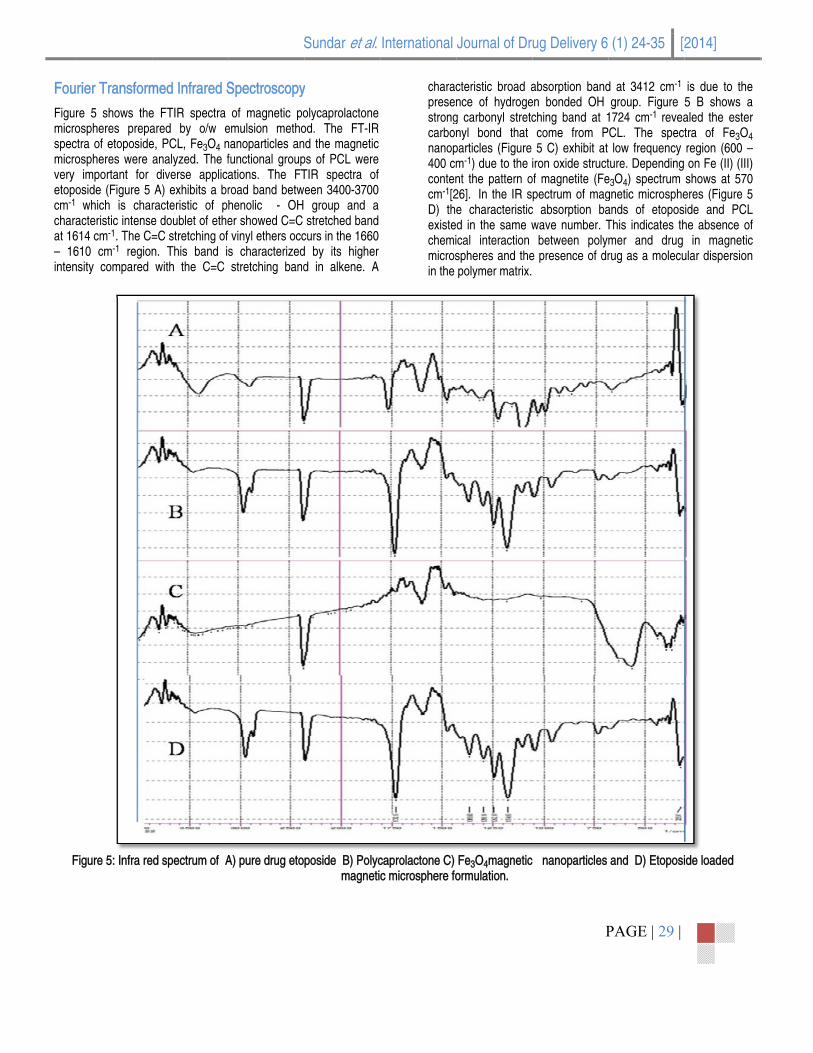
Fourier Transformed Infrared Spectroscopy (FTIR) analysis
The FTIR spectroscopy was used to characterize the drug, polymer, magnetite and the formulated magnetic microspheres. The samples were recorded on a Perkin Elmer-(Spectrum RX) using the conventional KBr pellet method. All samples were scanned in the IR range from 400- 5000cm-1at 25oC.
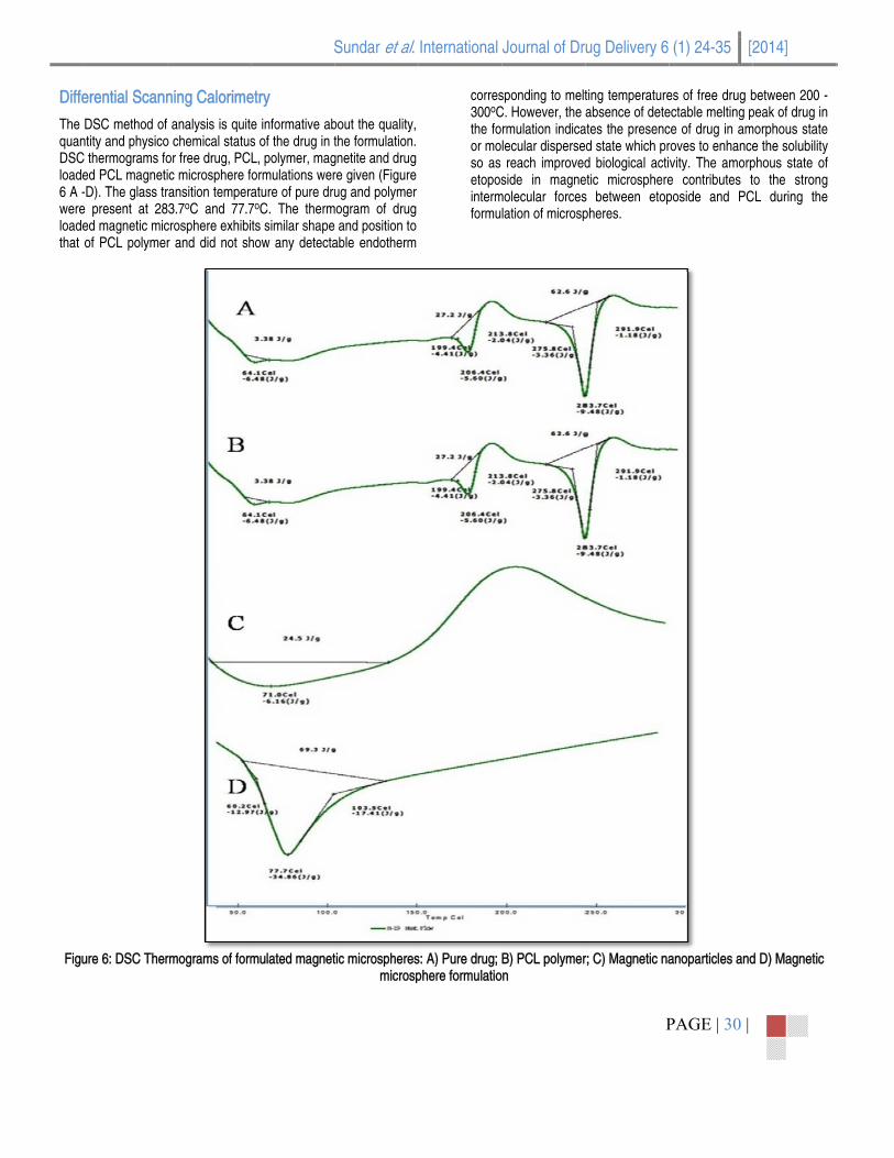
State of etoposide in magnetic microspheres
Differential scanning calorimetry was used to determine the thermal behavior of etoposide, polycaprolactone, magnetite and magnetic microsphere formulations. Samples were scanned for the melting temperatures in nitrogen atmosphere by Perkin Elmer DSC-7. Samples were placed in hermetically sealed aluminium pans and heated at a scan speed of 10 oC per min over a temperature range of 30- 500o C at a chart speed of 10 ml/min. The heat of fusion was calibrated with indium.
Sundar et al. International Journal of Drug Delivery 6 (1) 24-35 [2014]
PAGE | 26 |
Determination of magnetic property
The magnetic properties of Fe3O4/PCL microparticles were measured by using a Vibrating Sample Magnetometer (VSM) [DMS 1600]. The samples in the form of powder were placed in Teflon sample holder. The magnetic properties were then determined by an increasing magnetic field over the sample. The measurements were carried out in the field range of μ 1 T at room temperature.
Determination of magnetic content
The content of magnetite in formulations was estimated quantitatively by atomic absorption spectroscopy (AAS) [SL 173, Elico]. A preweighed (100 mg) amount of magnetic microspheres was digested with 5ml of HCl and 5ml of HNO3 in CEM microwave digester using MARSX press at 800psi and 200oC. The digested solution was made up to 500ml using de-ionized water and was thoroughly filtered using whatman 40 filter paper. The clear solution was assayed for iron by AAS at 248 nm. The weight percentage of iron content in the formulation can be calculated from the equation ppm (mg/L) X Volume in mL X dilution factor X 10-4
Wt % = ---------------------------------------------------------------------
Weight of sample in grams where, ppm (mg/L) is the result obtained from the instrument, Volume in ml- is the volume required for digestion, weight of sample in gm- 0.100gm for all the samples.
In vitro drug release studies
The in vitro drug release studies for formulated magnetic microspheres were carried out in phosphate buffer (pH 7.4) at 37.5 μ 0-5o C. Dried samples of drug loaded polycaprolactone magnetic microspheres (35 mg) were put into a dialysis bag immersed in 50
ml of phosphate buffer (pH 7.4) in a conical flask. [21, 22] The flask was placed in an orbital shaker incubator [C 24, Remi] and rotated at 50 rpm. At predetermined time intervals the flasks were taken out of the shaker, 5 ml aliquots of the medium were withdrawn and the same volume of fresh medium was added to the bulk to maintain the sink condition. The amount of drug released in media was analyzed by using UV- Visible spectrophotometer [Lambda 25, Perkin Elmer].
Histological studies
Presence of excipients in the formulation may sometimes produce toxicity which may be immediate or delayed toxicity. In order to ascertain the safety, the formulations were administered to group of Wistar Albino rats and the vital organs like liver, kidney, heart, brain and lungs were removed. The histopathological evaluation of the tissues with etoposide loaded PCL magnetic microspheres administered rats were fixed in 10% formalin, routinely processed and embedded in paraffin. Sections were cut on glass slides and stained with hematoxyllin and eosin.[23] The sections were examined under a light microscope to detect any damage occurred to the tissue and were compared with similar sections of tissues isolated from untreated rats. Studies were performed under OECD guidelines no 423.
Results and Discusssion
Characterization of magnetic microspheres
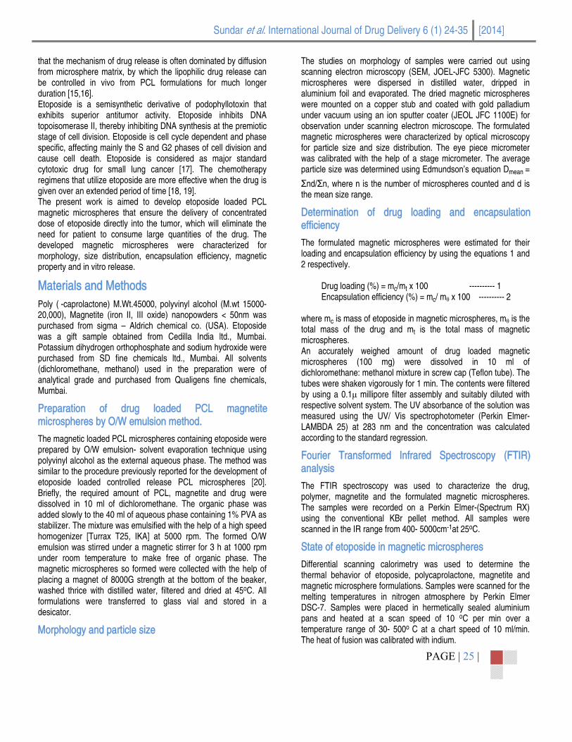
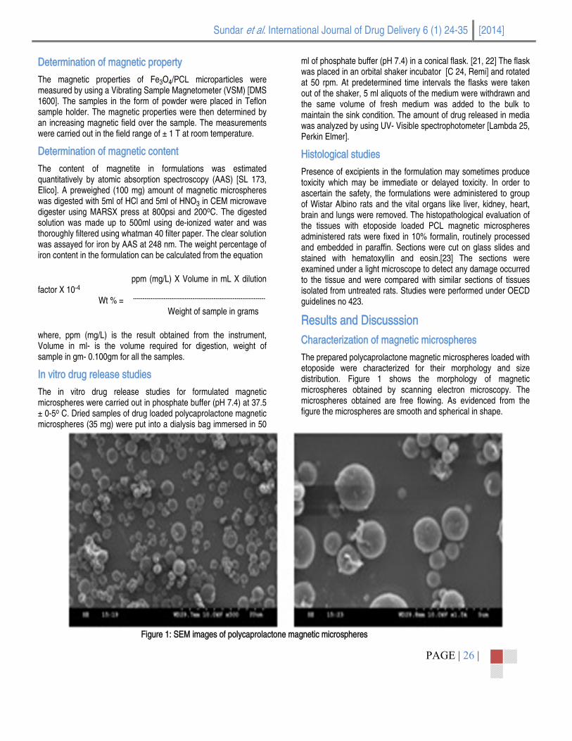
The prepared polycaprolactone magnetic microspheres loaded with etoposide were characterized for their morphology and size distribution. Figure 1 shows the morphology of magnetic microspheres obtained by scanning electron microscopy. The microspheres obtained are free flowing. As evidenced from the figure the microspheres are smooth and spherical in shape.
Figure 1: SEM images of polycaprolactone magnetic microspheres

The concis vaand magnratio distri9.08μincrehigheof co
For
magnetic miccentrations of PCaried in formulati
size distributionnetic microsphewas shown in t
ibution of prepμm. Increase i
ease in mean per concentrationollisions, resultin
Table 1: Compmulation code
F 1 F 2 F 3 F 4 F 5 F 6 F 7 F 8 F 9 F 10 F 11 F 12
Figure 2
crospheres weCL as shown in tons to investiga
n. The mean paeres of etopositable 1 and figurpared microsphin the concentrparticle size. It n of polymer mag in fusion of se
position, mean pDrug/polymer
ratio1:1:1:1:1:1:1:1:1:5:1:5:1:5:1:5:1:101:101:101:10
2: Particle size d
S
re formulated table 1. The amo
ate the influencearticle size of pode with differenre 2, 3 and 4. Teres ranged frration of polymcould be sugg
ay lead to increami particles and
article size, drugr/magnetite o 1 2 3 4 1 2 3 4 :1 :2 :3 :4
istribution of eto
Sundar et al.
with different ount of polymer of particle size olycaprolactone nt dug/polymer
The particle size rom 4.8μm to
mer resulted in gested that the ased frequency
d finally produce
g loading, encapMean Particle S
(μm) 4.805.105.274.686.056.196.406.809.088.227.888.42
oposide loaded p
International
biggerPolyviin thewere solvenviscosevapocoalesemulsother
psulation efficienSize Drug
Loading4.004.673.923.605.845.104.804.106.125.705.214.84
polycaprolactone
Journal of Dr
r particles therenyl alcohol at a
e formulation. Tcommon occu
nt evaporation sity and gradual oration [25]. Thscence and aggsion droplets, thand its consequ
ncy and magnetitg g (%)
Enca
072040002014
e magnetic micr
ug Delivery 6
P
eby increasing thconcentration of
The coalescencrrence in prepprocess were decrease in the
he presence of glomeration by fohus preventing tences.
te concentrationpsulation efficie
(%) 48.2 47.4 44.3 40.2 61.4 59.5 53.8 53.0 69.4 69.0 64.0 62.3
rospheres with d
(1) 24-35 [2
PAGE | 27 |
he size of microf 1% was used a
ce and agglomeparation of micprobably due to
e volume as a repolyvinyl alcohoorming a thin filtheir approach
n of prepared micncy M
Conce
drug/polymer rat
2014]
ospheres [24]. as a surfactant eration, which crospheres by o increase in
esult of solvent ol reduces the lm around the towards each
crospheres Magnetite entration (%)
13.614.713.515.214.317.719.322.717.721.931.530.8
tio 1:1

Effic
The microthat dichlencatable
Figure 3: P
Figure 4: P
ciency of enca
amount of drugospheres was dcould be reco
oromethane: apsulation of all e 1. An increase
Particle size distr
Particle size distr
apsulation
g encapsulated determined by covered after dismethanol mixformulations wee in concentrati
S
ribution of etopo
ribution of etopo
in each of polymcalculating the assolving the mxture. The
ere investigated ion of polymer
Sundar et al.
oside loaded poly
oside loaded poly
meric magnetic amount of drug
microspheres in efficiency of
and reported in resulted in the
International
ycaprolactone m
ycaprolactone m
increaBenoitincreamigrat
Journal of Dr
magnetic microsp
magnetic microsp
ased entrapmentt [24]. The enh
ase in viscosity tion of drug to th
ug Delivery 6
P
pheres with drug
pheres with drug
t efficiency. Thisancement in poof organic pha
he external wate
(1) 24-35 [2
PAGE | 28 |
g/polymer ratio 1
g/polymer ratio 1
s effect was alsoolymer concentrase which in tur phase.
2014]
:5
:10
o observed by ation leads to rn restrict the
Fou
Figurmicrospecmicrovery etopocm-1
charaat 16
16inten
F
rier Transform
re 5 shows theospheres prepactra of etoposideospheres were
important for oside (Figure 5
which is characteristic intense614 cm-1. The C=610 cm-1 regionnsity compared
Figure 5: Infra re
med Infrared S
e FTIR spectra ared by o/w ee, PCL, Fe3O4 nanalyzed. The fdiverse applicaA) exhibits a br
racteristic of pe doublet of ethe=C stretching of n. This band iwith the C=C
ed spectrum of A
S
Spectroscopy
of magnetic poemulsion methonanoparticles anfunctional groupations. The FTroad band betwhenolic - OH er showed C=C vinyl ethers occ
s characterizedstretching band
A) pure drug eto
Sundar et al.
y
olycaprolactone od. The FT-IR nd the magnetic ps of PCL were TIR spectra of een 3400-3700 group and a stretched band
curs in the 1660 d by its higher d in alkene. A
poside B) Polycmagneti
International
characpresenstrongcarbonnanop400 cmcontencm-1[2D) theexistechemimicrosin the
caprolactone C) c microsphere fo
Journal of Dr
cteristic broad ance of hydrogeg carbonyl stretcnyl bond that particles (Figurem-1) due to the int the pattern o26]. In the IR se characteristicd in the same wical interaction spheres and thepolymer matrix.
Fe3O4magneticormulation.
ug Delivery 6
P
absorption banden bonded OH ching band at 1come from P5 C) exhibit at
iron oxide structof magnetite (Fepectrum of mag
c absorption bawave number. T
between polyme presence of dr
nanoparticles
(1) 24-35 [2
PAGE | 29 |
d at 3412 cm-1
group. Figure 1724 cm-1 reveaCL. The spectlow frequency
ture. Depending e3O4) spectrum gnetic microspheands of etoposThis indicates thmer and drug rug as a molecu
and D) Etoposi
2014]
is due to the 5 B shows a aled the ester tra of Fe3O4 region (600 on Fe (II) (III) shows at 570
eres (Figure 5 ide and PCL
he absence of in magnetic
ular dispersion
ide loaded
Diffe
The quanDSCloade6 A -wereloadethat
Figu
erential Scann
DSC method ofntity and physicoC thermograms foed PCL magneti-D). The glass tre present at 28ed magnetic micof PCL polymer
ure 6: DSC Ther
ning Calorime
analysis is quito chemical statusor free drug, PCic microsphere fransition temper83.7oC and 77.7crosphere exhibir and did not sh
rmograms of form
S
etry
e informative abs of the drug in tL, polymer, magormulations werature of pure dru7oC. The thermts similar shape
how any detecta
mulated magnet
Sundar et al.
bout the quality, the formulation.
gnetite and drug re given (Figureug and polymer
mogram of drug and position to able endotherm
tic microspheresmicrosp
International
corres300oCthe foor moso as etoposintermformu
s: A) Pure drug; phere formulatio
Journal of Dr
sponding to meltC. However, the rmulation indicalecular dispersereach improved
side in magnemolecular forces
lation of microsp
B) PCL polymern
ug Delivery 6
P
ting temperatureabsence of dete
ates the presencd state which prd biological acti
etic microspheres between etoppheres.
r; C) Magnetic na
(1) 24-35 [2
PAGE | 30 |
es of free drug bectable melting pce of drug in amroves to enhancevity. The amorpe contributes tposide and PC
anoparticles and
2014]
between 200 - peak of drug in
morphous state e the solubility phous state of to the strong
CL during the
d D) Magnetic

Sundar et al. International Journal of Drug Delivery 6 (1) 24-35 [2014]
PAGE | 31 |
Magnetic property of magnetic microspheres
The superparamagnetic property of polymer magnetic microsphere is critical for their application in biomedical and bioengineering fields which prevents polymer magnetic microspheres from aggregation and enables them to redisperse rapidly when the magnetic field is removed [6]. The magnetization curves of the naked Fe3O4
particles (figure 7(a)) and Fe3O4 drug loaded polycaprolactone magnetic microspheres (figure 7(b)) recorded with VSM are illustrated in figure 7. As shown in the figure the magnetization of the samples would approach the saturation
values when the applied magnetic field increases to 10,000 Oe. The saturation magnetization ( s) of magnetite was found to be 24.82 emu/g and that of microsphere formulation was found to be 7.26 emu/g. The saturation magnetization of magnetic microspheres was much less than that of bulk magnetite as reported in the literature for this material [27]. Figure 7(a) and Fig 7(b) represents a typical characteristic of superparamagnetic material with no detected remainance or coacervity at room temperature indicated that the single domain magnetic Fe3O4
nanoparticles remained in the prepared magnetic microspheres.
Figure 7: Magnetization curves obtained by vibrating sample magnetometer (VSM) at room temperature. (a) Fe3O4 nanoparticles (b) Fe3O4 /
PCL magnetic microspheres.
Content of magnetic particles
The study includes the incorporation of Fe3O4 nanoparticles along
with drug and encapsulated with polycaprolactone. The presence of magnetic particles can carry the drug to a specific target site quickly under the external magnetic field. The concentration of magnetite included within the polymeric carriers was estimated in terms of weight percentage by atomic absorption spectroscopy. The amount of magnetite in the polymeric microspheres increases with the increase in the polymer concentration which was illustrated
in table 1. Microspheres fabricated with 1:10 drug/polymer ratio could accommodate highest amount of magnetite. On the other hand, the magnetite and drug compete with each other to dwell into the matrix space of the polymeric droplets during its formation. Accordingly the ability of the microspheres to entrap the drug decreases with increase in magnetite concentration. The average amount of magnetite within the spheres was found to be 19.4%w/w. This concentration of nanoparticulate magnetite within the polymeric vehicles could be sufficient to direct the microspheres to reach their destination target site. Such a

obsedemoachiemagnfindinhigheto wthe vmagnachie
In-v
Figurfrom time.drug/
ervation was pronstrated that eve 100% retenet for an artering was made wer amount of mithstand arterial
view of the abovnetite included eve the expected
itro drug relea
re 8 - 10 shows the polymeric m
. The release o/polymer ratio
Fig
reviously reporta 15 20% w
ention of the mio-capillary flow with gelatin maagnetite (upto 3pressure unde
ve mentioned resin microsphere
d degree of loca
ase studies
the percentagemagnetic microsof etoposide from
1:1, 1:5 and
gure 8: Drug rele
S
ed by Gupta aw/w magnetite magnetic carrier
of 0.005 0.1 gnetic microsph
30%) was considr a magnetic fiesearch findings tes in the presealization of the m
of accumulativepheres as a funm magnetic mic1:10 containing
ease profile of e
Sundar et al.
and Hung [28]is sufficient to
r using 8000G cm/s. A similar
heres, where a dered sufficient eld [1]. Thus in the 31.5 w/w of
ent study could microspheres.
e drug released ction of release
crospheres with g 1% polyvinyl
etoposide from P
International
alcohobecomreleasreportthe rediffusiburst encapdiffusewas freleasdue tototal micros76.25%
PCL magnetic mi
Journal of Dr
ol indicated a qmes slower andse for the formuted about the druelease involved on and polymerrelease was
psulated drug boed from the diafollowed by cose may be due to erosion of poly
amount of spheres with d%.
icrospheres of d
ug Delivery 6
P
uick release witd constant afterulations were foug release in mtwo different mer matrix degradaprobably due
ound to the microlysis bag [30, 3nstant slow reto diffusion of dymer [32]. At thetoposide reledrug/polymer ra
rug/polymer rati
(1) 24-35 [2
PAGE | 32 |
thin 48 hrs. Ther 48 hrs. The aound to be 38%icrospheres andechanisms of dration [29]. The cto small amou
oparticles surfac31]. Further the lease. The rearugs from polym
he end of 3rd weeased from tatio 1:10 was
o 1:1
2014]
e release rate average burst %. Zhou et al d revealed that rug molecules ause for initial unt of poorly ce which easily
burst release ason for slow mer as well as eek period the he magnetic found to be
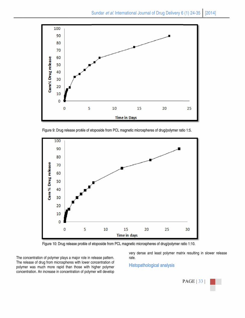
The The polymconc
Fig
Fig
concentration ofrelease of drug mer was much centration. An inc
gure 9: Drug rele
gure 10: Drug re
f polymer plays from microsphemore rapid tha
crease in conce
S
ease profile of e
elease profile of
a major role in reres with lower can those with ntration of polym
Sundar et al.
etoposide from P
etoposide from
release pattern. concentration of higher polymer
mer will develop
International
PCL magnetic mi
PCL magnetic m
very drate.
Histo
Journal of Dr
icrospheres of d
microspheres of
dense and leas
opathological a
ug Delivery 6
P
rug/polymer rati
drug/polymer ra
t polymer matri
analysis
(1) 24-35 [2
PAGE | 33 |
o 1:5.
atio 1:10.
ix resulting in s
2014]
slower release

Sundar et al. International Journal of Drug Delivery 6 (1) 24-35 [2014]
PAGE | 34 |
Histopathological analysis of various organs like brain, heart, liver, lungs and kidney of animals treated with encapsulated etoposide was illustrated in figure 11a-11e.The vital organs were assessed for toxic effects of etoposide loaded polycaprolactone magnetic microspheres. The cross section of brain, heart and liver of treated animals were examined for cell necrosis, cellular hoemostasis and inflammation on hepatic cells. None of these signs was detected, revealing there was no significant toxicity produced by the formulations (Figure 11a, 11b and 11c). Enlarged airway spaces and necrosis of alveoli on cross examination of the cells of the lungs were found to be absent, hence signifying no toxic effects (Figure 11d). A toxicity produced kidney is identified by reversible lesions such as interstitial fibrosis. The cross sections of the kidneys showed no such effects, thus signifying no toxic effects of the drug (Figure 11e). Hence it was concluded from the microphotographs that no histological alterations were observed in various organs of animals treated with encapsulated etoposide.
Conclusion The poly ( - caprolactone) microspheres containing magnetite and anticancer drug were successfully formulated by O/W emulsion solvent evaporation method with maximum drug encapsulation and desired release profile. The method adopted favored the formation of smooth, spherical shaped microspheres with optimal particle size. The formulated magnetic microspheres were magnetically responsive and this synthetic approach is applicable for producing magnetic polymer microspheres with high magnetite contents. The possession of sufficient paramagnetic property was confirmed by VSM. The histopathological studies proved that the etoposide loaded magnetic microspheres was nontoxic and safe. The results suggested that the magnetic PCL magnetic microspheres may have potential as a highly versatile carrier for targeted delivery approach.
References
[1]. Saravanan M, Bhaskara K, Gomathinayagam M, Sadasivan Pilai K. Ultrasonically controlled release and targeted delivery of diclofenac sodium via gelatin magnetic microspheres. Int J Pharm. 2004; 283: 71-82.
[2]. Widder KJ, Flouret G, Senyei A. Magnetic microspheres:synthesis of a novel parenteral drug carrier. J. Pharm. Sci. 1979; 68(1): 79-82.
[3]. Alexiou C, Arnold W, Klein RJ, Parak FJ, Hulin P, Bergemann C, Erhardt W, Wagenpfeil S, Lubbe AS. Locoregional cancer treatment with magnetic drug targeting. Cancer Res. 2000; 60: 6641 - 8.
[4]. Lubbe AS, Bergemann C, Brock J, McClure DG. Physiological aspects in magnetic drug-targeting. J. Magn. Magn. Mater. 1999; 194: 149-155.
[5]. Widder KJ, Senyei AE, Ranney DF. Magnetically responsive microspheres and other carriers for the biophysical targeting of antitumor agents. Adv Pharmacol Chemot. 1979; 16: 213-271.
[6]. Wu Y, Guo J, Yang WL, Wang CC, Fu SK. Preparation and characterization of chitosan-
poly(acrylic acid) polymer magnetic microspheres. Polymer. 2006; 47: 5287 - 94.
[7]. Cregg PJ, Murphy K, Mardinoglu A. Calculation of nanoparticle captures efficiency in magnetic drug targeting. J. Magn. Magn. Mater. 2008; 320: 3272 - 5.
[8]. Lin JJ, Chen JS, Haung SJ, Ko JH, Wang YM, Chen TL, Wang LF. Folic acid-pluronic F127 magnetic nanoparticale clusters for combined targeting, diagnosis, and therapy applications. Biomaterials 2009; 30: 5114 - 24.
[9]. Cabuil V. Preparation and properties of magnetic nanoparticles. Hubbard, A.T. (Ed), Encyclopedia of Surface and Colloid Science. Marcel Dekker Inc., New York, pp. 4306-21; 2002.
[10]. Iannone A, Margin R.L, Walczack T, Federico M, Swartz H.M, Tnansi A, Vannini V. Blood clearance of dextran magnetic particle determined by a non-invasive in vivo ESR method. Magn. Reson. Med. 1991; 22: 435 - 42.
[11]. Duhem N, Rolland J, Riva R, Guillet P, Schumers J.M, Gohy J.F, Preat V. Tocol modified glycol chitosan for the
oral delivery of poorly soluble drugs. Int. J. Pharm. 2012; 433: 453 - 60.
[12]. Yan S, Rao S, Zhu J, Wang Z, Duan Y, Chen X, Yin J. Nanoporous multilayer poly (L)-glutamic acid/chitosan microcapsules for drug delivery. Int.J.Pharm. 2012; 427: 443 - 51.
[13]. Sinha V.R, Bansal K, Kaushik R, Kumaria R, Trehan A. Poly-epsilon-caprolactone microspheres and nanospheres: an overview. Int. J.Pharm. 2004; 278 (1): 1 - 23.
[14]. Hakkarainen M, Albertsson A.C. Heterogenous biodegradation of polycaprolactone low molecular weight products and surface changes. Macromol. Chem. Phys. 2002; 203: 1357 63
[15]. Gorna K, Gogolewski S. In vitro degradation of novel medical biodegradable aliphatic polyurethanes based on epsilon-caprolactone and pluronics with various hydrophilicities. Polym. Degrad. Stab. 2002; 75: 113 - 22.
[16]. Perez M.H, Zinutti C, Lamprecht A, Ubrich N, Astier A, Hoffman M, Bodmeier R, Maincent P. The preparation and evaluation of poly

Sundar et al. International Journal of Drug Delivery 6 (1) 24-35 [2014]
PAGE | 35 |
(epsilon-caprolactone) microparticles containing both a lipophilic and a hydrophilic drug. J. Control. Release. 2000; 65(3): 429 - 38.
[17]. Ihde Dc. Small cell lung cancer. State-of-the-art therapy 1994. Chest 1995; 107(6) (Suppl): 2438 - 85.
[18]. Hande K.R. Etoposide: four decades of development of a topoisomerase II inhibitor. Eur. J. Cancer. 1998; 34: 1514 - 21.
[19]. Wolf S.N, Grosh W.W, Prater K. In vitro pharmacodynamic evaluation of VP-16213 and implications for chemotherapy. Cancer. Chemother. Pharmacol. 1987; 19: 246 - 9.
[20]. Wang S, Guo S. Formation mechanism and release behavior of poly( - caprolactone) microspheres containing disodium norcantharidate. Eur. J. Pharm and Biopharm. 2008; 69: 1176 81.
[21]. Yang Y, Park S.B, Ho-Geun Yoon, Huh Y-M, Haam S. Preparation of poly -caprolactone nanoparticles containing magnetite for magnetic drug carrier. Int. J. Pharm. 2006; 324: 185 - 90.
[22]. Fengxia Li, Xiaoli Li, Bin Li. Preparation of magnetic polylactic acid microspheres and investigation
of its releasing property for loading curcumin. J. Magn. Magn. Mater. 2011; 323: 2770 - 75.
[23]. Rita JM, Pradip KG, Manish LU, Rayasa SR. Thermoreversible mucoadhesive gel for nasal delivery of sumatryptan. AAPS Pharm Sci Tech. 2006; 7: E1 - E7.
[24]. Benoit MA, Baras B, Gillard J. Preparation and characterization of protein loaded poly ( -caprolactone) microparticles for oral vaccine delivery. Int.J.Pharm. 1999;184: 73 - 84.
[25]. Lamprecht A, Ubrich N, Hombriero Perez M, et al. Biodegradable monodispersed nano particles prepared by pressure homogenization emulsification. Int. J. Pharm. 1999; 184: 97 - 105.
[26]. Zaitsev VS, Filimonov DS, Presnyakov IA, Gazvbivo RF. Physical and chemical property of magnetite and magnetite-polymer nanoparticles and their colloidal dispersion. B.Colloidal Interface Sci. 2012; 49 - 57.
[27]. Yamaura M, Camilo RL, Sampaio LC, Macedo MA, Nakamura M, Toma HE. Preparation and characterization of (3-aminopropyl)triethoxysilane-coated magnetite nanoparticles. J.
Magn. Magn. Mater. 2004; 279: 210 - 217.
[28]. Gupta PK, Hung CT. Magnetically controlled targeted micro carrier systems. Life Sci. 1989; 44: 175 - 86.
[29]. Zhou SB, Deng XM, Li XH. Investigation on a novel core-coated microspheres protein delivery system. J. Controlled release. 2001; 75: 27 - 36.
[30]. Bodmeier R, Chem H. The preparation and characterization of microspheres containing the anti-inflammatory agents indomethacin, ibuprofen and ketoprofen. J. Controlled Release. 1989; 10: 167 - 75.
[31]. Ammoury N, Dubrasquet M, Fessi H, Devissaguet JP, Puisieux F, Beusio S. Indomethacin -loaded poly(D,L-lactide) nanocapsules, protection from gastrointestinal ulcerations and anti-inflammatory activity evaluation in rats. Clin. Mater. 13: 121 - 30.
[32]. Dhanaraju MD, Gopinath D, Rafiuddin Ahmed M, Jayakumar R, Vamsadhara C. Characterization of polymeric( -caprolactone) injectable implant delivery system for the controlled delivery of contraceptive steroids. J. Biomed. Matr. Research. 2006; 76(1): 63 - 72.