Exon/intron organization and complete nucleotide sequence ... · CommunicatedbyBarujBenacerraf,...
5
Proc. Nat! Acad. Sci. USA Vol. 79, pp. 893-897, February 1982 Immunology Exon/intron organization and complete nucleotide sequence of an HLA gene (genomic clone/variability/intraceliular segment) MARIE MALISSEN, BERNARD MALISSEN, AND BERTRAND R. JORDAN Centre d'Immunologie, Institut National de la Sante et de la Recherche Melicale, Centre National de la Recherche Scientifique de Marseille-Luminy, Case 906-13288, Marseille Cedex 9-France. Communicated by Baruj Benacerraf, October 19, 1981 ABSTRACT We have isolated and determined the sequence of a genomic clone containing the gene for a human class I trans- plantation antigen. The gene contains seven exons. The first five exons code respectively for a signal peptide, for the first, second, and third extracellular domains of the protein molecule, and for the transmembrane region. The cytoplasmic segment is encoded by part of the fifth and the sixth and the seventh exons. The struc- ture of the protein encoded by this unit is closely homologous with known class I transplantation antigens. Knowledge of the structure of the polymorphic class I trans- plantation antigens encoded in the major histocompatibility complex (MHC; HLA-A, -B, and -C in man, H-2K, D, and L in mouse) has made rapid progress recently. Complete protein sequences have been obtained by radiochemical sequence de- termination for mouse antigens (1); in the human MHC, the structure of the extracellular portion and cytoplasmic segment of two molecules, HLA-A2 and HLA-B7, has been determined (2, 3); partial data are also available on the transmembrane por- tion of the molecule. These results strengthen the assumption of a domain-like organization (4, 5) of class I antigens and have suggested possible variable regions in these molecules. More recently, advances have been made toward isolation of the cor- responding genes; cDNA clones have been obtained in both the H-2 (6, 7) and the HLA (8, 9) systems and used to probe the organization of these genes by Southern blotting (7, 10) and to isolate genomic clones (11, 12). We have recently isolated HLA genomic clones from a hu- man DNA library in A charon 4A, by using the mouse H-2 cDNA probes obtained by Kvist et al. (6). One of these clones, called A HLA 12, was shown to contain sequences coding for the third domain of the HLA molecule (11). We have now studied this clone in more detail; a 5.6-kilobase (kb) HindIII fragment was found to contain one complete HLA gene, and the sequence of this gene has been completely determined. These results reveal the general organization of an HLA transcription unit, support the domain model for class I antigens, provide some evidence against the involvement of DNA rearrangements in the expres- sion of these molecules, and show the unexpected complexity of the cytoplasmic coding region. They also provide a new com- plete class I antigen structure that can be compared with pre- viously determined protein sequences. MATERIALS AND METHODS Enzymes and Other Reagents. Restriction enzymes were obtained from Bethesda Research Laboratories, Gaithersburg, MD, New England Biolabs, and Boehringer Mannheim. Ter- minal transferase, polynucleotide kinase, and bacterial mono- phosphoesterase were from Bethesda Research Laboratories or P-L Biochemicals. Radiochemicals were obtained from Amersham. DNA Labeling and Fragment Preparation. 3'- and 5'-end labeling (with or without prior denaturation as appropriate) was carried out on either restriction digests or fragments eluted from acrylamide or agarose gels. Uniquely labeled fragments were then obtained by secondary restriction enzyme digestion, strand separation on 5% or 10% neutral acrylamide gels or both. DNA Sequence Analysis. Sequence determination was car- ried out according to Maxam and Gilbert (13); reaction products were analyzed on 20%, 8%, and 6% thin acrylamide/urea gels. Essentially all the sequence was determined independently on both strands, using either different restriction sites or 3'- and 5'-end labeling at the same site; overlapping sequences were obtained for all restriction sites except the Taq I site present ahead of the first exon at position 100 and the Sst I site in the transmembrane-first cytoplasmic region exon. RESULTS Isolation of Genomic Clones. A screen by plaque hybridiza- tion of the human DNA library in phage A charon 4A con- structed by Lawn et aL (14) was carried out with a mouse H-2 cDNA probe, the H2. 1 fragment from the pH-2d-3 plasmid (11, 15), containing the transmembrane, cytoplasmic, and 3' non- coding sequences of the corresponding mRNA. From 105 phage plaques, corresponding to approximately half a human haploid genome, 15 confirmed positive phages were obtained. Most of these clones are different from each other (unpublished). This suggests a surprisingly high number of coding sequences in human DNA, perhaps 30 per haploid genome, and is not in- consistent with results recently obtained in the H-2 system by Southern analysis of genomic DNA (7, 10). The A HLA 12 clone was chosen for further study, mapped, and shown by sequence analysis to contain authentic HLA coding sequences (11). Sequence Determination Strategy. Subclone pHLA 12.4-the 5.6-kb HindIII fragment from clone A HLA 12, which hybrid- ized with both third domain and transmembrane-cytoplasmic mouse probes (15) and was recloned in plasmid pBR328 (16)-is shown in Fig. 1. A complete set of HLA coding sequences was found in this segment. Its organization was determined mainly by DNA sequence determination and comparison of the pos- sible translation products with the HLA-B7 and -A2 protein sequences. This strategy allowed us to determine the organization of this transcription unit and to show that it contains all the expected coding sequences; 4.1 kb of DNA, containing the complete set Abbreviations: MHC, major histocompatibility complex; kb, kilobase(s). 893 The publication costs of this article were defrayed in part by page charge payment. This article must therefore be hereby marked "advertise- ment" in accordance with 18 U. S. C. §1734 solely to indicate this fact.
Transcript of Exon/intron organization and complete nucleotide sequence ... · CommunicatedbyBarujBenacerraf,...
Proc. Nat! Acad. Sci. USAVol. 79, pp. 893-897, February 1982Immunology
Exon/intron organization and complete nucleotide sequence of anHLA gene
(genomic clone/variability/intraceliular segment)
MARIE MALISSEN, BERNARD MALISSEN, AND BERTRAND R. JORDANCentre d'Immunologie, Institut National de la Sante et de la Recherche Melicale, Centre National de la Recherche Scientifique de Marseille-Luminy,Case 906-13288, Marseille Cedex 9-France.Communicated by Baruj Benacerraf, October 19, 1981
ABSTRACT We have isolated and determined the sequenceof a genomic clone containing the gene for a human class I trans-plantation antigen. The gene contains seven exons. The first fiveexons code respectively for a signal peptide, for the first, second,and third extracellular domains of the protein molecule, and forthe transmembrane region. The cytoplasmic segment is encodedby part of the fifth and the sixth and the seventh exons. The struc-ture of the protein encoded by this unit is closely homologous withknown class I transplantation antigens.
Knowledge of the structure of the polymorphic class I trans-plantation antigens encoded in the major histocompatibilitycomplex (MHC; HLA-A, -B, and -C in man, H-2K, D, and L inmouse) has made rapid progress recently. Complete proteinsequences have been obtained by radiochemical sequence de-termination for mouse antigens (1); in the human MHC, thestructure of the extracellular portion and cytoplasmic segmentof two molecules, HLA-A2 and HLA-B7, has been determined(2, 3); partial data are also available on the transmembrane por-tion of the molecule. These results strengthen the assumptionof a domain-like organization (4, 5) of class I antigens and havesuggested possible variable regions in these molecules. Morerecently, advances have been made toward isolation of the cor-responding genes; cDNA clones have been obtained in both theH-2 (6, 7) and the HLA (8, 9) systems and used to probe theorganization of these genes by Southern blotting (7, 10) and toisolate genomic clones (11, 12).We have recently isolated HLA genomic clones from a hu-
man DNA library in A charon 4A, by using the mouse H-2 cDNAprobes obtained by Kvist et al. (6). One of these clones, calledA HLA 12, was shown to contain sequences coding for the thirddomain of the HLA molecule (11). We have now studied thisclone in more detail; a 5.6-kilobase (kb) HindIII fragment wasfound to contain one complete HLA gene, and the sequence ofthis gene has been completely determined. These results revealthe general organization of an HLA transcription unit, supportthe domain model for class I antigens, provide some evidenceagainst the involvement ofDNA rearrangements in the expres-sion of these molecules, and show the unexpected complexityof the cytoplasmic coding region. They also provide a new com-plete class I antigen structure that can be compared with pre-viously determined protein sequences.
MATERIALS AND METHODSEnzymes and Other Reagents. Restriction enzymes were
obtained from Bethesda Research Laboratories, Gaithersburg,MD, New England Biolabs, and Boehringer Mannheim. Ter-
minal transferase, polynucleotide kinase, and bacterial mono-phosphoesterase were from Bethesda Research Laboratories orP-L Biochemicals. Radiochemicals were obtained fromAmersham.DNA Labeling and Fragment Preparation. 3'- and 5'-end
labeling (with or without prior denaturation as appropriate) wascarried out on either restriction digests or fragments eluted fromacrylamide or agarose gels. Uniquely labeled fragments werethen obtained by secondary restriction enzyme digestion,strand separation on 5% or 10% neutral acrylamide gels or both.DNA Sequence Analysis. Sequence determination was car-
ried out according to Maxam and Gilbert (13); reaction productswere analyzed on 20%, 8%, and 6% thin acrylamide/urea gels.Essentially all the sequence was determined independently onboth strands, using either different restriction sites or 3'- and5'-end labeling at the same site; overlapping sequences wereobtained for all restriction sites except the Taq I site presentahead of the first exon at position 100 and the Sst I site in thetransmembrane-first cytoplasmic region exon.
RESULTSIsolation of Genomic Clones. A screen by plaque hybridiza-
tion of the human DNA library in phage A charon 4A con-structed by Lawn et aL (14) was carried out with a mouse H-2cDNA probe, the H2. 1 fragment from the pH-2d-3 plasmid (11,15), containing the transmembrane, cytoplasmic, and 3' non-coding sequences ofthe corresponding mRNA. From 105 phageplaques, corresponding to approximately half a human haploidgenome, 15 confirmed positive phages were obtained. Most ofthese clones are different from each other (unpublished). Thissuggests a surprisingly high number of coding sequences inhuman DNA, perhaps 30 per haploid genome, and is not in-consistent with results recently obtained in the H-2 system bySouthern analysis ofgenomic DNA (7, 10). The A HLA 12 clonewas chosen for further study, mapped, and shown by sequenceanalysis to contain authentic HLA coding sequences (11).
Sequence Determination Strategy. Subclone pHLA 12.4-the5.6-kb HindIII fragment from clone A HLA 12, which hybrid-ized with both third domain and transmembrane-cytoplasmicmouse probes (15) and was recloned in plasmid pBR328 (16)-isshown in Fig. 1. A complete set ofHLA coding sequences wasfound in this segment. Its organization was determined mainlyby DNA sequence determination and comparison of the pos-sible translation products with the HLA-B7 and -A2 proteinsequences.
This strategy allowed us to determine the organization of thistranscription unit and to show that it contains all the expectedcoding sequences; 4.1 kb of DNA, containing the complete set
Abbreviations: MHC, major histocompatibility complex; kb, kilobase(s).
893
The publication costs ofthis article were defrayed in part by page chargepayment. This article must therefore be hereby marked "advertise-ment" in accordance with 18 U. S. C. §1734 solely to indicate this fact.
894 Immunology: Malissen et al.
R RS R BBSSS 5 B RXHLA 12 I
H Ba H Ba
H B Ba B $ S S F*1 I I I I I_1
L I 2
I . a I1
3 TMCI
lkb
I I
C2 C3 UT
2 3. I I
4
FIG. 1. Maps of genomic clone AHLA 12 and the pHLA 12.4 subclone.
H (Top) Partial restriction map of the 15-I kb insert present in clone 12. The
EcoRP sites at the end of the insertpHLA 12.4 were generated during construction of
-the library (14). The 5.6-kb Hindufragment was subcloned in plasmidpBR328 (20) and studied by Southernblotting and DNA sequence analysis.B, Bgi II; Ba, BamHI; R, EcoRi; S, SstI; P, Pvull. (Bottom) Map of subclonepHLA 12.4 (5.6-kb HindI fragmentfrom clone 12), *, Positions of exons;T, 3' Untranslated region. Arrows in-dicate individual sequences obtainedby the Maxam-Gilbert procedure; thosewith a dot indicate 3' labeling of theDNA strand, others indicate 5' labeling.
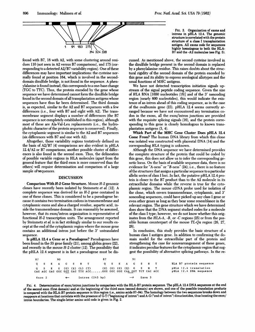
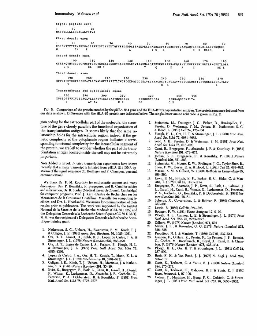
of exons, was analyzed by the Maxam-Gilbert chemical deg-radation procedure (13). The sequence determination strategyis summarized in Fig. 1 and the DNA sequence obtained isshown in Fig. 2. The locations of the exons and introns areshown in Fig. 1 and correlated with the HLA protein in Fig.3. The location of the exon/intron boundaries could be deter-mined from comparison of the possible translation products ofthe DNA sequences with the HLA-B7 protein sequences; G-T(beginning of intron) and A-G (end of intron) dinucleotides (17)were found in every case at the point at which the protein se-quence deduced from the DNA became unrelated to the HLA-B7 sequence. A check of these locations was provided by thesimilarly determined location ofthe starting point for next exon.One example is shown for the first-domain/intron/second-do-main junction in Fig. 4.A Complete HLA Gene'is Found in Genomic DNA. It has
been suggested that class I antigens could be synthesized froma small set of third-domain coding regions and a large set of"variable" first- and second-domain regions, assembled throughDNA rearrangements or differential RNA splicing (18). Thefinding of a complete HLA transcription unit containing codingsequences for all domains argues against the second version ofthis hypothesis, while the invariability ofDNA blots from spermor liver cells when probed with H-2 cDNA probes (7) providesevidence against the first version.A Probable Signal Peptide Exon is Found 125 Base Pairs
Upstream of the First Domain Exon. A signal sequence con-taining z--20 amino acids is known to exist in the initial trans-lation product of HLA mRNA (19). The corresponding regioncould not be identified directly because the protein sequencehas not been reported. However, we found a putative signalpeptide exon (see Figs. 2 and 5) that starts with a methioninecodon, encodes a stretch of 21 amino acids, 18 of which arehydrophobic or uncharged, and contains, at its 3' end, a "be-ginning ofintron" G-T dinucleotide that correlates with an "endof intron" A-G dinucleotide in the first codon of exon 2 (firstdomain). No other such structure is found in the 433 nucleotideswhose sequences have been determined upstream of the firstdomain exon.The First, Second, and Third Domains and the Transmem-
brane Segment of the HLA Antigen are Coded by DistinctExons. The exon/intron organization of the pHLA 12.4 tran-scription unit in this region follows closely (and confirms) the
domain structure proposed for class I transplantation antigens(4, 5): the first, second, and third domains and the transmem-brane segment are encoded by distinct exons (exons 2, 3, 4, and5, respectively) separated by introns of various lengths. Thistype oforganization is similar to that found for immunoglobulinsand suggests that these domains may have been assembled afterevolving independently, as originally suggested by Gilbert (20).Exon 5 deserves special attention because it actually codes forfirst a short hydrophilic segment (Glu-Pro-Ser-Ser), whichcould be considered as part of the third domain, then a stretchof28 hydrophobic amino acids, which constitute the transmem-brane segment, and finally a hydrophilic (Met-Trp-Arg-Lys-Lys-Ser-Ser) peptide, which is the beginning ofthe cytoplasmicregion and is considered to play a role in anchoring the antigenin the cell membrane (for review of the data on this region, seeref. 1).
The'Cytoplasmic Segment is Split Between Three Exons.We were surprised to find that the cytoplasmic segment codingsequences are found in three distinct exons: the first one, 7amino acids long, is continuous with the transmembrane exon;the second one, 438 nucleotides further to the 3' end of thegene, is 11 amino acids long and separated by a 141-nucleotide-long intron from the last, 14-amino acid-long exon, which iscontiguous with the 3' untranslated region. Recent results inthe mouse system indicate a similarly complex gene structurein this region (12) together with differences in the cDNA se-quence that suggest the existence of alternative processingpathways (12, 15). Although such data are not yet available inthe human system, the similarity in genetic organization of thecytoplasmic coding regions indicates that alternative splicingshould also be considered in this case. It is clear that much re-mains to be understood on the structure and function of the in-tracellular portion of transplantation antigens.
Comparison of the Protein Sequence Encoded by pHLA12.4 Confirms Regions of Variability Detected by HLA-B7Versus HLA-A2 Comparisons and Suggests Additional Vari-able Regions. The sequence of the protein encoded by thepHLA 12.4 exons is compared with the HLA-B7 protein in Fig.5. In the first domain, a small cluster of differences is seen be-tween positions 74 and 85. This cluster was apparent in HLA-B7 versus HLA-A2 comparisons. Altogether, the first domainofpHLA 12.4 shows 16 differences with the B7 protein and 22with the A2 protein. In the second domain, 15 differences are
i m lmmr'- M - ::: 0 r
Proc. Nad Acad. Sci. USA 79 (1982)
I
Immunology: Malissen et al. Proc. Nati Acad. Sci. USA 79 (1982) 895
CCCGAAGGCGGTGTATGGATTGGGGATGCCCCGCCTTGGGGATTCGCCACCTCCGCAGTTTCTCTTCTTCTCACAACCTGCGACGGGTCCTTCTTCCTCG 100
ATACTCACGAAGCGGACACAGTTCTCATTCCCACTAGGTGTCGGGTTTCTAGAGAAGCCAATCGGTGCCGCCGCGGTCCCGGTTCTAAAGTCCCCACGCA 200
CCCACCGGGACTCAGATTCTCCCCAGACGCCGAGGATGGTGCTCATGGCGCCCCGAACCCTCCTCCTGCTGCTCTCAGGGGCCCTGGCCCTGACCCAGAC 3UUEXON I M A P R T L L L L L S G A L A L T Q T
CTGGGCGCGTGAGTGCAGGGTCTGCAGGGAAATGGTCGGGAGGAGNGAGGGGCCCGCCCGGCGGGGTGCGCAGGACCCAGGGAGCCGCGCAGGGAGGAGG 400W A
GTCGGGCGGGTCTCAGCTCCTCCTCGCTCCCAGGTTCCCACTCCATGA~GGTATTTCTACACCACCATGTCCCGGCCCGGCGCCGGGGAGCCCCGCTTCAT 500EXON 2 R S H S M R Y F Y T T M S R P G A G E P R F I
CTCCGTCGGCTACGTGGACGATACGCAGTTCGTGCGGTTCGACAGCGACGACGCGAGTCCGAGAGAGGAGCCGCGGGCGCCGTGGATGGAGCGGGAGGGG 600S V G Y V D D T Q F V R F D S D D A S P R E E P R A P W M L R E G
CCAAAGTATTGGGACCGGAACACACAGATCTGCAAGGCCCAGGCACAGACTGAACGAGAGAACCTGCGGATCGCGCTCCGCTACTACAACCAGAGCGAGG 770P K Y W D R N T Q I C K A Q A Q T E R E N L R I A L R Y Y N Q S E
GCGGTGAGTTGACCCCGGCCCGGGGCGCAGGTCACGACCCCTCCCCATCCCCCACGGAGGGCCGGGTCGCCTCGAGTCTCTGGGTCCGAGATCCACCCCG ts00G
AAACCGCGGGATCCCGAGACCCTTGACCTGGGAGAGGCCCAGGCGCCTTTACCCGGTTTCATTTTCAGTTTAGGCCAAAATCCCCGCGGGTTGGTCGGGG 900
CGGGGCTGGGCTCGGGGGACCGGGCTGACCGCGGGGGCGGGCCAGGTTCTCACACCATGCAGGTGATGTATGGCTGCGACGTGGGGCCCGACGGGCCTTT 1000EXON 3 G S H T M Q V M Y G C D V G P D G P F
CCTCCGCGGGTATGAACAGCACGCCTACGACGGCAAGGATTACATCGCCCTGAACGAGGACCTGCGCTCCTGGACCGCGGCGGACATGGCAGCTCAGATC 1100L k G Y E Q H A Y D G K D Y I A L N E D L R S W T A A D M A A Q I
ACCAAGCGCAAGTGGGAGGCGGCCCGTCGGGCGGAGCAGCGGAGAGTCTACTTGGAGGGCGAGTTCGTGGAGTGGCTCCGCAGATACTTGGAGAACGGGA 1200T K R K W E A A R R A E Q R R V Y L E G E F V E W L R R Y L E N G
AGGAGACGCTGCAGCGCGCGGGTACCAGGGCCACAGGGCGCCTCCCTGATCGCCTGTAGATCTCCGGGGCTGGCCTCCCACAAGAAA.GGGAGACAAATGG 1300.& E T L Q R A
GACCAACACTATAATATCGCCCTCCCTCTGGTCTTGAGGGAGAAGAATCCTCTTGGGTTTCCAGAGAGTGACTCTGAGGGTCCGCCCTGCTCTCTGACAC 1400
AATTAAGGGATGAAATCTGTGAGGAAATGAAGGGAAGACAATCCCTGGAATACTGATGAGTGGTTCCCTTTGACACTGGCAGCAGCCTTGGGCCCCGTGA 1500
CTTTTCCTCTCAGGCCTTGTTCTCTGCTTCACACTCAATGTGCGTGGGGGTCTGAGTCCCTCAGCCTCCACTCAGGTCAGGACCAGAAGTCGCTGTTCCC 160U
TCTTCAGGGACTAGAATTTTCCACGGAATAGGAGATTATTCTAGGTGCCTCTGTCTAGGCTGTTGTCTGGGTTCTGTGCTCCCTTCCCCACCCTAGGCAT 17UU
UCTGTCAATTCTCAAGATGGCCACATGCGTGCTGGTGGAGTGTCCCATGACAGATGCAAAATGCCTGAATTTTCTGACTCTTTCCCGTCAGACCCCCCCA 180UEXON 4 D P P
AGACACATATGACCCACCACCCCATCTCTGACCATGAGGCCACCCTGAGGTGCTGGGCCCTGGGCTTCTACCCTGCGGAGATCACACTGACCTGGCAGCG 1900K T H M T H H P I S D H E A T L R C W A L G F Y P A E I T L T W Q R
GGATGGGGAGGACCAGACCCAGGACACGGAGCTCGTGGAGACCAGGCCTGCAGGGGATGGAACCTTCCAGAAGTGGGCGGCTGTGGTGGTGCCTTCTGGA 2000D G E D Q T Q D T E L V E T R P A G D G T F Q K W A A V V V P S G
GAGGAGCAGAGATACACCTGCCATGTGCAGCATGAGGGTCTGCCCGAGCCCCTCACCCTGAGATGGGGTAAGGAGGGAGATGGGGGTGTCATGTCTCTTA 2100
E E Q R Y T C H V Q H E G L P E P L T L R W
GGGAAAGCCGGAGACCTCTCTGGAGAGCTTAGCAGGGTCAGGGTTCCCTCACCTTCCCCCCTTTTCCCAGAGCCATCTTCCCAGCCCACCGTCCCCATCG 2200EXON S E P ES S Q P T V P I
TGGGCATCGTTGCTGGCTTGGTTCTACTTGTAGCTGTGGTCACTGGAGCTGTGGTCGCTGCTGTAATGTGGAGGAAGAAGAGCTCAGGTAAGGAAGGGGT 2300V G I V A G L V L L V A V V T G A V V AL V M W R K K S S
GAGGAGTGTGGTCTGAGAATTTCTTGTCTCACTGAGAGTTCCAAGCCCCAAGTAGAAGTGCCCTGCCTAGTTACTGGGAAGCACCATCCACACTCATGGG 2400
CCTACCCAGCCTGGGCCCTGTGTGCCAGCACTTACTCTTTTGTAAAGCACCTGTTACAATGAGGGACAGATTTATTACCTTGATGACTGTGGTGATGGGA 25OU
CCTGATCCCAGCAGTCACAAGTCACACGGGAAGGTCCCCGAGGACAGACCTCAGAAGGGCGGTTGGTCNAGGACCCACATCTGCTTTCTTCATGTTTCCT 2600
GATCCCGCCCTGGGTCTGCAGTTGCACATTTCTGGAAACTTCTCTGGGGTCCGAGACTTGGAGGTTCCTCTAGGACCTTATGGCCCTGGCTTCTTTCTGGC 27UU
ATCTCACAGGACATTTTCTTCCCACAGATAGArAAAGGAGGGAGCTACTCTCAGGCTGCAAGTAAGTATGAAGGAGGCTGATCCCTGAAATCCTTTGGATA 2800EXON 6 D R K G G S Y S Q A A
TTGTGTTTGGGAGCCCATGGGGGAGCTCACCCACCCCACAATTCTTCCTCTAGCCACATCTACTGTGGGATCTGACCAGGTCCTGTTTTTATTCTACTCC 2900EXON 7
_GGCAGCAACAGTGCCCAGGGCTCTGATGTGTCTCTCACGGCTTGAAACCTGAGACCTTGGGGGGCCTGATGTGTGGGGGATGTTGGGGGGGAACAGTGG 3000S S N S A Q G S D V S L T A
ACACAGCTGTGCTATGGGGTTCTTTGAATTTGATGTTTTGAGCATGCGATGGGCTGCCAAAGTGTCATCCATTACTGGGACAGATATGAATTTGTTCATG 3100
AATATTTTTTCTATAGTGTGAGACAGCTGCCTTGTGTGGGACTGAGAGGCAAGAGTTGTTCCTGCCTTCCCTTTGTGACTTGAAGAACCCTGACTTTCTT 3200
TCTACAAAGGCACCTGAATGTGTCTGTGTTCCTGTAGGCATAATGTGTGGAGGAGGGGAGACCAACCCACCCTCATGTCCACCATGACCCTCTTCCCCAC 3300
GCTGACTGTGTTCCCTCCCCAATCATCTTTCCTGTTCCAGAGAGGAGGGGCTGAGATGTCTCCATCTTTTTCTCAACTTTATGTGCACTGAGCTGTAATT 3400
CTTACTTCCCTCTTAAAATTGAATCTTGAGTAAACATTTACTTTTTCAAATTCTTGCCATGAGAGGGTTGATGACTTAATTAAAGGAGAAGATTCCTAAA 3500
ATTTGAGAGACAAAATAAATGGAACACATGAGAACCTTCCAGAGTCCATGTGTTTCTTGTGCTGATTTGTTGCAGGGGAGGAGAATAGGTGGGGCTGTGC 3600
CTAGTGGGTGCTCAGGCCCAGTATGGACTTTATGTGGTCACTGCTCAGCTGGGTCATCTTTGCTCCTTCATTCTCCTTGGCCCTTCAGTAGAACCTTGTC 3700
CTACCACCACCTGTGATCACAGGGACTTGGATGTCACCTACAGTGGTCCCTGCATACAAATCTCATTGTAGTATCAAGAGACTAATTTTCAGACCTGTCC 3800
AGCTCTTGCCCTCCTCCTAGGGCTCTTTCCTGGATTGTATTTTTCATCTTGCCTCCAATCTTTTTAAAGGAAGCAGATTCTAAAATTTGCAGAGAGGAGG 3900
GGCCCATAGTTTCTCATCATAGTGAACTTTCTGTTGGAGCTCCTCTTCTGCTCTCTTACTCTTCTTCCTTCCCTGAGTTGTAGTAATCCTAGTGCTGGCT 4000
CCAGTCCAAACTCATGGATTTACAAAGCAGAGTCTAATTTAGATTCATACGTGGTTGGAAAATTGGACCCATAAGCCTAGGGTTATCTTTCCTGAAGAGA 4100
AAAATATGGTTGTGTGCTGCAG
FIG. 2. DNA sequence of a 4.1-kb segment of pHLA 12.4 containing a completeHLA gene. Amino acid sequences encoded by the exons ofpHLA12.4 are shown below the DNA sequence. Splicing signals, the termination codon, and the polyadenylylation site are underlined. Amino acids: A,Alanine; C, Cysteine; D, aspartic acid; E, glutamic acid; F, phenylalanine; G, glycine; H, histidine; I, isoleucine; K, lysine; L, leucine; M, methionine;N, asparagine; P, proline; Q, glutamine; R, arginine; S, serine; T, threonine; V, valine; W, tryptophan; Y, tyrosine.
896 Immunology: Malissen etalP
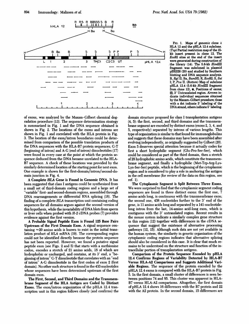
IFIG. 3. Organization of exons andintrons in pHLA 12.4. The genomicstructure is correlated with the proteinstructure of a class I transplantationantigen. All exons code for.sequenceshighly honologous to both the HLA-B7 and the -A2 molecules (see Fig. 5).
1 91 183314
found with B7, 18 with A2, with some clustering around resi-dues 110 (not seen in A2 versus B7 comparisons), and 175 (cor-responding to a detected A2/B7 variability region). One ofthesedifferences may have important implications: the cysteine nor-mally. found at position 164, which is involved in the second-domain disulfide bridge, is not-found in the sequence. A phen-ylalanine is found instead; this corresponds to a one-base change(TGC to TTC). Thus, the protein encoded by the gene whosesequence we have determined cannot form the disulfide bridgefound in the second domain ofall transplantation antigens whosesequences have thus far been determined. The third domainis, as expected, similar to the A2 and B7 sequences with a fewdifferences (i.e., four with B7 and eight with A2). The trans-membrane segment displays a number of differences (the B7sequence is not completely established in this region), althoughmost of these are Ala-Val-Leu replacements (i.e., the hydro-phobic character ofthe protein sequence is conserved). Finally,the cytoplasmic segment is similar to the A2 and B7 sequences(six differences with B7 and one with A2).
Altogether, those variability regions tentatively defined onthe basis of A2/B7 (4) comparisons are also evident in pHLA12.4/A2 or B7 comparisons; another possible cluster of differ-ences is also found in the second exon. Thus, the delineationof possible variable regions in HLA molecules (apart from thegeneral feature that the third exon is more conserved than theothers) will require determination and comparison of a largesample of&sequences.
DISCUSSIONComparison With H-2 Gene Structure. Mouse H-2 genomic
clones have recently been isolated by Steinmetz et at (12). Acomplete sequence was derived for an H-2 gene contained inone of these clones. This is considered to be a pseudogene be-cause it contains two termination codons in transmembrane andcytoplasmic exons and also.a charged residue, aspartic acid, in-side the transmembrane domain. It can reasonably be assumed,however, that its exon/intron organization is representative offunctional H-2 transcription units. The arrangement reportedby Steinmetz et at is in fact strikingly similar to our results ex-cept at the end ofthe cytoplasmic region where the mouse genecontains an additional intron just before the 3' untranslatedsequence.
Is pHLA 12.4 a Gene or a Pseudogene? Pseudogenes havebeen found in the 5S gene family (21), among globin genes (22),and recently in the mouse H-2 cluster (12). The possibility thatthe pHLA 12.4 segment is in fact a pseudogene must be dis-
87 90 87
cussed. As mentioned above, the second cysteine involved inthe disulfide bridge present in the second domain is replacedby a phenylalanine residue. This raises doubts about the struc-tural rigidity of the second domain of the protein encoded bythis gene and its ability to express serological allotypes and theusual functions of MHC antigens.We have not detected transcription initiation signals up-
stream of the signal peptide coding sequence. Given the sizeof HLA RNA [1650 nucleotides (18)] and of the 3' noncodingregion (nearly 600 nucleotides), this would indicate the exis-tence of an intron ahead of this coding sequence, as in the caseof the ovalbumin gene (23). pHLA 12.4 seems correctly ar-ranged because we have not encountered any termination co-don in the exons, all the exon/intron junctions are providedwith the requisite splicing signals (16), and the protein corre-sponding to this gene is closely homologous to known trans-plantation antigens (3, 4).Which Part of the MHC Gene Cluster Does pHLA 12.4
Come From? The human DNA library from which this clonewas isolated was constructed with placental DNA (14) and thecorresponding HLA typing is unknown.
Although the DNA sequence we have determined providesthe complete structure of the protein that could be coded bythis gene, this does not allow us to infer the corresponding ge-netic locus. On the basis of available sequence data, there is noevidence for "A-ness" or "B-ness" (24); i.e., there is no featureofthe structures that assigns a particular sequence to a particularallelic series of class I loci.-In fact, the putative pHLA 12.4 pro-tein is closer to the B7 product than to the A2 molecule in its.extracellular domains while the reverse is true for the cyto-plasmic region. The mouse cDNA probe used for isolation ofthe clone, which covers transmembrane, cytoplasmic, and 3'noncoding sequences, could have picked up any class I gene oreven other genes as long as they bear some resemblance in therelevant region. The gene structure which we have determineddoes show that the DNA segment studied codes for a moleculeof the class I type; however, we do not know whether this orig-inates from the HLA-A, -B, or -C regions (25) or from the pos-sible human counterpart of the mouse TL-Qa region (26, 27,28).
In conclusion, this study provides the basic structure of ahuman class I antigen gene. In addition to confirming the do-main model for the extracellular part of the protein andstrengthening the case for nonrearrangement of these genes,it indicates peculiar features for the cytoplasmic region that sug-gest the possibility of alternative splicing pathways. In the re-
91
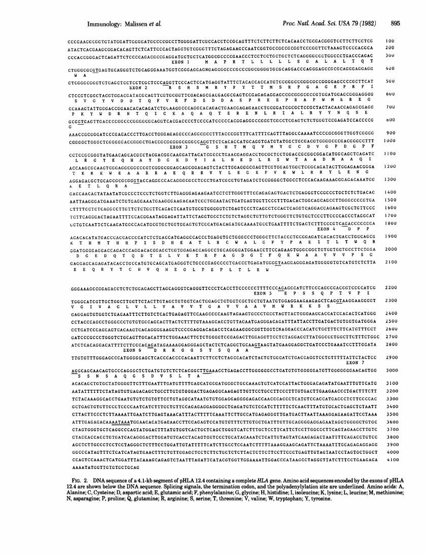
Q S E A G S H T
Q S E G (G) (E) (L) (T)CAG AGC GAG GGC GGT GAG TTG ACC ......(
Q S E A G S H T
(G) (G) (G) (Q) G S H TGGG GGC GGG CAA GGT TCT CAC ACC
.HLA B7 protein sequence
pHLA 12.4 translationpHLA 12.4 DNA sequence
Exon 2 4- Intron (245 bp) -* Exon 3
FIG. 4. Determination of exon/intron junctions by comparison with the HLA-B7 protein sequence. The pHLA 12.4 DNA sequences at the endof the second exon (first domain) and at the beginning of the third exon (second domain) are shown, and one of the possible translation productsis compared with the HLA-B7 protein sequence in this region (i.e., amino acids 87-94). The homology between the two sequences breaks down andreappears at locations that correlate with the presence of G-T ("beginning of intron") and A-G ("end of intron") dinucleotides, thus locating the exon/intron boundaries. The single-letter amino acid code is given in Fig. 2.
Proc. Nad Acad.,Sci. USA 79 (1982)
Proc. Natl. Acad. Sci. USA 79 (1982) 897
Signal peptide exon
1 10 20MAPRTLLLLLSGALALTQTWA
First domain exon
1 10 20 30 40 50 60 70 80 90RSHSMRYFYTTMSRPGAGEPRFISVGYVDDTQFVRFDSDDASPREEPRAPWMEREGPKYWDRNTQICKAQAQTERENLRIALRYYNQSEGG SV R A I Q E Y D S NLRG A
Second domain exon
100 110 120 130 140 150 160 170 180GSHTMQVMYGCDVGPDGPFLRGYEQHAYDGKDYIALNEDLRSWTAADMAAQITKRKWEAARRAEQRRVYLEGEFVEWLRRYLENGKETLQRA
L S RL HD Y T Q E A C DK E
Third domain exon
190 200 210 220 230 240 250 260 270DPPKTHMTHHPISDHEATLRCWALGFYPAEITLTWQRDGEDQTQDTELVETRPAGDGTFQKWAAVVVPSGEEQRYTCHVQHEGLPEPLTLRW
V R E K
Transmembrane and cytoplasmic exons
280 290 300 310EPSSQPTVPIVGIVAGLVLLVAVVTGAVVAAVMWRKKSS
S AV AV C R
320DRKGGSYSQAAGG
330 338SSNSAQGSDVSLTAC D
FIG. 5. Comparison of the protein encoded by thepHLA 12.4 gene and the HLA-B7 transplantation antigen. The protein sequence deduced fromour data is shown. Differences with the HLA-B7 protein are indicated below. The single-letter amino acid code is given in Fig. 2.
gion coding for the extracellular part of the molecule, the struc-ture of the gene closely parallels the functional organization ofthe transplantation antigen. It seems likely that the same re-lationship holds for the intracellular region: indeed, if the ge-netic complexity of the cytoplasmic region indicates a corre-sponding functional complexity for the intracellular segment ofthe protein, we are left to wonder whether the part ofthe trans-plantation antigen located inside the cell may not be extremelyimportant.
Note Added in Proof. In vitro transcription experiments have shownrecently that a major transcript is initiated from pHLA 12.4 DNA up-stream of the signal sequence (C. Kedinger and P. Chambon, personalcommunication).
We thank Dr. F. M. Kourilsky for enthusiastic support and manydiscussions; Drs. P. Kourilsky, F. Bregegere, and B. Cami for adviceand information; Dr. R. Staden (Medical Research Council, Cambridge)for computer programs; Prof. J. Kern (Centre de Recherches sur lesMecanismes de la Croissance Cristalline, Marseille) for computing fa-cilities; and Drs. L. Hood and S. Weissman for communication of theirresults prior to publication. This work was supported by the InstitutNational de la Sante et de la Recherche Medicale (CRL 80 1 027) andthe Delegation Generale a la Recherche Scientifique (ACC 80 E 0871).M. M. was the recipient ofa Delegation Generale a la Recherche Scien-tifique training grant.
1. Nathenson, S. G., Uehara, H., Ewenstein, B. M., Kindt, T. J.& Coligan, J. E. (1981) Annu. Rev. Biochem. 50, 1025-1051.
2. Orr, H. T., Lancet, D., Robb, R. J., Lopez de Castro, J. A. &Strominger, J. L. (1979) Nature (London) 232, 266-270.
3. Orr, H. T., Lopez de Castro, J. A., Parham, P., Pleogh, H. L.& Strominger, J. L. (1979) Proc. NatL Acad. Sci. USA 76,4395-4399.
4. Lopez de Castro, J. A., Orr, H. T., Kostyk, T., Mann, K. L. &Strominger, J. L. (1979) Biochemistry 18, 5704-5711.
5. Coligan, J. E., Kindt, T. J., Uehara, H., Martinko, J. & Nathen-son, S. G. (1981) Nature (London) 291, 35-39.
6. Kvist, S., Bregegere, F., Rask, L., Cami, B., Garoff, H., Daniel,F., Wiman, K., Larhammar, D., Abastado, J. P., Gachelin, G.,Peterson, P. A., Dobberstein, B. & Kourilsky, P. (1981) Proc.NatL Acad. Sci. USA 78, 2772-2776.
7. Steinmetz, M., Frelinger, J. G., Fisher, D., Hunkapiller, T.,Pereira, D., Weissman, F. M., Uehara, H., Nathenson, S. G.& Hood, L. (1981) Cell 24, 125-134.
8. Ploegh, H. L., Orr, H. T. & Strominger, J. L. (1980) Proc. NatiAcad. Sci. USA 77, 6081-6085.
9. Sood, A. K., Pereira, D. & Weissman, S. M. (1981) Proc. NatlAcad. Sci. USA 78, 616-620.
10. Cami, B., Bregegere, F., Abastado, J. P. & Kourilsky, P. (1981)Nature (London) 291, 673-675.
11. Jordan, B. R., Bregegere, F., & Kourilsky, P. (1981) Nature(London) 290, 521-523.
12. Steinmetz, M., Moore, K. W., Frelinger, J. G., Taylor Sher, B.,Shen, F. W., Boyse, E. A. & Hood, L. (1981) Cell 25, 683-692.
13. Maxam, A. M. & Gilbert, W. (1980) Methods in Enzymology 65,499-560.
14. Lawn, R. M., Fritsch, E. F., Parker, R. C., Blake, G. & Man-iatis, T. (1978) Cell 15, 1157-1174.
15. Bregegere, F., Abastado, J. P., Kivst, S., Rask, L., Lalanne, J.L., Garoff, H., Cami, B., Wiman, K., Larhammar, D., Peterson,P. A., Gachelin, G., Kourilsky, P. & Dobberstein, B. (1981) Na-ture (London) 292, 78-81.
16. Soberon, X., Covarrubias, L. & Bolivar, F. (1980) Genetics 9,287-305.
17. Lewin, B. (1980) Cell 22, 324-326.18. Bodmer, F. W. (1981) Tissue Antigens 17, 9-20.19. Ploegh, H. L., Cannon, L. E. & Strominger, J. L. (1979) Proc.
Natl, Acad. Sci. USA 76, 2273-2277.20. Gilbert, W. (1978) Nature (London) 271, 501.21. Miller, J. R., & Brownlee, G. G. (1978) Nature (London) 275,
556-558.22. Proudfoot, N. J. & Maniatis, T. (1980) Cell 21, 537-544.23. Gannon, F., O'Hare, K., Perrin, F., Le Pennec, J. P., Benoist,
C., Cochet, M., Breathnach, R., Royal, A., Cami, B. & Cham-bon, P. (1979) Nature (London) 278, 428-434.
24. Ploegh, H. L., Orr, H. T. & Strominger, J. L. (1981) Cell 24,287-299.
25. Bach, F. H. & Van Rood, J. J. (1976) N. EngL J. Med. 295,806-813.
26. Gazit, E., Terhorst, C. & Yunis, E. J. (1980) Nature (London)284, 275-277.
27. Gazit, E., Terhorst, C., Mahoney, R. J. & Yunis, E. J. (1980)Hum. Immunol 1, 97-109.
28. Cotner, T., Mashimo, H., Kung, P. C., Golstein, G. & Strom-inger, J. L. (1981) Proc. Natl Acad. Sci. USA 78, 3858-3862.
Immunology: Malissen et al.