Étude in vitro et in vivo de la virulence et du réassortiment génétique ...
197
Étud In DEPA SERVICE de in vitro vitro and se TH ACADEM FAC ARTEMENT DE VIROLO o et in vivo sérotyp in vivo st erotypes 4 Maria HESE PRESE MIE UNIVER UNIVE CULTE DE M T DES MALA OGIE VÉTÉR o de la vir es 4 et 9 d tudy of th 4 and 9 of Ana de la G ENTEE EN V Docteur e ANNEE AC RSITAIRE W ERSITE DE MEDECINE ADIES INFEC RINAIRE et rulence et du virus d he virulen f African Grandière VUE DE L’OB en Sciences V CADEMIQU WALLONIE- LIEGE VETERINA CTIEUSES e MALADIES t du réass de la peste ce and ge horse sic de Noronh BTENTION étérinaires E 2014-2015 -EUROPE AIRE et PARASITA VIRALES A sortiment e équine enetic reas kness viru ha Cotta DU GRADE AIRES ANIMALES génétiqu ssortmen us DE e des t of
Transcript of Étude in vitro et in vivo de la virulence et du réassortiment génétique ...
Étud
In
DEPA
SERVICE
de in vitro
vitro and se
TH
ACADEM
FAC
ARTEMENT
DE VIROLO
o et in vivosérotyp
in vivo sterotypes 4
Maria
HESE PRESE
MIE UNIVER
UNIVE
CULTE DE M
T DES MALA
OGIE VÉTÉR
o de la vires 4 et 9 d
tudy of th4 and 9 of
Ana de la G
ENTEE EN VDocteur e
ANNEE AC
RSITAIRE W
ERSITE DE
MEDECINE
ADIES INFEC
RINAIRE et
rulence etdu virus d
he virulenf African
Grandière
VUE DE L’OBen Sciences V
CADEMIQU
WALLONIE-
LIEGE
VETERINA
CTIEUSES e
MALADIES
t du réassde la peste
ce and gehorse sic
de Noronh
BTENTION étérinaires
E 2014-2015
-EUROPE
AIRE
et PARASITA
VIRALES A
sortiment e équine
enetic reaskness viru
ha Cotta
DU GRADE
AIRES
ANIMALES
génétiqu
ssortmenus
DE
e des
t of

REMERCIEMENTS
Je voudrais d’abord remercier mon promoteur, le Professeur Etienne Thiry, pour son accueil dans son
laboratoire et sa pleine confiance dans mon travail quotidien.
Je remercie toute l’équipe de virologie : Axel, pour ta patience sans fin à mes multiples questions ;
Damien, de m’avoir aidée à trouver mes marques à mon arrivée ; et surtout mon petit William : une
vraie amitié est née et ton soutien à toute épreuve, a été très précieux. Tu es un super gars ne l’oublie
jamais! Thank you Edmilson and Louisa for your support. Merci à tous d’avoir contribué à une
ambiance conviviale dans le bureau et aussi lors de nos sorties en congrès.
Un merci tout spécial à toi Fabiana, pour ton aide et tes conseils précieux. Tu m’as toujours remotivée
dans les moments difficiles et aidée à persévérer jusqu’au bout! Merci au reste de l’épidémiologie et
notamment à Ludo dont la bonne humeur est communicative. Je remercie également le Professeur
Claude Saegerman pour son aide dans mes statistiques.
Je remercie l’ensemble de l’équipe de l’immunologie : la Fish team (Michelle, Catherine, Léa, Yuan,
Joanna, Krys, …) et en particulier Maygane & Maxime avec qui j’ai passé de bons moments en intra-
et extra-laboratoire! Je remercie aussi les équipes de Laurent (Bilal, Céline, Béné, Caroline,
Mickaël,…) et de Benjamin (Françoise, Océane, Marion, Annette, …).Grâce à la convivialité de
chacun, il règne une bonne ambiance dans les laboratoires partagés.
Je remercie nos techniciens pour leur aide au quotidien : Cédric, Lorène, Christine et Justine.
Merci à vous deux mes blondies, Christina et Lorène, pour votre soutien et votre écoute. Je suis
souvent sortie de votre bureau apaisée et requinquée prête à tout affronter.
Je remercie l’équipe ENZOREM du CERVA : Brigitte Caij, merci pour vos nombreux mots
d’encouragements ; Marylène Tignon et Nick de Regge pour vos conseils avisés et votre temps dédié
au projet.
Je remercie le service d’Entomologie fonctionnelle et évolutive de l’Agro BioTech de Gembloux. En
particulier, je remercie mon co-promoteur, Frédéric Francis, pour son accompagnement au cours du
projet, et ensuite Slimane, qui m’a initié au microcosme belge au cours des piégeages et élevages de
moustiques.

Ce travail a été subventionné par le Service Public Fédéral Santé Publique, Sécurité de la Chaîne
alimentaire et Environnement (projets INDEVIREQ 1.0 et 2.0 et PupWelfare) ainsi que par
l’Université de Liège.
Je remercie ma maman et ma sœur pour leur soutien tout au long de mon cursus et leur présence lors
de mes choix de carrière.
Et enfin, je te remercie Luc, pour toujours m’encourager à chaque étape, pour ta bonne humeur, ta
patience au quotidien. Ça y est : une autre aventure, et pas des moindre, nous attend avec la venue
prochaine de notre mini nous. J’ai hâte et je suis sûre que tu feras un super papa !
iii
TABLE DES MATIERES
Table des matières…………………………………………………………......iii
Liste des abréviations…………………………………………………………..v
Chapitre I : Introduction………………………………………………………1
1.1 Historique………………………………………………………………………..……..2
1.2 Le virus de la peste équine au sein de la famille des Reoviridae………………………2
1.3 Virologie moléculaire du virus de la peste équine…………………………………..…4
1.4 Variabilité du virus de la peste équine…………………………………………………8
1.4.1 Diversité génétique des populations virales de virus à ARN…………………..8
1.4.2 Diversité génétique du virus de la peste équine………………………………..9
1.5 Epidémiologie de la peste équine……………………………………………………..10
1.5.1 Cycle de transmission………………………………………………………...10
1.5.2 Peste équine en Afrique………………………………………………………12
1.5.3 Peste équine en Europe……………………………………………………….13
1.6 Risque d’introduction de la peste équine en Belgique………………………………..13
1.7 Pathogénie et aspects cliniques de la peste équine……………………………………15
1.7.1 Pathogénie……………….……………………………………………………15
1.7.2 Réponse immunitaire…………………………………………………………16
1.7.3 Signes cliniques……………………………………………………………….17
1.7.4 Lésions nécropsiques…………………………………………………………18
1.8 Diagnostic de l’infection par le virus de la peste équine……………………………..19
1.8.1 Diagnostic moléculaire……………………………………………………….19
1.8.2 Diagnostic sérologique………………………………………………………..19
1.8.3 Isolement en culture cellulaire………………………………………………..20
1.9 Vaccination…………………………………………………………………………21
1.9.1 Vaccins commercialisés………………………………………………………21
1.9.2 Vaccins expérimentaux……………………………………………………….21
1.10 Modèle murin d’infection par le virus de la peste équine……………..…………….22
Chapitre II : Objectifs………………………………………………………...24
iv
Chapitre III : Section expérimentale………………………………………...27
Étude 1 : Modèle murin expérimental d’infection par le virus de la peste équine……...28
Study of the virulence of serotypes 4 and 9 of African horse sickness virus in
IFNAR -/-, Balb/C and 129 Sv/Ev mice
Étude 2 : Étude du réassortiment génétique du virus de la peste équine………………..43
In vitro and in vivo characterization of in vitro generated African horse
sickness reassortant viruses
Partie 1 : Étude in vitro du réassortiment génétique entre les sérotypes 4 et 9………………48
du virus de la peste équine
Partie 2 : Étude de la virulence in vivo des réassortants obtenus in vitro……………………61
à partir des sérotypes 4 et 9 du virus de la peste équine
Chapitre IV : Discussion……………………………………………………...70
Chapitre V : Conclusions et perspectives……………………………………82
Chapitre VI : Résumé – Summary…………………………………………...85
Chapitre VII : Références bibliographiques………………………………...90
Annexes……………………………………………………………………….106
Préambule…………………………………………………………………………………...107
Risk profiling of introduction and dissemination of Eastern, Western and Venezuelan equine
encephalitis virus in Belgium………………………….…………………………………………….108
Diversity and ecology survey of mosquitoes potential vectors in Belgian equestrian farms:
a threat prevention of mosquito-borne equine arboviruses….………………………………140

v
LISTE DES ABREVIATIONS
AAE : acides aminés essentiels
ADN : acide désoxyribonucléique
ADNc : ADN complémentaire
AHSV : African horse sickness virus, virus de la peste équine
ARN : acide ribonucléique
BHK : Baby Hamster Kidney, rein de jeunes hamsters
CERVA : Centre d’Etude et de Recherche Vétérinaires et Agrochimiques
Ct : cycle seuil de détection
CPE : cytopathic effect, effet cytopathogène
DICC50 : dose infectieuse en culture de cellules à 50 %
DMEM : Dulbecco’s Modified Eagle Medium, Gibco®Invitrogen
dNTPs : désoxyribonucléotides triphosphates
EDTA : acide éthylène diamine tétraacétique
ELISA : enzyme-linked immunosorbent assay, dosage immuno-enzymatique sur support
solide
ENSO : El Niño Southern Oscillation, oscillation australe associée à El Niño
FCO : fièvre catarrhale ovine
FCS : Fetal Calf Serum, sérum de veau fœtal
IFNAR : Interferon-α Receptor knock-out, déficient en récepteur à l’interféron α
MDA5 : melanoma differenciation-associated gene 5
MOI : Multiplicity Of Infection, multiplicité d’infection
vi
MS : Monkey Kidney, rein de singe
NGS : next-generation sequecing, séquençage haut débit
NS : non structural protein, protéine non structurale
OIE : Office international des épizooties
ORF : open reading frame, cadre ouvert de lecture
PAMP : pathogen-associated molecular patterns, motif moléculaire associés aux pathogènes
PRR : pattern recognition receptors, récepteurs de reconnaissance de motifs moléculaires
Pb : paires de bases nucléotidiques
PBS : Phosphate Buffered Saline, tampon phosphate salin
PCR : Polymerase Chain Reaction, réaction de polymérisation en chaîne
PS : Pénicilline-Streptomycine
qRT-PCR : réaction de polymérisation en chaîne quantitative après transcription inverse
RIG : retinoid acid-inducible gene-I
rpm : Rotation per minute, tour par minute
Spf : Service public fédéral
TCID50 : Tissue Culture Infectious Dose, dose infectieuse en culture de tissu à 50 %
TLR : toll-like recepteur, récepteur de type toll
VP : (structural) viral protein, protéine virale (de structure)
Introduction
Chapitre 1 : Introduction – Le virus de la peste équine
2
1.1 Historique
Le premier épisode clinique d’un syndrome ressemblant à la peste équine a été
rapporté au Yémen en 1327. Cependant, le virus de la peste équine ou AHSV (African horse
sickness virus) est avant tout un virus africain et y a été observé les premières fois à la suite de
l’introduction de chevaux en provenance d’Inde pendant l’exploration de l’Afrique de l’Est et
centrale par les colons portugais en 1569. En Afrique du Sud, le virus était déjà probablement
présent mais des signes cliniques concordant avec la peste équine n’ont pas été décrits avant
1657, après la première introduction de chevaux par les colons néerlandais. Une épidémie
majeure y eut d’ailleurs lieu en 1719 pendant laquelle quelques 1700 chevaux moururent.
Ensuite, différents épisodes de peste équine furent rapportés et des dizaines de milliers de
chevaux périrent de la maladie (Maclachlan et Guthrie, 2010). Ces faits historiques ne se
basent que sur la présence de signes cliniques similaires à ceux observés lors d’une infection
de peste équine confirmée (Mellor et Hamblin, 2004).
En effet, le premier isolement du virus de la peste équine date du début des années
1900 et la distinction antigénique en 9 sérotypes connus date de 1962. Depuis lors, tous les
sérotypes de la peste équine ont été retrouvés en Afrique sub-saharienne et le sérotype 9 a
provoqué des épidémies en dehors des zones endémiques et notamment en Egypte, Turquie,
Liban, Jordanie, Iran, Afghanistan, Inde, Maroc, Tunisie, Algérie mais également en 1965 en
Espagne. L’exception s’est avérée en 1987 lors d’une épidémie de peste équine en Espagne et
au Portugal (Portas et al., 1999) qui, cette fois, a été attribuée au sérotype 4 de l’AHSV. Le
virus s’y est maintenu pendant les quatre étés suivants, provoquant des épidémies à répétition
et montrant la capacité de l’infection virale à se maintenir en dehors des zones endémiques
africaines (Wilson et al., 2009; Johnson et al., 2012).
1.2 Le virus de la peste équine au sein de la famille des Reoviridae
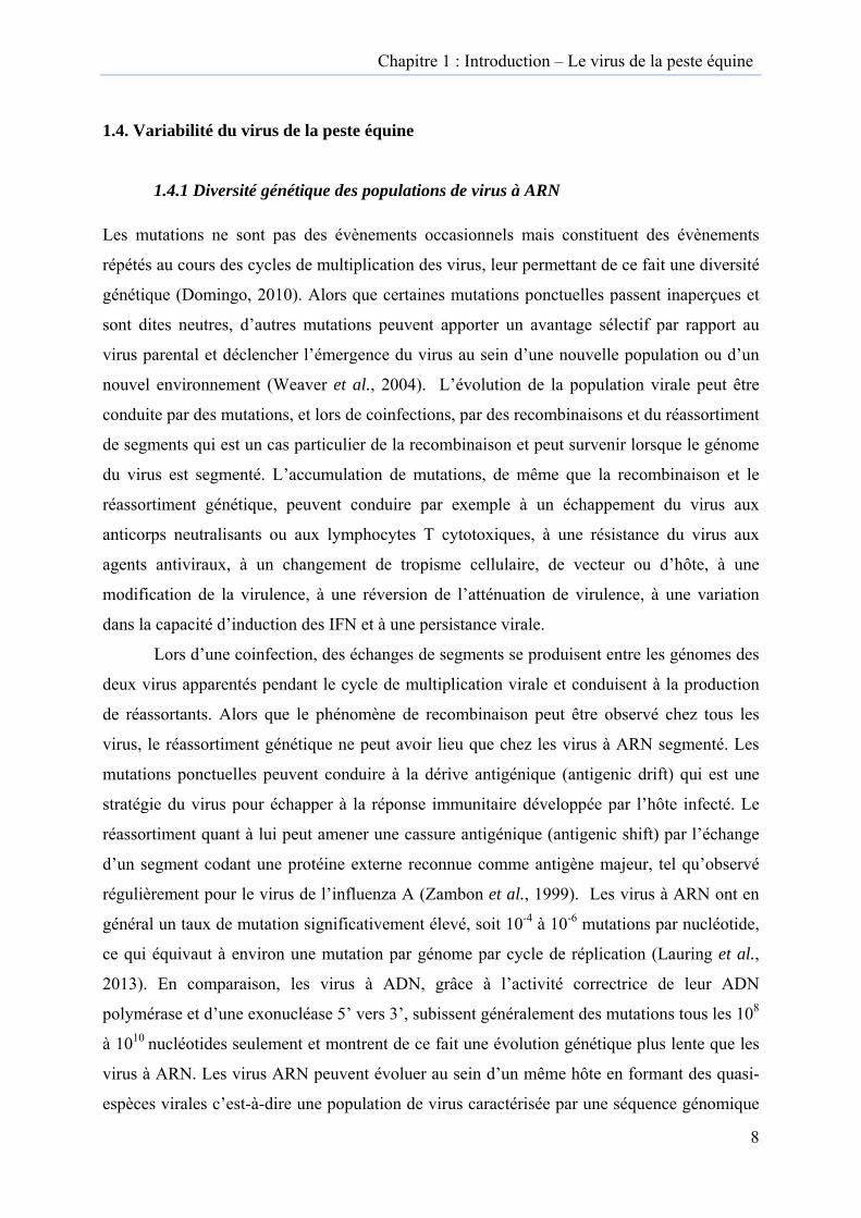
Le virus de la peste équine appartient à la famille des Reoviridae, genre Orbivirus.
La famille des Reoviridae comprend deux sous-familles : Sedoreovirinae et Spinareovirinae
avec 14 genres répartis entre ces deux sous-familles (Figure 1)
(http://www.ictvonline.org/virusTaxonomy.asp). Le genre Orbivirus est un des 6 genres que
comporte la sous-famille des Sedoreovirinae. La famille des Reoviridae inclut des pathogènes
des vertébrés, des arthropodes et des plantes.

Figure polyméReovirid(Blueton(YunnanPhytore(Kadipi(RotavirparasiticFijivirucytoplasLdCPV pseudosMRV (M(Chum (D’aprè
1 : Arbre pérase (segmdae. Mimorngue virus)n orbivirus)eovirus : RDro virus) ; Crus) ; Mycoca reovirus)
us : NLRV (smic polyhe (Lymantriascutellaris reMammaliansalmon reov
ès Attoui et
phylogénétiment 1). La
reovirus : M, AHSV (A), PHSV (PeDV (Rice dwCardoreoviroreovirus : R) ; Coltiviru(Nilaparvataedrosis virua dispar cytoeovirus) ; O
n orthoreovivirus), GCRal., 2009)
Cha
ique de la ffigure repr
MpRV (MicAfrican horse
ervuvian howarf virus) rus : ESRVRaRV (Roseus : EYAV (a lugens reo
us), DsCPV oplasmic po
Oryzavirus :irus) ; AquaRV (Grass C
apitre 1 : In
famille des résente 14
cromonas pue sickness vorse sicknes; Seadorna
V (Eriocheir ellinia anti-(Eyach viruovirus) ; Cyp(Dendrolim
olyhedrosis: RRSV (Riareovirus : SCarp reoviru
ntroduction –
Reoviridaedes 15 gen
usilla reovirvirus), CHUs virus), SCvirus : BAVsinensis reorot virus), C
us), CTFV (Cpovirus : Bm
mus punctatu) ; Dinovernce ragged sSBRV (Strius), GSRV (
– Le virus d
e sur base dres de la farus) ; Orbivi
UV (Palyam CRV (St CroV (Banna viovirus) ; RoCPRV (CrypColorado ticmCPV (Bomus cytoplasmnavirus : Atunt virus) ;ped bass reo(Golden shi
de la peste é
des séquencamille des irus : BTVvirus), YU
oix river virirus), KDV otavirus : RVphonectria ck fever virmbyx mori mic polyhed
ApRV (Aede; Orthoreovovirus), CSiner reoviru
équine
3
ces de la
V UOV rus) ;
V
rus) ;
drosis), es virus : SRV us).
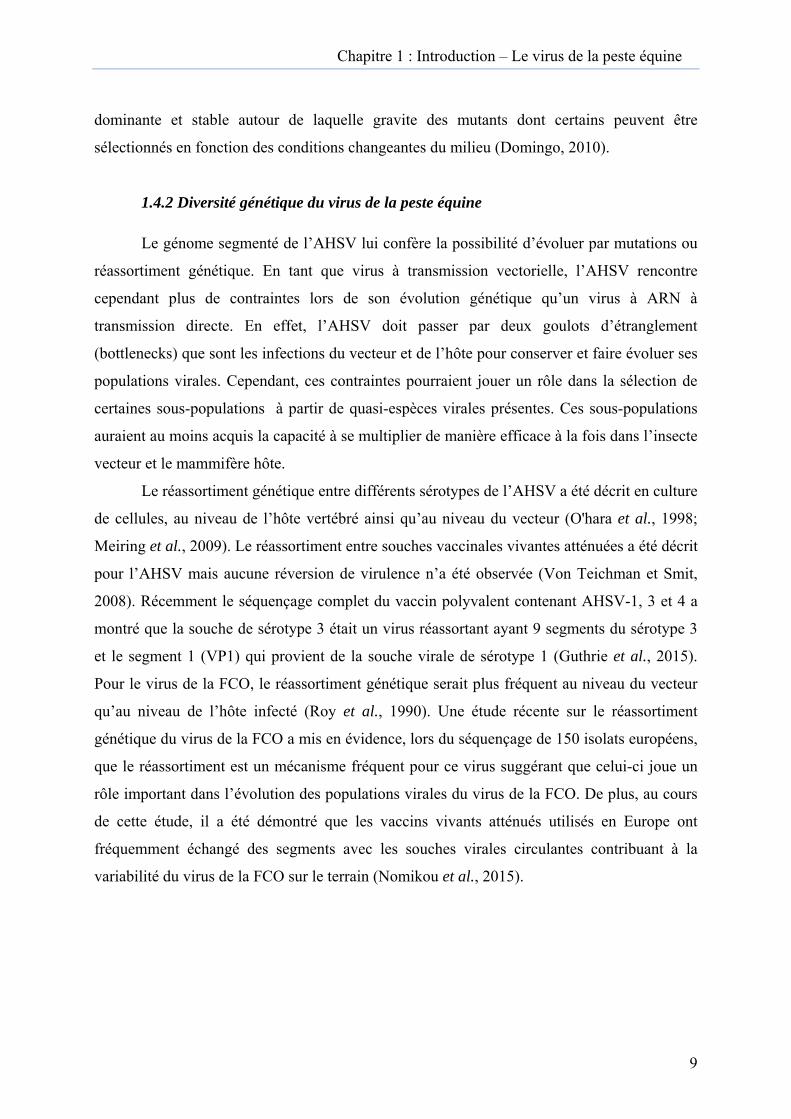
Figure Orbivirudécrits; African
2 : Analyseus sont reprEHDV, Ep
n horse sickn
e phylogénérésentées ici
pizootic hemness virus a
étique du si. BTV, Blu
morrhagic di9 sérotypes
segment 2 duetongue virisease viruss décrits. (D
du genre Orrus compren compte ent
D’après Hof
rbivirus. Sixnd actuellemtre 8 et 10 sfmann et al,
ix espèces dment 27 sérsérotypes; A 2008).
du genre otypes
AHSV,
Chapitre 1 : Introduction – Le virus de la peste équine
4
Même si certaines similarités avec les autres membres de la famille des Reoviridae peuvent
être trouvées, les Orbivirus sont assez différents du point de vue de leur structure, de leurs
propriétés physico-chimiques, de leur cycle de réplication, de leur pathogénie et de leur
épidémiologie. La première différence majeure est que les Orbivirus sont des arbovirus donc
transmis par des vecteurs, que les Reovirus ou encore les Rotavirus n’en sont pas et sont
transmis par voie féco-orale.
Le sérotype est la catégorie dans laquelle on classe les virus selon leur réaction en présence
de sérums contenant des anticorps spécifiques ; pour les Orbivirus, les anticorps neutralisants induits
par VP2 (protéine de capside externe) définissent le sérotype. Le sérogroupe est l’ensemble de
plusieurs sérotypes possédant en commun un facteur caractéristique qui est la protéine VP7 (protéine
de capside interne) pour le genre Orbivirus. L’isolat est un virus produit en culture pure autant
que cela est connu. L’isolat peut ultérieurement être un mélange de virus et nécessite un
isolement du virus en culture de cellules. La souche virale se définit comme un isolat viral qui
ressemble au virus prototype de l’espèce dans les propriétés majeures qui définissent l’espèce,
mais qui diffère dans des propriétés mineures comme la spécificité de l’espèce de vecteur, les
signes cliniques induits, les propriétés sérologiques et génétiques (Thiry et al., 2008). Sur
base de leur profil sérologique, 22 sérogroupes, différents ont été identifiés pour les Orbivirus
et certains isolats sont encore non classés (Figure 2). A l’intérieur de ces sérogroupes,
différents sérotypes peuvent être différenciés, notamment 9 pour l’AHSV et 27, pour le virus
de la fièvre catarrhale ovine (FCO) (Jenckel et al., 2015) qui est le virus le plus apparenté à
l’AHSV et est le virus de référence pour le genre Orbivirus.
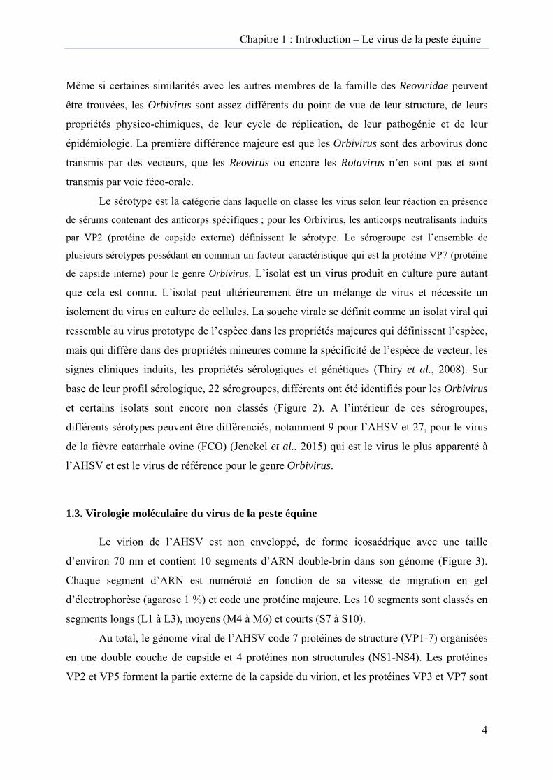
1.3. Virologie moléculaire du virus de la peste équine
Le virion de l’AHSV est non enveloppé, de forme icosaédrique avec une taille
d’environ 70 nm et contient 10 segments d’ARN double-brin dans son génome (Figure 3).
Chaque segment d’ARN est numéroté en fonction de sa vitesse de migration en gel
d’électrophorèse (agarose 1 %) et code une protéine majeure. Les 10 segments sont classés en
segments longs (L1 à L3), moyens (M4 à M6) et courts (S7 à S10).
Au total, le génome viral de l’AHSV code 7 protéines de structure (VP1-7) organisées
en une double couche de capside et 4 protéines non structurales (NS1-NS4). Les protéines
VP2 et VP5 forment la partie externe de la capside du virion, et les protéines VP3 et VP7 sont
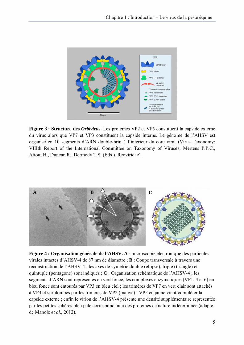
Figure du viruorganiséVIIIth RAttoui H
Figure virales ireconstrquintupsegmenbleu fonà VP3 ecapside par les pde Man
A
3 : Structus alors queé en 10 segReport of H., Duncan
4 : Organisintactes d’Aruction de lle (pentago
nts d’ARN sncé sont entet surplombé
externe ; enpetites sphèole et al., 2
ure des Orbe VP7 et Vgments d’Athe InternaR., Dermod
sation généAHSV-4 de ’AHSV-4 ; ne) sont indont représentourés par Vés par les trnfin le virio
ères bleu pâl012).
Ch
bivirus. Les VP3 constituARN doubleational Comdy T.S. (Eds
érale de l’A87 nm de dles axes de
diqués ; C : ntés en vert
VP3 en bleu rimères de Von de l’AHSle correspon
B
hapitre 1 : In
protéines Vuent la cape-brin à l’in
mmittee on s.), Reovirid
AHSV. A : mdiamètre ; B
symétrie doOrganisatio
t foncé, les c ciel ; les tri
VP2 (mauveSV-4 présenndant à des
ntroduction
VP2 et VP5pside internentérieur du Taxonomy
dae).
microscopie : Coupe traouble (ellipon schématicomplexes imères de Ve) ; VP5 en jnte une densprotéines d
C
n – Le virus
5 constituene. Le génomcore viral
y of Viruse
e électroniquansversale àse), triple (tique de l’AHenzymatiqu
VP7 en vert jaune vient
sité supplémde nature ind
de la peste
nt la capsideme de l’AH(Virus Tax
es, Mertens
que des partià travers unetriangle) et HSV-4 ; lesues (VP1, 4 clair sont a
t compléter mentaire repdéterminée
équine
5
e externe HSV est xonomy: s P.P.C.,
icules e
s et 6) en
attachés la
présentée (adapté

Bou
Figure de fixatiest indudécapsidsoutientassemblprotéinesortie hofait sur maturatdésormabourgeo
urgeonnement de virion
Exocyt
5 : Représeion du virus
uite par VP5dation est nt la traductiole les corps es virales etors des corpdes vésicule
tion du virioais une partonnement m
tose du virion
entation scs sur son réc5 qui possèdnécessaire à on des ARNd’inclusion
t les nouveaps d’inclusioes d’exocyt
on est achevticule infect
membranaire
Attachement du virion
Vésicule
hématique cepteur cellde des proprl’activation
N simples brn cytoplasmaux brins d’Aon cytoplastose grâce à vée par l’acqieuse compe (adapté de
Assemblage du vir
Pénétra
T
e du cycle dlulaire est siriétés déstabn de la transrins et form
miques au cœARN synthésmiques, le tà l’interactioquisition de
plète. Enfin Ne Mohl et R
rion
Décapation
Transport du core
de multiplicitué sur VP2bilisantes description vir
me les tubuleœur desquelétisés. Aprètransport du
on de NS3 aes protéines NS3 favorisoy, 2014).
psidation
Virop
cation des O2. La décape la membrarale dans le es dans le cys sont rasseès l’assemblu core dans avec des calpVP2 et VP5se la libérat
plasme
Orbivirus. Lpsidation duane. Cette core viral. Nytoplasme.
emblées les lage du corele cytoplasm
lpectines. La5, le virion tion des viri
Le site u virus
NS1 NS2
e et sa me se a étant
ions par
Chapitre 1 : Introduction – Le virus de la peste équine
6
les constituants majeurs de la couche interne de la capside (Roy, 1996) (Figure 3 et 4) alors
que VP1, VP4 et VP6 sont des constituants mineurs de la partie interne de la capside.
La protéine VP2, codée par le segment 2, est la protéine la plus variable et est
spécifique aux différents sérotypes viraux rencontrés. VP2 est la protéine la plus variable
entre les différents sérotypes avec environ 60 % d’homologie nucléotidique entre les
différents sérotypes et environ 90 % d’homologie nucléotidique entre souches au sein du
même sérotype. Avec VP5 (segment 6), VP2 est également responsable de la neutralisation
virale. VP2 et VP5 ensemble sont les médiateurs de l’attachement viral à la cellule et de sa
pénétration dans la cellule. L’attachement de VP2 sur des récepteurs cellulaires contenant de
l’acide sialique est suivi par une endocytose contrôlée par des molécules de clathrine.
La capside externe est enlevée après pénétration grâce aux propriétés de déstabilisation
de la membrane de VP5 (pH=5), ce qui expose le core viral constitué par la capside interne et
l’acide nucléique (VP1, 3, 4, 6 et 7) (Figure 5 et Tableau 1).
VP7 codée par le segment 7, est la protéine la plus conservée entre les différents
sérotypes et elle est spécifique du sérogroupe. VP7 est le constituant majeur de la capside
interne de l’AHSV. Une infectiosité similaire du virus de la FCO est décrite pour des
particules virales composées uniquement du core et des particules virales complètes, lors de
l’infection de cellules d’insectes, suggérant que VP7 participe à l’entrée du virus dans la
cellule d’insecte et joue un rôle dans l’infectiosité de la particule de core pour des vecteurs
adultes et les cellules d’insectes (Mertens et al., 1996; Xu et al., 1997).
VP3 (segment 3) présente un rôle de protection des enzymes actives du core viral.
VP1 (segment 1), VP4 (segment 4) et VP6 (segment 9) sont des protéines associées aux
enzymes du core du virion et sont responsables de la transcription avec VP1 de l’ARN
Polymérase ARN dépendante, de l’acquisition de la coiffe des nouveaux brins d’ARN avec
VP4 et de l’activité hélicase avec VP6 qui déroule les brins d’ARN en présence d’ATP. La
transcription précoce a lieu dans le core viral et produit des ARNm coiffés qui permettront la
synthèse des protéines.
La protéine non structurale NS1 codée par le segment 5 est très conservée entre les
différents sérotypes et forme les tubules caractéristiques dans le cytoplasme de la cellule
infectée. Le rôle de ces tubules n’est pas connu précisément mais leur association aux
protéines du cytosquelette suggère leur participation dans les mécanismes de transfert
intracellulaire. Récemment des études sur le virus de la FCO suggère également que la
Segment ARN Taille segment (pb) Protéine codée Fonction
1 3965 VP1 ARN Polymérase ARN dépendante
2 3203 VP2
Constituant externe de la capside externe ; séquence très variable ; détermine le sérotype
viral ; induit anticorps neutralisants ; médiateur de l’attachement viral et de la pénétration dans la
cellule
3 2792 VP3 Constituant mineur de la capside interne ;
protection des protéines avec activités enzymatiques actives (VP1, VP4 et VP6) ;
4 1978 VP4 Guanyl-transférase ; coiffe et polyadényle les ARNm précoces
5 1748 NS1
Forme des tubules dans le cytoplasme de la cellule infectée ; implication dans la
morphogenèse de la particule virale ; participe aux mécanismes de transport intracellulaire
6 1566 VP5
Composant mineure de la capside externe ; propriétés de déstabilisation de la capside virale
ce qui permet au core d’être relargué dans le cytoplasme
7 1050 VP7 Composante majeure de la capside interne ; séquence très conservée entre les différents sérotypes, antigène spécifique du groupe ;
8 1165 NS2 Constituant majeur des corps d’inclusion
cytoplasmique ; favorise la traduction des ARNm au sein de ces corps d’inclusion
9 1169 VP6 (NS4)
ARN hélicase ; déroule les brins d’ARN en présence d’ATP ; active dans le core viral ; un ORF alternatif code NS4, pour le virus de la
FCO, cette protéine participerait à la modulation de réponse aux voies de l’IFN de type I
10 758 NS3-NS3a
(NS5-virus de la FCO)
Protéines associées à la membrane des vésicules intracellulaires et dans la membrane cellulaire ;
favorise la libération des virions par bourgeonnement ; pour le virus de la FCO,
participe à la modulation de la production des IFN suite à l’infection ; des études avec le virus
de la FCO ont mis en évidence un ORF alternatif codant NS5 dont la fonction doit encore être
explorée
Tableau 1 : Segments génomiques et protéines du virus de la peste équine. VP : viral protein ; NS : non structural protein ; ORF : open reading frame (cadre ouvert de lecture) ; sérotype : catégorie dans laquelle on classe les virus selon leur réaction en présence de sérums contenant des anticorps spécifiques. Subdivision de l'espèce ; pour l’AHSV, le sérotype est défini par VP2 ; sérogroupe : ensemble de plusieurs sérotypes possédant en commun un facteur caractéristique ; pour l’AHSV, le sérogroupe est définit par VP7 ; core ou nucléocapside : ensemble formé par la capside interne et l’acide nucléique viral.
Chapitre 1 : Introduction – Le virus de la peste équine
7
protéine NS1 joue un rôle crucial dans la régulation de l’expression des gènes viraux (Matsuo
et Roy, 2013 ; Boyce et al., 2012) . NS2 (segment 8) est le constituant majeur des corps
d’inclusion cytoplasmiques observés dans les cellules infectées. NS2 serait responsable du
recrutement des différents segments d’ARN de manière à n’en avoir qu’une copie par génome
grâce à un mécanisme encore inconnu. L’assemblage du core viral a lieu dans ces corps
d’inclusion cytoplasmiques. Les particules de core viral une fois assemblées, quittent ensuite
ces corps d’inclusion et sont transportés sur des vésicules d’exocytose grâce à l’interaction de
NS3 avec des calpectines. D’autre part, pendant ce processus, les protéines VP2 et VP5 sont
acquises afin de former des particules virales complètes. NS3-NS3a (segment 10) est une
protéine associée à la membrane qui est impliquée dans le relâchement du virion par altération
de la perméabilité de la membrane cellulaire (Meiring et al., 2009). D’autre part, par analogie
avec le virus de la FCO, on reconnaît à NS3, un rôle dans la virulence. En effet, des propriétés
de modulation de NS3 dans la production d’interférons suite à l’infection par le virus de la
FCO ont été reconnues (Chauveau et al., 2013).
Récemment, un cadre de lecture ouvert (open reading frame ou ORF) alternatif au
niveau du segment 9 (VP6) a été mis en évidence et code une protéine non structurale
supplémentaire NS4 mais son rôle dans le cycle de réplication de l’AHSV est encore inconnu
(Zwart et al., 2015). Pour le virus de la FCO, NS4 module la réponse IFN de type I de l’hôte
car cette protéine favorise la réplication virale in vitro en cellules pré-traitées à l’IFN de type I
(Ratinier et al., 2011).
De même, un ORF alternatif au niveau du segment 10 du virus de la FCO a été mis en
évidence et code une cinquième protéine non-structurale, NS5. Cependant, le rôle précis de
cette dernière n’est pas encore connu (Stewart et al., 2015) et des études spécifiques pour
l’AHSV sont nécessaires pour confirmer la présence de cet ORF alternatif pour ce virus
également.
Quelques réactions sérologiques croisées entre les sérotypes ont été observées : entre
les sérotypes 1 et 2, entre les sérotypes 3 et 7, entre les sérotypes 6 et 9 et entre les sérotypes 5
et 8 mais aucune réaction croisée avec d’autres Orbivirus connus n’a encore été mise en
évidence (Von Teichman et al., 2010). Les caractéristiques physico-chimiques de l’AHSV
sont sa sensibilité aux acides, son inactivation à un pH inférieur à 6,0, sa résistance aux
solvants lipidiques et sa résistance relative à la chaleur. Son infectiosité reste relativement
stable à 4°C (Mellor et Hamblin, 2004).
Chapitre 1 : Introduction – Le virus de la peste équine
8
1.4. Variabilité du virus de la peste équine
1.4.1 Diversité génétique des populations de virus à ARN
Les mutations ne sont pas des évènements occasionnels mais constituent des évènements
répétés au cours des cycles de multiplication des virus, leur permettant de ce fait une diversité
génétique (Domingo, 2010). Alors que certaines mutations ponctuelles passent inaperçues et
sont dites neutres, d’autres mutations peuvent apporter un avantage sélectif par rapport au
virus parental et déclencher l’émergence du virus au sein d’une nouvelle population ou d’un
nouvel environnement (Weaver et al., 2004). L’évolution de la population virale peut être
conduite par des mutations, et lors de coinfections, par des recombinaisons et du réassortiment
de segments qui est un cas particulier de la recombinaison et peut survenir lorsque le génome
du virus est segmenté. L’accumulation de mutations, de même que la recombinaison et le
réassortiment génétique, peuvent conduire par exemple à un échappement du virus aux
anticorps neutralisants ou aux lymphocytes T cytotoxiques, à une résistance du virus aux
agents antiviraux, à un changement de tropisme cellulaire, de vecteur ou d’hôte, à une
modification de la virulence, à une réversion de l’atténuation de virulence, à une variation
dans la capacité d’induction des IFN et à une persistance virale.
Lors d’une coinfection, des échanges de segments se produisent entre les génomes des
deux virus apparentés pendant le cycle de multiplication virale et conduisent à la production
de réassortants. Alors que le phénomène de recombinaison peut être observé chez tous les
virus, le réassortiment génétique ne peut avoir lieu que chez les virus à ARN segmenté. Les
mutations ponctuelles peuvent conduire à la dérive antigénique (antigenic drift) qui est une
stratégie du virus pour échapper à la réponse immunitaire développée par l’hôte infecté. Le
réassortiment quant à lui peut amener une cassure antigénique (antigenic shift) par l’échange
d’un segment codant une protéine externe reconnue comme antigène majeur, tel qu’observé
régulièrement pour le virus de l’influenza A (Zambon et al., 1999). Les virus à ARN ont en
général un taux de mutation significativement élevé, soit 10-4 à 10-6 mutations par nucléotide,
ce qui équivaut à environ une mutation par génome par cycle de réplication (Lauring et al.,
2013). En comparaison, les virus à ADN, grâce à l’activité correctrice de leur ADN
polymérase et d’une exonucléase 5’ vers 3’, subissent généralement des mutations tous les 108
à 1010 nucléotides seulement et montrent de ce fait une évolution génétique plus lente que les
virus à ARN. Les virus ARN peuvent évoluer au sein d’un même hôte en formant des quasi-
espèces virales c’est-à-dire une population de virus caractérisée par une séquence génomique
Chapitre 1 : Introduction – Le virus de la peste équine
9
dominante et stable autour de laquelle gravite des mutants dont certains peuvent être
sélectionnés en fonction des conditions changeantes du milieu (Domingo, 2010).
1.4.2 Diversité génétique du virus de la peste équine
Le génome segmenté de l’AHSV lui confère la possibilité d’évoluer par mutations ou
réassortiment génétique. En tant que virus à transmission vectorielle, l’AHSV rencontre
cependant plus de contraintes lors de son évolution génétique qu’un virus à ARN à
transmission directe. En effet, l’AHSV doit passer par deux goulots d’étranglement
(bottlenecks) que sont les infections du vecteur et de l’hôte pour conserver et faire évoluer ses
populations virales. Cependant, ces contraintes pourraient jouer un rôle dans la sélection de
certaines sous-populations à partir de quasi-espèces virales présentes. Ces sous-populations
auraient au moins acquis la capacité à se multiplier de manière efficace à la fois dans l’insecte
vecteur et le mammifère hôte.
Le réassortiment génétique entre différents sérotypes de l’AHSV a été décrit en culture
de cellules, au niveau de l’hôte vertébré ainsi qu’au niveau du vecteur (O'hara et al., 1998;
Meiring et al., 2009). Le réassortiment entre souches vaccinales vivantes atténuées a été décrit
pour l’AHSV mais aucune réversion de virulence n’a été observée (Von Teichman et Smit,
2008). Récemment le séquençage complet du vaccin polyvalent contenant AHSV-1, 3 et 4 a
montré que la souche de sérotype 3 était un virus réassortant ayant 9 segments du sérotype 3
et le segment 1 (VP1) qui provient de la souche virale de sérotype 1 (Guthrie et al., 2015).
Pour le virus de la FCO, le réassortiment génétique serait plus fréquent au niveau du vecteur
qu’au niveau de l’hôte infecté (Roy et al., 1990). Une étude récente sur le réassortiment
génétique du virus de la FCO a mis en évidence, lors du séquençage de 150 isolats européens,
que le réassortiment est un mécanisme fréquent pour ce virus suggérant que celui-ci joue un
rôle important dans l’évolution des populations virales du virus de la FCO. De plus, au cours
de cette étude, il a été démontré que les vaccins vivants atténués utilisés en Europe ont
fréquemment échangé des segments avec les souches virales circulantes contribuant à la
variabilité du virus de la FCO sur le terrain (Nomikou et al., 2015).
T
Figure
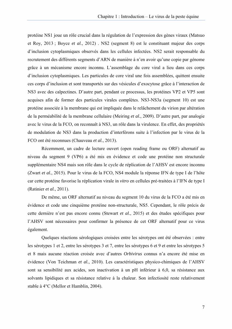
vecteur
d’incub
°C, l’EI
Transmissiol’infection
vecteur
5 : Cycle n
r Culicoides
ation extrin
IP dure de 7
on de n au r
naturel de t
s imicola, v
nsèque (EIP
7 à 10 jours.
ransmissio
vecteur prin
) est fortem
. Adapté d’
Périoe
d’iin
on du virus
ncipal et le
ment dépend
’après Mello
ode d’incubextrinsèqu
Période incubation
ntrinsèque
de la peste
zèbre, hôte
dante de la te
or et al., 200
bation e
e équine (A
e réservoir.
empérature
09.
AHSV) entr
. La période
extérieure
Trade l
e le
e
; à 25
ansmission’infection àl’hôte
n à
Chapitre 1 : Introduction – Le virus de la peste équine
10
1.5. Épidémiologie de la peste équine
1.5.1. Cycle de transmission
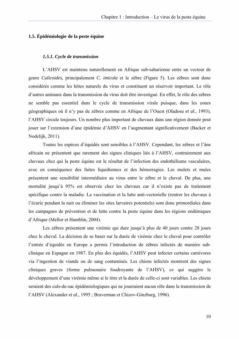
L’AHSV est maintenu naturellement en Afrique sub-saharienne entre un vecteur de
genre Culicoides, principalement C. imicola et le zèbre (Figure 5). Les zèbres sont donc
considérés comme les hôtes naturels du virus et constituent un réservoir important. Le rôle
d’autres animaux dans la transmission du virus doit être investigué. En effet, le rôle des zèbres
ne semble pas essentiel dans le cycle de transmission virale puisque, dans les zones
géographiques où il n’y pas de zèbres comme en Afrique de l’Ouest (Oladosu et al., 1993),
l’AHSV circule toujours. Un nombre plus important de chevaux dans une région donnée peut
jouer sur l’extension d’une épidémie d’AHSV en l’augmentant significativement (Backer et
Nodelijk, 2011).
Toutes les espèces d’équidés sont sensibles à l’AHSV. Cependant, les zèbres et l’âne
africain ne présentent que rarement des signes cliniques liés à l’AHSV, contrairement aux
chevaux chez qui la peste équine est le résultat de l’infection des endothéliums vasculaires,
avec en conséquence des fuites liquidiennes et des hémorragies. Les mulets et mules
présentent une sensibilité intermédiaire au virus entre le zèbre et le cheval. De plus, une
mortalité jusqu’à 95% est observée chez les chevaux car il n’existe pas de traitement
spécifique contre la maladie. La vaccination et la lutte anti-vectorielle (rentrer les chevaux à
l’écurie pendant la nuit ou éliminer les sites larvaires potentiels) sont donc primordiales dans
les campagnes de prévention et de lutte contre la peste équine dans les régions endémiques
d’Afrique (Mellor et Hamblin, 2004).
Les zèbres présentent une virémie qui dure jusqu’à plus de 40 jours contre 28 jours
chez le cheval. La décision de se baser sur la durée de virémie chez le cheval pour contrôler
l’entrée d’équidés en Europe a permis l’introduction de zèbres infectés de manière sub-
clinique en Espagne en 1987. En plus des équidés, l’AHSV peut infecter certains carnivores
via l’ingestion de viande ou de sang contaminés. Les chiens infectés montrent des signes
cliniques graves (forme pulmonaire foudroyante de l’AHSV), ce qui suggère le
développement d’une virémie même si le titre et la durée de celle-ci sont variables. Les chiens
seraient des culs-de-sac épidémiologiques qui ne joueraient aucun rôle dans la transmission de
l’AHSV (Alexander et al., 1995 ; Braverman et Chizov-Ginzburg, 1996).
Chapitre 1 : Introduction – Le virus de la peste équine
11
Chez les éléphants, des anticorps reconnus par la fixation du complément et des
anticorps neutralisants ont été retrouvés à de faibles taux chez des individus sauvages.
Cependant, des infections expérimentales ont été réalisées et n’ont pas permis de conclure
clairement sur leur rôle dans l’épidémiologie du virus. En effet, aucune virémie n’a été
détectée et leurs réponses immunitaires étaient limitées à des taux insignifiants d’anticorps
(Barnard et al., 1995; Barnard, 1997). L’infection des dromadaires est rare et inapparente.
Aucun détail n’est disponible sur la durée de la virémie chez ces derniers (Mellor et Hamblin,
2004).
Une étude en Afrique du Sud, Namibie et Kenya, visant à connaître les maladies
auxquelles les rhinocéros sont exposés, a mis en évidence des anticorps envers l’AHSV avec
un taux variable selon la répartition géographique suggérant qu’ils y sont régulièrement
exposés (Fischer-Tenhagen et al., 2000). Cependant, une étude réalisée plus récemment dans
la même région africaine, n’a pas détecté d’anticorps envers l’AHSV indiquant une
prévalence variable au cours du temps (Miller et al., 2011). Le rôle des rhinocéros dans
l’épidémiologie de la peste équine n’a pas été investigué mais semble insignifiant.
Culicoides imicola est considéré comme étant le vecteur principal dans la transmission
de l’AHSV en Afrique sub-saharienne, mais d’autres Culicoides peuvent être des vecteurs
potentiels aussi bien en Afrique qu’en Europe (Mellor et al., 2000; Meiswinkel et Paweska,
2003). En effet, lors des épidémies espagnoles et portugaises, le virus de la peste équine a été
isolé également de C. obsoletus et C. pulicaris (Portas et al., 1999; Capela et al., 2003), deux
espèces de Culicoides qui, contrairement à C. imicola, sont retrouvées fréquemment et en
grande proportion en Belgique. En outre, C. imicola est déjà présent dans le Bassin
méditerranéen (Rawlings et Mellor, 1994; Capela et al., 2003; Ramilo et al., 2012) et au sud
de la France et son nombre pourrait augmenter dans le nord de l’Europe avec le
réchauffement climatique (Acevedo et al., 2010; Cornell et al., 2010; Ramilo et al., 2012). En
conditions de laboratoire, C. sonorensis, le vecteur nord-américain du virus de la FCO, a été
trouvé capable de transmettre l’AHSV. Le deuxième vecteur compétent de l’AHSV dans le
sud de l’Afrique est C. bolitinos (Meiswinkel et Paweska, 2003; Venter et al., 2009; Venter et
al., 2010). L’AHSV a également été isolé de moustiques comme Aedes spp. dont on ignore
l’espèce (Mellor et al., 1990) pendant l’épisode espagnol-portugais et la compétence
vectorielle a été testée en laboratoire pour Anopheles stephensi, Culex pipiens, et Aedes
aegypti (Mellor, 1993) qui s’avèrent être capables de transmettre l’AHSV en conditions de
laboratoire. Les tiques pourraient aussi servir de vecteurs pour l’AHSV.

Chapitre 1 : Introduction – Le virus de la peste équine
12
L’isolement de l’AHSV d’Hyalomma dromedarii (Mellor, 1993; Wilson et al., 2009) en
conditions naturelles en Egypte ainsi que le test positif de sa capacité vectorielle en
laboratoire, sont des facteurs à prendre en compte. En effet, en comparaison des culicoïdes et
des moustiques, la durée de vie d’une tique est plus longue et en fait des candidates pour
l’ « overwintering » de l’AHSV. Cependant, l’overwintering du virus de la FCO-8 en Europe
occidentale, suggèrent que les culicoïdes femelles seraient de meilleures candidates pour
l’overwintering de l’AHSV dans nos contrées.
1.5.2. Peste équine en Afrique
L’AHSV est endémique dans les régions tropicales et subtropicales de l’Afrique au
sud du Sahara. Le désert du Sahara semble être une barrière géographique qui protège le nord
de l’Afrique du virus. A ce jour, la peste équine est endémique en Afrique sub-saharienne,
avec des épisodes épidémiques annuels principalement en Ethiopie et en Afrique du Sud
(Bitew et al., 2011; Aklilu et al., 2014). Tous les sérotypes de l’AHSV circulent en Afrique
sub-saharienne et en 2015, des épidémies sont en cours au Mozambique et en Ethiopie
(http://www.oie.int/wahis_2/public/wahid.php/Diseaseinformation/Diseasedistributionmap,
consulté le 5 mai 2015). En Afrique du Sud, les épidémies de grande ampleur d’AHSV ont
lieu tous les 10-15 ans mais leur cause a été incertaine jusqu’à récemment où une corrélation
étroite entre ces épidémies et le phénomène climatique nommé El Niño a été trouvée. El Niño
est un courant côtier chaud, qui associé à des variations de pressions atmosphériques entre le
Pacifique Est et Ouest (El Niño Southern Oscillation, ENSO), aurait des répercussions
climatiques jusqu’en Afrique du Sud et amène des périodes de sécheresse suivies de fortes
pluies. Dans le sol humide et dans les années à forte pluie, la population de C. imicola peut
augmenter jusqu’à 200 fois (Baylis et al., 1999). En plus de ces épidémies de peste équine,
des cycles endémiques de transmission sont présents dans différentes régions de l’Afrique
sub-saharienne comme l’Éthiopie et en Afrique du Sud où le virus peut être isolé de mars à
septembre voire toute l’année selon les régions d’Afrique.
Chapitre 1 : Introduction – Le virus de la peste équine
13
1.5.3. Peste équine en Europe
La première épidémie de peste équine en Europe a fait suite à l’introduction du
sérotype 9 dans le nord de l’Afrique (Tunisie, Maroc et Algérie) en 1965. L’AHSV a remonté
dans le nord du continent africain par l’intermédiaire d’ânes infectés et transportés à travers le
Sahara. Le vent aurait été alors responsable de l’introduction du virus dans le sud de
l’Espagne, en emmenant des vecteurs infectés à travers le détroit de Gibraltar (Wilson et al.,
2009).
L’incursion la plus récente de l’AHSV en Europe a eu lieu en Espagne en 1987.
L’introduction du virus est due à l’importation de zèbres infectés par le sérotype 4 provenant
de Namibie dans un parc animalier de la région de Madrid. Jusqu’au mois d’octobre 1987, des
cas furent recensés en Espagne. Pendant l’hiver suivant, le cycle du virus semblait interrompu
mais le virus a survécu grâce au climat tempéré de l’Espagne et a continué à sévir au cours
des 3 années suivantes (Mellor, 1993; Rawlings et Mellor, 1994). Après la notification de la
présence du virus à l’OIE par les autorités espagnoles, les services vétérinaires portugais ont
mis sur pied un plan d’éradication du virus en cas d’introduction du virus sur leur territoire
ainsi que des mesures préventives. Ce plan a dû être activé 2 ans plus tard, puisque l’AHSV a
été isolé en Algarve, dans un village frontalier de l’Espagne. Grâce à ce plan comprenant une
campagne de vaccination et des mesures de restriction des mouvements des équidés, la peste
équine a pu être éradiquée du Portugal après 13 semaines. La campagne de vaccination a été
poursuivie en 1990 et 1991. Ensuite, le Portugal a été déclaré indemne d’AHSV. Des enquêtes
menées dans les écuries où les premiers cas de peste équine ont été déclarés ont conclu à
l’absence de mouvement de chevaux depuis ou vers l’Espagne. L’hypothèse avancée pour
expliquer l’émergence de l’AHSV au Portugal est l’introduction de vecteurs infectés depuis
l’Espagne grâce au vent (Portas et al., 1999).
1.6. Risque d’introduction de la peste équine en Belgique
Dans le contexte actuel de réchauffement climatique et d’intensification des échanges
internationaux, le risque d’introduction de l’AHSV en Belgique doit être envisagé. L’AHSV
peut être introduit par plusieurs voies, dont l’introduction d’un hôte ou d’un vecteur infecté
depuis une région endémique. Les recommandations très strictes de l’OIE sur le transport
d’équidés au départ de zones endémiques réduisent de façon importante le risque
d’introduction par un hôte infecté. En effet, le code terrestre de l’OIE (Chapitre 12.1) prévoit
Chapitre 1 : Introduction – Le virus de la peste équine
14
une période de quarantaine avec au moins une épreuve sérologique réalisée pendant cette
période et des mesures de lutte contre le vecteur pendant la quarantaine et au moment du
chargement vers le lieu d’exportation :
http://www.oie.int/index.php?id=169&L=1&htmfile=chapitre_ahs.htm. Malgré tout, il
convient de rester vigilant au moment de l’importation d’équidés, car si les chevaux
développent une virémie de durée assez courte (entre 4 et 8 jours avec un maximum de
28jours), les ânes ou les zèbres développent une virémie durant une période plus longue.
L’AHSV pourrait également être introduit par l’importation de vecteurs infectés exotiques ou
indigènes par transport de bateau ou d’avion. Cette voie d’introduction est beaucoup plus
difficile à contrôler que l’importation d’un hôte infecté. De plus, lors de la première épidémie
européenne d’AHSV, l’introduction de Culicoides infectés via le détroit de Gibraltar a été
incriminée. Cette hypothèse a également été avancée pour expliquer l’incursion du virus de la
FCO de sérotype 8 en Europe (Maclachlan et Guthrie, 2010). C. imicola, le vecteur principal
de l’AHSV, est déjà présent dans le bassin méditerranéen, dans le sud de la France, en
Espagne, en Grèce, en Italie, au Portugal, en Turquie et à Chypre.
En plus des éléments présents dans le cycle de transmission de l’AHSV, il faut prendre
en compte des facteurs d’émergence comme le climat, le commerce international, les
transports internationaux d’équidés dans le cadre d’épreuves sportives ou d’expositions, les
activités humaines et le virus en lui-même. Pour le climat, un temps chaud et humide est
favorable à la création d’un environnement propice au développement des populations de
vecteurs. Le virus en lui-même, de par son génome à ARN segmenté, a une capacité
d’évolution importante par un taux de mutation élevé et un potentiel de réassortiment de ces
différents segments. Des publications récentes ont mis par ailleurs en avant le fait que le
vaccin atténué avait la capacité d’infecter le culicoïde vecteur et de s’y répliquer (Venter et
Paweska, 2007; Venter et al., 2009).
En conclusion, le risque d’introduction de l’AHSV en Belgique peut être qualifié de
très faible grâce notamment aux mesures strictes imposées par l’OIE. Cependant, au vu de la
gravité des conséquences économiques générées par une épidémie d’AHSV, il convient de
rester vigilant et d’inclure l’AHSV dans le diagnostic différentiel avec d’autres maladies telles
que l’encéphalose virale équine (equine encephalosis virus, Orbivirus), l’artérite virale équine
(equine viral arteritis, Arteriviridae), l’infection par le virus Hendra (Hendra virus,
Henipavirus), l’anémie infectieuse des équidés (equine anemia infectious virus,
Chapitre 1 : Introduction – Le virus de la peste équine
15
Lentivirus), le charbon bactéridien (Bacillus anthracis), la trypanosomose (Trypanosoma
evansi) , la piroplasmose (Babesia caballi ou Theileria equi) et le purpura hémorragique
(infection respiratoire à Streptococcus equi subsp. equi ou suite à un épisode de grippe équine,
d’anémie infectieuse équine, de rhodococcose, d’entérite, d’abcès streptococcique, de gourme
ou suite à une vaccination contre la gourme, la grippe équine ou le tétanos (Thibert, 2007 ;
Fiche technique de l’OIE, http://www.oie.int/fr/sante-animale-dans-le-monde/fiches-
techniques/).
1.7. Pathogénie et aspects cliniques de la peste équine
1.7.1 Pathogénie
Même si l’AHSV peut être isolé à partir de beaucoup d’organes, le tropisme cellulaire
du virus semble limité aux cellules endothéliales (Laegreid et al., 1992), à certaines lignées de
macrophages et aux réticulocytes au niveau des organes lymphoïdes (Carrasco et al., 1999).
La réplication virale au niveau des cellules endothéliales entraîne des lésions cellulaires avec
altération de leur jonction et une augmentation de la perméabilité capillaire. Chez les chevaux,
les signes cliniques caractéristiques de l’AHSV sont le résultat de ces lésions causées aux
appareils respiratoire et circulatoire, provoquant des œdèmes et des hémorragies dans
différents organes et tissus (Skowronek et al., 1995). La pathogénie de l’AHSV comprend
deux virémies : la virémie primaire débute par l’infection du nœud lymphatique drainant le
site de la piqûre du vecteur et est suivie par la dissémination de l’infection dans les poumons,
la rate et d’autres organes lymphoïdes. La multiplication du virus dans ces organes donne la
virémie secondaire. Le temps entre l’infection et la virémie secondaire peut varier de 2 à 21
jours mais dure en général moins de 9 jours. Chez les chevaux, la virémie dure environ de 4 à
8 jours avec un maximum de 28 jours tandis que chez l’âne africain ou le zèbre, la virémie
peut persister jusqu’à 40 jours. Les zèbres constituent le réservoir principal de l’AHSV et ne
développent pas ou très peu de signes cliniques. La virémie chez les zèbres peut durer jusqu’à
40 jours avec un pic de virémie apparaissant au 4ème jour après l’infection (Barnard et al.,
1994).
Chapitre 1 : Introduction – Le virus de la peste équine
16
1.7.2 Réponse immunitaire
La réponse immunitaire innée est la première ligne de défense contre les virus, l’ARN
double brin et simple brin constituant deux motifs moléculaires spécifiques (PAMP) majeurs
reconnus par les récepteurs cellulaires spécialisés (PRR). La liaison de ces molécules sur les
récepteurs active notamment la production d’interférons de type I (IFN α/β) et d’autres
cytokines pro-inflammatoires qui maîtrisent l’infection. À leur tour, en se liant sur les
récepteurs, les cytokines activent la cascade de signalisation Jak/STAT permettant à
l’organisme d’établir un état antiviral dans les cellules infectées et avoisinantes non infectées
et régulant la réponse immunitaire adaptative générée par les lymphocytes B et T.
Jusqu’à ce jour, aucune donnée sur la réponse immunitaire liée aux voies de l’IFN
type I, n’est disponible pour l’AHSV. Cependant, plusieurs études récentes ont générés des
données sur l’induction des IFN de type I par le virus de la FCO (Vitour et al., 2015). Dans
les cellules non hématopoïétiques le virus de la FCO active les ARN hélicases RIG-1 (retinoic
acid-inductible gene-I) et MDA5 (melanoma differentiation-associated gene 5) et la
réplication virale est nécessaire à cette activation, alors qu’au niveau des cellules dendritiques
plasmocytoïdes primaires ovines, l’induction de la production des IFN α/β par le virus de la
FCO requiert la protéine MyD88 mais pas la réplication virale. Cette différence entre les voies
de signalisation entre les deux types cellulaires pourrait être expliquée par une activation de
récepteurs différents selon le moment de l’infection. D’autre part, des évidences sur le rôle de
NS3 dans le blocage de l’initiation de la réponse innée dans les cellules non hématopoïétiques
en inhibant la voie de signalisation des récepteurs RIG-I a été démontrée. Cependant, les
mécanismes d’action précis de NS3 restent à élucider de même que le rôle des 2 autres
protéines NS4 et NS5 qui pourraient également être impliquées dans cette inhibition (Stewart
et al., 2015 ; Doceul et al., 2014 ; Chauveau et al., 2013 ; Ratinier et al., 2011).
Les marqueurs sérologiques précoces correspondent principalement à VP5, VP6 et
NS2 et minoritairement à VP3 et NS3. Les anticorps neutralisants dirigés contre VP2
apparaissent plus tard (environ 3 semaines après l’infection) ainsi que les anticorps
spécifiques de VP7 (environ 15 jours après l’infection) (Martinez-Torrecuadrada et al., 1997).
En plus des anticorps neutralisants, la réponse immunitaire cellulaire joue un rôle important
dans le contrôle de l’infection. Les cellules cytotoxiques (CD8+) répondant in vitro aux
antigènes du virus de la FCO, ont été décelées dans le sang de bovins ou moutons pendant la
1ère semaine suivant l’infection et ont atteint un pic deux semaines post-infection (Roy, 2007).

Figure
A : œdè
hémorra
pulmon
cardiaqu
A
C
6 : signes c
ème supra-o
agique (form
aire (forme
ue). Photos
cliniques pr
orbitaire (ma
me cardiaqu
pulmonaire
Institut de
résents che
aladie fébril
ue) ; C : pro
e) ; D : abat
santé, Pirbr
ez le cheval
le) ; B : œd
oductions sp
ttement sév
right et U.S.
B
D
lors d’une
dème supra-
pumeuses ca
ère et œdèm
. Departmen
infection p
orbitaire av
ausée par un
me cou et po
nt of Agricu
par l’AHSV
vec conjonct
n œdème
oitrail (form
ulture.
V.
tivite
me
Chapitre 1 : Introduction – Le virus de la peste équine
17
Comme pour le virus de la FCO, la prolifération de cellules T CD8+ virus-spécifique,
parmi les cellules mononucléées sanguines (PBMC) a été mise en évidence lors de leur
infection avec l’AHSV suggérant qu’elles pouvaient conférer une protection contre le virus
(Pretorius et al., 2012). Pour le virus de la FCO, les lymphocytes T cytotoxiques (CTL) virus-
spécifiques inhibent la réplication virale au niveau des fibroblastes cutanés. Les récepteurs des
CTL reconnaissent d’abord les protéines non-structurales suivi de VP3 et pour finir des
protéines VP7, VP2 et VP5 (Schwartz-Cornil et al., 2008).
1.7.3 Signes cliniques
Il existe 4 formes cliniques différentes de l’infection par l’AHSV (Mellor et Hamblin,
2004) qui débutent après une période d’incubation de 3 à 15 jours correspondant à
l’apparition de la virémie secondaire :
- la maladie fébrile : elle atteint principalement le zèbre et l’âne africain, chez qui la
peste équine passe le plus souvent inaperçue ou lors d’une infection par une souche moins
virulente. Les signes cliniques sont une fièvre modérée durant 5 à 8 jours et un œdème de la
fosse supra-orbitaire parfois accompagnés de congestion de la conjonctive. Les animaux
guérissent de cette forme subaiguë et il n’y a pas de létalité constatée avec cette forme
clinique (Figure 6 A).
- la forme cardiaque ou subaiguë : le premier signe est une fièvre s’installant
progressivement et qui peut persister pendant plusieurs semaines. Lorsque la baisse de la
température est amorcée, l’œdème sous-cutané apparaît principalement au niveau de la tête,
de l’encolure, du poitrail et de la fosse supra-orbitaire. Les conjonctives peuvent être
congestionnées et des pétéchies peuvent apparaître au niveau des yeux ainsi que des
ecchymoses sur la face ventrale de la langue. On observe parfois des coliques en fin de
maladie et le taux de létalité est observé 3 à 10 jours après le développement des œdèmes
sous-cutanés (environ 50 %). (Figure 6 B et D).
- la forme pulmonaire ou aiguë : cette forme peut être fulgurante au point que l’animal
meure sans signes cliniques précurseurs ni présence de fièvre. Une fièvre élevée (39-41°C) est
notée, accompagnée d’un abattement marqué, de détresse respiratoire et d’une dyspnée avec
Chapitre 1 : Introduction – Le virus de la peste équine
18
un jetage nasal mousseux très abondant suite à l’œdème pulmonaire. Une toux forte,
spasmodique et douloureuse secoue l’animal. Le pronostic est alors très réservé et le taux de
létalité peut dépasser les 95 % (Figure 6 C). Cette forme aiguë pulmonaire est rencontrée chez
l’animal sensible infecté par une souche très virulente.
- la forme mixte : cette forme est un mélange entre la forme cardiaque et la forme
pulmonaire. La forme mixte est la forme la plus observée chez les chevaux infectés. Les
signes cliniques apparaissent en ordre différent selon les cas. Le taux de létalité approche les
70 % et la mort survient en 3 à 6 jours après le début de la fièvre.
1.7.4 Lésions nécropsiques
Les lésions macroscopiques relevées à l’autopsie sont dépendantes de la forme
clinique développée chez le cheval atteint. Avec la forme cardiaque, les lésions
prédominantes sont des exsudats gélatineux au niveau des tissus sous-cutané et musculaire et
des nœuds lymphatiques. De l’hydropéricarde, des hémorragies et des pétéchies sont présents
sur les surfaces de l’épicarde et l’endocarde et sur les séreuses du colon et du caecum. Comme
pour la forme pulmonaire, de l’hydrothorax et de l’œdème pulmonaire peuvent être observés
mais l’œdème est cependant beaucoup moins marqué dans cette forme.
La forme pulmonaire présente les lésions les plus visibles à l’autopsie. Ce sont surtout
de l’œdème interlobulaire des poumons et de l’hydrothorax qui sont observés. Un exsudat
gélatineux jaune envahit les espaces interlobulaires. L’arbre bronchique, la trachée, le larynx
et les cavités nasales sont remplis par un liquide blanc mousseux. De l’ascite peut être observé
au niveau abdominal ou de l’hydrothorax au niveau thoracique et l’estomac, le foie, la rate et
les reins présentent de la congestion à des degrés divers (Mellor et Hamblin, 2004). De
l’hémorragie présente au niveau de la région fundique de l’estomac est pathognomonique de
l’infection par l’AHSV chez le cheval développant la forme pulmonaire de la maladie.
La forme mixte combine les lésions des formes cardiaque et pulmonaire. Les lésions
microscopiques sont le résultat de l’augmentation de la perméabilité des parois des vaisseaux
capillaires et de la conséquente détérioration de la circulation sanguine. Les lésions
histologiques observées sont en rapport avec les lésions macroscopiques observées
(congestion, œdème, pétéchies et hémorragies) (Zientara, 2010).
Chapitre 1 : Introduction – Le virus de la peste équine
19
1.8. Diagnostic de l’infection par le virus de la peste équine
Malgré des signes cliniques assez pathognomoniques, le diagnostic de la peste équine est
essentiel pour faire la distinction avec d’autres maladies virales équines telles que
principalement l’encéphalose équine, l’anémie infectieuse des équidés ou l’artérite virale
équine. Il existe des méthodes de diagnostic moléculaire et sérologique pour la mise en
évidence de l’AHSV. En général, l’échantillon analysé est le sang total prélevé sur tube
EDTA (acide éthylène diamine tétraacétique) pendant la phase fébrile de l’infection. Le virus
peut aussi être isolé à partir de différents organes comme le foie, la rate, les poumons et les
nœuds lymphatiques. L’isolement viral se fait soit par inoculation intracérébrale à des
souriceaux nouveau-nés ou par injection d’œufs embryonnés ou en culture cellulaire. Des
tests sérologiques spécifiques sont commercialisés.
1.8.1 Diagnostic moléculaire
Les tests moléculaires (réaction de polymérisation en chaîne, PCR) permettent la détection de
l’ARN du virus de la peste équine lors d’une PCR conventionnelle ou une PCR en temps réel.
Différentes séquences conservées entre les différents sérotypes ont été utilisées comme cibles
après rétro-transcription dans des PCR conventionnelles. Il s’agit des séquences segments 3,
5, 7 et 8 codant VP3, NS1, VP7 et NS2 (Mizukoshi et al., 1994; Zientara et al., 1998;
Aradaib, 2009). Une PCR conventionnelle ciblant le segment 2 codant VP2 a été développée
et permet la distinction entre les différents sérotypes (Sailleau et al., 2000). Différentes PCR
en temps réel ont été développées pour détecter l’AHSV en ciblant soit les séquences du
segment 7 (VP7) (Aguero et al., 2008) soit des segments 7 et 8 (VP7 et NS2) (Quan et al.,
2010), du segment 5 (NS1) (Rodriguez-Sanchez et al., 2008) ou du segment 8 (NS2) (Monaco
et al., 2011) mais ne permettent pas le sérotypage de l’AHSV. D’autres PCR en temps réel
sont disponibles pour le sérotypage de l’AHSV et ciblent les séquences du segment 2 (VP2)
(Koekemoer, 2008; Bachanek-Bankowska et al., 2014). Enfin, dans le cadre de cette thèse,
une PCR en temps réel et en mode duplex a été développée en ciblant les séquences des
segments 2 et 6 (VP2 et VP5) et permet la distinction entre les sérotypes 4 et 9 de l’AHSV (de
la Grandière et al., en préparation).
1.8.2 Diagnostic sérologique
Les anticorps sont décelés dans le sérum des chevaux infectés à partir de 10 à 14 jours après
l’infection. Lors des formes aiguës de l’infection, la progression de la maladie est très rapide
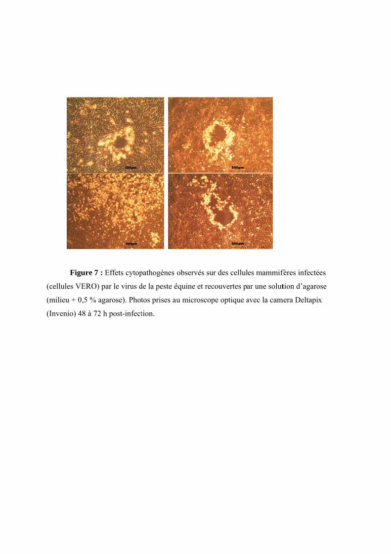
F
(cellules
(milieu
(Invenio
Figure 7 : E
s VERO) pa
+ 0,5 % aga
o) 48 à 72 h
Effets cytop
ar le virus d
arose). Phot
h post-infect
pathogènes
de la peste é
tos prises au
tion.
observés su
équine et rec
u microscop
ur des cellul
couvertes p
pe optique a
es mammif
ar une solut
avec la cam
fères infecté
tion d’agaro
mera Deltapi
ées
ose
x
Chapitre 1 : Introduction – Le virus de la peste équine
20
et les anticorps ne sont pas détectés par les techniques ELISA ou Western Blot avant la mort
de l’animal. Quand les animaux infectés survivent au-delà de 9 jours, quelle que soit la forme
développée de l’infection, une forte réponse sérologique est observée. L’OIE reconnaît
officiellement plusieurs techniques de diagnostic sérologique dont la réaction de fixation au
complément, l’ELISA et la séroneutralisation (OIE, Manuel Terrestre, 2012). La
séroneutralisation est utilisée pour le sérotypage de l’AHSV et est dès lors intéressante dans
les régions endémiques où plusieurs souches virales circulent. Il existe deux kits commerciaux
pour la détection sérologique de l’AHSV. Il s’agit d’un kit ELISA de compétition ou indirect
(Ingenasa, Ingezim) avec une détection spécifique des anticorps envers VP7 dans le sérum des
équidés ou un kit ELISA en double sandwich avec l’anticorps envers VP7 et qui détecte cette
fois-ci les antigènes dans les rates d’animaux infectés (Maree et Paweska, 2005).
Un ELISA indirect a également été développé en utilisant comme antigène la protéine
NS3 du sérotype 4 de l’AHSV. Cet ELISA développé au moment de l’épidémie espagnole-
portugaise, permettait de distinguer les sérums d’animaux infectés, ou vaccinés avec un
vaccin à virus modifié, des sérums d’animaux vaccinés avec des vaccins inactivés.
1.8.3 Isolement en culture cellulaire
L’isolement de l’AHSV est possible sur différentes lignées cellulaires de mammifères
(cellules de reins de singe vert africain ou VERO, cellules de reins de jeune hamster ou BHK-
21, et cellules de singe ou MS) et sur différentes lignées cellulaires d’insectes (cellules
d’Aedes albopictus ou C6/36, cellules de Culicoides variipennis ou K3, et cellules de C.
sonorensis ou W3).
Des effets cytopathogènes (CPE) peuvent être observés sur cellules de mammifères
entre 2 à 8 jours post-infection (Figure 7). En culture de cellules d’insectes, aucun CPE n’est
observable. Ce phénomène a été attribué au fait que la voie de signalisation pour l’induction
de l’apoptose n’est pas déclenchée par l’infection des cellules d’insectes par l’AHSV (Stassen
et al., 2011).

Chapitre 1 : Introduction – Le virus de la peste équine
21
1.9. Vaccination
1.9.1 Vaccins commercialisés
Les premiers vaccins contre l’AHSV étaient des vaccins atténués par plusieurs
passages en cerveau de souris. Ils donnaient une bonne protection mais avaient
occasionnellement de sévères effets secondaires y compris des cas fatals d’encéphalites chez
les chevaux et les ânes, surtout après la primovaccination. Ce problème fut résolu après
passages successifs en culture de cellules (VERO). Les vaccins actuels sont deux vaccins
atténués multivalents dont l’un contient les sérotypes 1, 3 et 4, et dont l’autre contient les
sérotypes 2, 6, 7 et 8 de l’AHSV. Le sérotype 5 n’est pas compris dans le vaccin à cause de
réactions secondaires violentes et le sérotype 9 n’est pas inclus grâce à une forte réaction
croisée avec le sérotype 6. La vaccination se compose d’une primovaccination et d’un rappel
dans la première année, puis de rappels annuels. Il existe également certains vaccins atténués
monovalent, par exemple pour le sérotype 9 en Afrique de l’Est. Les vaccins atténués
conviennent pour la vaccination dans les zones endémiques mais ne sont pas autorisés dans
les zones indemnes. En effet, des cas de retour de virulence des souches atténuées ou de
réassortiment avec la souche sauvage circulant lors d’une épidémie ont déjà été rapportés
(Oura et al., 2012; Aklilu et al., 2014).
Au début des années 1990, un vaccin inactivé monovalent contenant le sérotype 4 a
été commercialisé. Actuellement, il n’est plus disponible sur le marché à cause de coûts de
production élevés et d’une durée d’immunité insuffisante.
1.9.2 Vaccins expérimentaux
La génération de virus recombinant utilisant le virus Vaccinia (MVA) comme vecteur
d’expression et exprimant les gènes de différentes protéines de l’AHSV est prometteur pour l’
obtention de vaccins efficaces. Une première étude insérant les gènes codant VP2, VP7 et
NS3 dans le vecteur Vaccinia a confirmé l’induction d’une réponse immunitaire chez des
poneys inoculés par le virus recombinant. Alors que NS3 n’induisait pas d’anticorps, une
concentration élevée en anticorps neutralisants a été observée lors de l’immunisation de
poneys avec le MVA-VP2 (Chiam et al., 2009). Ces résultats ont été confirmés lors de
l’immunisation de souris IFNAR -/- chez qui une immunisation avec le MVA-VP2 protégeait
Chapitre 1 : Introduction – Le virus de la peste équine
22
contre l’inoculation du virus homologue avec notamment l’absence de détection de virémie
chez les animaux vaccinés (Castillo-Olivares et al., 2011). De la Poza et al. (2013) ont ensuite
comparé l’efficacité de MVA recombinant et ADNc recombinant exprimant soit VP2 ou NS1
ou soit VP2 et NS1 chez des souris IFNAR -/-. Ils ont observé que le MVA recombinant
protégeait plus efficacement contre l’infection par le virus homologue (AHSV-4) que le
vaccin d’ADNc alors que, pour l’infection par le virus hétérologue AHSV-9, la protection
induite par les deux types de vaccins était similaire. D’autre part, l’inclusion de NS1 dans la
composition vaccinale ensemble avec VP2 conférait une protection optimale contre
l’infection par virus homologue et hétérologue. NS1 étant très conservée entre les différents
sérotypes de l’AHSV, son incorporation dans le vaccin est importante pour induire une
protection croisée avec les autres sérotypes. Des chevaux immunisés avec un MVA
recombinant VP2-9 étaient protégés contre l’infection par le virus homologue (AHSV-9), ne
développaient pas de signes cliniques et n’ont également pas produit de virémie (Alberca et
al., 2014). Les sérums de souris immunisés avec un vaccin MVA recombinant VP2
protégeaient les souris à qui le sérum était administré jusqu’à 48 h avant ou après l’infection
par le virus homologue (AHSV-4) (Calvo-Pinilla et al., 2014 ; Calvo-Pinilla et al., 2015). La
production de sérums issus d’animaux immunisés pourrait dès lors être utilisée en cas
d’urgence lors d’une épidémie.
Le canarypoxvirus a également été utilisé comme vecteur vaccinal pour l’AHSV. Des
chevaux immunisés avec un canarypoxvirus recombinant exprimant les deux protéines de
capside externe (VP2 et VP5) ont développé des anticorps neutralisants et ont résisté à
l’infection par le virus homologue (AHSV-4) (Guthrie et al., 2009 ; Garch et al., 2012)..
1.10 Modèle murin d’infection pour le virus de la peste équine
L’établissement d’un modèle d’étude sur souris est crucial pour l’étude de l’infection
d’un virus comme l’AHSV. En effet, outre les problèmes d’éthique qui seraient posés,
l’utilisation de chevaux pour une infection in vivo rencontre des problèmes de logistique
(nécessité d’une animalerie A3 assez spacieuse pour pouvoir détenir des grands animaux,
manipulations très lourdes) et donc un coût de mise en place très important. Peu d’études scientifiques se sont intéressées jusqu’à présent à la mise en place d’un modèle
d’étude de l’AHSV sur souris. Dans l’étude de Wade-Evans et al. (1998), un premier modèle
Chapitre 1 : Introduction – Le virus de la peste équine
23
a été établi en utilisant les souris immunocompétentes Balb/C et utilisé pour l’étude de la
protection conférée par un vaccin sous-unitaire. La seule technique utilisée au cours de cette
étude pour déterminer la virémie sanguine est la méthode de dénombrement des particules
infectieuses permettant de calculer des doses infectieuses en culture de cellules à 50 %
(TCID50) et peu d’informations sont donc disponibles. De même, dans l’étude d’O’Hara et al.
(1998), seule la mortalité liée au sérotype, à la voie d’inoculation et au titre viral est
investiguée pour les souris Balb/C. En se basant sur une étude d’infection par le virus de la
FCO sur un modèle de souris déficiente en récepteur à l’interféron α (Interferon-α Receptor
knock-out, IFNAR -/-), Castillo-Olivares et al. (2011) ont mis en place le modèle d’étude
animal IFNAR -/- en utilisant le sérotype 4 uniquement. Mais comme pour les études
précédentes, la virémie n’est définie que sur base de titrage (en TCID50) à certains moments
de l’expérience. De plus, de ces 3 études, il ressort une description très succinte des signes
cliniques observés chez la souris infectée par l’AHSV. Plus récemment, le modèle souris
IFNAR -/- a été utilisé dans plusieurs études testant l’efficacité de vaccins sous-unitaires
contre l’AHSV-4 ou l’AHSV-9 (De La Poza et al., 2013; Calvo-Pinilla et al., 2014; Calvo-
Pinilla et al., 2015). De plus, la première étude de cette thèse est consacrée à la comparaison
de 3 souches de souris pour l’infection par les AHSV-4 et -9 (De La Grandiere et al., 2014).

Objectifs
Chapitre 2 : Objectifs
25
Les maladies virales transmises par les arthropodes ou arbovirus représentent une
menace pour la santé humaine et animale. Les émergences inattendues et la persistance du
virus de la FCO et du virus Schmallenberg (SBV), en Europe du Nord et centrale (Gould et
Higgs, 2009; Saegerman et al., 2010; Tabachnick, 2010), sont des exemples évidents de la
possibilité que des maladies exotiques peuvent émerger et devenir endémiques dans ces
régions (Weaver et Reisen, 2009). De plus, l’adaptation de ces virus aux vecteurs Culicoides
indigènes ont mis en évidence la nécessité d’étudier les mécanismes de mutation et donc
d’évolution des populations virales qui permettraient d’expliquer leur adaptation à de
nouvelles niches écologiques (Domingo, 2010; Maclachlan et Guthrie, 2010; Backer et
Nodelijk, 2011).
Les virus à ARN se répliquent avec un taux de mutation très élevé et montrent de ce
fait une diversité génétique significative (Worobey et Holmes, 1999). L’évolution rapide de
ces virus à ARN complique la prédiction d’émergence virale de virus comme l’AHSV. Les
deux « bottlenecks » auxquels doivent faire face les virus à transmission vectorielle sont
d’une part la réplication dans un vecteur arthropode et d’autre part la réplication dans un hôte
vertébré, ce qui implique donc une plasticité du virus au niveau du passage dans ces deux
systèmes différents. Chez les Orbivirus, les mutations ponctuelles ont déjà été analysées en
revue (Von Teichman et Smit, 2008; Meiring et al., 2009; Matsuo et al., 2010), alors que le
réassortiment a été peu étudié. Pourtant le réassortiment est un important mécanisme
d’évolution pour les virus à génome segmenté comme cela est déjà observé avec les virus
Influenza A (Zambon, 1999) ou encore avec les Rotavirus (Ghosh et Kobayashi, 2011). Le
réassortiment peut conduire à des conséquences au niveau de l’épidémiologie du virus :
interaction entre les différentes souches virales, les équidés et les vecteurs, et adaptation du
virus à un nouvel environnement. D’autres effets possibles du réassortiment peuvent être une
modification de la virulence de l’AHSV, du profil antigénique ou du tropisme pour l’hôte ou
le vecteur. Les virus réassortants ne peuvent pas être identifiés par les méthodes diagnostiques
conventionnelles qui portent sur le segment 2 codant la protéine VP2 responsable du sérotype.
D’autres approches diagnostiques sont donc nécessaires pour leur mise en évidence sur le
terrain.
Chapitre 2 : Objectifs
26
Cette thèse a été réalisée dans l’objectif d’approfondir les connaissances sur l’AHSV
par la mise au point d’un modèle animal expérimental et de contribuer à l’étude du
phénomène de réassortiment pour l’AHSV.
Dans un premier temps, la mise en place et le développement d’un modèle murin s’est
révélé un outil nécessaire pour l’étude de l’infection par l’AHSV. La comparaison de
l’infection entre trois souches de souris et deux voies d’inoculation différentes a été effectuée
dans la première étude de la thèse. Ces trois souches de souris ont été testées pour étudier la
pathogénie et les signes cliniques de la maladie après infection par les sérotypes 4 et 9 de
l’AHSV. L’influence des voies d’administration (sous-cutanée et intranasale) sur l’issue de la
maladie a été étudiée. D’autre part, trois souches de souris ont été utilisées, dont une était une
souche de souris déficiente en récepteur à l’interféron α/β (souris IFNAR -/-) et l’influence de
la présence ou non de récepteur à l’interféron α/β est discutée en comparant les signes
cliniques présentés par ces souris avec ceux des souris immunocompétentes (souris Balb/C et
129 WT).
Deux volets composent la deuxième étude présentée dans cette thèse. Dans le premier
volet, le phénomène de réassortiment génétique lors de coinfections entre les sérotypes 4 et 9
de l’AHSV a été étudié. Tout d’abord, des outils de discrimination des virus réassortants
potentiels ont été développés. Ensuite des expériences de coinfection in vitro ont été réalisées
pour l’obtention de virus réassortants. Ces derniers ont été caractérisés par PCR et
séquençage. Leurs propriétés de multiplication en culture de cellules ont été comparées aux
virus parentaux. Dans le deuxième volet, l’application concrète du modèle murin développé
dans la première étude a permis d’évaluer la virulence in vivo de virus réassortants obtenus in
vitro et préalablement sélectionnés grâce aux résultats obtenus dans la première partie de cette
étude.
Section Expérimentale
Chapitre 3 : Section expérimentale – Étude 1
28
Section expérimentale
Étude 1
Modèle murin expérimental d’infection par le virus de la peste équine
Chapitre 3 : Section expérimentale – Étude 1
29
Préambule
L’objectif de cette étude est l’analyse in vivo de la virulence de deux différentes
souches de l’AHSV et de comparer les conséquences de l’infection sur trois souches de souris
différentes. Dans ce but, les sérotypes 4 (AHSV-4) et 9 (AHSV-9 de l’AHSV ont été inoculés
par voies sous-cutanée (SC) et en intranasale (IN) à deux souches de souris
immunocompétentes (Balb/C 129 Sv/Ev (129 WT)) ainsi qu’à des souris de souche
IFNAR -/-, déficientes en récepteur à l’interféron α (avec un background génétique de
129 Sv/Ev).
Chez les souris IFNAR -/-, une mortalité s’élevant à 50 % a été relevée et des signes
cliniques significativement plus importants ont été observés en comparaison avec les souris
immunocompétentes inoculées par voie SC. Les signes cliniques observés étaient
significativement plus prononcés après l’infection par l’AHSV-4, surtout chez les souris
immunocompétentes inoculées par voie IN. Le nombre de copies génomiques mesurées dans
les échantillons sanguins était significativement plus élevé après l’infection par l’AHSV-4.
Dans les organes des souris 129 WT inoculées en IN, le nombre de copies génomiques était
significativement plus élevé après infection avec l’AHSV-4. Ensemble et dans les conditions
de cette étude, ces différents résultats démontrent une plus grande virulence de l’AHSV-4 par
rapport à l’AHSV-9 et la présence de signes cliniques plus graves chez les souris inoculées
par la voie IN, du moins pour les 2 sérotypes inoculés. L’étude apporte aussi des évidences
indirectes du rôle de la voie interféron de type I au cours de l’infection par l’AHSV.
Cette étude a permis de mettre au point différents modèles murins pour l’étude de
l’infection par l’AHSV au sein du laboratoire, et d’établir des critères de sélection du modèle
murin à utiliser en fonction des objectifs poursuivis.
Chapitre 3 : Section expérimentale – Étude 1
30
Maria Ana de la Grandière, Fabiana Dal Pozzo, Marylène Tignon, William Zonta,
Damien Thiry, Axel Mauroy, Élisabeth Mathijs, Ann-Brigitte Caij, Claude Saegerman
and Etienne Thiry
Study of the virulence of serotypes 4 and 9 of African horse sickness virus in IFNAR -/-, Balb/C and 129 Sv/Ev mice
Veterinary Microbiology (2014) 174 : 322-332
Chapit
tre 3 : Sectioon expérimementale – Étuude 1
31
Chap
pitre 3 : Section expérimmentale – ÉÉtude 1
32
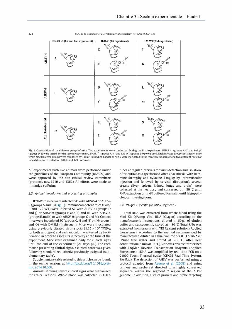
Chap
pitre 3 : Section expérimmentale – ÉÉtude 1
33
Cha
apitre 3 : Seection expérrimentale – Étude 1
34
Cha
apitre 3 : Secction expériimentale – ÉÉtude 1
35
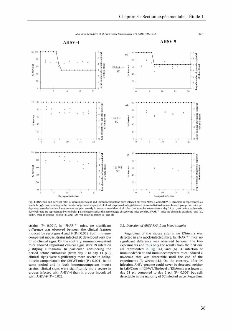
Cha
apitre 3 : Secction expériimentale – ÉÉtude 1
36
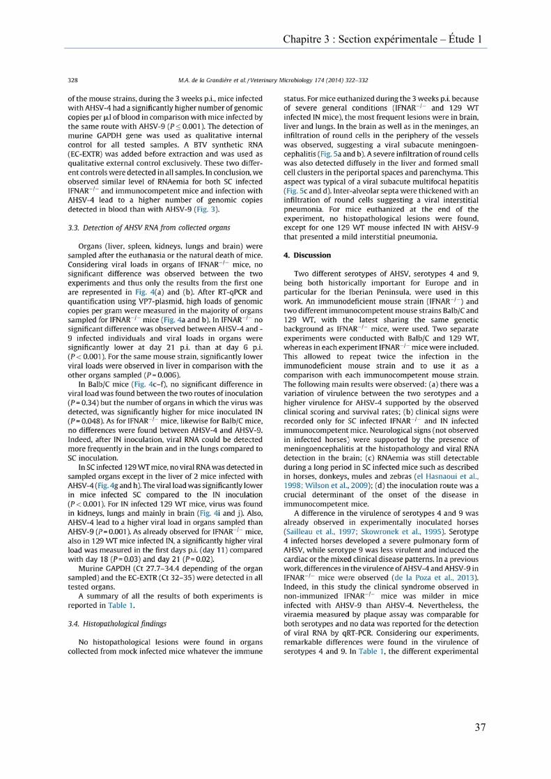
Cha
apitre 3 : Section expérrimentale – Étude 1
37
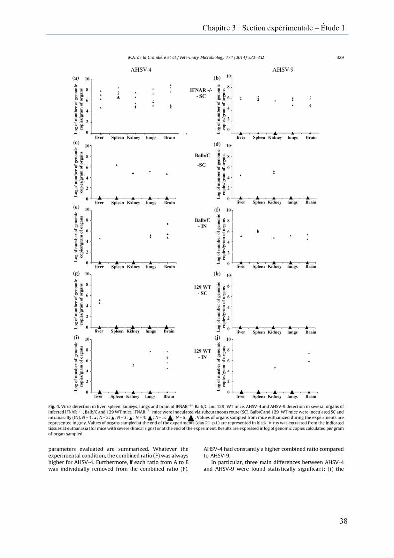
Cha
-SC
apitre 3 : Section expérrimentale – Étude 1
38
Cha
apitre 3 : Seection expérrimentale – Étude 1
39
Cha
apitre 3 : Seection expérrimentale – Étude 1
40

Ch
apitre 3 : Seection expérrimentale –– Étude 1
41
Chapitre 3 : Section expérimentale – Étude 1
42
Supplementary table for the article “Study of the virulence of serotypes 4 and 9 of African horse sickness virus in IFNAR -/-, Balb/C and 129 Sv/Ev mice”, available on http://www.sciencedirect.com/science/article/pii/S0378113514004829
Supplementary table : Evaluation and scoring of the different clinical parameters. Description of the scoring of the different clinical signs observed during the two experiments. The clinical signs were evaluated twice a day during the 3 weeks post-infection.
Scores Parameters 0 1 2 3
Activity Normal Isolated, abnormal posture
Hunched posture, inactive or hyperactive
Moribund or convulsions
Awareness Normal Depression Little response to manipulations
Unconscious, lethargy
Movement/gait Normal Reduction of mobility
Ataxia, paresia of one to three limbs
Circling gait, paresia of 4 limbs, paralysis
Vocalisation Normal Squeaks during palpation
Strongly squeaks and struggles during
palpation
Abnormal vocalization
Consistency of faeces Normal Wet
Flaccid faeces, dirty perineum or
unusually dry faeces +/- mucus
Diarrhea during manipulation or lack of faeces during 48 hours or presence of
blood
Urine Normal / Abnormal colour or volume
Lack or urine during 24 hours or
incontinence with dirty perineum
Water intake Normal Increase or decrease of watering during
24 hours
Increase or decrease of watering during
48 hours
Permanent watering or lack of watering
during 24 hours
Food intake Normal Increase or decrease of feeding during 24
hours
Increase or decrease of feeding during 48
hours
Lack of feeding during 24 hours
Body weight Normal Slow growth curve Chronic weight loss >15 %, or no growth
Acute weight loss > 10 %, chronic weight
loss > 15 % or no growth
Body condition Normal Thin Loss of body fat, stunted growth Muscle wasting
Respiration Normal Rapid, discrete Rapid, abdominal Hard, irregular, dyspnea
Aspect of coat Normal Rough hair coat Injured or thin coat Hair loss,
automutilation, suppuration
Dehydration None Less elastic skin Persistent skin fold Persistent skin fold and enophtalmos
Ocular signs Normal Ocular discharge, conjunctivitis Lacrimation Closed eyelid
Nasal signs Normal Wet Nasal discharge Coagulated Other
Chapitre 3 : Résultats – Section expérimentale – Étude 2
43
Section expérimentale
Etude 2 :
Etude du réassortiment génétique du virus de la peste équine
Chapitre 3 : Résultats – Section expérimentale – Étude 2
44
Préambule
Après avoir mis en place un outil pour l’étude de l’infection par l’AHSV dans la
première étude grâce au développement de modèles murins, le réassortiment génétique est au
cœur de la deuxième étude de cette thèse. En effet, en plus des mutations ponctuelles et de la
recombinaison, le réassortiment génétique est un mécanisme d’évolution supplémentaire chez
les virus à ARN segmenté tels l’AHSV. La circulation de plusieurs souches virales peut
générer des virus réassortants lorsqu’elles infectent la même cellule.
L’objectif de cette étude est de préciser la nature du réassortiment entre les sérotypes 4
et 9 de l’AHSV au niveau des segments échangés ainsi que la virulence des virus réassortants
qui a été étudiée in vitro et également in vivo en utilisant un modèle murin mis au point lors
de l’étude précédente.
Cette étude a été divisée en deux parties : la première partie décrit l’étude du
réassortiment génétique des sérotypes 4 et 9 du virus de la peste équine in vitro. À la suite des
coinfections, des virus réassortants entre les sérotypes 4 et 9 de l’AHSV ont été générés. Six
virus réassortants parmi ceux obtenus ont été caractérisés au niveau génétique par la
détermination de l’origine des différents segments du génome par qRT-PCR et PCR. Au
regard des résultats, l’échange de segments entre les deux sérotypes s’est avéré être multiple
pour les six virus réassortants analysés. Malgré une multiplication virale plus lente du
sérotype 9 par rapport au sérotype 4, un phénomène de dominance du sérotype 9 sur le
sérotype 4 a été observé lors des coinfections en culture de cellules.
Dans la deuxième partie, l’étude de la virulence in vivo des virus réassortants obtenus
in vitro est décrite. Le modèle souris Balb/C mis au point lors de la première étude, a été
utilisé pour évaluer la virulence de 3 virus sélectionnés parmi les virus réassortants obtenus, et
comparer cette virulence aux virus parentaux selon les paramètres suivants : score clinique,
détection du virus dans les échantillons de sang et d’organes. Parmi les trois virus réassortants
testés, un virus réassortant était plus virulent que les virus parentaux, confirmant que le
réassortiment génétique fait partie des mécanismes d’évolution de l’AHSV qui peut conduire
à une modification de sa virulence.
Chapitre 3 : Résultats – Section expérimentale – Étude 2
45
Soumis pour publication
In vitro and in vivo characterization of in vitro generated African horse sickness reassortant viruses
Maria Ana de la Grandière1, Fabiana Dal Pozzo2, Claude Saegerman2 and Etienne Thiry1
(1) Veterinary Virology and Animal Viral Diseases, Fundamental and Applied Research on Animal and Health (FARAH), Faculty of Veterinary Medicine, University of Liège, B-4000 Liège, Belgium.
(2) Research Unit of Epidemiology and Risk Analysis applied to veterinary sciences (UREAR-ULg), Fundamental and Applied Research on Animal and Health (FARAH), Faculty of Veterinary Medicine, University of Liège, B-4000 Liège, Belgium.
Chapitre 3 : Section expérimentale – Étude 2 – Introduction
46
INTRODUCTION
The rapid evolution of RNA viruses makes the prediction of viral emergence of such
viruses as African horse sickness virus (AHSV) difficult, especially when additional
mechanisms like reassortment take place. Arboviruses meet two bottlenecks for their
evolution: in fact, the viral cycle of an arbovirus involves replication in an arthropod vector
and a vertebrate host, necessitating a plasticity of the virus at the transition between the two
different systems. In this context, point mutations have already been investigated but only few
studies exist regarding the genetic reassortment within the Orbivirus genus (Von Teichman et
Smit, 2008; Meiring et al., 2009; Matsuo et al., 2010), in spite of the fact that it is a very
important mechanism of evolution for viruses with a segmented genome and has already been
observed for other segmented viruses (Zambon, 1999; Matthijnssens et al., 2006).
Reassortment can lead to changes in the epidemiology of a virus such as interaction between
different strains, horses and insect vectors and virus adaptation to a new environment. Other
possible effects of reassortment may be a change in the virulence, in the antigenic profile or
the tropism for host or vector of AHSV.
African horse sickness virus is an exotic arbovirus in Europe and, like Bluetongue virus
(BTV), belongs to the Reoviridae family within the Orbivirus genus. The unexpected
emergence of BTV in Northern and Central Europe in 2006 (Thiry et al., 2006; Gould et
Higgs, 2009; Saegerman et al., 2010; Tabachnick, 2010) when the virus replicated in an
indigenous arthropod competent vector (Maclachlan et al., 2007; Wilson et al., 2009;
Maclachlan et Guthrie, 2010; Pfeffer et Dobler, 2010; Martinez-Lopez et al., 2011), is an
obvious example for the possibility for exotic arboviruses to emerge and become endemic in
these areas. AHSV is endemic in sub-Saharan Africa and for some serotypes in West Africa.
In recent decades, AHSV has caused severe epidemics in Europe. AHSV serotype 9 (AHSV-
9) was introduced in 1966 in Spain via infected Culicoides through the Strait of Gibraltar. In
1987, AHSV serotype 4 (AHSV-4) appeared in Spain and in Portugal and remained there for
the next four summers, causing epidemics, thus demonstrating the virus’s ability of survival
for several years outside of South Africa.
The AHSV virion is non-enveloped with a size of about 70 nm. As for BTV, the virion
is composed of seven structural proteins (VP1-7) and four non-structural proteins (NS1-3a),
arranged in a double layer of capsid which contains 10 segments of double-stranded RNA.
VP2 and VP5 proteins form the external part of the virion capsid and VP3 and VP7 proteins

Chapitre 3 : Section expérimentale – Étude 2 – Introduction
47
are the major elements of the inner layer of the capsid. The VP2 protein is the most variable
and, is responsible for the different viral serotypes identified until now. Some cross-reactions
among serotypes are observed, for example between serotypes 1 and 2, 3 and 7, 5 and 8 and 6
and 9, but no cross-reaction with other known Orbiviruses is documented (Roy, 1996).
In this study, coinfections with AHSV-4 and AHSV-9 were performed and led to the
generation of reassortant viruses. The aim of the study was to analyse the nature of
reassortment, with regards to the exchanged segments and to study the virulence of the
obtained reassortant viruses in vivo. Characteristics of these reassortant viruses were studied
and compared to those of the parental strains. Viral growth curves and identification of the
origin of the different segments were described. Three reassortant viruses were selected to
inoculate Balb/C mice and to compare their virulence in vivo to that of the virulence of the
parental strains.
Chapitre 3 : Section expérimentale – Étude 2 – Partie 1
48
Étude in vitro du réassortiment génétique entre les sérotypes 4 et 9 du virus de la peste équine
Étude 2
Partie 1 :
Figure virus; A
1 : CoinfecAHSV-9 : Se
ction assay erotype 9 of
protocol. Af African ho
AHSV -4 : Sorse sicknes
Serotype 4 oss virus
of African h
horse sickneess
Chapitre 3 : Section expérimentale – Étude 2 – Partie 1
49
MATERIALS AND METHODS
Cells and viruses
Vero cells were grown in Dulbecco’s modified Eagle medium (DMEM; Sigma-
Aldrich) supplemented with 10 % heat inactivated foetal calf serum (FCS) (Invitrogen), 2% of
penicillin-streptomycin combination (PS) (Invitrogen) and 1 % of a non-essential amino acids
preparation (NEAA) (Invitrogen). Vero cells were incubated at 37 °C in a humidified
incubator with 5 % CO2. The origin of cell passages for the AHSV serotype 4 (AHSV-4) and
AHSV serotype 9 (AHSV-9) strains used in this study was previously described (Agüero et
al., 2008; de la Grandière et al., 2014). Virus stocks were generated by infection of confluent
Vero cells with these viruses at a multiplicity of infection (MOI) of 0.1. When a total
cytopathic effect (CPE) was visible, mainly at 48 or 72 hours post- infection, supernatants
were harvested and centrifuged. The virus was released from the cells by three freezing
/thawing cycles. Supernatants were stored at -80 °C until use.
Standard virus titration was performed in Vero cells while viral titres were expressed
as TCID50/ml (Reed & Muench, 1938).
Coinfection assays
Vero cells were grown in 12-well plates the day before the coinfection. First, the cells
were coinfected in duplicate wells with AHSV-4 and AHSV-9 at an equal MOI of 1 or 5 for 2
h. In a second experiment, synchronous coinfections were performed in duplicate wells with
AHSV-4 at MOI of 5 or 10 and with AHSV-9 with a MOI of 1 for 2 h (Fig.1). After 48 h
post-infection (p.i.) when CPE was obvious, the supernatants were collected and centrifuged
to remove cellular debris. A volume of 10 µl of the viral mixed population was passaged four
times on Vero cells and each time in the presence of viral CPE, 10 µl of the supernatant from
the previous passage were used to inoculate the cell monolayer. The mixed population of
passage 0 and 4 was inoculated onto Vero cells grown in 6-well plates, using five 10-fold
successive dilutions (10-1 to 10 -5). After 2 h of incubation, the inoculum was removed and
replaced by 2 ml of agar overlay (0.7 %). When an individual well-isolated plaque was
Chapitre 3 : Section expérimentale – Étude 2 – Partie 1
50
visible, it was picked through the agar overlay with a 27g needle (Terumo) and suspended in
500 µl of unmodified DMEM. Each suspension was then inoculated onto Vero cells grown in
12-well plates in order to control the appearance of the characteristic viral CPE. Only from
the suspensions showing CPE, were supernatants harvested and centrifuged before viral RNA
extraction via QIAamp Viral RNA Mini Kit (Qiagen), according to manufacturer’s
instructions.
Genotyping of new progeny virions isolated from coinfection experiments
After heat denaturation (5 minutes at 95 °C), RNA was reverse transcribed with a
Reverse transcriptase Core kit (Eurogentec). The initial characterization of the potential
reassortant viruses was performed using two duplex real-time PCR assays in which VP2 and
VP5 serotype-specific primers and probes were used (Table 3). cDNA was amplified using
Takyon NoRox Probe MasterMix dTTP Blue (Eurogentec) according to the manufacturer’s
instructions, on a C1000 Touch Thermal cycler (CFX96 Real Time System, Bio-Rad). The
reaction mixtures contained 10 µl 2x master mix, 250 nm of each primer and probe, 2 µl of
cDNA sample and water for a final volume of 20 µl. The qPCR program was: 10 min at 95 °C
and 45 cycles of 15 s at 95 °C and 30 s at 60 °C.
A reassortant virus was identified as a virus with seg-2 from parental AHSV-4 and
seg-6 from parental AHSV-9 or a virus with seg-2 from AHSV-9 and seg-6 from AHSV-4.
When potential reassortant viruses were detected, they were allowed to amplify in vitro (4
passages in Vero cells). One of the potential reassortant viruses was not used for the further
genetic characterization as it did not achieve as high viral titres as the six other candidates.
Conventional PCRs were developed to characterize the other segments of reassortant viruses
and to differentiate between AHSV-4 and -9 origin. For the ten segments, serotype-specific
primer sets were designed and are listed in Table 1. Conventional PCRs were performed using
a Taq Polymerase (Taq New Englands, BioLab). The reaction mixtures contained 5 µl 10x
master mix (New Englands BioLab), 5 µl dNTPs (250 µM), 300 nm of each primer, 5 µl
sample cDNA and water to a final volume of 50 µl. The PCR program was: 2 min at 94 °C,
40 cycles of 30 s at 94 °C, 30 s at 56 °C and 45 s at 72°C, and a final elongation of 7 min at
72 °C. Amplicons were loaded onto a conventional gel (2 % of TBE) and migrated for about
45 min at 100 V.
Table 1 : Primers and Probes used for the detection of AHSV-4 and AHSV-9 genome segments
Segment Oligonucleotide position (strain) * Sequence (5’-3’) 5’ fluor
1 (VP1) 1780-1798 (4) CAATCTAACCATTGCGATC 2275-2254 (4) ACCAAAAATACGCTTGACTTCT 1780-1802 (9) TAACTTAACTATCGCGATT 2275-2254 (9) GCCAAAAATCCGTTTTACCTCC
2 (VP2) 1769-1789 (4) † TTAGGACGAAAGTGGGAGGA
1875-1856 (4) † ACATCCTTTCCTGCGCTTAG 1836-1854 (4) † ATGAGCCCCACAGAGTGTTT FAM 1753-1774 (9) † GAGCATTTTGGATCATGAGGA 1843-1822 (9) † TGTCTCTGGTGATGCGTTAAA 1785-1806 (9) † TCGTTGATGATCCTGAGAGGT Texas Red
3 (VP3) 1445-1465 (4) ACGTGTAATCCGTTACTGTGA
1904-1922 (4) CACAGTGTCCGGGACTCTG 1445-1465 (9) GCGCATAATTCGTTACTGTGA 1904-1922 (9) TACGGTGTCCGGAACTCTA
4 (VP4) 377-401 (4) TTATGCCTTCCAACACGTGACTGTC
1181-1158 (4) CTCAATATGTATATCTTCATATAA 377-401 (9) CTATGCTTTCCAACACGTAACCGTT
1181-1158 (9) TTCAATATGTATATTTTCATACAG
5 (NS1) 612-631 (4) TTAAAGGATCTGATCGAAGG 824-844 (4) TCCGTTGCGTCCTCGATCGCG 612-631 (9) CTAAAAGATTTGATTGAAGG 824-844 (9) CCGCAAGCGTCTCCGATCGCA
6 (VP5) 689-709 (4) † AAGCGCGGAAGTAATTGAGA
779-761 (4) † CTGTATTGCGCGGGTTGT 719-739 (4) † AGGAGGTACCAATCTTCGGC FAM 684-704 (9) † GATTTAAGCGCCGAAGTCAT 794-776 (9) † CAACTTCAGGCCCCCTTG 714-734 (9) † CTGAGGAGGTGCCAGTCTTT Texas Red
7 (VP7) 128-149 (4/9) TAATAGGTACAATGGTTTAACG
512-476 (4/9) TGCTCCTGCATAAGGTCCCGTCTGT 105-125 (9) CGCAATCAATAGGTATAATG 444-464 (9) AATTGATGTATCCACCACGC
8 (NS2)
291-321 (4/9) TTTTGAAGCGTTAACACCAGTGCCGATGGCA 663-630 (4/9) ACTCTCATTGCTCTGAACAGAAAGTTTCCTTGGT 291-321 (4) CTTTGAGGCGTTAACACCAGTACCAATGGCT
663-630 (4) GCTTTCATTGCTCTGAACAGAAAGTCTCCTTGGC
9 (VP6)
867-887 (4)
ATTGCGGAATTTGGAGGTC
1078-1058 (4) TCCCCTTCCCCTTCATGAAC 421-441 (9) CTAAGACCGGAGCAGATCGT 623-603 (9) CTTCGCCTCCTCTCTCTCAG
10 (NS3) 31-51 (4)
447-426 (4) 369-388 (9)
ACAATCGCCAAGAATTATAGC TCTTAATATTTCGTCGTCAACG
TGATTTTTATTAGTGGGTGC
481-460 (9) ACTCTTCATCAACCAATCGTTT * Numbers in parentheses refer to AHSV strain (4 indicates AHSV-4, 9 AHSV-9). Genome positions are provided according to GenBank accession numbers for AHSV-4 (JQ796724.1 to JQ796733.1) and for AHSV-9 (KF860036.1 to KF860045.1) ; † These primers are used in qPCR assays.
Chapitre 3 : Section expérimentale – Étude 2 – Partie 1
51
The genome segments of reassortant and parental viruses were sequenced, in order to
confirm the results of the genotyping of the reassortant viruses according to the conventional
and the real time PCRs. Plasmids containing the amplicons obtained after the segment-
specific conventional PCRs, were constructed. Each PCR product was purified using a High
Pure PCR Product Purification Kit (Roche) and then cloned using the pGEM-T Easy Vector
System I (Promega). Plasmids containing the inserted sequence were purified with the High
Pure Plasmid Isolation kit (Roche) and sequenced using T7 and SP6 primers to determine the
orientation of the insert and to verify the integrity and the specificity of the nucleotide
sequence.
Reassortant nomenclature
The nomenclature for reassortant viruses proposed by Meiring et al., 2009 was subsequently
adapted for this study. Thus, for example, R4-92, 10 is a reassortant virus with an AHSV-4
backbone with VP2 and NS3 genomic segments originating from AHSV-9. When equal
number of segments originated from parental viruses, the backbone of the generated AHSV
reassortant was considered as the parental origin of segment 2.
Viral growth curves
For the analysis of the in vitro virus growth, 1x10⁵ Vero cells/well were grown in 12-
well plates in 1 ml of growth medium the day before infection. The cells were then incubated
with 500 µl medium containing the relevant virus in duplicate wells at a MOI of 5 for 2 h at
37 °C (5 % CO2). After 2 h, the inocula were discarded and the cells were washed three times
with Phosphate-buffered Saline (PBS) and then incubated with 1 ml of fresh DMEM
supplemented with 2 % of FCS, 2 % PS and 1 % NEAA. At 0, 4, 8, 24, 48 and 72 h post-
infection, the supernatants were harvested and replaced with 1 ml of PBS. The plates and the
supernatants were then stored at – 80 °C. The collected supernatants were clarified by
centrifugation (5000 rpm for 15 min) and extracellular viral titres were determined by
standard titration and expressed in TCID50/ml. After three freezing /thawing cycles, 1ml of
PBS were collected from the plates, clarified by centrifugation (5000 rpm for 15 min) and
intracellular viral titres were obtained by standard titration and expressed in TCID50/ml.
Chapitre 3 : Section expérimentale – Étude 2 – Partie 1
52
Statistical analyses
All statistical analyses (viral growth curves of reassortant viruses compared to those of
parental viruses for both intracellular and extracellular virus titration of viruses) were
performed using a linear regression with AHSV-4 as the reference group. For reassortant
viruses, the frequency of the origin of the different segments was assessed using a Fisher’s
exact test. For all tests, P-values of < 0.05 were considered to be significant (Dohoo et al.,
2010)
Table 2 : Results of the screening of reassortant African horse sickness viruses by qRT-PCR targeting VP2 and VP5.
Coinfection conditions Passage AHSV-4 AHSV-9 Mixed
population Reassortant
virus
Equal MOI
MOI = 1
0
0 %
82 %
18 %
0 %
4 0 % 100 % 0 % 0 %
MOI = 5 0 10 % 85 % 5 % 0 %
4 0 % 100 % 0 % 0 %
Different MOI
AHSV-4 (MOI = 5) AHSV-9 (MOI = 1)
0
43 %
20 %
27 %
10 %
4 0 % 93 % 7 % 0 %
AHSV-4 (MOI = 10) AHSV-9 (MOI = 1)
0 50 % 0 % 50 % 0 %
4 20 % 32 % 43 % 5 %
MOI : multiplicity of infection; AHSV -4 : Serotype 4 of African horse sickness virus; AHSV-9 :
Serotype 9 of African horse sickness virus
Chapitre 3 : Section expérimentale – Étude 2 – Partie 1
53
RESULTS
Production of AHSV reassortant viruses by coinfection of Vero cells with AHSV-4 and
AHSV-9
Using synchronous coinfection with an equal multiplicity of infection (MOI), the
parental AHSV-9 was predominantly detected (Table 2). No reassortant viruses could be
isolated under these experimental conditions after screening of 40 and 73 plaque-isolated
viruses (with equal MOI of 1 and 5, respectively). Indeed, when an equal MOI of 1 was used,
82 % of plaque-isolated viruses, characterized directly after coinfection (passage 0),
corresponded to the AHSV-9 parental strain, and after four in vitro cell passages, 100% of
plaque-isolated viruses were identified to be AHSV-9 (Table 2). When an equal MOI of 5 was
used, although 10% of plaque-isolated viruses characterized at passage 0 corresponded to the
AHSV-4 parental virus, after 4 passages, 100% of plaque-isolated viruses were AHSV-9
parental strain (Table 2). During synchronous coinfections using higher MOIs for serotype 4
than for serotype 9, a total of seven reassortant viruses was obtained (Table 2). During the
course of the first condition with MOI of 5 for serotype 4 and 1 for serotype 9, 3 reassortants
(from a total number of 119 plaque-isolated viruses) were characterized at passage 0.
However, after four passages of the mixed populations, no reassortant virus could be detected
and serotype 9 was predominantly identified (93% of tested plaque-purified viruses) (Table
1). During the coinfection with MOI of 10 for serotype 4 and 1 for serotype 9, four reassortant
viruses were isolated (from a total number of 82 plaque-isolated viruses) at passage 4 of the
mixed populations (Table 1). Using these experimental conditions, only AHSV-4 could be
identified at passage 0 from plaque-isolated viruses. The AHSV-9 parental strain could only
be detected at passage 0 together with the serotype 4 parental strain in non-isolated plaques,
while in isolated plaques only at passage 4 of the mixed populations.

Table 3 : Origin of the different genome segments of the analysed reassortant African horse sickness viruses
Reassortant virus
Origin of the different genome segments
Seg1 Seg2 Seg3 Seg4 Seg5 Seg6 Seg7 Seg8 Seg9 Seg10
(VP1) (VP2) (VP3) (VP4) (NS1) (VP5) (VP7) (NS2) (VP6) (NS3)
*R9-45, 6, 7, 9 9 9 9 9 4 4 4 9 4 9
*R9-41, 4, 6, 7, 9
(R1) 4 9 9 4 9 4 4 9 4 9
†R4-91, 2, 8, 10
(R2) 9 9 4 4 4 4 4 9 4 9
†R4-92, 3, 8, 10 (R3)
4 9 9 4 4 4 4 9 4 9
†R4-92,10 4 9 4 4 4 4 4 9 4 9
†R4-92,8,10 4 9 4 4 4 4 4 9 4 9
* R9-4 : reassortant virus between African horse sickness virus (AHSV) serotypes 4 and 9 with a
predominant number of segments from AHSV serotype 9
†R4-9 : reassortant virus between serotypes 4 and 9 of AHSV with a predominant number of segments
from AHSV serotype 4
The indices report the segment number originating from the less represented parental virus (Meiring et
al., 2009).
Chapitre 3 : Section expérimentale – Étude 2 – Partie 1
54
Genotyping of new progeny virions isolated from coinfection experiments
Potential reassortant viruses were plaque-purified and amplified four times in Vero
cells. The third passage of six potential reassortant viruses was used for further genetic and
biological characterization.
To investigate the eight other segments of the genome, conventional PCRs were used.
Because of the high level of homology between the serotypes 4 and 9 regarding segment 7
(VP7; 97.3 % nucleotide sequence homology) and segment 8 (NS2; 96.6 % nucleotide
sequence homology), sequencing only allowed determination of their origin (Supplementary
Table). The results showed that for the six reassortant viruses investigated, there were
multiple exchanges of segments between AHSV-4 and -9 (Table 3). The distribution of the
origin of the different segments of the reassortant viruses was not homogeneous (Fisher’s
exact test, p<0.001). The screening by qRT-PCR showed that VP2 always originated from
AHSV-9 and VP5 from AHSV-4. VP6 was always derived from AHSV-4 and NS3 from
AHSV-9. The origin of the other segments was mostly from AHSV-4 for VP1 (66.7 %) and
AHSV-9 for VP3 (66.7 %).
Viral growth curves
Viral growth curves of reassortant viruses were compared to those of parental viruses
for both intracellular and extracellular viruses (Fig. 1). For extracellular virus titrations,
AHSV-9 and R4-91, 2, 8, 10 had significantly lower viral titres (p<0.001) while the five other
reassortant viruses had a similar growth curve to that of AHSV-4. Similar observations were
made for intracellular virus titrations, with R4-91, 2, 8, 10 having significantly lower viral titres
(p=0.01) and AHSV-9 lower viral titres (p=0.05) than AHSV-4. In addition the intracellular
virus titrations of R4-92, 10 and R4-92, 8, 10 showed significantly lower viral titres (p=0.01) than
AHSV-4.

Figure 1 : Viral growth curves of reassortant viruses and parental viruses. Titration of extracellular viruses ((a) to (f)) and intracellular viruses ((g) to (l)) as described in Materials and Methods. AHSV -4 : Serotype 4 of African horse sickness virus; virus AHSV-9 : Serotype 9 of African horse sickness virus; (a) et (g) : R9-45, 6, 7, 9 ; (b) et (h) : R9-41, 4, 6, 7, 9 ; (c) et (i) : R4-91, 2, 8, 10 ; (d) et (j) : R4-92, 3, 8, 10 ; (e) et (k) : R4-92,10 ; (f) et (l) : R4-92,8,10
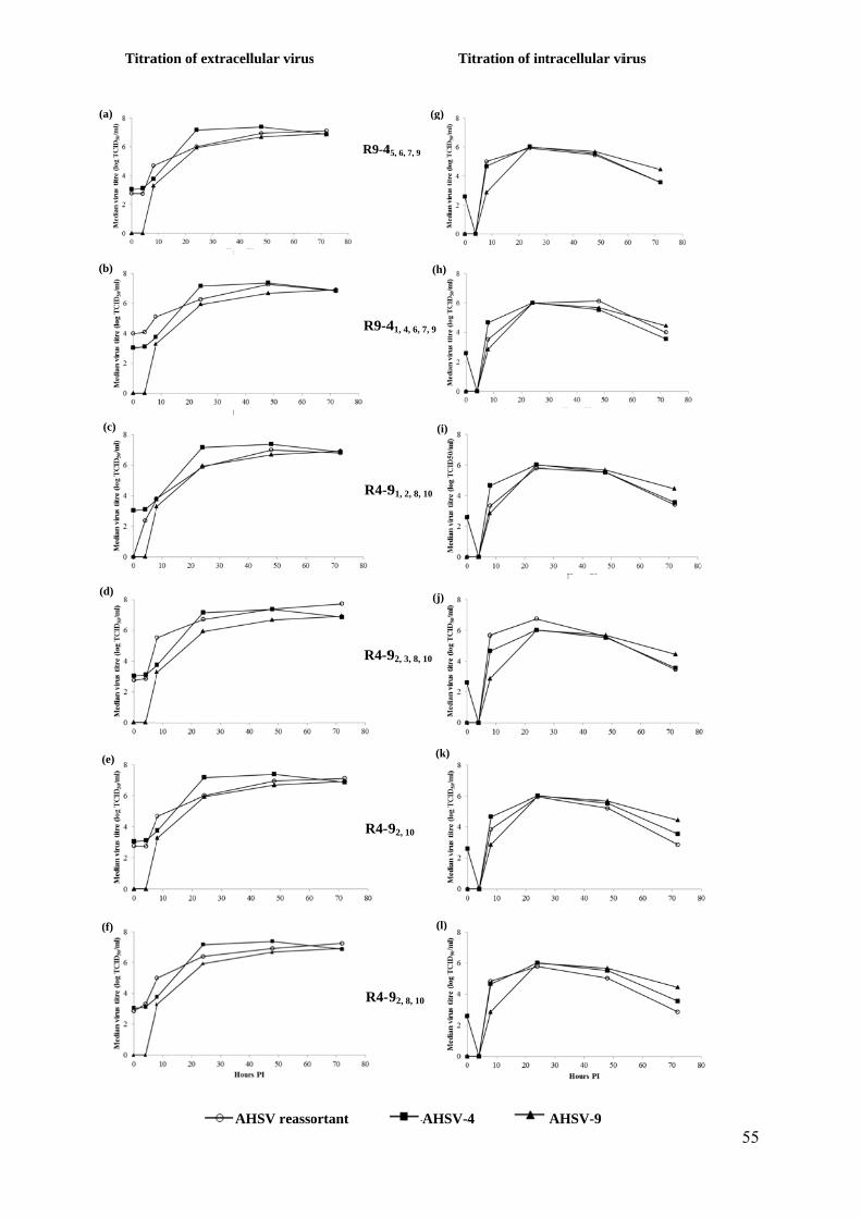
Titra
(a)
(b)
(c)
(d)
(e)
(f)
ation of extra
cellular virus
AHSV reasso
s
ortant
R9-4
R9-4
R4-9
R4-9
R4-9
R4-9
T
(g)
(h)
(i)
(j)
(k)
AHSV-
(l)
45, 6, 7, 9
41, 4, 6, 7, 9
91, 2, 8, 10
92, 3, 8, 10
92, 10
92, 8, 10
Titration of in
-4
tracellular vi
AHSV-9
irus
55
Chapitre 3 : Section expérimentale – Étude 2 – Partie 1
56
DISCUSSION
AHSV reassortants were generated during synchronous in vitro co-infection
experiments with serotype 4 at a higher MOI than serotype 9. The obtained results showed :
(a) multiple exchanges of genomic segments with a non-homogenous distribution of the
segments depending on the parental virus; (b) differences in the in vitro viral growth for
some reassortants compared to the parental viruses.
For this study, we decided to screen for potential reassortants based on the
determination of the origin of segments 2 and 6 of AHSV. Indeed, the majority of BTV1-
BTV8 reassortants have a Seg-2 from BTV-8 and a Seg-6 from BTV-1, making highly
probable to detect reassortant viruses only by targeting these two segments (Shaw et al.,
2013). Secondly, it was also wanted to study the virulence of AHSV reassortants in which
these two segments originated from different parental viruses.
Different MOI were used to perform coinfections. First, equal MOI for both serotypes
have been used as previously described for other serotypes of AHSV (O'hara et al., 1998;
Meiring et al., 2009) or for BTV (Shaw et al., 2013). In these studies, reassortant viruses were
obtained during coinfections with equal MOI. However, in our study, dominance of serotype
9 over serotype 4 was observed during coinfection assays and no reassortant viruses could be
obtained in these conditions. The use of a higher MOI for serotype 4 led to the isolation of
several reassortant viruses. Using a MOI of 5, reassortant viruses could be detected at passage
0 whereas with a MOI of 10, reassortant viruses were obtained at the fourth passage. Using a
MOI of 10, no reassortant virus could be detected at passage 0. It can be suggested that with a
ten-fold higher MOI, serotype 4 was rendered artificially dominant thus preventing the
exchange of segments with serotype 9. During the four consecutive passages, the conditions
enabling reassortment of the genome segments were probably re-established, allowing
detection of some reassortants at the fourth passage. The three reassortant viruses obtained at
passage 0 corresponded to 10% of the characterised progeny viruses. The other three
reassortant viruses obtained at passage 4 corresponded to 5% of the characterized progeny
viruses. This indicates that a two-fold increase of the AHSV-4 MOI (from 5 to 10) did not
lead to a proportional increase in the number of generated reassortant viruses.
Chapitre 3 : Section expérimentale – Étude 2 – Partie 1
57
In other studies, performing in vitro coinfections of AHSV or BTV, no dominance of
serotypes has been described (O'hara et al., 1998; Meiring et al., 2009; Shaw et al., 2013).
However, during in vivo coinfections with BTV-8 and BTV-1, a predominant detection of
BTV-8 was demonstrated in coinfected calves (Dal Pozzo et al., 2013).
Genetic characterization of the six reassortant viruses by qRT-PCR and RT-PCR
showed multiple segment exchanges between the two parental viruses complementing with
previous observations on AHSV (O'hara et al., 1998) and BTV (Shaw et al., 2013) reassortant
viruses. In our conditions of work, the origin of the different segments was almost equally
distributed. A genome constellation per serotype was demonstrated among the six generated
reassortant AHSV. Some segments originated systematically from serotype 4, namely
segments 6 (VP5), 7 (VP7) and 9 (VP6) while on the other hand some segments, such as
segments 2 (VP2) and 10 (NS3), originated exclusively from serotype 9. These results suggest
a conserved segment constellation during the reassortment events under these conditions.
AHSV serotype is characterized both by VP2 and VP5 (Roy, 1996) although the current
diagnostic methods rely only on VP2 for serotyping (Sailleau et al., 1997). In this study, the
segments encoding these two proteins originated from AHSV4 and AHSV9 for segments 6
and 2, respectively (Table 2), complicating the detection and genotyping of the reassortant
viruses when current diagnostic procedures are used.
58
Supplementary Table : Results from genome sequencing of reassortant African horse sickness viruses and parental African horse sickness viruses. Expressed as percentage of homology (calculated by BioEdit Sequence Alignment Editor).
32/62* 90/61† Clone 4 Clone 9 R9-45, 6, 7, 9 R9-41, 4, 6, 7, 9 R4-91, 2, 8, 10 R4-92, 3, 8, 10 R4-92,10 R4-92,8,10
Seg-1 (VP1)
32/62* 90.0 100.0 86.3 87.1 100.0 86.9 100.0 100.0 100.0 90/61† 86.9 99.0 99.4 86.9 99.6 86.9 86.9 86.9 Clone 4 86.3 87.1 100.0 86.9 100.0 100.0 100.0 Clone 9 99.2 86.3 99.4 86.3 86.3 86.3
R9-45, 6, 7, 9 87.1 99.8 87.1 87.1 87.1 R9-41, 4, 6, 7, 9 86.9 100.0 100.0 100.0 R4-91, 2, 8, 10 86.9 86.9 86.9 R4-92, 3, 8, 10 100.0 100.0
R4-92,10 100.0 R4-92,8,10
Seg-2 (VP2)
32/62* 58.7 100 61.3 62.8 58.2 60.4 61.3 61.3 61.3 90/61† 62.1 100.0 98.3 97.8 98.9 100.0 100.0 100.0 Clone 4 61.8 62.8 58.2 60.4 61.8 61.8 61.8 Clone 9 98.3 97.8 98.9 100.0 100.0 100.0
R9-45, 6, 7, 9 96.1 96.5 97.0 97.0 97.0 R9-41, 4, 6, 7, 9 96.7 97.8 97.8 97.8 R4-91, 2, 8, 10 98.9 98.9 98.9 R4-92, 3, 8, 10 100.0 100.0
R4-92,10 100.0 R4-92,8,10
Seg-3 (VP3)
32/62* 95.1 100.0 93.9 93.7 93.3 99.4 93.7 99.2 99.6 90/61† 93.9 100.0 99.4 99.4 93.3 99.8 93.1 93.5 Clone 4 93.9 93.7 93.3 99.4 93.7 99.2 99.6 Clone 9 99.4 99.4 93.3 99.8 93.1 93.5
R9-45, 6, 7, 9 98.7 93.1 99.2 92.9 93.3 R9-41, 4, 6, 7, 9 92.7 93.1 98.5 99.0 R4-91, 2, 8, 10 93.1 98.5 99.0 R4-92, 3, 8, 10 92.9 93.3
R4-92,10 99.2 R4-92,8,10
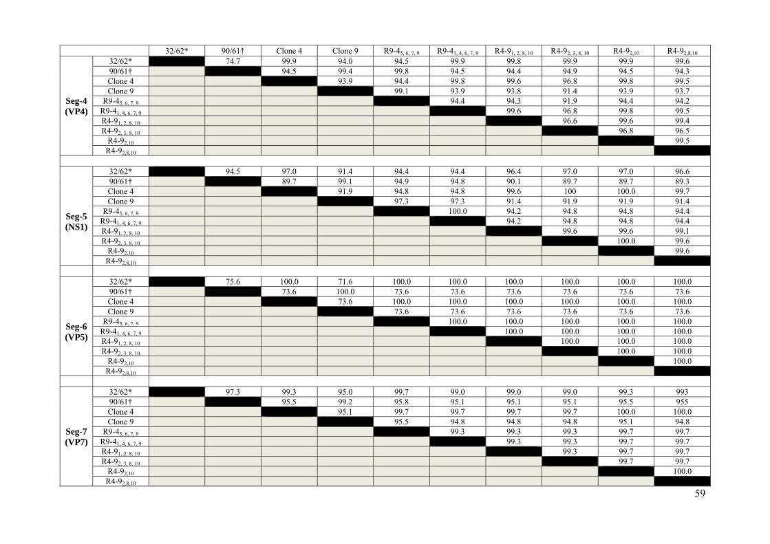
59
32/62* 90/61† Clone 4 Clone 9 R9-45, 6, 7, 9 R9-41, 4, 6, 7, 9 R4-91, 2, 8, 10 R4-92, 3, 8, 10 R4-92,10 R4-92,8,10
Seg-4 (VP4)
32/62* 74.7 99.9 94.0 94.5 99.9 99.8 99.9 99.9 99.6 90/61† 94.5 99.4 99.8 94.5 94.4 94.9 94.5 94.3 Clone 4 93.9 94.4 99.8 99.6 96.8 99.8 99.5 Clone 9 99.1 93.9 93.8 91.4 93.9 93.7
R9-45, 6, 7, 9 94.4 94.3 91.9 94.4 94.2 R9-41, 4, 6, 7, 9 99.6 96.8 99.8 99.5 R4-91, 2, 8, 10 96.6 99.6 99.4 R4-92, 3, 8, 10 96.8 96.5
R4-92,10 99.5 R4-92,8,10
Seg-5 (NS1)
32/62* 94.5 97.0 91.4 94.4 94.4 96.4 97.0 97.0 96.6 90/61† 89.7 99.1 94.9 94.8 90.1 89.7 89.7 89.3 Clone 4 91.9 94.8 94.8 99.6 100 100.0 99.7 Clone 9 97.3 97.3 91.4 91.9 91.9 91.4
R9-45, 6, 7, 9 100.0 94.2 94.8 94.8 94.4 R9-41, 4, 6, 7, 9 94.2 94.8 94.8 94.4 R4-91, 2, 8, 10 99.6 99.6 99.1 R4-92, 3, 8, 10 100.0 99.6
R4-92,10 99.6 R4-92,8,10
Seg-6 (VP5)
32/62* 75.6 100.0 71.6 100.0 100.0 100.0 100.0 100.0 100.0 90/61† 73.6 100.0 73.6 73.6 73.6 73.6 73.6 73.6 Clone 4 73.6 100.0 100.0 100.0 100.0 100.0 100.0 Clone 9 73.6 73.6 73.6 73.6 73.6 73.6
R9-45, 6, 7, 9 100.0 100.0 100.0 100.0 100.0 R9-41, 4, 6, 7, 9 100.0 100.0 100.0 100.0 R4-91, 2, 8, 10 100.0 100.0 100.0 R4-92, 3, 8, 10 100.0 100.0
R4-92,10 100.0 R4-92,8,10
Seg-7 (VP7)
32/62* 97.3 99.3 95.0 99.7 99.0 99.0 99.0 99.3 993 90/61† 95.5 99.2 95.8 95.1 95.1 95.1 95.5 955 Clone 4 95.1 99.7 99.7 99.7 99.7 100.0 100.0 Clone 9 95.5 94.8 94.8 94.8 95.1 94.8
R9-45, 6, 7, 9 99.3 99.3 99.3 99.7 99.7 R9-41, 4, 6, 7, 9 99.3 99.3 99.7 99.7 R4-91, 2, 8, 10 99.3 99.7 99.7 R4-92, 3, 8, 10 99.7 99.7
R4-92,10 100.0 R4-92,8,10
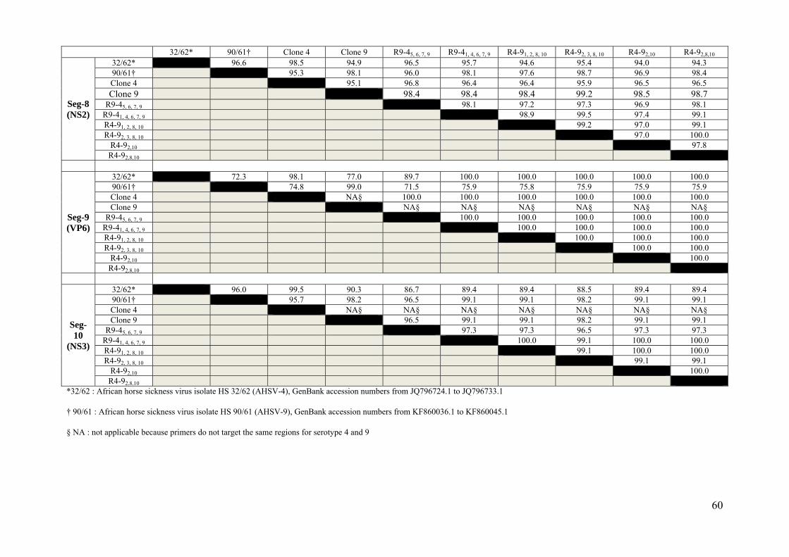
60
32/62* 90/61† Clone 4 Clone 9 R9-45, 6, 7, 9 R9-41, 4, 6, 7, 9 R4-91, 2, 8, 10 R4-92, 3, 8, 10 R4-92,10 R4-92,8,10
Seg-8 (NS2)
32/62* 96.6 98.5 94.9 96.5 95.7 94.6 95.4 94.0 94.3 90/61† 95.3 98.1 96.0 98.1 97.6 98.7 96.9 98.4 Clone 4 95.1 96.8 96.4 96.4 95.9 96.5 96.5 Clone 9 98.4 98.4 98.4 99.2 98.5 98.7
R9-45, 6, 7, 9 98.1 97.2 97.3 96.9 98.1 R9-41, 4, 6, 7, 9 98.9 99.5 97.4 99.1 R4-91, 2, 8, 10 99.2 97.0 99.1 R4-92, 3, 8, 10 97.0 100.0
R4-92,10 97.8 R4-92,8,10
Seg-9 (VP6)
32/62* 72.3 98.1 77.0 89.7 100.0 100.0 100.0 100.0 100.0 90/61† 74.8 99.0 71.5 75.9 75.8 75.9 75.9 75.9 Clone 4 NA§ 100.0 100.0 100.0 100.0 100.0 100.0 Clone 9 NA§ NA§ NA§ NA§ NA§ NA§
R9-45, 6, 7, 9 100.0 100.0 100.0 100.0 100.0 R9-41, 4, 6, 7, 9 100.0 100.0 100.0 100.0 R4-91, 2, 8, 10 100.0 100.0 100.0 R4-92, 3, 8, 10 100.0 100.0
R4-92,10 100.0 R4-92,8,10
Seg-10
(NS3)
32/62* 96.0 99.5 90.3 86.7 89.4 89.4 88.5 89.4 89.4 90/61† 95.7 98.2 96.5 99.1 99.1 98.2 99.1 99.1 Clone 4 NA§ NA§ NA§ NA§ NA§ NA§ NA§ Clone 9 96.5 99.1 99.1 98.2 99.1 99.1
R9-45, 6, 7, 9 97.3 97.3 96.5 97.3 97.3 R9-41, 4, 6, 7, 9 100.0 99.1 100.0 100.0 R4-91, 2, 8, 10 99.1 100.0 100.0 R4-92, 3, 8, 10 99.1 99.1
R4-92,10 100.0 R4-92,8,10
*32/62 : African horse sickness virus isolate HS 32/62 (AHSV-4), GenBank accession numbers from JQ796724.1 to JQ796733.1
† 90/61 : African horse sickness virus isolate HS 90/61 (AHSV-9), GenBank accession numbers from KF860036.1 to KF860045.1
§ NA : not applicable because primers do not target the same regions for serotype 4 and 9
Chapitre 3 : Section expérimentale – Étude 2 – Partie 2
61
Étude de la virulence in vivo des réassortants obtenus in vitro à partir des sérotypes 4 et 9 du
virus de la peste équine
Étude 2
Partie 2 :
Figure virusesAfrican
1 : Schéma. AHSV -4
n horse sickn
atic represe: Serotype 4ness virus; R
entation of 4 of AfricanR1 : R9-41, 4
animal inon horse sick4, 6, 7, 9 ; R2
oculations wknes; virus A: R4-91, 2, 8,
Subcutainocula
with parentAHSV-9 : S10 ; R3 : R4
aneous or iations of Ba
tal or reassSerotype 9 o4-92, 3, 8, 10
intranasal alb/c mice
sortants of
Chapitre 3 : Section expérimentale – Étude 2 – Partie 2
62
MATERIALS AND METHODS
Animal inoculation and processing of the samples
BALB/cByJ (Balb/C) mice were purchased from Charles River France. Two in vivo
experiments were performed simultaneously using six-week-old female mice. In the first
experiment, Balb/C mice were inoculated subcutaneously (SC), while for the second
experiment, Balb/C mice were inoculated intranasally (IN). In both experiments six groups
were created, in which five were infected and one was mock-infected. Each infected group
was composed of six mice, while for ethical reasons only three mice made up the mock-
infected groups. Mice were allowed to acclimatize to the biosafety level 3 animal facilities
(A3) at the Faculty of Veterinary Medicine, Liège, for one week before the begin of the
experiments. All experiments with live animals were performed respecting the guidelines of
the European Community (86/609) and were approved by the site ethical review committee
(protocol n°1633). All efforts were made to minimize animal suffering.
Control mice were inoculated SC (group A) or IN (group G) with unmodified DMEM.
Mice were inoculated SC (250 µl) or IN (50 µl) using previously titrated virus stocks of
AHSV-4 (1,36 x10⁶ TCID50/ml; groups B and H) or AHSV-9 (4.74 x 10⁶ TCID50/ml; groups
C and I) or reassortant virus 1 (R9-41, 4, 6, 7, 9; 8.43 x 10⁶ TCID50/ml; groups D and J) or
reassortant virus 2 (R4-91, 2, 8, 10; 6.32 x10⁶ TCID50/ml; groups E and K) or reassortant virus 3
(R4-92, 3, 8, 10; 1.36 x10⁷ TCID50/ml; groups F and L) (Figure 1). Each inoculum was tested by
back-titration in order to assess its infectivity at the time of the experiment. Mice were
examined daily for clinical signs until the end of the experiment (21 days p.i.). For each
mouse presenting clinical signs, a clinical score was given following previously assigned
standardised criteria (De La Grandiere et al., 2014). Animals showing severe clinical signs
were euthanized for ethical reasons. One mouse from group G and two mice from group H
were euthanized for organ collection, at day 14 p.i. having shown no clinical signs. Whole
blood was collected in EDTA tubes at regular intervals (days 2, 4, 7, 11, 14, 16, 18 and 21 p.i.
for SC infected mice or days 7, 14 and 21 p.i. for IN infected mice) for virus detection. After
euthanasia (performed after anaesthesia with ketamine 50 mg/kg and xylazine 5 mg/kg by
intramuscular injection and followed by cervical disruption), several organs (liver, spleen,
kidney, lungs and brain) were collected at the necropsy and conserved at – 80°C until RNA
extraction.
Chapitre 3 : Section expérimentale – Étude 2 – Partie 2
63
qRT-PCR specific for AHSV segment 7
Total RNA was extracted from whole blood using the Mini Kit QIAamp Viral RNA (Qiagen)
according to the manufacturer’s instructions, diluted in 60 µl of elution buffer and
subsequently stored at – 80 °C. Total RNA was extracted from organs with TRI Reagent
solution (Applied Biosystems), according to the method recommended by manufacturer,
diluted in a final volume of 60 µl of RNAse/DNAse free water and stored at – 80 °C. After
heat denaturation (5 minutes at 95 °C), RNA was reverse transcribed with the Reverse
transcriptase Core kit (Eurogentec). cDNA was amplified by qPCR on a C1000 Touch
Thermal cycler (CFX96 Real Time System, Bio-Rad). The detection of AHSV was performed
using a protocol adapted from Agüero et al., 2008 and previously described (de la Grandière
et al., 2014). In addition, a set of primers and probes targeting murine RNA GAPDH were
included as an internal control (IC) for all samples (Gangisetty and Reddy, 2009). Moreover,
a synthetic external control RNA (EC-EXTR) with a known and defined concentration was
added to the sample prior to extraction, allowing standardisation and monitoring of the
efficiency of the extraction and the RT-qPCR (Vandenbussche et al., 2010). For each sample,
viral VP7 amplification, as well as murine GAPDH and EC-EXTR detection were performed
in duplicate and in separate wells. The results were expressed as mean values of the replicates.
In order to quantify the number of viral genomic copies per µl of blood (RNAemia) or gram
of tissue, a plasmid containing the VP7 target sequence amplified during the RT-qPCR was
constructed (De La Grandiere et al., 2014). These VP7 plasmid dilutions were included in
duplicate at each qPCR to provide a standard curve.
Statistical analyses
All statistical analyses (ratios between clinical score and the survival time for each
mouse, RNAemia and detection of the virus in organs) were performed using a linear
regression with AHSV-4 as the reference group (except for the clinical ratio where the mock-
infected group of mice was the reference group). For all tests, P-values of < 0.05 were
considered to be significant (Dohoo et al., 2010).
Table 2 : Origin of the different genome segments of the analysed reassortant African horse sickness viruses
Reassortant virus
Origin of the different genome segments
Seg1 Seg2 Seg3 Seg4 Seg5 Seg6 Seg7 Seg8 Seg9 Seg10
(VP1) (VP2) (VP3) (VP4) (NS1) (VP5) (VP7) (NS2) (VP6) (NS3)
*R9-45, 6, 7, 9 9 9 9 9 4 4 4 9 4 9
*R9-41, 4, 6, 7, 9
(R1) 4 9 9 4 9 4 4 9 4 9
†R4-91, 2, 8, 10
(R2) 9 9 4 4 4 4 4 9 4 9
†R4-92, 3, 8, 10 (R3)
4 9 9 4 4 4 4 9 4 9
†R4-92,10 4 9 4 4 4 4 4 9 4 9
†R4-92,8,10 4 9 4 4 4 4 4 9 4 9
* R9-4 : reassortant virus between African horse sickness virus (AHSV) serotypes 4 and 9 with a
predominant number of segments from AHSV serotype 9
†R4-9 : reassortant virus between serotypes 4 and 9 of AHSV with a predominant number of segments
from AHSV serotype 4
The indices report the segment number originating from the less represented parental virus (Meiring et
al., 2009).
Chapitre 3 : Section expérimentale – Étude 2 – Partie 2
64
RESULTS
Selection of three reassortant viruses for in vivo studies
The three reassortant viruses to be used for the in vivo studies were selected based on
the results of viral growth curves and the characterization of genome segments. In particular,
the viruses R1, namely R9-41, 4, 6, 7, 9, and R3, namely R4-92, 3, 8, 10 had a similar growth curve
to that of the parental AHSV-4 with an equal origin of the segments between both serotypes
for the first and a major part of their segments originating from AHSV-4 (60 %) for the
second ; the virus R2, namely R4-91, 2, 8, 10, had significantly lower titres for both intracellular
and extracellular virus and its segments were derived mostly from AHSV-4 (60 %) (Table 1).
Clinical signs and mortality
Two experiments were conducted simultaneously in Balb/c mice: the first one
comparing reassortant and parental AHSV strains after subcutaneous (SC) infection, the
second one after intranasal (IN) infection. The back titration of the inocula for both
experiments was 9.28 x 10⁶ TCID50/ml for AHSV-4, 4.31 x 10⁶ TCID50/ml for AHSV-9, 7.78
x 106 TCID50/ml for R1, 5.37 x 10⁶ TCID50/ml for R2 and 4.74 x 10⁷ TCID50/ml for R3.
No mock infected mice showed clinical signs during the three weeks of the
experiment. As previously described (De La Grandiere et al., 2014), no clinical signs were
observed in all groups of SC infected mice (data not shown).
Clinical signs observed in IN infected mice were very similar to those previously observed
(rough hair coat, severe and acute weight loss, weakness and dehydration) (de la Grandière et
al., 2014). One mouse IN infected with R1 already lost between 15 and 20% of its body
weight at day 7 post infection (p.i.). Both the high weight loss and the presence of other
clinical signs led to euthanasia of all mice (Fig. 2). The observation of a euthanasia rate of
100 %, confirmed the higher virulence for R1 in comparison with both other reassortant and
parental viruses. In the group IN infected with R2 (group K), the first clinical signs were
observed from day 11 p.i. onwards and the final mortality rate observed at the end of the
experiment was 33 % (Fig.2). Less clinical signs were reported in the group infected IN with
R3 (group L), with only one mouse showing severe clinical signs necessitating subsequent
euthanasia at day 10 p.i. (Fig.2).
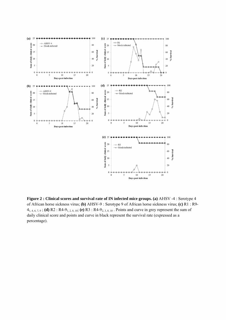
Figure 2of Africa41, 4, 6, 7, 9
daily clinpercenta
(a)
(b)
2 : Clinical san horse sick9 ; (d) R2 : Rnical score a
age).
scores and skness virus; (
R4-91, 2, 8, 10; (eand points an
urvival rate(b) AHSV-9 e) R3 : R4-92
nd curve in bl
(
(d
e of IN infec: Serotype 9
2, 3, 8, 10 . Poinlack represen
(c)
d)
(e)
ted mice gro9 of African hnts and curvent the surviva
oups. (a) AHhorse sicknese in grey repral rate (expre
HSV -4 : Serss virus; (c) resent the sumessed as a
otype 4 R1 : R9-m of
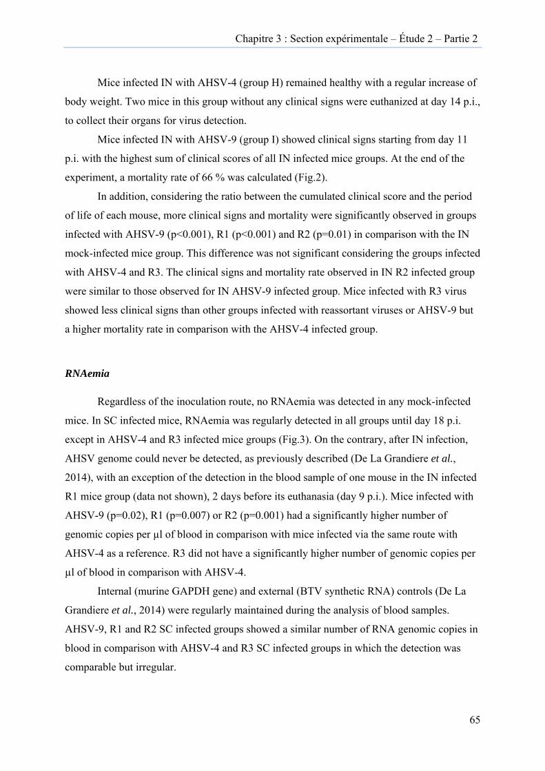
Chapitre 3 : Section expérimentale – Étude 2 – Partie 2
65
Mice infected IN with AHSV-4 (group H) remained healthy with a regular increase of
body weight. Two mice in this group without any clinical signs were euthanized at day 14 p.i.,
to collect their organs for virus detection.
Mice infected IN with AHSV-9 (group I) showed clinical signs starting from day 11
p.i. with the highest sum of clinical scores of all IN infected mice groups. At the end of the
experiment, a mortality rate of 66 % was calculated (Fig.2).
In addition, considering the ratio between the cumulated clinical score and the period
of life of each mouse, more clinical signs and mortality were significantly observed in groups
infected with AHSV-9 (p<0.001), R1 (p<0.001) and R2 (p=0.01) in comparison with the IN
mock-infected mice group. This difference was not significant considering the groups infected
with AHSV-4 and R3. The clinical signs and mortality rate observed in IN R2 infected group
were similar to those observed for IN AHSV-9 infected group. Mice infected with R3 virus
showed less clinical signs than other groups infected with reassortant viruses or AHSV-9 but
a higher mortality rate in comparison with the AHSV-4 infected group.
RNAemia
Regardless of the inoculation route, no RNAemia was detected in any mock-infected
mice. In SC infected mice, RNAemia was regularly detected in all groups until day 18 p.i.
except in AHSV-4 and R3 infected mice groups (Fig.3). On the contrary, after IN infection,
AHSV genome could never be detected, as previously described (De La Grandiere et al.,
2014), with an exception of the detection in the blood sample of one mouse in the IN infected
R1 mice group (data not shown), 2 days before its euthanasia (day 9 p.i.). Mice infected with
AHSV-9 (p=0.02), R1 (p=0.007) or R2 (p=0.001) had a significantly higher number of
genomic copies per µl of blood in comparison with mice infected via the same route with
AHSV-4 as a reference. R3 did not have a significantly higher number of genomic copies per
µl of blood in comparison with AHSV-4.
Internal (murine GAPDH gene) and external (BTV synthetic RNA) controls (De La
Grandiere et al., 2014) were regularly maintained during the analysis of blood samples.
AHSV-9, R1 and R2 SC infected groups showed a similar number of RNA genomic copies in
blood in comparison with AHSV-4 and R3 SC infected groups in which the detection was
comparable but irregular.
Figure
4 of Afr
(c) R1 :
number
rate (exp
(a)
(b)
3 : RNAem
rican horse
: R9-41, 4, 6, 7
r of genomic
pressed in p
AHSV-9
AHSV-4
▲
♦
mia and sur
sickness vir
7, 9 ; (d) R2
c copies/µl
percentage)
▲ RNAemia
Survival
Ch
rvival rate i
rus; (b) AH
: R4-91, 2, 8,
of whole bl
.
hapitre 3 : S
in SC infec
HSV-9 : Sero
, 10 ; (e) R3
lood; points
(c)
(d)
(e)
Section exp
cted mice g
otype 9 of A
: R4-92, 3, 8,
s and curve
R1
R2
R3
érimentale –
roups. (a) A
African hors
10. Points in
in black rep
– Étude 2 –
AHSV-4 : S
se sickness
n grey repre
present the s
– Partie 2
66
Serotype
virus;
esent the
survival
Figure
brain).
Serotyp
(f) ; R3
(a)
(b)
(c)
4 : Detectio
AHSV -4 :
pe 9 of Afric
: R4-92, 3, 8,
AHSV-4
AHSV-4
AHSV-9
on of the vi
Serotype 4
can horse si
, 10 (g). N=1
4-SC
4-IN
9-IN
irus in diffe
4 of African
ckness viru
: ▲ ; N=2 :
erent organ
n horse sickn
us (c); R1 : R
▲ ; N=3 : ▲
(d)
(e)
(f)
(g)
ns (liver, sp
ness virus (a
R9-41, 4, 6, 7,
▲ ; N=4 : ▲
R2
R
R1-IN
R2-SC
)
pleen, kidne
a) and (b); v
9 (d) ; R2 :
; N=5 : ▲
2-IN
R3-IN
N
C
eys, lungs a
virus AHSV
R4-91, 2, 8, 10
; N=6 : ▲
and
V-9 :
0 (e) and
▲
Chapitre 3 : Section expérimentale – Étude 2 – Partie 2
67
Detection of the virus in organs
Organs (liver, spleen, kidneys, lungs and brain) were sampled after euthanasia.
Considering viral loads in organs of SC infected mice, virus was exceptionally detected only
in the AHSV-4 infected group in lungs (Fig.4a), and from the kidneys of one mouse in R2 SC
infected group (Fig.4f).
The one organ, in which viral detection was regular in all IN infected mice, was the
brain. Indeed, viral loads were detectable in all brain samples from mice euthanized during
the experiment and even in AHSV-4 infected mice euthanized without previously having
shown any clinical signs (day 14 p.i.). In correlation with the clinical signs observed, the
highest viral load was found in the brain of R1 IN infected mice (p=0.07) and over all groups,
the brain had a significantly higher viral load than other organs (p<0.001). At day 21 p.i. (end
of the experiment), no viral load was detected from the others organs in all IN infected mice
groups except in one kidney in the R3 IN infected group (Fig.4g). Murine GADPH and the
EC-EXTR were detected in all tested organs.
Chapitre 3 : Section expérimentale – Étude 2 – Partie 2
68
DISCUSSION
AHSV reassortant previously in vitro generated, were tested during this in vivo
experiment. The obtained results showed : (a) differences in the in vivo virulence of some
reassortants when compared to parental viruses, with one reassortant being more virulent than
parental viruses; (b) and a correlation between the severity of clinical signs and the
quantification of viral genome in blood and brain.
Three reassortant viruses were selected in order to study their virulence in vivo in
comparison with the virulence of the parental strains. The SC route was used in order to
measure viraemia and the IN route was used to observe clinical signs after viral infection (De
La Grandiere et al., 2014).
A higher virulence compared to the parental viruses was observed for one reassortant
virus (R1 namely R9-41, 4, 6, 7, 9 IN infected group) demonstrating that reassortment can lead to
modifications in the virulence of AHSV. In a previous study with AHSV-3 and AHSV-8
(O'hara et al., 1998) in Balb/c mice, no higher virulence was found for reassortant viruses
tested in vivo but similar virulence or less virulence was measured in comparison to parental
strains. In horses, the natural host of AHSV, reassortment between vaccine strains for
AHSV1, -3 and -4 has been demonstrated. However, none of the isolated reassortant viruses
induced clinical signs when inoculated to horses (Von Teichman et Smit, 2008), probably
because the vaccine strains were already attenuated. In a study using BTV-1 and BTV-8 in
IFNAR -/- mice, despite a slower viral growth in vitro, reassortant viruses tested in vivo
showed similar virulence in comparison with both parental strains (Shaw et al., 2013). Results
from a study testing in vitro virulence of BTV-1, -6 and 8 showed increased rates of CPE and
a fast replication curve of reassortants in comparison to parental viruses. Additional study is
needed to confirm a suggestion of higher virulence of these reassortant viruses in vivo
(Coetzee et al., 2014).
A qRT-PCR targeting VP7 was used to compare the levels of viremia from reassortant
and parental viruses. RNAemia was correlated with the observation of clinical signs even if
the R1 SC group was not different from the R2 and AHSV-9 SC infected groups. Indeed, R1,
R2 and AHSV-9 SC infected groups had similar levels of genomic copies in blood but higher
numbers in comparison to R3 and AHSV-4 SC infected groups. These data nicely
complement previous studies on AHSV (O'hara et al., 1998) and BTV (Shaw et al., 2013)
where viral genome equivalent quantities were not estimated.
Chapitre 3 : Section expérimentale – Étude 2 – Partie 2
69
The detection of the virus from different sampled organs (liver, spleen, kidneys, lungs
and brain) was performed using the same qRT-PCR as used for the blood and targeting VP7.
As previously described (De La Grandiere et al., 2014), virus was regularly detected in the
brains of IN infected mice. In particular, brains of mice euthanized during the experiment and
of mice showing clinical signs had higher viral loads. Indeed, in organs from mice euthanized
at the end of the experiment (3 weeks post-infection) no viral load could be detected except in
the kidneys of one mouse in the R3 IN infected group and R2 SC infected group, and from
lungs of one mouse in the AHSV-4 SC infected group. Once again, the higher viral load in the
brain was correlated to the higher clinical score found in the R1 IN infected group.
The in vitro characterization of the three in vivo-tested reassortants of AHSV show a
difference in the origin of segment 5 (NS1) for the most virulent, namely R1 (Table 2). No
specific property of virulence is associated to Seg-5 in the literature, thus the hypothesis of the
combination of all the segments together could more likely explain the higher virulence
exhibited by R1. Furthermore, the occurrence of point mutations during the successive
passages in VERO cells having led to a modification of virulence could be an additional
explanation.

Discussion
Chapitre 4 : Discussion
71
Le virus de la peste équine (AHSV) est un virus à transmission vectorielle à potentiel
d’émergence en Europe. Ce virus endémique en Afrique sub-saharienne trouve son principal
réservoir chez le zèbre mais il est pathogène pour tous les autres équidés. Le zèbre ne présente
pas ou peu de signes cliniques mais l’AHSV peut provoquer jusqu’à plus de 95 % de létalité
chez le cheval infecté avec des conséquences économiques très importantes pour la filière
équine. La circulation de plusieurs souches virales dans les pays endémiques a mené à
l’isolement de réassortants et notamment entre souches vaccinale et sauvage. Peu d’études se
sont intéressées jusqu’à présent au phénomène de réassortiment génétique de l’AHSV alors
que celui-ci peut constituer un mécanisme d’évolution important pour les virus à ARN
segmenté comme l’AHSV.
La première étude a abouti au développement d’un modèle murin d’infection par
l’AHSV. Trois souches de souris et deux voies d’inoculation ont été testées : des souris
déficientes en récepteur à l’interféron α/β ou IFNAR -/-, des souris Balb/C et des souris 129
WT, ces dernières partageant le même background génétique avec les souris IFNAR -/- , ainsi
que les voies sous-cutanée (SC) et intranasale (IN) avec des propriétés différentes en ce qui
concerne la distribution du virus dans l’organisme. Cette première étude a donc permis
l’établissement d’un outil repris et utilisé dans la deuxième étude.
La deuxième étude s’est intéressée au réassortiment génétique de l’AHSV. Dans une
première partie, l’obtention et la caractérisation de virus réassortants in vitro ont été étudiées.
Des virus réassortants ont été générés suite à la coinfection par les sérotypes 4 et 9 de
l’AHSV. Une dominance du sérotype 9 par rapport au sérotype 4 a été observée au cours de la
coinfection. Après la caractérisation des virus réassortants obtenus in vitro, trois virus
réassortants ont été sélectionnés pour en étudier la virulence in vivo en utilisant un des
modèles murins mis en place dans la première étude. Dans cette deuxième partie, la virulence
des virus réassortants a été comparée à celle des virus parentaux par la mesure de plusieurs
paramètres expérimentaux (scores cliniques et mortalité, ARNémie et détection du virus dans
les organes). Les différents résultats de ces deux études seront discutés.
Modèle murin expérimental d’infection par le virus de la peste équine
Deux sérotypes différents de virus de la peste équine, les sérotypes 4 et 9, tous les
deux historiquement importants pour l'Europe et en particulier pour la péninsule ibérique, ont
Chapitre 4 : Discussion
72
été utilisés dans ce travail. Une souche de souris déficiente en récepteur à l’interféron α
(IFNAR -/-) et deux souches différentes de souris immunocompétentes les souris Balb/C et les
souris 129 WT, cette dernière partageant le même fond génétique que les souris IFNAR -/-,
ont été utilisées.
Deux expériences distinctes ont été réalisées avec des souris Balb/C et 129 WT, alors
que dans chaque expérience, des souris IFNAR -/- ont été utilisées. L’utilisation des souris
IFNAR -/- dans les deux expériences a permis de répéter deux fois l'infection dans cette
souche de souris et de la comparer avec chaque souche de souris immunocompétentes. Les
principaux résultats observés sont : (a) une variation de la virulence entre les deux sérotypes
et une virulence plus importante du sérotype 4 de l’AHSV ont été démontrées par un score
clinique plus élevé chez les souris inoculées par AHSV-4 et également un taux de survie plus
faible; (b) des signes cliniques ont été observés uniquement chez les souris IFNAR -/-
infectées par voie SC et chez les souris immunocompétentes infectées par voie IN. Les signes
neurologiques (non observés chez les chevaux infectés) ont été confirmés par la présence
d’une méningo-encéphalite à l'histopathologie et la détection d'ARN viral dans le cerveau; (c)
l’ARNémie était détectable pendant une longue période dans le sang des souris infectées en
SC tel que décrit pour les chevaux, les ânes, les mulets et les zèbres (El Hasnaoui et al., 1998;
Wilson et al., 2009); (d) la voie d'inoculation est un déterminant crucial de l'apparition de la
maladie chez les souris immunocompétentes.
Une différence de la virulence des sérotypes 4 et 9 a déjà été observée chez les
chevaux inoculés expérimentalement (Skowronek et al., 1995; Sailleau et al., 1997). En effet,
dans ces études, les chevaux infectés par le sérotype 4 ont développé une forme pulmonaire
sévère de la peste équine, tandis que le sérotype 9 a été moins virulent et a induit la forme
cardiaque ou la forme mixte de la maladie. Dans une étude utilisant des souris IFNAR -/-, des
différences dans la virulence entre AHSV-4 et -9 ont également été observées (De La Poza et
al., 2013). En effet, dans cette étude, les signes cliniques observés chez les souris IFNAR -/-
non vaccinées était plus modérés que chez des souris infectées par l’AHSV-9 par rapport à
celles infectées par AHSV-4. Néanmoins, la virémie mesurée par la méthode de plages de
lyse était comparable pour les deux sérotypes sans données sur la détection de l'ARN viral par
qRT-PCR.
Chapitre 4 : Discussion
73
Dans la première étude de cette thèse, des différences significatives ont été observées au
niveau de la virulence des sérotypes 4 et 9. Tout d’abord, au niveau de l’ARNémie exprimée
en log du nombre de copies génomiques, les valeurs sont significativement plus élevées pour
le sérotype 4 que pour le sérotype 9, aussi bien lors de l’infection de souris IFNAR -/- que de
souris immunocompétentes. Ensuite, les scores cliniques relevés entre J0 et J11 post-infection
chez les deux souches de souris immunocompétentes étaient plus élevés pour le sérotype 4.
Enfin, la charge virale mesurée dans les organes des souris 129 WT inoculées par voie IN,
était significativement plus élevée pour le sérotype 4. La peste équine est caractérisée chez les
chevaux par quatre formes cliniques de la maladie: la maladie de la forme fébrile, la forme
cardiaque (subaiguë), la forme pulmonaire (suraiguë) et la forme mixte (la plus fréquemment
observée). Les signes cliniques présents chez la souris infectée ne font partie ni de la forme
cardiaque ni de la forme pulmonaire ou mixte, indiquant les limites du modèle murin comme
modèle d’étude de la pathogénie de la peste équine. Même si, lors de chaque expérience, une
souris IFNAR -/- a été retrouvée morte sans présenter de signes cliniques préalables comme
cela peut être observé chez le cheval souffrant de la forme pulmonaire de la maladie. De
même les signes nerveux développés par les souris IFNAR-/- ne sont pas présents chez le
cheval infecté. Les souches virales d’inoculation ont pu acquérir un neurotropisme au cours
de leurs passages successifs en cerveau de souriceaux, ce qui permet d’expliquer ces signes
nerveux.
Chez les souris développant des signes cliniques, une chronologie dans l’apparition de
ceux-ci a pu être mise en évidence : d’abord les souris présentent une conjonctivite et/ou un
pelage rugueux, ensuite une perte de poids pouvant aller jusqu’à 15 ou 20 %, de l’apathie et
enfin des signes neurologiques observés uniquement chez les souris IFNAR -/-. Ceux-ci
étaient une ataxie, une paralysie d’un ou plusieurs membres ainsi qu’une démarche en cercle.
Ils ne sont pas observés chez les chevaux. Ils pourraient être expliqués par une adaptation des
virus utilisés dans ces deux expériences, à travers des passages successifs sur cerveaux de
souriceaux nouveau-nés, méthode utilisée pour l’isolement viral (O'hara et al., 1998).
L’inoculation IN est la voie qui conduit à l’apparition de mortalité chez les souris Balb/C.
Cependant, les résultats de l’étude de O’Hara et al. (1998) sont difficiles à comparer avec les
résultats obtenus dans cette thèse, à cause de l’origine différente des souches virales utilisées.
Avec les souris immunocompétentes, l'apparition de signes cliniques a été observée seulement
après infection IN. Deux hypothèses pourraient expliquer cette observation : d'abord le
Chapitre 4 : Discussion
74
neurotropisme acquis par les souches virales après leurs passages en cerveau de souriceaux
(O'hara et al., 1998; Lubroth et al., 2007) et ensuite la neuro-invasion par voie rétrograde au
départ du nerf olfactif. Pour le virus de la fièvre du Nil occidental (WNV), ainsi que pour
d'autres arbovirus neurotropes, une controverse repose sur la possibilité de neuroinvasion
rétrograde du cerveau (Monath et al., 1983). De cette façon, l'infection de neurones olfactifs a
été proposée comme un mode d'entrée pour les souches du WNV neurotropes et très
virulentes dans le système nerveux central (Murray et al., 2010). De toute évidence, le fait que
les neurones olfactifs ne soient pas protégés par la barrière hémato-encéphalique pourrait
expliquer un accès plus facile du virus au cerveau. De ce fait, le choix de la voie d'inoculation
a une importance capitale de même que la souche de souris utilisée (immunocompétente ou
IFNAR -/-) et le potentiel neuroinvasif du virus inoculé. Après l'infection, l'ARN viral peut
être détecté dans certains organes (foie, les reins, la rate, etc.) malgré l'absence d’ARNémie.
L'hypothèse la plus probable est l'absence de détection d'une ARNémie transitoire en raison
de limitations du point de vue éthique dans la fréquence de la collecte de sang chez la même
souris.
Après l'infection par voie SC, seules les souris IFNAR -/- présentaient des signes cliniques (à
partir de J2 post-infection), alors que les deux souches de souris immunocompétentes ont
présenté une infection subclinique en dépit d'une ARNémie constante et élevée. La
comparaison entre les souris IFNAR -/- et les souris immunocompétentes infectées en SC
permet de postuler le rôle de l’IFN de type I au moment de l’infection par l’AHSV. Après
l'infection en SC, les niveaux d’ARNémie élevés sont similaires alors que les souris
immunocompétentes n'ont pas développé de signes cliniques comme le souris IFNAR -/- dès
le J2 post-infection. Aucune donnée sur le rôle des voies IFN de type I dans l'infection par
l’AHSV n’est disponible jusqu'à présent, mais il existe des données pour un autre Orbivirus, à
savoir le BTV (Ruscanu et al., 2012; Chauveau et al., 2013) et d'autres virus appartenant à la
famille des Reoviridae (Johansson et al., 2007; Deal et al., 2013). L'infection virale induit une
forte production d'IFN de type I. Chez les moutons infectés par le BTV, le pic de la
production d'IFN a été corrélé avec le pic de la virémie mesurée et ensuite associée à une forte
diminution de titres de virus (Foster et al., 1991). Plus récemment, il a été démontré que la
protéine non structurale NS3 du BTV interfère avec la production d'IFN de type I (Chauveau
et al., 2013). Les souris IFNAR - / - ont une plus grande sensibilité aux infections virales
(Bereczky et al., 2010; Wernike et al., 2012) qui est liée à l'absence de récepteurs à l'IFN α/β
et, par conséquent, elles présentent une déficience de l’immunité innée. Les résultats des
Chapitre 4 : Discussion
75
études avec le BTV et ceux obtenus à partir l’étude de cette thèse supportent l'hypothèse du
rôle de l'IFN de type I dans le contrôle de l'infection virale, et en particulier dans la réduction
de la sévérité de l'expression clinique de la maladie. Une expression plus sévère de la maladie
chez les souris IFNAR - / - a été observée, tandis que 50% de ces souris ont pu survivre à
l'infection, malgré l'absence de l'activité antivirale innée liée aux IFN de type I. Cela suggère
l’implication d'autres mécanismes immunitaires dans le contrôle de l'infection.
En conclusion de la première étude de cette thèse, les résultats fournissent de nombreuses
données sur la virulence des sérotypes 4 et 9 de l’AHSV chez deux souches de souris
immunocompétentes et une souche de souris IFNAR -/-. Cette première étude a été initiée
dans le but de développer un outil pour l’étude du réassortiment génétique de l’AHSV sur
base de certains éléments déjà rapportés dans la littérature. Les souris de souche 129 WT
n’avaient pas encore été testées pour l’étude de l’infection par l’AHSV, des données de
quantification du virus par qPCR dans le sang et les organes n’étaient pas encore disponibles
et la comparaison de l’infection chez trois souches de souris différentes n’avait pas encore été
réalisée en parallèle.
Le sérotype 4 peut être considéré comme plus virulent que le sérotype 9, au moins
pour les souches virales utilisées dans cette étude. Les différences observées dans la virulence
ont été obtenues par l'utilisation de deux sérotypes qui ont subi les mêmes passages
historiques de laboratoire. Cela confirme l'hypothèse d'une diversité génétique entre les deux
sérotypes, et donc de marqueurs génétiques de virulence qui devraient être étudiés.
L’utilisation de ces modèles murins pour l’étude de la pathogénie de la peste équine est
limitée par l’absence de lésions endothéliales lors des analyses histopathologiques ainsi que
par la présence de signes cliniques de type nerveux chez les souris IFNAR -/- et absents chez
l’hôte naturel. Le tropisme nerveux acquis par ces souches lors de leur passage en cerveau de
souriceaux est une hypothèse pouvant expliquer ces signes neurologiques. D’autre part, la
présence de virus en quantité importante dans le cerveau des souris inoculés par voie IN
suggère l’invasion du virus par voie rétrograde via le nerf olfactif. Les seuls accidents
humains de laboratoire dus à l’AHSV ont d’ailleurs été provoqués par l’inhalation de souches
neurotropes lors de leur manipulation en vue de la production de vaccins et avaient provoqué
des encéphalites et des choriorétinites chez les travailleurs (Taylor et al., 1992).
L'infection de souris Balb/C et de souris 129 WT pourrait être utilisée comme un
modèle valable dans l'identification de marqueurs génétiques de neurotropisme de ces souches
Chapitre 4 : Discussion
76
de l’AHSV ainsi que pour d'autres arbovirus neurotropes grâce à la voie d’inoculation IN. Les
souris IFNAR -/- sont un excellent modèle pour les études sur l'efficacité du vaccin en raison
de leur sensibilité clinique élevée à l’infection par ce virus. Néanmoins, des souches de souris
immunocompétentes ayant une réponse immunitaire antivirale innée conservée doivent
également être prises en considération pour les études sur l'efficacité du vaccin et aussi pour
d'autres fins, telles que l'étude de l'évolution des populations virales de l’AHSV. En effet, les
modèles de souris Balb/C et 129 WT comme les souris IFNAR -/- permettent une
multiplication efficace du virus. Ce sont donc des modèles intéressants pour la génération de
virus réassortants in vivo et peuvent compléter l’analyse du réassortiment in vitro.
Étude in vitro du réassortiment génétique entre les sérotypes 4 et 9 de l’AHSV
Des virus réassortants ont été obtenus au cours de coinfections in vitro synchrones
utilisant un MOI supérieur pour le sérotype 4 par rapport au sérotype 9. De multiples
échanges des segments entre les deux sérotypes ont été observés chez chaque virus réassortant
isolé. Des différences dans les courbes de multiplication virale de certains virus réassortants
ont pu être mises en évidence lorsqu’elles étaient comparées aux courbes de réplication des
virus parentaux.
La décision de se baser sur les segments 2 (VP2) et 6 (VP5) pour développer une qRT-PCR
de criblage des surnageants de culture cellulaire issus des coinfections a été prise pour
plusieurs raisons. D’une part, ces segments codent les deux protéines de capside externe de
l’AHSV et ont un rôle dans l’attachement du virus sur les cellules et son internalisation.
Obtenir des virus réassortants, dont ces deux segments en particulier étaient originaires, d’un
virus parental différent était intéressant pour en étudier la virulence. D’autre part, une étude
récente sur un virus très analogue à l’AHSV, à savoir le virus de la FCO, a démontré que, lors
d’expériences de coinfections entre les sérotypes 1 et 8 du virus de la FCO, la majorité des
virus réassortants isolés avait des segments 2 et 6 provenant chacun d’un virus parental
(Shaw et al., 2013). Ces résultats rendent donc très probable l’identification des virus
réassortants grâce à une qRT-PCR ciblant ces deux segments. À l’initiation de ce travail, les
séquences du génome complet de l’AHSV-4 et -9 n’étaient pas disponibles sur GenBank,
rendant difficile, le développement de qPCR de criblage sur les 10 segments. D’autre part, le
criblage de virus infectieux issus d’une centaine de plages de lyse pour chaque condition
expérimentale, nécessitait le développement d’un outil de discrimination rapide.
Chapitre 4 : Discussion
77
Cependant, cette décision a priori peut restreindre la détection d’autres virus réassortants,
sachant que 1024 combinaisons sont théoriquement possibles lors du réassortiment aléatoire.
Dès lors, les nouvelles méthodes disponibles actuellement comme le séquençage à haut débit
(Next-Generation Sequencing ou NGS) sont à envisager pour des études futures.
Différents MOI ont été utilisés pour réaliser les coinfections in vitro. Tout d’abord, des
MOI identiques pour les deux sérotypes ont été testés comme cela avait été réalisé lors
d’études utilisant d’autres sérotypes d’AHSV (O'hara et al., 1998; Meiring et al., 2009) ou le
virus de la FCO (Shaw et al., 2013). Dans ces différentes études, des virus réassortants ont pu
être générés dans les conditions expérimentales de coinfection in vitro synchrones utilisant un
MOI identique pour les deux virus inoculés. Cependant, dans l’étude de cette thèse, aucun
virus réassortant n’a été obtenu en utilisant des MOI identiques car une dominance du
sérotype 9 sur le sérotype 4 était présente dans ces conditions. En augmentant le MOI du
sérotype 4 par rapport au sérotype 9, plusieurs virus réassortants ont pu être générés. Lorsque
le sérotype 4 était inoculé avec un MOI de 5, des virus réassortants ont pu être identifiés au
cours de l’isolement viral réalisé tout de suite après la coinfection. Par contre, avec un MOI
de 10 pour le sérotype 4, des virus réassortants ont pu être mis en évidence seulement au
cours de l’isolement réalisé après 4 passages du surnageant de la culture cellulaire issu de la
coinfection. Ces résultats suggèrent que le sérotype 4 avec un MOI 10 fois supérieur au MOI
du sérotype 9, était rendu artificiellement dominant et n’a donc pas permis l’échange de
segments avec le sérotype 9. Les 4 passages successifs ont probablement permis au sérotype 9
de récupérer progressivement le titre viral plus élevé, aboutissant alors à l’échange de
segments entre les deux sérotypes et la génération de virus réassortants. Les trois réassortants
identifiés au passage 0 avec le MOI de 5 correspondent à 10 % des surnageants de culture
cellulaire issus des coinfections tandis que les 4 virus réassortants isolés au passage 4 avec le
MOI de 10 correspondent à 5 % du total des criblages par qRT-PCR. Cette observation
indique que la génération de virus réassortant n’augmente pas de façon proportionnelle avec
le MOI utilisé. La dominance du sérotype 9 et l’utilisation de MOI différents apportent un
biais important à connaître puisqu’il peut jouer dans l’obtention de virus réassortants dans les
conditions naturelles.
Dans d’autres études sur le réassortiment de l’AHSV (O'hara et al., 1998; Meiring et
al., 2009) ou le virus de la FCO (Shaw et al., 2013), aucun phénomène de dominance d’un
sérotype par rapport à un autre n’est décrit pendant les expériences de coinfection in vitro.
Chapitre 4 : Discussion
78
Cependant, dans une étude de coinfection in vivo utilisant les sérotypes 1 et 8 du virus de la
FCO, une détection prédominante du sérotype 8 a été mise en évidence dans des échantillons
provenant de veaux coinfectés (Dal Pozzo et al., 2013). La caractérisation des six virus
réassortants analysés par qRT-PCR et PCRs conventionnelles a montré que l’échange de
segments entre les deux sérotypes était de nature multiple et hétérogène dans la répartition de
leurs origines. L’échange multiple de segments a également été mis en évidence pour l’AHSV
(O'hara et al., 1998) et le virus de la FCO (Shaw et al., 2013). Certains segments étaient
systématiquement issus du sérotype 4, à savoir les segments 6 (VP5), 7 (VP7) et 9 (VP6)
tandis que d’autres provenaient toujours du sérotype 9, les segments 2 (VP2), 8 (NS2) et 10
(NS3). Le segment 2 provenait systématiquement du sérotype 9 et conférait donc ce sérotype
au virus réassortant, cependant, l’exploration des autres segments montrent que pour la
majorité des virus réassortants analysés, le « backbone » était de sérotype 4 (66 %).
Ces résultats suggèrent l’existence de constellations conservées dans le génome selon
les sérotypes pendant les événements de réassortiment. Une étude récente sur le réassortiment
génétique pour le BTV, a mis en évidence des associations de segments durant les échanges
entre les différents virus en conditions naturelles (Nomikou et al., 2015). Ce résultat contraste
avec les résultats obtenus par Shaw et al., 2013, qui ont réussi à démontrer par génétique
inverse que le réassortiment pouvait impliquer tous les segments sans association spécifique
de ceux-ci pendant les échanges. Ces résultats suggèrent l’existence de contraintes réelles
dans l’assemblage de segments génomiques dans le cadre d’obtention de virus réassortants
lors de coinfection. Ces observations peuvent être étendues aux rotavirus humains où
l’existence de constellations de segments conservés a été mise en évidence. Ceci est expliqué
par une évolution virale avec une adaptation à l’espèce, en l’occurrence l’homme (Patton,
2012). Pour l’AHSV, d’autres études sont nécessaires pour dégager le rôle des différents
segments à partir de ces constellations génomiques et déterminer si l’avantage sélectif ou non
conféré par cette association de segments est lié à un effet individuel d’une protéine ou à un
effet coopératif de différentes protéines entre elles.
Étude de la virulence in vivo des virus réassortants issus des coinfections in vitro
Après avoir généré et caractérisé des virus réassortants in vitro, trois virus réassortants
ont été sélectionnés pour être inoculés à des souris Balb/C. Les résultats principaux sont :
Chapitre 4 : Discussion
79
l’observation de différences au niveau de la virulence in vivo des virus réassortants avec les
virus parentaux, avec un virus réassortant plus virulent, et la mise en évidence d’une
corrélation entre la sévérité des signes cliniques relevés et les résultats de détection du virus
dans les échantillons de sang et d’organes.
Suite aux résultats de la première étude de cette thèse (De La Grandiere et al., 2014),
les voies SC et IN ont été utilisées pour inoculer les souris avec les 3 virus réassortants
générés et sélectionnés in vitro. En effet, alors que la voie SC apporte l’avantage de permettre
la détection du virus dans les échantillons sanguins pendant une longue période (jusqu’à 21
jours post-infection), les souris inoculées par voie IN développent des signes cliniques dont la
sévérité peut amener à procéder à leur euthanasie et du virus peut être détecté à partir de leurs
échantillons d’organes. Les deux voies d’inoculation sont donc complémentaires.
Des résultats différents ont été obtenus au cours de cette deuxième étude par rapport à
ce qui avait été observé dans la première étude. En effet, les conclusions de la première étude
aboutissait à une virulence plus élevée pour l’AHSV-4 par rapport à l’AHSV-9 alors que
l’inverse a été relevé au cours de la deuxième étude. Pourtant, le même protocole
expérimental ainsi que les mêmes méthodes de détection du virus ont été utilisés pour les
deux études. L’historique des passages des souches virales utilisées est légérement différent,
avec un nombre inférieur de passages en culture cellulaire pour les souches virales utilisées
dans la deuxième étude. Au cours des passages supplémentaires, des mutations ont pu
engendrer une virulence plus importante pour l’AHSV-4 par rapport à l’AHSV-9 pour l’étude
1 ; dans ce contexte, il serait pertinent d’envisager un séquençage complet des inoculums de
départ afin d’expliquer la différence de virulence observée pour l’AHSV-4 entre les deux
études.
Une plus grande virulence a été observée pour un des virus réassortants testés en
comparaison avec les virus parentaux ; il s’agissait du R9-41, 4, 6, 7, 9 ou R1 dans le groupe de
souris inoculé en IN avec ce dernier. Cette observation démontre bien que le réassortiment
génétique peut modifier la virulence de l’AHSV. Dans une précédente étude ayant généré des
virus réassortants entre les sérotypes 3 et 8 de l’AHSV, aucune différence de virulence in vivo
n’a été constatée lorsque ces virus réassortants étaient inoculés en IN à des souris Balb/C
(O'hara et al., 1998). Chez les chevaux, hôtes naturels pour l’AHSV, du réassortiment
génétique a pu être mis en évidence lors de vaccination avec des vaccins multivalents. Des
Chapitre 4 : Discussion
80
virus réassortants entre les sérotypes 1, 3 et 4 ont été isolés mais aucun de ces derniers n’a
montré une différence de virulence lorsqu’ils étaient inoculés à leur tour à des chevaux (Von
Teichman et Smit, 2008). De même, dans l’étude ayant généré des virus réassortants entre les
sérotypes 1 et 8 du virus de la FCO et ce, malgré une courbe de croissance ralentie in vitro,
aucun des virus réassortants inoculés à des souris IFNAR -/- n’a montré de différence de
virulence par rapport aux virus parentaux (Shaw et al., 2013). Enfin, des différences dans les
courbes de multiplication virale et dans la production de CPE ont été observées pour des virus
réassortants obtenus lors de coinfections avec les sérotypes 1, 6 et 8 du virus de la FCO
lorsqu’ils étaient comparés aux virus parentaux (Coetzee et al., 2014). La caractérisation du
virus réassortant R1 qui a démontré une plus grande virulence in vivo que les virus parentaux,
met en évidence une différence dans l’origine du segment 5 codant la protéine NS1.
Cependant, aucune propriété de virulence spécifique n’est associée au segment 5 dans la
littérature, donc la virulence plus importante de R1 doit plutôt être associée à la combinaison
des différents segments composant son génome plutôt qu’à NS1 seul. De plus, des mutations
ponctuelles ont pu se produire pendant les passages successifs en culture de cellules VERO ou
lors de la réplication dans la souris et pourraient également contribuer à expliquer une
différence dans la virulence in vivo.
Une qRT-PCR ciblant le segment 7 (VP7) a été utilisée pour comparer le niveau de
virémie des virus réassortants et parentaux. La RNAémie était correlée aux signes cliniques
observés dans les groupes correspondants inoculés en IN. Cependant, le nombre de copies
génomiques mesurées était identique dans les groupes inoculés en SC avec R1, R2 et le
sérotype 9 de l’AHSV, tout en étant significativement plus élevé que dans les groupes
inoculés en SC avec R3 et le sérotype 4 de l’AHSV. Les autres études sur le réassortiment de
l’AHSV et du virus de la FCO n’ont pas procédé à la détection du virus dans le sang (O'hara
et al., 1998; Shaw et al., 2013).
La détection du virus dans les échantillons provenant des différents organes prélevés
(foie, rate, reins, poumons et cerveau) a été effectuée en utilisant la même qRT-PCR que pour
la RNAémie. Comme décrit dans la première étude (De La Grandiere et al., 2014), le virus
était le plus souvent détecté à partir des échantillons de cerveau des groupes de souris
inoculées en IN. En particulier, les souris euthanasiées en cours d’expérience suite à la
sévérité des signes cliniques présentés, sont celles dont le cerveau avait la charge virale la

Chapitre 4 : Discussion
81
plus importante. En effet, dans les organes des souris euthanasiées à la fin de l’expérience (3
semaines post-infection), aucune charge virale n’a pu être mise en évidence excepté dans les
reins d’une souris inoculée en IN avec R3 et une souris inoculée en SC avec R2 et dans les
poumons d’une souris inoculée en SC avec le sérotype 4. Pour le cerveau aussi, la charge
virale la plus élevée a été mesurée dans le groupe inoculé en IN avec R1 et est donc aussi
correlée aux signes cliniques relevés dans ce groupe.
Le modèle d’infection expérimental de souris, même avec la souche
immunocompétente Balb/C, a démontré son efficacité en permettant de comparer la virulence
des virus réassortants par rapport aux virus parentaux et à mettre en évidence la plus grande
virulence d’un virus réassortant.

Conclusions –Perspectives

Chapitre 5 : Conclusions et perspectives
83
Les résultats présentés dans cette thèse alimentent les connaissances sur un virus
possédant un potentiel d’émergence en Europe, tel l’AHSV. Ils participent à consolider un
état de préparation souhaitable pour être prêt à enrayer une éventuelle épidémie en cas
d’introduction réussie du virus.
Au cours de la première étude, différents modèles murins ont été testés pour
l’infection par le virus de la peste équine. Différentes applications et perspectives peuvent être
envisagées dès le moment où ce modèle animal est disponible. Les études récentes de
l’infection par l’AHSV n’utilisent que les souris IFNAR -/- car elles présentent l’avantage de
développer des signes cliniques et une virémie détectable pendant une longue période.
Cependant, il semble que le modèle souris immunocompétente constitué aussi bien par des
Balb/c que des 129 WT, présente des caractéristiques semblables à savoir le développement
de signes cliniques et de la virémie en fonction de la voie d’inoculation choisie et ce modèle
peut donc être également utilisé.
Une première application du modèle a été réalisée au cours de la deuxième étude dans
laquelle la virulence de virus réassortants obtenus in vitro a été étudiée sur des souris
immunocompétente Balb/C. Cette étude a démontré que le réassortiment génétique pouvait
conduire à une modification de virulence. Il était intéressant de pouvoir tester la virulence in
vivo des virus réassortants obtenus in vitro. En effet, les expériences in vitro impliquent
l’infection d’un type particulier de cellule alors que le modèle animal est un système
éminemment complexe et l’extrapolation des résultats in vitro vers la situation in vivo peut
être difficile. Néanmoins, les manipulations in vitro restent une étape indispensable pour
l’étude du virus.
Une première perspective pour une autre utilisation du modèle souris est de procéder à
une expérience de coinfection in vivo dans le but de générer des virus réassortants directement
in vivo. Différents paramètres comme le MOI ou encore le moment d’inoculation (coinfection
synchrone ou non) devront être réglés pour arriver à obtenir des virus réassortants. Les temps
de prélèvement des échantillons sanguins et/ou d’organes devront être adaptés pour permettre
l’isolement viral.
Ensuite, l’AHSV étant un arbovirus, il serait très intéressant d’étudier la compétence et
la capacité vectorielle des vecteurs indigènes belges en utilisant les modèles souris mis au
point dans la première étude. Jusqu’à présent, les culicoïdes sont un frein à ce type
d’expérience à cause de la difficulté que représente leur élevage mais la compétence d’autres
Chapitre 5 : Conclusions et perspectives
84
vecteurs potentiels comme les moustiques pourraient être d’abord testée et servirait de mise
au point à appliquer dès que l’élevage des culicoïdes sera disponible.
La deuxième étude avait pour sujet l’étude du réassortiment génétique entre les
sérotypes 4 et 9 de l’AHSV. Des virus réassortants ont pu être générés in vitro et suggèrent
que le réassortiment génétique n’est pas un évènement rare pour l’AHSV, du moins entre les
deux sérotypes utilisés dans cette étude. De plus, lorsque trois virus réassortants ont été
inoculés in vivo, une virulence modifiée a pu être mise en évidence. Ces résultats soutiennent
le rôle du réassortiment dans l’évolution des populations virales des Orbivirus en conditions
de circulation de plusieurs sérotypes dans une même région. Dans ces conditions
épidémiologiques et comme cela a déjà été démontré pour le virus de la FCO (Maan et al.,
2010), le réassortiment génétique pourrait également survenir pour l’AHSV en conditions de
terrain. Cela impliquerait d’adapter les techniques de diagnostic courantes pour l’AHSV pour
pouvoir détecter la présence de virus réassortants. En effet, jusqu’à présent le sérotypage pour
l’AHSV est réalisé en se basant sur des techniques de détection qui ciblent le segment 2
(VP2) et ne permettent donc pas de mettre en évidence la variabilité génotype et phénotypique
existant au sein des différents sérotypes. Cependant, la détermination de l’origine de VP2
reste capitale pour le choix des stratégies de vaccination et de contrôle.
Le réassortiment génétique pourrait être utilisé pour investiguer plus en profondeur le
rôle des différents gènes dans la virulence de l’AHSV. En fait, peu de données sont
disponibles sur les facteurs de virulence de l’AHSV et, même si certaines données générées
par des études sur le virus de la FCO peuvent être transposables à l’AHSV, il est important de
connaître les déterminants de la virulence spécifiquement pour l’AHSV. Ce type d’étude
mettrait à profit les méthodes développées dans cette thèse, à savoir le modèle animal et la
production de virus réassortants in vitro.

Résumé - Summary
Chapitre 6 : Résumé - Summary
86
RÉSUMÉ
Les maladies à transmission vectorielle représentent un enjeu important en termes de santé
publique et animale. Le virus de la peste équine (African horse sickness virus ; AHSV) est un
virus à double brin d’ARN segmenté, appartenant à la famille des Reoviridae et au genre
Orbivirus. L’AHSV se différencie en 9 sérotypes distincts et est transmis par la piqûre d’un
vecteur, principalement de l’espèce Culicoides imicola. Chez les chevaux, l’AHSV cause une
morbidité sévère s’accompagnant d’un taux de létalité pouvant atteindre 95 % ainsi que de
lourdes conséquences économiques.
L’établissement d’un modèle d’étude en souris est nécessaire pour plusieurs champs d’étude
de ce virus, comme l’investigation de la pathogénie de ce virus, l’étude de la virulence,
l’étude d’efficacité de nouveaux vaccins. Dans la première étude de cette thèse, trois modèles
murins, soit une lignée de souris déficientes en récepteur à l’interféron α (A129 KO ou
IFNAR -/-), et deux lignées immunocompétentes (Balb/C et 129 WT) ont été testées. Les
virus de sérotypes 4 et 9 de l’AHSV ont été utilisés pour les inoculations des souris ; ces deux
sérotypes ont été à l’origine des épidémies observées en Espagne en 1966 et à la fin des
années 80 en Espagne et au Portugal. Les voies d’inoculation sous-cutanée (SC) et/ou intra-
nasale (IN) ont été choisies et un groupe de souris témoin (mock-infected) a été utilisé pour
les trois modèles testés. Des échantillons de sang ont été prélevés de chaque souris infectée et
témoin à intervalles réguliers. Les organes (foie, rate, reins, poumons et cerveau) ont été
prélevés à la fin de l’expérience ou après euthanasie. Tous les échantillons, sang et organes,
ont été analysés par qRT-PCR avec comme cible le segment 7 codant la protéine VP7 de
l’AHSV, cette dernière étant la protéine de structure la plus conservée entre les différents
sérotypes. Les deux sérotypes (4 et 9) de l’AHSV ont été détectés par qRT-PCR jusqu’à 3
semaines post-infection (correspondant à la fin de l’expérience) dans le sang de toutes les
souris infectées par voie SC. Le virus de sérotype 4 atteint des niveaux de virémie légèrement
plus élevés par rapport au virus de sérotype 9. Le pic de virémie a été mesuré entre les jours 2
et 4 post-infection pour les trois lignées de souris. Les souris Balb/C et 129 WT infectées par
voie IN n’ont à aucun moment de l’expérience montré de virémie détectable par qRT-PCR.
La mise au point de ce modèle sur souris permet de disposer d’un outil efficace et nécessaire
pour l’étude de l’infection par l’AHSV, afin de caractériser in vivo la virulence de ce virus et
de suivre l’évolution des populations virales pendant la multiplication virale in vivo.
Chapitre 6 : Résumé - Summary
87
Dans la deuxième étude, le phénomène de réassortiment génétique pour l’AHSV a été
investigué. Dans un premier temps des coinfections in vitro ont été réalisées et ont permis de
générer des virus réassortants entre les sérotypes 4 et 9 de l’AHSV. Malgré une vitesse de
réplication plus lente pour le sérotype 9, une dominance sur le sérotype 4 a été observée et a
nécessité l’adaptation des conditions de coinfections. La caractérisation des virus réassortants
par qRT-PCR et PCR a montré de multiples échanges de segments et des constellations
génomiques de segments en fonction du sérotype. Trois virus réassortants obtenus in vitro ont
été inoculés à des souris Balb/C afin de comparer leur virulence in vivo avec celle des
parentaux. Un virus réassortant a montré une virulence plus importante que les virus
parentaux et les autres virus réassortants. Une corrélation entre le développement des signes
cliniques et les résultats de détection de charge virale (nombre de copies génomiques
d’ADNc) dans les échantillons de sang et d’organes a été mise en évidence. Ces résultats
supportent l’hypothèse que le réassortiment peut conduire à une modification de la virulence
de l’AHSV qui pourrait avoir des répercussions sur de l’épidémiologie du virus.
En conclusion, un modèle murin expérimental a été mis au point dans la première partie de
cette thèse. Il a prouvé son efficacité par son utilisation lors de l’évaluation de la virulence des
virus réassortants générés au cours de la deuxième partie de la thèse. Les virus réassortants de
cette étude possèdent tous des segments 2 (VP2) et 6 (VP5) d’origine parentale différente.
Cette constatation montre les limites des méthodes de sérotypage conventionnelles qui portent
exclusivement sur l’identification du segment 2 et qui ne permettent donc pas la mise en
évidence de virus réassortants.
Chapitre 6 : Résumé - Summary
88
SUMMARY
Vector-borne diseases are a major threat for public and animal health. African horse sickness
virus (AHSV) has a segmented genome composed of ten double-stranded RNA segments and
belongs to the family Reoviridae and genus Orbivirus. The virus has nine known antigenically
distinct serotypes and is transmitted by culicoides biting midges, principally Culicoides
imicola. African horse sickness causes severe morbidity and mortality of up to 95 % in horses
leading to severe economic losses.
The establishment of an experimental mouse model is needed for the investigation of the
pathogenesis of this infection, the study of its virulence and the efficacy of vaccines. Three
mouse models, interferon-α receptor knock-out mice (IFNAR -/- or A129 KO) and
immunocompetent mice (Balb/C and 129 WT), were tested. The virus used for inoculation of
mice belongs to the two serotypes, serotype 4 and 9, which have been known to cause
epidemics in Europe, The virus was inoculated via subcutaneous route (SC) and/or
intranasally (IN). Samples of whole blood were taken for both infected and knock-out mice at
regular intervals. Organs (liver, spleen, kidney, lung and brain) were sampled at the end of the
experiments, when the most affected mice were euthanized. All these samples were tested by
a qRT-PCR, targeting African horse sickness genome segment 7, the most conserved segment
between serotypes. Both serotypes (4 and 9) of African horse sickness were detected by qRT-
PCR until three weeks post-infection (corresponding with the end of the experiments) in
blood from SC infected mice. Serotype 4 showed a higher peak of RNAemia than serotype 9.
The peak of RNAemia was measured between days 2 and 4 post-infection for the three strains
of mice. No RNAemia was detected in blood from IN infected mice during the 3 weeks of the
experiment. The setting up of this mouse model has developed a tool for efficient in vivo
study to characterize the in vivo virulence of this virus, to monitor the evolution of viral
populations during in vivo replication cycles.
In the second study, genetic reassortment for AHSV was investigated. First, in vitro
coinfections were performed and reassortant viruses between AHSV serotypes 4 and 9 were
generated. Despite a slower viral growth curve for serotype 9 in comparison to serotype 4,
dominance of serotype 9 over serotype 4 was observed during coinfection assays and required
adaptations of the conditions for coinfections assays. Genetic characterization of the six
Chapitre 6 : Résumé - Summary
89
reassortant viruses by qRT-PCR and RT-PCR showed multiple segment exchanges and a
genome constellation per serotype was demonstrated. Three reassortant viruses were selected
in order to study their virulence in vivo in comparison with the virulence of the parental
strains in Balb/C mice. A higher virulence compared to the parental viruses and other
reassortant viruses was observed for one reassortant virus. A correlation between severity of
clinical signs and virus detection in blood and organs was found. These data support the
modification of virulence by genetic reassortment with possible consequences in virus
epidemiology.
In conclusion, in the first part of this thesis, an experimental mouse model was developed. It
has proven its effectiveness by its use in the assessment of the virulence of reassortant viruses
generated during the second part of the thesis. All the reassortant viruses from this study
possess segments 2 (VP2) and 6 (VP5) originating from different parental viruses. This result
shows the limitations of conventional serotyping methods which focus exclusively on the
identification of segment 2 and therefore do not allow the detection of reassortant viruses.

Références bibliographiques
Chapitre 7 : Références bibliographiques
91
A
ACEVEDO P., RUIZ-FONS F., ESTRADA R., MARQUEZ A.L., MIRANDA M.A., GORTAZAR C., LUCIENTES J. A broad assessment of factors determining Culicoides imicola abundance: modelling the present and forecasting its future in climate change scenarios. PloS one, 2010, 5, e14236.
AGARWAL A., DASH P.K., SINGH A.K., SHARMA S., GOPALAN N., RAO P.V., PARIDA M.M., REITER P. Evidence of experimental vertical transmission of emerging novel ECSA genotype of Chikungunya Virus in Aedes aegypti. PLoS Negl Trop Dis, 2014, 8, e2990.
AGUERO M., GOMEZ-TEJEDOR C., ANGELES CUBILLO M., RUBIO C., ROMERO E., JIMENEZ-CLAVERO A. Real-time fluorogenic reverse transcription polymerase chain reaction assay for detection of African horse sickness virus. J. Vet. Diagn. Invest., 2008, 20, 325-328.
AGUILAR P.V., ESTRADA-FRANCO J.G., NAVARRO-LOPEZ R., FERRO C., HADDOW A.D., WEAVER S.C. Endemic Venezuelan equine encephalitis in the Americas: hidden under the dengue umbrella. Future Virol, 2011, 6, 721-740.
AKLILU N., BATTEN C., GELAYE E., JENBERIE S., AYELET G., WILSON A., BELAY A., ASFAW Y., OURA C., MAAN S., BACHANEK-BANKOWSKA K., MERTENS P.P. African horse sickness outbreaks caused by multiple virus types in Ethiopia. Transbound Emerg Dis, 2014, 61, 185-192.
ALEXANDER K.A., KAT P.W., HOUSE J., HOUSE C., O'BRIEN S.J., LAURENSON M.K., MCNUTT J.W., OSBURN B.I. African horse sickness and African carnivores. Vet. Microbiol., 1995, 47, 133-140.
ANISHCHENKO M., BOWEN R.A., PAESSLER S., AUSTGEN L., GREENE I.P., WEAVER S.C. Venezuelan encephalitis emergence mediated by a phylogenetically predicted viral mutation. Proc. Natl. Acad. Sci. U.S.A., 2006, 103, 4994-4999.
ANYAMBA A., CHRETIEN J.P., FORMENTY P.B., SMALL J., TUCKER C.J., MALONE J.L.,
EL BUSHRA H., MARTIN V., LINTHICUM K.J. Rift Valley Fever potential, Arabian Peninsula. Emerging Infect. Dis., 2006, 12, 518-520.
ARADAIB I.E. PCR detection of African horse sickness virus serogroup based on genome segment three sequence analysis. J. Virol. Methods, 159, 1-5.
ARRIGO N.C. Cotton Rats and House Sparrows as Hosts for North and South American Strains of Eastern Equine Encephalitis Virus. Emerging Infect. Dis., 2010, 16.
ARRIGO N.C., ADAMS A.P., WEAVER S.C. Evolutionary patterns of eastern equine encephalitis virus in North versus South America suggest ecological differences and taxonomic revision. J. Virol., 2010, 84, 1014-1025.
B
BACHANEK-BANKOWSKA K., MAAN S., CASTILLO-OLIVARES J., MANNING N.M., MAAN N.S., POTGIETER A.C., DI NARDO A., SUTTON G., BATTEN C., MERTENS P.P. Real time RT-PCR assays for detection and typing of African horse sickness virus. PloS one, 2014, 9, e93758.
BACKER J.A., NODELIJK G. Transmission and control of african horse sickness in the Netherlands: a model analysis. PloS one, 2011, 6, e23066.
BARKER C.M., JOHNSON W.O., ELDRIDGE B.F., PARK B.K., MELTON F., REISEN W.K. Temporal connections between Culex tarsalis abundance and transmission of western equine encephalomyelitis virus in California. Am. J. Trop. Med. Hyg., 2010, 82, 1185-1193.
BARNARD B.J., BENGIS R., KEET D., DEKKER E.H. Epidemiology of African horsesickness: duration of viraemia in zebra (Equus burchelli). Onderstepoort J. Vet. Res., 1994, 61, 391-393.
BARNARD B.J., BENGIS R.G., KEET D.F., DEKKER E.H. Epidemiology of African horsesickness: antibodies in free-living elephants (Loxodonta africans) and their response to experimental infection. Onderstepoort J. Vet. Res., 1995, 62, 271-275.

Chapitre 7 : Références bibliographiques
92
BARNARD B.J. Antibodies against some viruses of domestic animals in southern African wild animals. Onderstepoort J. Vet. Res., 64, 95-110.
BATAILLE A., CUNNINGHAM A.A., CEDENO V., CRUZ M., EASTWOOD G., FONSECA D.M., CAUSTON C.E., AZUERO R., LOAYZA J., MARTINEZ J.D., GOODMAN S.J. Evidence for regular ongoing introductions of mosquito disease vectors into the Galapagos Islands. Proc. Biol. Sci., 2009, 276, 3769-3775.
BAYLIS M., MELLOR P.S., MEISWINKEL R. Horse sickness and ENSO in South Africa. Nature, 1999, 397, 574.
BECKER, N., PETRIC, D., ZGOMBA, M., BOASE, C., DAHL, C., MADON, M., KAISER, A. Mosquitoes and their control. 2nd Edition, Springer Berlin Heidelberg, Berlin, 2010. BEER M., CONRATHS F.J., VAN DER POEL W.H. 'Schmallenberg virus'--a novel orthobunyavirus emerging in Europe. Epidemiol. Infect., 2013, 141, 1-8.
BERECZKY S., LINDEGREN G., KARLBERG H., AKERSTROM S., KLINGSTROM J., MIRAZIMI A. Crimean-Congo hemorrhagic fever virus infection is lethal for adult type I interferon receptor-knockout mice. J. Gen. Virol., 2010, 91, 1473-1477.
BERGER-PREISS E., KOCH W., BEHNKE W., GERLING S., KOCK H., ELFLEIN L., APPEL K.E. In-flight spraying in aircrafts: determination of the exposure scenario. Int J Hyg Environ Health, 2004, 207, 419-430. BERGER-PREISS E., KOCH W., GERLING S., KOCK H., KLASEN J., HOFFMANN G., APPEL K.E. Aircraft disinsection: exposure assessment and evaluation of a new pre-embarkation method. Int J Hyg Environ Health, 2006, 209, 41-56.
BIRON C.A. Role of early cytokines, including alpha and beta interferons (IFN-alpha/beta), in innate and adaptive immune responses to viral infections. Semin. Immunol., 1998, 10, 383-390.
BITEW M., ANDARGIE A., BEKELE M., JENBERIE S., AYELET G., GELAYE E. Serological survey of African horse sickness in
selected districts of Jimma zone, Southwestern Ethiopia. Trop Anim Health Prod, 2011, 43, 1543-1547. BOUKRAA S., RAHARIMALALA F.N., ZIMMER J.Y., SCHAFFNER F., BAWIN T., HAUBRUGE E., FRANCIS F. Reintroduction of the invasive mosquito species Aedes albopictus in Belgium in July 2013. Parasite, 2013, 20, 54.
BOYCE M., CELMA C.C, ROY P. Bluetongue virus non-structural protein 1 is a positive regulator of viral protein synthesis. Virol. J., 2012, 9, 178.
BRAULT A.C., POWERS A.M., HOLMES E.C., WOELK C.H., WEAVER S.C. Positively charged amino acid substitutions in the e2 envelope glycoprotein are associated with the emergence of venezuelan equine encephalitis virus. J. Virol., 2002, 76, 1718-1730.
BRAVERMAN Y., CHIZOV-GINZBURG A. Role of dogs (Canis domesticus) as hosts for African horse sickness virus. Vet. Microbiol., 1996, 51, 19-25.
BROWN E.B., ADKIN A., FOOKS A.R., STEPHENSON B., MEDLOCK J.M., SNARY E.L. Assessing the risks of West Nile virus-infected mosquitoes from transatlantic aircraft: implications for disease emergence in the United Kingdom. Vector Borne Zoonotic Dis., 2012, 12, 310-320.
BUNNING M.L., BOWEN R.A., CROPP C.B., SULLIVAN K.G., DAVIS B.S., KOMAR N., GODSEY M.S., BAKER D., HETTLER D.L., HOLMES D.A., BIGGERSTAFF B.J., MITCHELL C.J. Experimental infection of horses with West Nile virus. Emerging Infect. Dis., 2002, 8, 380-386.
BURKETT-CADENA N.D., GRAHAM S.P., HASSAN H.K., GUYER C., EUBANKS M.D., KATHOLI C.R., UNNASCH T.R. Blood feeding patterns of potential arbovirus vectors of the genus culex targeting ectothermic hosts. Am. J. Trop. Med. Hyg., 2008, 79, 809-815.
BURKETT-CADENA N.D., WHITE G.S., EUBANKS M.D., UNNASCH T.R. Winter biology of wetland mosquitoes at a focus of eastern equine encephalomyelitis virus transmission in Alabama, USA. J. Med. Entomol., 2011, 48, 967-973.
Chapitre 7 : Références bibliographiques
93
C
CALISHER C.H. Medically important arboviruses of the United States and Canada. Clin. Microbiol. Rev., 1994, 7, 89-116.
CALISTRI P., GIOVANNINI A., HUBALEK Z., IONESCU A., MONACO F., SAVINI G., LELLI R. Epidemiology of west nile in europe and in the mediterranean basin. Open Virol J, 2010, 4, 29-37.
CALVO-PINILLA E., DE LA POZA F., GUBBINS S., MERTENS P.P., ORTEGO J., CASTILLO-OLIVARES J. Vaccination of mice with a modified Vaccinia Ankara (MVA) virus expressing the African horse sickness virus (AHSV) capsid protein VP2 induces virus neutralising antibodies that confer protection against AHSV upon passive immunisation. Virus Res., 2014, 180, 23-30.
CALVO-PINILLA E., DE LA POZA F., GUBBINS S., MERTENS P.P., ORTEGO J., CASTILLO-OLIVARES J. Antiserum from mice vaccinated with modified vaccinia Ankara virus expressing African horse sickness virus (AHSV) VP2 provides protection when it is administered 48h before, or 48h after challenge. Antiviral Res., 2015, 116, 27-33.
CALZOLARI M., BONILAURI P., BELLINI R., ALBIERI A., DEFILIPPO F., TAMBA M., TASSINARI M., GELATI A., CORDIOLI P., ANGELINI P., DOTTORI M. Usutu virus persistence and West Nile virus inactivity in the Emilia-Romagna region (Italy) in 2011. PloS one, 2013, 8, e63978.
CAMPBELL G.L., MARFIN A.A., LANCIOTTI R.S., GUBLER D.J. West Nile virus. Lancet Infect Dis, 2002, 2, 519-529.
CAPELA R., PURSE B.V., PENA I., WITTMAN E.J., MARGARITA Y., CAPELA M., ROMAO L., MELLOR P.S., BAYLIS M. Spatial distribution of Culicoides species in Portugal in relation to the transmission of African horse sickness and bluetongue viruses. Med. Vet. Entomol., 2003, 17, 165-177.
CARRASCO L., SANCHEZ C., GOMEZ-VILLAMANDOS J.C., LAVIADA M.D., BAUTISTA M.J., MARTINEZ-
TORRECUADRADA J., SANCHEZ-VIZCAINO J.M., SIERRA M.A. The role of pulmonary intravascular macrophages in the pathogenesis of African horse sickness. J. Comp. Pathol., 1999, 121, 25-38.
CASTILLO-OLIVARES J., CALVO-PINILLA E., CASANOVA I., BACHANEK-BANKOWSKA K., CHIAM R., MAAN S., NIETO J.M., ORTEGO J., MERTENS P.P. A modified vaccinia Ankara virus (MVA) vaccine expressing African horse sickness virus (AHSV) VP2 protects against AHSV challenge in an IFNAR -/- mouse model. PloS one, 2011, 6, e16503.
CAUSTON C.E., PECK S.B., SINCLAIR B.J., ROQUE-ALBELO L., HODGSON C.J., LANDRY B. Alien insects: threats and implications for conservation of Galápagos Islands. Ann. Entomol. Soc. Am., 2006, 99, 121-143.
CENTERS FOR DISEASE C., PREVENTION. Locally acquired Dengue--Key West, Florida, 2009-2010. MMWR Morb. Mortal. Wkly. Rep., 2010, 59, 577-581.
CHAUVEAU E., DOCEUL V., LARA E., BREARD E., SAILLEAU C., VIDALAIN P.O., MEURS E.F., DABO S., SCHWARTZ-CORNIL I., ZIENTARA S., VITOUR D. NS3 of bluetongue virus interferes with the induction of type I interferon. J. Virol., 2013, 87, 8241-8246.
CHIAM R., SHARP E., MAAN S., RAO S., MERTENS P., BLACKLAWS B., DAVIS-POYNTER N., WOOD J., CASTILLO-OLIVARES J. Induction of antibody responses to African horse sickness virus (AHSV) in ponies after vaccination with recombinant modified vaccinia Ankara (MVA). PloS one, 2009, 4, e5997.
COETZEE P., VAN VUUREN M., STOKSTAD M., MYRMEL M., VAN GENNIP R.G., VAN RIJN P.A., VENTER E.H. Viral replication kinetics and in vitro cytopathogenicity of parental and reassortant strains of bluetongue virus serotype 1, 6 and 8. Vet. Microbiol., 2014, 171, 53-65.
COFFEY L.L., FORRESTER N., TSETSARKIN K., VASILAKIS N., WEAVER S.C. Factors shaping the adaptive landscape for arboviruses: implications for the emergence of disease. Future Microbiol, 2013, 8, 155-176.
Chapitre 7 : Références bibliographiques
94
CORNELL S.J., ACEVEDO P., RUIZ-FONS F., ESTRADA R., MÁRQUEZ A.L., MIRANDA M.A., GORTÁZAR C., LUCIENTES J. A Broad Assessment of Factors Determining Culicoides imicola Abundance: Modelling the Present and Forecasting Its Future in Climate Change Scenarios. PloS one, 2010, 5, e14236.
D
DAL POZZO F., MARTINELLE L., THYS C., SARRADIN P., DE LEEUW I., VAN CAMPE W., DE CLERCQ K., THIRY E., SAEGERMAN C. Experimental co-infections of calves with bluetongue virus serotypes 1 and 8. Vet. Microbiol., 2013, 165, 167-172.
DAVIS L., BECKHAM J., TYLER K. North American Encephalitic Arboviruses. Neurol Clin, 2008, 26, 727-757.
DE LA GRANDIERE M.A., DAL POZZO F., TIGNON M., ZONTA W., THIRY D., MAUROY A., MATHIJS E., CAIJ A.B., SAEGERMAN C., THIRY E. Study of the virulence of serotypes 4 and 9 of African horse sickness virus in IFNAR(-/-), Balb/C and 129 Sv/Ev mice. Vet. Microbiol., 2014, 174, 322-332.
DE LA POZA F., CALVO-PINILLA E., LOPEZ-GIL E., MARIN-LOPEZ A., MATEOS F., CASTILLO-OLIVARES J., LORENZO G., ORTEGO J. Ns1 Is a Key Protein in the Vaccine Composition to Protect Ifnar(-/-) Mice against Infection with Multiple Serotypes of African Horse Sickness Virus. PloS one, 2013, 8, e70197.
DEAL E.M., LAHL K., NARVAEZ C.F., BUTCHER E.C., GREENBERG H.B. Plasmacytoid dendritic cells promote rotavirus-induced human and murine B cell responses. J. Clin. Invest., 2013, 123, 2464-2474.
DEARDORFF E.R., FORRESTER N.L., TRAVASSOS-DA-ROSA A.P., ESTRADA-FRANCO J.G., NAVARRO-LOPEZ R., TESH R.B., WEAVER S.C. Experimental infection of potential reservoir hosts with Venezuelan equine encephalitis virus, Mexico. Emerging Infect. Dis., 2009, 15, 519-525.
DEARDORFF E.R., WEAVER S.C. Vector competence of Culex (Melanoconion) taeniopus for equine-virulent subtype IE strains of Venezuelan
equine encephalitis virus. Am. J. Trop. Med. Hyg., 2010, 82, 1047-1052.
DEKONINCK W., HENDRICKX F., VASN BORTEL W., VERSTEIRT V., COOSEMANS M., DAMIENS D., HANCE T., DE CLERCQ E.M., HENDRICKX G., SCHAFFNER F., GROOTAERT P. Human-induced expanded distribution of Anopheles plumbeus, experimental vector of West Nile virus and a potential vector of human malaria in Belgium. J. Med. Entomol., 2011, 48, 924-928.
DEKONINCK, W., DE KEYSER, R., HENDRICKX, F., KERKHOF, S., VAN BORTEL,W., VERSTEIRT, V., GROOTAERT, P. Mosquito (Culicidae) voucher specimens in the RBINS collection: remnants of a past glory or hidden treasure. J Eur Mosq Control Assoc, 2011, 29, 13-21. DELFRARO A., BURGUEÑO A., MOREL N., GONZÁLEZ G., GARCÍA A., MORELLI J., PÉREZ W., CHIPARELLI H., ARBIZA J. Fatal Human Case of Western Equine Encephalitis, Uruguay. Emerging Infect. Dis., 2011, 17, 952-954.
DELMONT J., BROUQUI P., POULLIN P., BOURGEAADE A. Harbour-acquired Plasmodium falciparum malaria. Lancet, 1994, 344, 330-331.
DEMEULEMEESTER, J., DEBLAUWE, I., DE WITTE, J., JANSEN, F., HENDY, A., MADDER, M.. First interception of Aedes (Stegomyia) albopictus in Lucky Bamboo shipments in Belgium. J Eur Mosq Control Assoc, 2014, 32, 14-16. DI SABATINO D., BRUNO R., SAURO F., DANZETTA M.L., CITO F., IANETTI S., NARCISI V., DE MASSIS F., CALISTRI P. Epidemiology of West Nile disease in Europe and in the Mediterranean Basin from 2009 to 2013. Biomed Res Int, 2014, 2014: 907852.
DOHM D.J., O'GUINN M.L., TURELL M.J. Effect of environmental temperature on the ability of Culex pipiens (Diptera: Culicidae) to transmit West Nile virus. J. Med. Entomol., 2002, 39, 221-225.
Chapitre 7 : Références bibliographiques
95
DOHOO, I., MARTIN, W. STRYHN H., Veterinary Epidemiologic Research, 2nd edn. VER Inc., 2009. DOMINGO E. Mechanisms of viral emergence. Vet. Res., 2010, 41, 38.
DOUGLAS K.O., KILPATRICK A.M., LEVETT P.N., LAVOIE M.C. A quantitative risk assessment of West Nile virus introduction into Barbados. West Indian Med J, 2007, 56, 394-397.
DURAND B., LECOLLINET S., BECK C., MARTINEZ-LOPEZ B., BALENGHIEN T., CHEVALIER V. Identification of hotspots in the European union for the introduction of four zoonotic arboviroses by live animal trade. PloS one, 2013, 8, e70000.
E
EL HASNAOUI H., EL HARRAK M., ZIENTARA S., LAVIADA M., HAMBLIN C. Serological and virological responses in mules and donkeys following inoculation with African horse sickness virus serotype 4. Arch. Virol. Suppl., 1998, 14, 29-36.
ENGLER O., SAVINI G., PAPA A., FIGUEROLA J., GROSCHUP M.H., KAMPEN H., MEDLOCK J., VAUX A., WILSON A.J., WERNER D., JOST H., GOFFREDO M., CAPELLI G., FEDERICI V., TONOLLA M., PATOCCHI N., FLACIO E., PORTMANN J., ROSSI-PEDRUZZI A., MOURELATOS S., RUIZ S., VAZQUEZ A., CALZOLARI M., BONILAURI P., DOTTORI M., SCHAFFNER F., MATHIS A., JOHNSON N. European surveillance for West Nile virus in mosquito populations. Int J Environ Res Public Health, 2013, 10, 4869-4895.
F
FERRO C., BOSHELL J., MONCAYO A.C., GONZALEZ M., AHUMADA M.L., KANG W., WEAVER S.C. Natural enzootic vectors of Venezuelan equine encephalitis virus, Magdalena Valley, Colombia. Emerging Infect. Dis., 2003, 9, 49-54.
FIGUEIREDO L.T. Emergent arboviruses in Brazil. Rev. Soc. Bras. Med. Trop., 2007, 40, 224-229.
FISCHER-TENHAGEN C., HAMBLIN C., QUANDT S., FROLICH K. Serosurvey for
selected infectious disease agents in free-ranging black and white rhinoceros in Africa. J. Wildl. Dis., 2000, 36, 316-323.
FORRESTER N.L., KENNEY J.L., DEARDORFF E., WANG E., WEAVER S.C. Western Equine Encephalitis submergence: lack of evidence for a decline in virus virulence. Virology, 2008, 380, 170-172.
FOSTER N.M., LUEDKE A.J., PARSONSON I.M., WALTON T.E. Temporal relationships of viremia, interferon activity, and antibody responses of sheep infected with several bluetongue virus strains. Am. J. Vet. Res., 1991, 52, 192-196.
FULHORST C.F., HARDY J.L., ELDRIDGE B.F., PRESSER S.B., REEVES W.C. Natural vertical transmission of western equine encephalomyelitis virus in mosquitoes. Science, 1994, 263, 676-678.
FYODOROVA M.V., SAVAGE H.M., LOPATINA J.V., BULGAKOVA T.A., IVANITSKY A.V., PLATONOVA O.V., PLATONOV A.E. Evaluation of potential West Nile virus vectors in Volgograd region, Russia, 2003 (Diptera: Culicidae): species composition, bloodmeal host utilization, and virus infection rates of mosquitoes. J. Med. Entomol., 2006, 43, 552-563. G
GANGISETTY O., REDDY D.S. The optimization of TaqMan real-time RT-PCR assay for transcriptional profiling of GABA-A receptor subunit plasticity. J. Neurosci. Methods, 2009, 181, 58-66.
GARDNER A.M., HAMER G.L., HINES A.M., NEWMAN C.M., WALKER E.D., RUIZ M.O. Weather variability affects abundance of larval Culex (Diptera: Culicidae) in storm water catch basins in suburban Chicago. J. Med. Entomol., 2012, 49, 270-276.
GHOSH S., KOBAYASHI N. Whole-genomic analysis of rotavirus strains: current status and future prospects. Future Microbiol, 2011, 6, 1049-1065.
GJENERO-MARGAN I., ALERAJ B., KRAJCAR D., LESNIKAR V., KLOBUCAR A., PEM-NOVOSEL I., KURECIC-FILIPOVIC S.,
Chapitre 7 : Références bibliographiques
96
KOMPARAK S., MARTIC R., DURICIC S., BETICA-RADIC L., OKMADZIC J., VILIBIC-CAVLEK T., BABIC-ERCEG A., TURKOVIC B., AVSIC-ZUPANC T., RADIC I., LJUBIC M., SARAC K., BENIC N., MLINARIC-GALINOVIC G. Autochthonous dengue fever in Croatia, August-September 2010. Euro Surveill., 2011, 16. GO Y.Y., BALASURIYA U.B., LEE C.K. Zoonotic encephalitides caused by arboviruses: transmission and epidemiology of alphaviruses and flaviviruses. Clin Exp Vaccine Res, 2014, 3, 58-77.
GODDARD, J. 2008. Mosquito-Borne Diseases In : Goddard J. (Ed.) Infectious Diseases and Arthropods, Springer Science & Business Media, Humana Press, New York, 2009, 267 p. GOH K.T., NG S.K., KUMARAPATHY S. Disease-bearing insects brought in by international aircraft into Singapore. Southeast Asian J. Trop. Med. Public Health, 1985, 16, 49-53.
GOULD E.A., HIGGS S. Impact of climate change and other factors on emerging arbovirus diseases. Trans. R. Soc. Trop. Med. Hyg., 2009, 103, 109-121.
GOULD E.A., GALLIAN P., DE LAMBALLERIE X., CHARREL R.N. First cases of autochthonous dengue fever and chikungunya fever in France: from bad dream to reality! Clin. Microbiol. Infect., 2010, 16, 1702-1704.
GRATZ N.G., STEFFEN R., COCKSEDGE W. Why aircraft disinsection? Bull. World Health Organ., 2000, 78, 995-1004.
GREENLEE J.E. The equine encephalitides. Handb Clin Neurol, 2014, 123, 417-432.
GRIFFIN D.E. Alphaviruses. In: Knipe D. M.,Howley P. M. (Eds), Fields Virology. Lippincott Williams & Wilkins: Philadelphia, 2007, 1023-1067. H
HARDY J.L. The ecology of western equine encephalomyelitis virus in the Central Valley of California, 1945-1985. Am. J. Trop. Med. Hyg., 1987, 37, 18S-32S.
HASSAN H.K., CUPP E.W., HILL G.E., KATHOLI C.R., KLINGLER K., UNNASCH T.R. Avian host preference by vectors of eastern equine encephalomyelitis virus. Am. J. Trop. Med. Hyg., 2003, 69, 641-647.
HAYES E.B., KOMAR N., NASCI R.S., MONTGOMERY S.P., O'LEARY D.R., CAMPBELL G.L. Epidemiology and transmission dynamics of West Nile virus disease. Emerging Infect. Dis., 2005, 11, 1167-1173.
HESSON J.C., RETTICH F., MERDIC E., VIGNJEVIC G., OSTMAN O., SCHAFER M., SCHAFFNER F., FOUSSADIER R., BESNARD G., MEDLOCK J., SCHOLTE E.J., LUNDSTROM J.O. The arbovirus vector Culex torrentium is more prevalent than Culex pipiens in northern and central Europe. Med. Vet. Entomol., 2014, 28, 179-186.
HOFMANN M.A., RENZULLO S., MADER M., CHAIGNAT V., WORWA G., THUER B. Genetic characterization of toggenburg orbivirus, a new bluetongue virus, from goats, Switzerland. Emerging Infect. Dis.,2008, 14, 1855-61. HOFFMANN B., SCHEUCH M., HOPER D., JUNGBLUT R., HOLSTEG M., SCHIRRMEIER H., ESCHBAUMER M., GOLLER K.V., WERNIKE K., FISCHER M., BREITHAUPT A., METTENLEITER T.C., BEER M. Novel orthobunyavirus in Cattle, Europe, 2011. Emerging Infect. Dis., 2012, 18, 469-472.
HOLLIDGE B.S., GONZALEZ-SCARANO F., SOLDAN S.S. Arboviral encephalitides: transmission, emergence, and pathogenesis. J Neuroimmune Pharmacol, 2010, 5, 428-442.
HOWARD J.J., WHITE D.J., MULLER S.L. Mark-recapture studies on the Culiseta (Diptera: Culicidae) vectors of eastern equine encephalitis virus. J. Med. Entomol., 1989, 26, 190-199.
HUBALEK Z., HALOUZKA J. West Nile fever--a reemerging mosquito-borne viral disease in Europe. Emerging Infect. Dis., 1999, 5, 643-650.
J
JENCKEL M., BREARD E., SCHULZ C., SAILLEAU C., VIAROUGE C., HOFFMANN B., HOPER D., BEER M., ZIENTARA S. Complete coding genome sequence of putative novel bluetongue virus serotype 27. Genome Announc, 2015, 3.
Chapitre 7 : Références bibliographiques
97
JOHANSSON C., WETZEL J.D., HE J., MIKACENIC C., DERMODY T.S., KELSALL B.L. Type I interferons produced by hematopoietic cells protect mice against lethal infection by mammalian reovirus. J. Exp. Med., 2007, 204, 1349-1358.
JOHNSON N., VOLLER K., PHIPPS L.P., MANSFIELD K., FOOKS A.R. Rapid molecular detection methods for arboviruses of livestock of importance to northern Europe. J. Biomed. Biotechnol., 2012, 2012, 719402.
JONES K.E., PATEL N.G., LEVY M.A., STOREYGARD A., BALK D., GITTLEMAN J.L., DASZAK P. Global trends in emerging infectious diseases. Nature, 2008, 451, 990-993.
JOSE J., SNYDER J.E., KUHN R.J. A structural and functional perspective of alphavirus replication and assembly. Future Microbiol, 2009, 4, 837-856.
JOSHI V., MOURYA D.T., SHARMA R.C. Persistence of dengue-3 virus through transovarial transmission passage in successive generations of Aedes aegypti mosquitoes. Am. J. Trop. Med. Hyg., 2002, 67, 158-161.
K
KILPATRICK A.M., GLUZBERG Y., BURGETT J., DASZAK P. Quantitative risk assessment of the pathways by which West Nile virus could reach Hawaii. Ecohealth, 2004, 1, 205-209.
KILPATRICK A.M., KRAMER L.D., CAMPBELL S.R., ALLEYNE E.O., DOBSON A.P., DASZAK P. West Nile virus risk assessment and the bridge vector paradigm. Emerging Infect. Dis., 2005, 11, 425-429.
KILPATRICK A.M., DASZAK P., GOODMAN S.J., ROGG H., KRAMER L.D., CEDENO V., CUNNINGHAM A.A. Predicting pathogen introduction: West Nile virus spread to Galaipagos. Conserv. Biol., 2006, 20, 1224-1231.
KOOPMANS, M., MARTINA, B., REUSKEN, C., VAN MAANEN, K. 2007. West Nile virus in Europe: waiting for the start of the epidemic? In: Takken, W., Knols, B. G. J., Emerging pests and vector- borne diseases in Europe, Vol. 1, Ecology and control of vector-borne diseases,
Wageningen Academic Pub. Wageningen. KOEKEMOER J.J. Serotype-specific detection of African horsesickness virus by real-time PCR and the influence of genetic variations. J. Virol. Methods, 2008, 154, 104-110.
KONONCHIK J.P., JR., HERNANDEZ R., BROWN D.T. An alternative pathway for alphavirus entry. Virol. J., 2011, 8, 304.
KUHN R.J. Togaviridae: The Viruses and Their Replication. In: Knipe D. M.,Howley P. M. (Eds), Fields Virology. Lippincott Williams & Wilkins: Philadelphia, 2007, 1002-1022.
KUNO G., CHANG G.J. Biological transmission of arboviruses: reexamination of and new insights into components, mechanisms, and unique traits as well as their evolutionary trends. Clin. Microbiol. Rev., 2005, 18, 608-637.
L
LA LINN M., GARDNER J., WARRILOW D., DARNELL G.A., MCMAHON C.R., FIELD I., HYATT A.D., SLADE R.W., SUHRBIER A. Arbovirus of marine mammals: a new alphavirus isolated from the elephant seal louse, Lepidophthirus macrorhini. J. Virol., 2001, 75, 4103-4109.
LAEGREID W.W., BURRAGE T.G., STONE-MARSCHAT M., SKOWRONEK A. Electron microscopic evidence for endothelial infection by African horsesickness virus. Vet. Pathol., 1992, 29, 554-556.
LAURING A.S., FRYDMAN J., ANDINO R. The role of mutational robustness in RNA virus evolution. Nat. Rev. Microbiol., 2013, 11, 327-336.
LIEBHOLD A.M., WORK T.T., MCCULLOUGH D.G., CAVEY J.F. Airline baggage as a pathway for alien insect species invading the United States. Am. Entomol, 2006, 52, 48-54.
LOUNIBOS L.P. Invasions by insect vectors of human disease. Annu. Rev. Entomol., 2002, 47, 233-266.
LUBROTH J., RWEYEMAMU M.M., VILJOEN G., DIALLO A., DUNGU B., AMANFU W.

Chapitre 7 : Références bibliographiques
98
Veterinary vaccines and their use in developing countries. Rev. - Off. Int. Epizoot., 2007, 26, 179-201.
M
MAAN S., MAAN N.S., VAN RIJN P.A., VAN GENNIP R.G., SANDERS A., WRIGHT I.M., BATTEN C., HOFFMANN B., ESCHBAUMER M., OURA C.A., POTGIETER A.C., NOMIKOU K., MERTENS P.P. Full genome characterisation of bluetongue virus serotype 6 from the Netherlands 2008 and comparison to other field and vaccine strains. PloS one, 2010, 5, e10323.
MACLACHLAN N.J., BALASURIYA U.B., DAVIS N.L., COLLIER M., JOHNSTON R.E., FERRARO G.L., GUTHRIE A.J. Experiences with new generation vaccines against equine viral arteritis, West Nile disease and African horse sickness. Vaccine, 2007, 25, 5577-5582.
MACLACHLAN N.J., GUTHRIE A.J. Re-emergence of bluetongue, African horse sickness, and other orbivirus diseases. Vet. Res., 2010, 41, 35.
MADANI T.A., AL-MAZROU Y.Y., AL-JEFFRI M.H., MISHKHAS A.A., AL-RABEAH A.M., TURKISTANI A.M., AL-SAYED M.O., ABODAHISH A.A., KHAN A.S., KSIAZEK T.G., SHOBOKSHI O. Rift Valley fever epidemic in Saudi Arabia: epidemiological, clinical, and laboratory characteristics. Clin. Infect. Dis., 2003, 37, 1084-1092.
MANOLE V., LAURINMÄKI P., VAN WYNGAARDT W., POTGIETER C.A., WRIGHT I.M., VENTER G.J., VAN DIJK A.A., SEWELL B.T., BUTCHER S.J. Structural insight into African horsesickness virus infection. J. Virol., 2012, 86, 7858-66.
MAREE S., PAWESKA J.T. Preparation of recombinant African horse sickness virus VP7 antigen via a simple method and validation of a VP7-based indirect ELISA for the detection of group-specific IgG antibodies in horse sera. J. Virol. Methods, 2005, 125, 55-65.
MARTINEZ-LOPEZ B., PEREZ A.M., SANCHEZ-VIZCAINO J.M. Identifying equine premises at high risk of introduction of vector-borne diseases using geo-statistical and space-time analyses. Prev. Vet. Med., 2011.
MARTINEZ-TORRECUADRADA J.L., DIAZ-LAVIADA M., ROY P., SANCHEZ C., SANCHEZ-VIZCAINO J.M., CASAL J.I. Serologic markers in early stages of African horse sickness virus infection. J. Clin. Microbiol., 1997, 35, 531-35.
MATSUO E., ROY P. Minimum requirements for bluetongue virus primary replication in vivo. J.Virol., 2013, 87, 882-9.
MATSUO E., CELMA C.C., ROY P. A reverse genetics system of African horse sickness virus reveals existence of primary replication. FEBS Letter, 2010, 584, 3386-3391.
MATTHIJNSSENS J., RAHMAN M., YANG X., DELBEKE T., ARIJS I., KABUE J.P., MUYEMBE J.J., VAN RANST M. G8 rotavirus strains isolated in the Democratic Republic of Congo belong to the DS-1-like genogroup. J. Clin. Microbiol., 2006, 44, 1801-1809.
MCCULLOUGH D.G., WORK T.T., CAVEY J.F., LIEBHOLD A.M., MARSHALL D. Interceptions of nonindigenous plant pests at US ports of entry and border crossings over a 17-year period. In : Simberloff D. (Ed.) Biological Invasions., 2006, 8 611-630.
MEIRING T.L., HUISMANS H., VAN STADEN V. Genome segment reassortment identifies non-structural protein NS3 as a key protein in African horsesickness virus release and alteration of membrane permeability. Arch. Virol., 2009, 154, 263-271. MEISWINKEL R., PAWESKA J.T. Evidence for a new field Culicoides vector of African horse sickness in South Africa. Prev. Vet. Med., 2003, 60, 243-253.
MELLOR P.S., BONED J., HAMBLIN C., GRAHAM S. Isolations of African horse sickness virus from vector insects made during the 1988 epizootic in Spain. Epidemiol. Infect., 1990, 105, 447-454.
MELLOR P.S. African horse sickness: transmission and epidemiology. Vet. Res., 1993, 24, 199-212.
Chapitre 7 : Références bibliographiques
99
MELLOR P.S., BOORMAN J., BAYLIS M. Culicoides biting midges: their role as arbovirus vectors. Annu. Rev. Entomol., 2000, 45, 307-340.
MELLOR P.S., HAMBLIN C. African horse sickness. Vet. Res., 2004, 35, 445-466.
MERTENS P.P., BURROUGHS J.N., WALTON A., WELLBY M.P., FU H., O'HARA R.S., BROOKES S.M., MELLOR P.S. Enhanced infectivity of modified bluetongue virus particles for two insect cell lines and for two Culicoides vector species. Virology, 1996, 217, 582-593.
MILLER M., BUSS P., JOUBERT J., MASEKO N., HOFMEYR M., GERDES T. Serosurvey for selected viral agents in white rhinoceros (Ceratotherium simum) in Kruger National Park, 2007. J. Zoo Wildl. Med., 2011, 42, 29-32.
MIZUKOSHI N., SAKAMOTO K., IWATA A., UEDA S., KAMADA M., FUKUSHO A. Detection of African horsesickness virus by reverse transcriptase polymerase chain reaction (RT-PCR) using primers for segment 5 (NS1 gene). J. Vet. Med. Sci., 1994, 56, 347-352.
MONACO F., POLCI A., LELLI R., PINONI C., DI MATTIA T., MBULU R.S., SCACCHIA M., SAVINI G. A new duplex real-time RT-PCR assay for sensitive and specific detection of African horse sickness virus. Mol. Cell. Probes, 2011, 25, 87-93.
MONATH T.P., CROPP C.B., HARRISON A.K. Mode of entry of a neurotropic arbovirus into the central nervous system. Reinvestigation of an old controversy. Lab. Invest., 1983, 48, 399-410.
MORRIS C.D., SRIHONGSE S. An evaluation of the hypothesis of transovarial transmission of eastern equine encephalomyelitis virus by Culiseta melanura. Am. J. Trop. Med. Hyg., 1978, 27, 1246-1250.
MOURAO M.P., BASTOS MDE S., DE FIGUEIREDO R.P., GIMAQUE J.B., GALUSSO EDOS S., KRAMER V.M., DE OLIVEIRA C.M., NAVECA F.G., FIGUEIREDO L.T. Mayaro fever in the city of Manaus, Brazil, 2007-2008. Vector Borne Zoonotic Dis., 2012, 12, 42-46.
MUNOZ M., NAVARRO J.C. [Mayaro: a re-emerging Arbovirus in Venezuela and Latin America]. Biomedica, 2012, 32, 286-302.
MURRAY K.O., MERTENS E., DESPRES P. West Nile virus and its emergence in the United States of America. Vet. Res., 2010, 41, 67.
N
NATHANSON N. Epidemiology. In: Knipe D. M. H., P. M. (Ed.), Fields Virology. 2007, 424-446.
NETT R.J., CAMPBELL G.L., REISEN W.K. Potential for the emergence of Japanese encephalitis virus in California. Vector Borne Zoonotic Dis., 2009, 9, 511-517.
O
O'HARA R.S., MEYER A.J., BURROUGHS J.N., PULLEN L., MARTIN L.A., MERTENS P.P. Development of a mouse model system, coding assignments and identification of the genome segments controlling virulence of African horse sickness virus serotypes 3 and 8. Arch. Virol. Suppl., 1998, 14, 259-279.
OIE (2014a). Chapter 12.11. - Venezuelan Equine Encephalomyelitis. Terrestrial Animal Health Code.
OIE (2014b). Chapter 12.4. - Equine encephalomyelitis (Eastern and Western). Terrestrial Animal Health Code.
OIE (2014c). Chapter 5.7.- Animal health measures applicable on arrival. Terrestrial Animal Health Code.
OLADOSU L.A., OLAYEYE O.D., BABA S.S., OMILABU S.A. Isolation and identification of African horse sickness virus during an outbreak in Lagos, Nigeria. Rev. - Off. Int. Epizoot., 1993, 12, 873-877.
OSORIO H.C., ZE-ZE L., AMARO F., NUNES A., ALVES M.J. Sympatric occurrence of Culex pipiens (Diptera, Culicidae) biotypes pipiens, molestus and their hybrids in Portugal, Western Europe: feeding patterns and habitat determinants. Med. Vet. Entomol., 2014, 28, 103-109.

Chapitre 7 : Références bibliographiques
100
OURA C.A., IVENS P.A., BACHANEK-BANKOWSKA K., BIN-TARIF A., JALLOW D.B., SAILLEAU C., MAAN S., MERTENS P.C., BATTEN C.A. African horse sickness in The Gambia: circulation of a live-attenuated vaccine-derived strain. Epidemiol. Infect., 2012, 140, 462-465.
P
PATTON J. Rotavirus diversity and evolution in the post-vaccine world. Discov Med, 2012, 13, 85-97.
PELEMAN R., BENOIT D., GOOSSENS L., BOUTTENS F., PUYDT H.D., VOGELAERS D., COLARDYN F., VAN DE WOUDE K. Indigenous malaria in a suburb of Ghent, Belgium. J Travel Med, 2000, 7, 48-49.
PETERS C.J. Emerging Viral Diseases. In: Knipe D. M.,Howley P. M. (Eds), Fields Virology. Lippincott Williams & Wilkins, 2007, 605-625.
PFEFFER M., DOBLER G. Emergence of zoonotic arboviruses by animal trade and migration. Parasit Vectors, 2010, 3, 35.
PORTAS M., BOINAS F.S., OLIVEIRA E.S.J., RAWLINGS P. African horse sickness in Portugal: a successful eradication programme. Epidemiol. Infect., 1999, 123, 337-346.
POWERS A.M., BRAULT A.C., SHIRAKO Y., STRAUSS E.G., KANG W., STRAUSS J.H., WEAVER S.C. Evolutionary relationships and systematics of the alphaviruses. J. Virol., 2001, 75, 10118-10131.
PRETORIUS A., VAN KLEEF M., VAN WYNGAARDT W., HEATH J. Virus-specific CD8⁺ T-cells detected in PBMC from horses vaccinated against African horse sickness virus. Vet. Immunol. Immunopathol., 2012, 146, 81-6.
Q
QUAN M., LOURENS C.W., MACLACHLAN N.J., GARDNER I.A., GUTHRIE A.J. Development and optimisation of a duplex real-time reverse transcription quantitative PCR assay targeting the VP7 and NS2 genes of African horse sickness virus. J. Virol. Methods, 2010, 167, 45-52.
R
RAMILO D.W., DIAZ S., PEREIRA DA FONSECA I., DELECOLLE J.C., WILSON A., MEIRELES J., LUCIENTES J., RIBEIRO R., BOINAS F. First report of 13 species of culicoides (Diptera: ceratopogonidae) in mainland portugal and azores by morphological and molecular characterization. PloS one, 2012, 7, e34896.
RAWLINGS P., MELLOR P.S. African horse sickness and the overwintering of Culicoides spp. in the Iberian peninsula. Rev. - Off. Int. Epizoot., 1994, 13, 753-761.
RAYMAN R.B. Aircraft disinsection. Aviat Space Environ Med, 2006, 77, 733-736.
REED L.J., MUENCH H. A simple method of estimating fifty percent endpoints. Am J Hyg, 1938, 27.
REINERT J.F. New classification for the composite genus Aedes (Diptera: Culicidae: Aedini), elevation of subgenus Ochlerotatus to generic rank, reclassification of the other subgenera, and notes on certain subgenera and species. J. Am. Mosq. Control Assoc., 2000, 16, 175-188.
REUSKEN C.B., DE VRIES A., BUIJS J., BRAKS M.A., DEN HARTOG W., SCHOLTE E.J. First evidence for presence of Culex pipiens biotype molestus in the Netherlands, and of hybrid biotype pipiens and molestus in northern Europe. J. Vector Ecol., 2010, 35, 210-212.
REZZA G., NICOLETTI L., ANGELINI R., ROMI R., FINARELLI A.C., PANNING M., CORDIOLI P., FORTUNA C., BOROS S., MAGURANO F., SILVI G., ANGELINI P., DOTTORI M., CIUFOLINI M.G., MAJORI G.C., CASSONE A., GROUP C.S. Infection with chikungunya virus in Italy: an outbreak in a temperate region. Lancet, 2007, 370, 1840-1846.
ROCHELEAU J.P., ARSENAULT J., LINDSAY L.R., DIBERNARDO A., KULKARNI M.A., COTE N., MICHEL P. Eastern Equine Encephalitis Virus: High Seroprevalence in Horses from Southern Quebec, Canada, 2012. Vector Borne Zoonotic Dis., 2013.
Chapitre 7 : Références bibliographiques
101
RODRIGUEZ-SANCHEZ B., FERNANDEZ-PINERO J., SAILLEAU C., ZIENTARA S., BELAK S., ARIAS M., SANCHEZ-VIZCAINO J.M. Novel gel-based and real-time PCR assays for the improved detection of African horse sickness virus. J. Virol. Methods, 2008, 151, 87-94.
ROQUE ALBELO L., BERG M., GALARZA M. (2006). ‘Polizontes peligrosos’, dispersion de insectos entre las Islas Galapagos en barcos de turismo. Puerto Ayora, Galapagos, Charles Darwin Foundation.
ROSEN G.E., SMITH K.F. Summarizing the evidence on the international trade in illegal wildlife. EcoHealth, 2010, 7, 24-32.
ROY P., MARSHALL J.J., FRENCH T.J. Structure of the bluetongue virus genome and its encoded proteins. Curr. Top. Microbiol. Immunol., 1990, 162, 43-87.
ROY P. Orbivirus structure and assembly. Virology, 1996, 216, 1-11.
RUDOLF M., CZAJKA C., BORSTLER J., MELAUN C., JOST H., VON THIEN H., BADUSCHE M., BECKER N., SCHMIDT-CHANASIT J., KRUGER A., TANNICH E., BECKER S. First nationwide surveillance of Culex pipiens complex and Culex torrentium mosquitoes demonstrated the presence of Culex pipiens biotype pipiens/molestus hybrids in Germany. PloS one, 2013, 8, e71832.
RUSCANU S., PASCALE F., BOURGE M., HEMATI B., ELHMOUZI-YOUNES J., URIEN C., BONNEAU M., TAKAMATSU H., HOPE J., MERTENS P., MEYER G., STEWART M., ROY P., MEURS E.F., DABO S., ZIENTARA S., BREARD E., SAILLEAU C., CHAUVEAU E., VITOUR D., CHARLEY B., SCHWARTZ-CORNIL I. The double-stranded RNA bluetongue virus induces type I interferon in plasmacytoid dendritic cells via a MYD88-dependent TLR7/8-independent signaling pathway. J. Virol., 2012, 86, 5817-5828.
RUSSELL R.C. Survival of insects in the wheel bays of a Boeing 747B aircraft on flights between tropical and temperate airports. Bull. World Health Organ., 1987, 65, 659-662.
RUSSELL R.C., PATON R. In-flight disinsection as an efficacious procedure for preventing international transport of insects of public health importance. Bull. World Health Organ., 1989, 67, 543-547.
S
SAEGERMAN C., MELLOR P., UYTTENHOEF A., HANON J.B., KIRSCHVINK N., HAUBRUGE E., DELCROIX P., HOUTAIN J.Y., POURQUIER P., VANDENBUSSCHE F., VERHEYDEN B., DE CLERCQ K., CZAPLICKI G. The most likely time and place of introduction of BTV8 into Belgian ruminants. PloS one, 2010, 5, e9405.
SAILLEAU C., MOULAY S., CRUCIERE C., LAEGREID W.W., ZIENTARA S. Detection of African horse sickness virus in the blood of experimentally infected horses: comparison of virus isolation and a PCR assay. Res. Vet. Sci., 1997, 62, 229-232.
SAILLEAU C., HAMBLIN C., PAWESKA J.T., ZIENTARA S. Identification and differentiation of the nine African horse sickness virus serotypes by RT-PCR amplification of the serotype-specific genome segment 2. J. Gen. Virol., 2000, 81, 831-837.
SAILLEAU C., BREARD E., VIAROUGE C., DESPRAT A., DOCEUL V., LARA E., LANGUILLE J., VITOUR D., ATTOUI H., ZIENTARA S. Acute Schmallenberg virus infections, France, 2012. Emerging Infect. Dis., 2013, 19, 321-322.
SAVAGE H.M., CEIANU C., NICOLESCU G., KARABATSOS N., LANCIOTTI R., VLADIMIRESCU A., LAIV L., UNGUREANU A., ROMANCA C., TSAI T.F. Entomologic and avian investigations of an epidemic of West Nile fever in Romania in 1996, with serologic and molecular characterization of a virus isolate from mosquitoes. Am. J. Trop. Med. Hyg., 1999, 61, 600-611.
SCHWARTZ-CORNIL I., MERTENS P.P., CONTRERAS V., HEMATI B., PASCALE F., BRÉARD E., MELLOR P.S., MACLACHLANN.J., ZIENTARA S. Bluetongue virus: virology, pathogenesis and immunity. Vet. Res., 2008, 39, 46.
SCHERRER, B. 1984. Biostatistique. Gaëtan Morris, Québec.
Chapitre 7 : Références bibliographiques
102
SCHAFFNER, F., ANGEL, G., GEOFFROY, B., HERVY, J.P., RHAIEM, A., BRUNHES, J. The mosquitoes of Europe. An identification and training programme. In : IRD Éditions & EID Méditerrannée, Montpellier, France, 2001. SCHAFFNER F., VAN BORTEL W., COOSEMANS M. First record of Aedes (Stegomyia) albopictus in Belgium. J. Am. Mosq. Control Assoc., 2004, 20, 201-203.
SCOTT T.W., WEAVER S.C. Eastern equine encephalomyelitis virus: epidemiology and evolution of mosquito transmission. Adv. Virus Res., 1989, 37, 277-328.
SEBASTIAN M.M., STEWART I., WILLIAMS N.M., POONACHA K.B., SELLS S.F., VICKERS M.L., HARRISON L.R. Pathological, entomological, avian and meteorological investigation of a West Nile virus epidemic in a horse farm. Transbound Emerg Dis, 2008, 55, 134-139.
SEYMOUR C., DICKERMAN R.W., MARTIN M.S. Venezuelan encephalitis virus infection in neotropical bats. I. Natural infection in a Guatemalan enzootic focus. Am. J. Trop. Med. Hyg., 1978a, 27, 290-296.
SEYMOUR C., DICKERMAN R.W., MARTIN M.S. Venezuelan encephalitis virus infection in neotropical bats. II. Experimental infections. Am. J. Trop. Med. Hyg., 1978b, 27, 297-306.
SHAW A.E., RATINIER M., NUNES S.F., NOMIKOU K., CAPORALE M., GOLDER M., ALLAN K., HAMERS C., HUDELET P., ZIENTARA S., BREARD E., MERTENS P., PALMARINI M. Reassortment between two serologically unrelated bluetongue virus strains is flexible and can involve any genome segment. J. Virol., 2013, 87, 543-557.
SILVER, J.B. 2008. Mosquito Ecology: Field Sampling Methods. In : 3rd Edition, Springer, Dordrecht. SIMMONS D.T., STRAUSS J.H. Replication of Sindbis virus. I. Relative size and genetic content of 26 s and 49 s RNA. J. Mol. Biol., 1972, 71, 599-613.
SKOWRONEK A.J., LAFRANCO L., STONE-MARSCHAT M.A., BURRAGE T.G., REBAR A.H., LAEGREID W.W. Clinical pathology and hemostatic abnormalities in experimental African horsesickness. Vet. Pathol., 1995, 32, 112-121.
STAPLES J.E., BREIMAN R.F., POWERS A.M. Chikungunya fever: an epidemiological review of a re-emerging infectious disease. Clin. Infect. Dis., 2009, 49, 942-948.
STASSEN L., HUISMANS H., THERON J. African horse sickness virus induces apoptosis in cultured mammalian cells. Virus Res., 2011.
SULLIVAN W.N., AZURIN J.C., WRIGHT J.W., GRATZ N.G. Studies on Aircraft Disinsection at "Blocks Away" in Tropical Areas. Bull. World Health Organ., 1964, 30, 113-118.
SULLIVAN W.N., PAL R., WRIGHT J.W., AZURIN J.C., OKAMOTO R., MCGUIRE J.U., WATERS R.M. Worldwide studies on aircraft disinsection at "blocks away". Bull. World Health Organ., 1972, 46, 485-491.
T
TABACHNICK W.J. Challenges in predicting climate and environmental effects on vector-borne disease episystems in a changing world. J. Exp. Biol., 2010, 213, 946-954.
TAKASAKI T. Imported dengue fever/dengue hemorrhagic fever cases in Japan. Trop Med Health, 2011, 39, 13-15.
TAKUMI K., SCHOLTE E.J., BRAKS M., REUSKEN C., AVENELL D., MEDLOCK J.M. Introduction, scenarios for establishment and seasonal activity of Aedes albopictus in The Netherlands. Vector Borne Zoonotic Dis., 2009, 9, 191-196.
TATEM A.J., ROGERS D.J., HAY S.I. Estimating the malaria risk of African mosquito movement by air travel. Malar. J., 2006, 5, 57.
TATEM A.J., HAY S.I. Climatic similarity and biological exchange in the worldwide airline transportation network. Proc. Biol. Sci., 2007, 274, 1489-1496.
Chapitre 7 : Références bibliographiques
103
TAYLOR M.B., VAN DER MEYDEN C.H., ERASMUS B.J., REID R., LABUSCAGNE J.H., DREYER L., PROZESKY O.W., Encephalitis and chorioretinitis associated with neurotropic African horse sickness virus infection in laboratory workers. S. Afr. Med. J., 1992, 81, 462-467.
TERZIAN A.C., AUGUSTE A.J., VEDOVELLO D., FERREIRA M.U., DA SILVA-NUNES M., SPERANCA M.A., SUZUKI R.B., JUNCANSEN C., ARAUJO J.P., JR., WEAVER S.C., NOGUEIRA M.L. Isolation and characterization of Mayaro virus from a human in Acre, Brazil. Am. J. Trop. Med. Hyg., 2015, 92, 401-404.
THIBERT S. L’expression cutanée des affections systémiques chez le cheval. Thèse présentée à Lyon, 2007
THIBOUTOT M.M., KANNAN S., KAWALEKAR O.U., SHEDLOCK D.J., KHAN A.S., SARANGAN G., SRIKANTH P., WEINER D.B., MUTHUMANI K. Chikungunya: a potentially emerging epidemic? PLoS Negl Trop Dis, 2010, 4, e623.
THIRY E., SAEGERMAN C., GUYOT H., KIRTEN P., LOSSON B., ROLLIN F., BODMER M., CZAPLICKI G., TOUSSAINT J.F., DE CLERCQ K., DOCHY J.M., DUFEY J., GILLEMAN J.L., MESSEMAN K. Bluetongue in northern Europe. Vet. Rec., 2006, 159, 327.
THIRY E., DAL POZZO F., MUYLKENS B., THIRY J., MAUROY A., MATHIJS E., SCIPIONI A., ZICOLA A. Spécificité et diversités virales : implications en médecine vétérinaire. Bulletin des GTV, hors série, 2008.
TOUSSAINT J.F., VANDENBUSSCHE F., MAST J., DE MEESTER L., GORIS N., VAN DESSEL W., VANOPDENBOSCHE E., KERKHOFS P., DE CLERCQ K., ZIENTARA S., SAILLEAU C., CZAPLICKI G., DEPOORTER G., DOCHY J.M. Bluetongue in northern Europe. Vet. Rec., 2006, 159, 327.
TRAVASSOS DA ROSA A.P., TURELL M.J., WATTS D.M., POWERS A.M., VASCONCELOS P.F., JONES J.W., KLEIN T.A., DOHM D.J., SHOPE R.E., DEGALLIER N., POPOV V.L., RUSSELL K.L., WEAVER S.C., GUZMAN H., CALAMPA C., BRAULT A.C., LEMON A.P., TESH R.B. Trocara virus: a newly recognized Alphavirus (Togaviridae) isolated from mosquitoes in the Amazon Basin. Am. J. Trop. Med. Hyg., 2001, 64, 93-97.
U
UBICO S.R., MCLEAN R.G. Serologic survey of neotropical bats in Guatemala for virus antibodies. J. Wildl. Dis., 1995, 31, 1-9.
UNNASCH R.S., SPRENGER T., KATHOLI C.R., CUPP E.W., HILL G.E., UNNASCH T.R. A dynamic transmission model of eastern equine encephalitis virus. Ecol Modell, 2006, 192, 425-440.
V
VAN DEN BROM R., LUTTIKHOLT S.J., LIEVAART-PETERSON K., PEPERKAMP N.H., MARS M.H., VAN DER POEL W.H., VELLEMA P. Epizootic of ovine congenital malformations associated with Schmallenberg virus infection. Tijdschr Diergeneeskd, 2012, 137, 106-111.
VAN DEN HURK A.F., RITCHIE S.A., MACKENZIE J.S. Ecology and geographical expansion of Japanese encephalitis virus. Annu. Rev. Entomol., 2009, 54, 17-35.
VANDENBUSSCHE F., VANDEMEULEBROUCKE E., DE CLERCQ K. Simultaneous detection of bluetongue virus RNA, internal control GAPDH mRNA, and external control synthetic RNA by multiplex real-time PCR. Methods Mol. Biol., 2010, 630, 97-108.
VASCONCELOS P.F.C., TRAVASSOS DA ROSA A.P.A., PINHEIRO F.P., TRAVASSOS DA ROSA J.F.S. Arboviroses. In : VERONESI: Tratado de Infectologia. Atheneu: São Paulo, 2005, 289-302.
VENTER G.J., PAWESKA J.T. Virus recovery rates for wild-type and live-attenuated vaccine strains of African horse sickness virus serotype 7 in orally infected South African Culicoides species. Med. Vet. Entomol., 2007, 21, 377-383.
VENTER G.J., WRIGHT I.M., VAN DER LINDE T.C., PAWESKA J.T. The oral susceptibility of South African field populations of Culicoides to African horse sickness virus. Med. Vet. Entomol., 2009a, 23, 367-378.
VENTER G.J., WRIGHT I.M., PAWESKA J.T. A comparison of the susceptibility of the biting midge Culicoides imicola to infection with recent and
Chapitre 7 : Références bibliographiques
104
historical isolates of African horse sickness virus. Med. Vet. Entomol., 2010, 24, 324-328.
VENTER M., HUMAN S., ZAAYMAN D., GERDES G.H., WILLIAMS J., STEYL J., LEMAN P.A., PAWESKA J.T., SETZKORN H., ROUS G., MURRAY S., PARKER R., DONNELLAN C., SWANEPOEL R. Lineage 2 west nile virus as cause of fatal neurologic disease in horses, South Africa. Emerging Infect. Dis., 2009b, 15, 877-884.
VERSTEIRT, V., BOYER, S., DAMIENS, D., DE CLERCQ, E.M., DEKONINCK, W., DUCHEYNE, E., GROOTAERT, P., GARROS, C., HANCE, T., HENDRICKX, G., COOSEMANS, M., VAN BORTEL, W. 2013. Nationwide inventory of mosquito biodiversity (Diptera: Culicidae) in Belgium, Europe. Bull. Entomol. Res. 103, 193-203. VON TEICHMAN B.F., SMIT T.K. Evaluation of the pathogenicity of African Horsesickness (AHS) isolates in vaccinated animals. Vaccine, 2008, 26, 5014-5021.
VON TEICHMAN B.F., DUNGU B., SMIT T.K. In vivo cross-protection to African horse sickness Serotypes 5 and 9 after vaccination with Serotypes 8 and 6. Vaccine, 2010, 28, 6505-6517.
VOS C.J.D.H., M.R.; FISCHER, E.A.J.; KOEIJER, A.A. DE; BREMMER, J. . Risk assessment framework for emerging vector-borne livestock diseases. Wageningen UR, Central Veterinary Institute, 2012, 76.
W
WEAVER S.C., KANG W., SHIRAKO Y., RUMENAPF T., STRAUSS E.G., STRAUSS J.H. Recombinational history and molecular evolution of western equine encephalomyelitis complex alphaviruses. J. Virol., 1997, 71, 613-623.
WEAVER S.C., POWERS A.M., BRAULT A.C., BARRETT A.D. Molecular epidemiological studies of veterinary arboviral encephalitides. Vet. J., 1999, 157, 123-138.
WEAVER S.C., ANISHCHENKO M., BOWEN R., BRAULT A.C., ESTRADA-FRANCO J.G.,
FERNANDEZ Z., GREENE I., ORTIZ D., PAESSLER S., POWERS A.M. Genetic determinants of Venezuelan equine encephalitis emergence. Arch. Virol. Suppl., 2004a, 43-64.
WEAVER S.C., FERRO C., BARRERA R., BOSHELL J., NAVARRO J.C. Venezuelan equine encephalitis. Annu. Rev. Entomol., 2004b, 49, 141-174.
WEAVER S.C., REISEN W.K. Present and future arboviral threats. Antiviral Res., 2009, 85, 328-345.
WEAVER S.C., WINEGAR R., MANGER I.D., FORRESTER N.L. Alphaviruses: Population genetics and determinants of emergence. Antiviral Res., 2012, 94, 242-257.
WEITZEL, T., BRAUN, K., COLLADO, A., BECKER, N. Distribution and frequency of Culex pipiens and Culex torrentium (Culicidae) in Europe and diagnostic allozyme markers. J Eur Mosq Control Assoc, 2011, 29, 22-37. WERNIKE K., BREITHAUPT A., KELLER M., HOFFMANN B., BEER M., ESCHBAUMER M. Schmallenberg virus infection of adult type I interferon receptor knock-out mice. PloS one, 2012, 7, e40380.
WHITE G., OTTENDORFER C., GRAHAM S., UNNASCH T.R. Competency of reptiles and amphibians for eastern equine encephalitis virus. Am. J. Trop. Med. Hyg., 2011, 85, 421-425.
WHO (1983). INTERNATIONAL HEALTH REGULATIONS (1969). Organization W. H. Geneva.
WHO. Recommendations on the disinsecting of aircraft. Weekly Epidemiological Record. 1985, 60, 45-47.
WHO/HQ. Report of the informal consultation on aircraft disinsection. 1995.
WILSON A., CARPENTER S., GLOSTER J., MELLOR P. Re-emergence of bluetongue in northern Europe in 2007. Vet. Rec., 2007, 161, 487-489.
Chapitre 7 : Références bibliographiques
105
WILSON A., MELLOR P.S., SZMARAGD C., MERTENS P.P. Adaptive strategies of African horse sickness virus to facilitate vector transmission. Vet. Res., 2009, 40, 16.
WILSON A.J., MELLOR P.S. Bluetongue in Europe: past, present and future. Philos. Trans. R. Soc. Lond., B, Biol. Sci., 2009, 364, 2669-2681.
WOROBEY M., HOLMES E.C. Evolutionary aspects of recombination in RNA viruses. The J. Gen. Virol., 1999, 80 ( Pt 10), 2535-2543.
X
XU G., WILSON W., MECHAM J., MURPHY K., ZHOU E.M., TABACHNICK W. VP7: an attachment protein of bluetongue virus for cellular receptors in Culicoides variipennis. J. Gen. Virol., 1997, 78 ( Pt 7), 1617-1623.
Y
YU W., DALE P., TURNER L., TONG S. Projecting the impact of climate change on the transmission of Ross River virus: methodological challenges and research needs. Epidemiol. Infect., 2014, 1-11.
Z
ZACKS M.A., PAESSLER S. Encephalitic alphaviruses. Vet. Microbiol., 2010, 140, 281-286.
ZAMBON M.C. Epidemiology and pathogenesis of influenza. J. Antimicrob. Chemother., 1999, 44 Suppl B, 3-9.
ZHANG M., FANG Y., BRAULT A.C., REISEN W.K. Variation in western equine encephalomyelitis viral strain growth in mammalian, avian, and mosquito cells fails to explain temporal changes in enzootic and epidemic activity in California. Vector Borne Zoonotic Dis., 2011, 11, 269-275.
ZIENTARA S., SAILLEAU C., MOULAY S., CRUCIERE C., EL-HARRAK M., LAEGREID W.W., HAMBLIN C. Use of reverse transcriptase-polymerase chain reaction (RT-PCR) and dot-blot hybridisation for the detection and identification of African horse sickness virus nucleic acids. Arch. Virol. Suppl., 1998, 14, 317-327.
ZIENTARA S. African horse sickness. In: P.-C. Lefèvre, J. Blancou, R. Chermette,G. Uilenberg, Lavoisier (Eds), Infectious and parasitic diseases of livestock, 2010, 689-704.
ZIENTARA S., PONSART C. Viral emergence and consequences for reproductive performance in ruminants: two recent examples (bluetongue and Schmallenberg viruses). Reprod. Fertil. Dev., 2014, 27, 63-71.
ZWART L., POTGIETER C.A., CLIFT S.J., VAN STADEN V. Characterising Non-Structural Protein NS4 of African Horse Sickness Virus. PloS one, 2015, 10, e0124281.
Annexes
Annexes - Préambule
107
Préambule
Cette thèse a été réalisée dans le cadre de deux projets financés par le service public
fédéral santé publique, sécurité de la chaîne alimentaire et environnement (INDEVIREQ 1.0
et 2.0). Un des premiers thèmes de ces projets était le virus de la peste équine avec la mise au
point de méthodes de diagnostic, d’un modèle murin, l’étude du réassortiment génétique et la
détermination qualitative des risques d’introduction du virus en Belgique. Le deuxième thème
du premier projet s’est intéressé en plus à l’évaluation du risque d’introduction des virus
d’encéphalites équines américaines de l’Est (EEEV), de l’Ouest (WEEV) et vénézuélienne
(VEEV). Dans ce cadre, deux autres volets ont été étudiés et correspondent aux deux annexes.
Tout d’abord une recherche bibliographique des voies d’introduction potentielle de ces
virus en Belgique a été effectuée et a permis l’étude qualitative du profil de risque
d’introduction de ces virus. De même un recensement bibliographique des espèces de vecteurs
d’hôtes potentiels et de vecteurs compétents pour ces virus présents en Belgique a été effectué
et est venu compléter l’évaluation de risque au niveau de l’exposition de ces espèces aux virus
une fois introduits en Belgique. L’ensemble des résultats obtenus est rassemblé dans la
première annexe et correspond à un article intitulé : Risk profiling of introduction and
dissemination of Eastern, Western and Venezuelan equine encephalitis viruses in Belgium.
Ensuite des campagnes de piégeage des vecteurs autour et dans des fermes équines ont
été réalisées pendant deux années consécutives de manière à faire l’inventaire des espèces de
vecteurs présentes près des chevaux et d’identifier les espèces potentiellement vecteurs pour
les 3 virus. Ce travail a été effectué en collaboration étroite avec l’unité d’entomologie
fonctionnelle et évolutive de la Faculté de Gembloux-Agro Bio Tech. Dans ce cadre, des
pièges ont été placés à proximité de fermes équines afin de capturer les adultes moustiques et
des prélèvements des points d’eau ou d’environnements humides ont été réalisés afin de
récolter les larves de moustiques. Une fois les échantillons ramenés au laboratoire, les
identifications morphologiques et moléculaires des vecteurs ont été effectuées. Les résultats
obtenus sont présentés dans la deuxième annexe et constituent un article intitulé : Diversity
and ecology survey of mosquitoes potential vectors in Belgian equestrian farms: a threat
prevention of mosquito-borne equine arboviruses.
Annexe 1
108
Annexe 1
“Risk profiling of introduction and dissemination of Eastern, Western and Venezuelan equine encephalitis
viruses in Belgium.”
En préparation
de la Grandière MA1, Terzian ACB3, de Oliveira-Filho EF1, Dal Pozzo F2,
Nogueira ML3, Thiry E1
(1) Veterinary Virology and Animal Viral Diseases, Fundamental and Applied
Research on Animal and Health (FARAH), Faculty of Veterinary Medicine,
University of Liège, B-4000 Liège, Belgium.
(2) Research Unit of Epidemiology and Risk Analysis applied to veterinary sciences
(UREAR-ULg), Fundamental and Applied Research on Animal and Health
(FARAH), Faculty of Veterinary Medicine, University of Liège, B-4000 Liège,
Belgium.
(3) Faculdade de Medicina de São José do Rio Preto, São José do Rio Preto, SP,
Brazil; Institute for Human Infections and Immunity, Center for Tropical
Diseases and Department of Pathology, University of Texas Medical Branch,
Galveston, Texas; Instituto de Ciências Biológicas/Universidade de São Paulo,
São Paulo, Brazil; Universidade Federal do ABC, Santo André, São Paulo,
Brazil; Universidade Estadual Paulista, Botucatu, São Paulo, Brazil
Annexe 1
109
Abstract
Assessment of the introduction and spread of Eastern, Western and Venezuelan equine
encephalitis viruses in Belgium was performed by establishing a risk profiling. First a
scenario tree was built to identify the main routes of potential introduction and spread of
vectors and hosts. After considering the epidemiological status of countries and biological
factors affecting the risk of introduction, the different transportation forms were discussed.
The probabilities and uncertainties associated with each step of the introduction and spread
risk profiling were qualified by expert opinion. The probability of introduction of these three
viruses was estimated from very low to moderate but all elements of their cycle are present in
Belgium. This risk profiling is valid for the current period but may help decision makers for
implementing measures preventing the introduction of these arboviruses.
Annexe 1
110
Introduction
Arthropod-borne diseases transmitted by arboviruses are often associated with human
outbreaks and represent a serious public health issue (Figueiredo, 2007). They are distributed
worldwide and represented nearly 30% of all emerging infectious diseases in the last decade
(Jones et al., 2008). The majority of these are RNA viruses of medical and veterinary
importance belong to the families Flaviviridae (genus Flavivirus), Bunyaviridae (genus
Nairovirus, Orthobunyavirus, Phlebovirus, and Tospovirus), Togaviridae (genus Alphavirus),
Rhabdoviridae (genus Vesiculovirus), Orthomyxoviridae (genus Thogotovirus), and
Reoviridae (genus Orbivirus and Coltivirus) (Figueiredo, 2007, Vasconcelos et al., 2005, Go
et al., 2014).
In humans, arboviral infections are usually asymptomatic or presented with a mild influenza-
like illness but in some cases they can be very severe and life threatening. However, the
importance of several arboviruses as human and veterinary pathogens has increased, causing
respiratory illness, arthritis, febrile illness, encephalitis, hemorrhagic syndrome, shock, and
death (Hollidge et al., 2010). Humans and domestic animals (horses and pigs) usually are
dead-end hosts to most of arboviruses as a result of an incidental “spillover” when they can
become infected after entering enzootic areas (Anishchenko et al., 2006). Nevertheless, in
some cases, horses and human can act as amplifying hosts by presenting with high viremia,
enough to infect the arthropod vector (Nathanson, 2007, Unnasch et al., 2006, Anishchenko et
al., 2006, Coffey et al., 2013, Hollidge et al., 2010, van den Hurk et al., 2009, Brault et al.,
2002, Weaver et al., 1999).
The number of arboviruses-related outbreaks increased dramatically during the years
due to ecological changes in the natural habitat of the virus leading to changes in their life
cycles, contributing to the adaptation to new host and reservoirs (Peters, 2007, Figueiredo,
2007). Indeed, arboviruses outbreaks have been reported in several regions e.g. West Nile
Fever virus (WNV) in North America, Mediterranean area, Romania, Central Europe and
Russia (Weaver and Reisen, 2010, Fyodorova et al., 2006, Savage et al., 1999); Rift Valley
Fever virus (RVFV) expanding in Africa and in the Arabian Peninsula (Anyamba et al., 2006,
Madani et al., 2003); Chikungunya Fever virus (CHIKV) in North of Italy and France
(Thiboutot et al., 2010, Staples et al., 2009, Rezza et al., 2007, Gould et al., 2010);
Bluetongue-8 (BTV-8) in Netherlands, Belgium, Germany, France and Luxembourg and
others European countries (Wilson and Mellor, 2009, Toussaint et al., 2006, Thiry et al.,
2006, Wilson et al., 2007); Schmallenberg virus (SBV) first in Germany and Netherlands and
Annexe 1
111
then spreading through Europe (Hoffmann et al., 2012, Sailleau et al., 2013, van den Brom et
al., 2012) and Dengue in Croatia, France, USA and Japan (Gjenero-Margan et al., 2011,
Gould et al., 2010, Centers for Disease and Prevention, 2010, Takasaki, 2011).
Equine encephalitis Alphavirus are part of these already occurred or potential arbovirus
infections, especially Eastern equine encephalitis virus (EEEV), Western equine encephalitis
virus (WEEV) and Venezuelan equine encephalitis virus (VEEV). They belong to the genus
Alphavirus of the family Togaviridae. The virions are 60 to 70 nm in diameter, enveloped
containing spikes in the surface. The genus comprises 28 virus species classified antigenically
into 7 complexes (Zacks and Paessler, 2010, Travassos da Rosa et al., 2001, Powers et al.,
2001, La Linn et al., 2001, Griffin, 2007). They can be divided into two groups: those which
cause encephalitis and are known as New World Alphaviruses and those which cause
systemic human disease with rash and arthralgia, been known as Old World Alphaviruses
(Greenlee, 2014, Griffin, 2007). EEEV, WEEV and VEEV are the representative species of
the New World Alphavirus group whereas the Old World group is represented by Sindbis
virus (SINV), Chikungunya (CHIKV), O’nyong-nyong virus (ONNV), Ross River virus
(RRV), Barmah Forest virus (BFV) and Semliki Forest virus (SFV) and Mayaro virus
(MAYV). However, RRV, SINV and CHIKV have been occasionally associated with
encephalitis (Go et al., 2014). It should be also pointed that even MAYV is classified as Old
World Alphavirus, it was described in Central America and is mostly found in the Amazon
Basin (Terzian et al., 2015, Munoz and Navarro, 2012, Mourao et al., 2012). Among the
viruses described above, the Equine Encephalitis, represented by three zoonotic RNA
Alphaviruses named WEEV, EEEV and VEEV, could represent a new threat to Europe
causing deaths in humans and horses (Durand et al., 2013). The introduction of these viruses
can be caused by the intense traffic of goods and animals, increasing the risk of introduction
of infected vectors and reservoirs (Tatem and Hay, 2007). Recently hotspots for the
introduction of zoonotic arboviruses in Europe were identified. Accordingly, Belgium
presents high risk areas to the introduction of EEEV, VEEV and JEV (Durand et al., 2013).
The aim of this study was to provide a qualitative risk profiling of EEEV, WEEV and
VEEV introduction in Belgium. These three viruses were chosen because they are
representative of the New World Alphavirus group and different elements of their cycle are
already present in Europe.
Annexe 1
112
This study identified the vulnerabilities that can lead to the introduction of these three viruses.
Hazard identification
EEEV, WEEV and VEEV belong to Togaviridae family, genus Alphavirus. Alphaviruses are
small, spherical, enveloped, positive sense ssRNA (Kononchik et al., 2011). The genome is
composed by positive single-strand RNA, linear of approximately 11 kb in size containing
two ORFs and it is organized in non-structural proteins (nsP1 to nsP4) at the 5’ end and in
structural proteins (C, E3, E2, 6K, and E1) at the 3’ end (Simmons and Strauss, 1972, Kuhn,
2007, Hollidge et al., 2010). The RNA encodes four non-structural proteins involved in virus
replication and pathogenesis and five structural proteins that compose the virion (Weaver et
al., 2012). EEEV, WEEV and VEEV are considered such as New World viruses and their
infected hosts may succumb with encephalitis (Powers et al., 2001). The viruses are
transmitted by blood sucking arthropods (most of the time mosquitoes), and replicate in both
arthropod and vertebrate hosts (Jose et al., 2009).
a. Eastern equine encephalitis
EEEV is probably the most pathogenic of the encephalitic alphaviruses and can be
distinguished into two strains: the South American and the North American strains, the latter
being more pathogenic for humans. These two strains may themselves be differentiated
antigenically and genetically. Among the four groups of EEE viruses, group I is endemic in
North America and the Caribbean, and is responsible for most human diseases. The other
three groups (IIA, IIB and III) are primarily responsible for diseases in horses in South and
Central America. The genotypes of the viruses from the North and South American EEEV
strains have significant differences in their transmission cycle and their virulence (Arrigo et
al., 2010, Arrigo, 2010). EEEV causes sporadic infections in the human population during the
summer months and occasionally causes epidemics. Enzootic transmission cycle in North
America involves birds and Culiseta melanura. The majority of the mosquito vectors
implicated in the transmission of the South American EEEV strains, belong to Culex
(Melanoconion) species, which have a broad host range (Arrigo, 2010). Unlike the North
American strains, susceptibility of a vertebrate species as a preferred host for the South
Annexe 1
113
American strain of EEEV has not been clearly established. Viral isolations and seroprevalence
show that wild birds, rodents, marsupials and reptiles are susceptible to infection (Arrigo,
2010). Humans and horses are infected during outbreaks through bridge vectors but they do
not develop a sufficient level of viremia to infect new vectors (Hassan et al., 2003).
Recently the South America strain was proposed to be classified as a distinct species called
Madariaga Virus due to its genetic and biological characteristics. Evolutionary patterns of
EEEV in North versus South America suggest ecological differences and taxonomic revision
(Arrigo et al., 2010).
a. Western equine encephalitis
WEEV is found in the Western part of North and South America. The WEEV is a
recombinant between Sindbis virus and EEEV (Weaver et al., 1997). WEEV has two
transmission cycles. The main transmission cycle occurs in North America and remains
between a vector, Culex tarsalis and a bird host. The birds include the Passeriformes order,
pheasants and domestic fowl which have the highest level of antibodies against WEEV
(Hardy, 1987). The secondary cycle of transmission occurs in South America and involves
Aedes melanimom and a mammal and/or a rodent host (Pfeffer and Dobler, 2010). This cycle
is less known but still seems to have an important role in the transmission of WEEV.
Similarly, some species of reptiles such as turtles could play a role in the overwintering of the
virus.
b. Venezuelan equine encephalitis
VEEV is an emerging and re-emerging neotropical pathogen of great importance for animal
and public health. The VEEV virus complex includes at least 14 different subtypes and is
divided into epizootic (types IA, IB and IC) and enzootic strains (type ID, IE, IF and II-IV)
(Aguilar et al., 2011). A wide range of vectors may be involved in the transmission cycle and,
as a consequence, VEEV epizootic strains can be opportunistic about their choice of potential
vectors. Enzootic strains are almost exclusively transmitted by mosquitoes of the genus Culex
and subgenus Melanoconion (Weaver et al., 2004b, Deardorff and Weaver, 2010). Wild
rodents seem to be the main reservoir hosts for most endemic strains because they are
frequently infected in the nature, have high rates of immunity, develop a moderate to high
viremia after experimental inoculation and less or no apparent clinical signs (Weaver et al.,
2004b, Weaver et al., 2004a, Deardorff et al., 2009, Pfeffer and Dobler, 2010)
Figure 1 : Scenari
by the tran
2.2 to 2.4 r
io tree for th
nsportation
refere to the
he introduc
of infected
steps descr
ction and ex
vector or h
ribed in Tab
xposure of E
host from e
ble 4.
EEEV, WEE
ndemic cou
EV and VE
untries. A, B
EV into Be
B, 1.1 to 1.
elgium
.4 and
Annexe 1
114
VEEV uses bats and shorebirds (herons) as alternative host reservoirs, with evidence of the
involvement of bats in the spread of epizootic and enzootic infection (Seymour et al., 1978a,
Seymour et al., 1978b, Ubico and McLean, 1995, Ferro et al., 2003, Weaver et al., 2004a,
Weaver et al., 2004b). Horses are efficient amplifying hosts for epizootic strains of VEEV and
a strong association exists between the importance of viremia in horse and the size of
outbreaks (Weaver et al., 2004b, Pfeffer and Dobler, 2010). Finally, any major outbreak has
never been reported in the absence of an infected equine population. Humans are also
amplifying hosts for the virus but their lower exposure to mosquito bites makes them less
efficient amplifying hosts than horses (Weaver et al., 2004b).
Risk profiling
a. Qualitative import risk assessment method
A qualitative import risk assessment (IRA) based on the recommendations of OIE terrestrial
code was used here for the assessment of the risk profiling of introduction of these three
viruses in Belgium. This risk assessment method includes 4 steps: entry assessment, exposure
assessment, consequence assessment and risk estimation. In this work, the risk profiling
focused on the two first steps of the risk assessment, namely introduction and
exposure/spread. The risk of entry is identified as the probability of introduction of an
infected vector or animal host in Belgium because they are the two main elements of the
arbovirus cycle. Many parameters are considered such as the lifecycle of the vector or the
conditions during transport. Exposure starts with the arriving of an element of the equine
encephalitis cycle in Belgium (vector or animal host). The importance of the point of arrival
of the infected element or the season of importation is discussed in this part. Virus spread is
evaluated as the establishment of the viral cycle and virus dissemination in Belgium.
To describe the events that could lead to the introduction of the viruses, a scenario tree was
build (Figure 1). Definitions of the different levels of probability and uncertainty for the
different steps from the introduction to the spread of the viruses, are given in Tables 1 and 2.
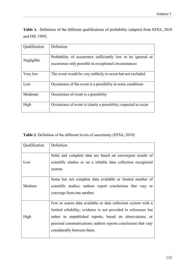
Annexe 1
115
Table 1. Definition of the different qualifications of probability (adapted from EFSA, 2010
and OIE 1999)
Qualification Definition
Negligible Probability of occurrence sufficiently low to be ignored or
occurrence only possible in exceptional circumstances
Very low The event would be very unlikely to occur but not excluded
Low Occurrence of the event is a possibility in some conditions
Moderate Occurrence of event is a possibility
High Occurrence of event is clearly a possibility; expected to occur
Table 2. Definition of the different levels of uncertainty (EFSA, 2010)
Qualification Definition
Low
Solid and complete data are based on convergent results of
scientific studies or on a reliable data collection recognized
system.
Medium
Some but not complete data available or limited number of
scientific studies; authors report conclusions that vary or
converge from one another.
High
Few or scarce data available or data collection system with a
limited reliability; evidence is not provided in references but
rather in unpublished reports, based on observations, or
personal communications; authors reports conclusions that vary
considerably between them.

Annexe 1
116
b. Legislation for animal importation in Belgium (Europe)
Legislations for horse importation are found in the Terrestrial Animal Health Code of
OIE (chapter 12.4 for EEEV and WEEV and chapter 12.11 for VEEV). The recommendations
concern only the importations of horses and no specific legislation exists for other animal
species. However, for birds, there are specific legislations concerning other diseases that
could be useful for the prevention of these three viruses. Indeed, for WNV, strict rules (e.g.
quarantine) are applicable for exporting countries depending on their disease status. Similar
recommendations are described in the Terrestrial Animal Health Code for avian influenza and
New castle disease viruses. CITES treaty resolutions provide strict measures for the
importation of exotic species in Europe. The CITES text is applicable to threatened species
but also for other exotic species.
All these regulations exist in the case of legal trade. The evaluation of illegal trade is very
difficult to perform because the exact numbers are not known. However, the illegal trade
could be an important source of introduction of the above mentioned diseases and is
expressed here as uncertainty.
c. Entry
1. Country factors
The EEEV North American viral subtype I is endemic from Canada to Florida and outbreaks
occur from June to November corresponding with the peak of vector activity (Cs. Melanura).
South American strains of EEEV (IIA, IIB and III) are widely distributed in Central and
South America (Figure 2) and epizootics can occur all year round in these countries. For both
strains, the first signs of a new outbreak appear with the first equine clinical case. For that
reason, horses could act as sentinel host for EEEV early detection in the surveillance program
(Go et al., 2014, Weaver et al., 1999, Greenlee, 2014). Every year, during summer and
autumn, presence of EEEV outbreaks in animals and/or humans, are notified to health
authorities in the USA mostly in the East Coast.
WEEV have been detected from Canada to Argentina mainly in the western regions of
Americas (Figure 2). The number of human cases was very high in the years 1940 to 1950
and has since heavily declined without a clear explanation (Forrester et al., 2008, Zhang et al.,
2011).
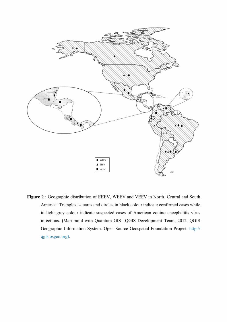
Figure
A
i
i
G
q
2 : Geograp
America. T
in light gre
infections.
Geographic
qgis.osgeo.
phic distrib
Triangles, sq
ey colour in
(Map build
c Informatio
org).
ution of EE
quares and c
ndicate susp
d with Quan
on System.
EEV, WEEV
circles in bl
pected case
ntum GIS –
Open Sour
V and VEE
ack colour i
es of Amer
–QGIS Dev
rce Geospat
V in North
indicate con
ican equine
velopment T
tial Founda
h, Central an
nfirmed cas
e encephali
Team, 2012
ation Projec
nd South
ses while
itis virus
2. QGIS
ct. http://
Annexe 1
117
This decrease of infections could be due to a reduction in the virulence of currently circulating
viruses in comparison with those circulating previously, to a better irrigation of agricultural
land, and to an enhanced policy to fight against vectors (Barker et al., 2010). In comparison
with EEEV, WEEV outbreaks occurred every 10 years with sometimes fatal issues in humans
(Delfraro et al., 2011).
VEEV was isolated for the first time in 1938 in South America (Venezuela) (Weaver et al.,
2004b). Seven antigenic complexes including enzootic and epizootic strains of VEEV were
already isolated in several countries in Central (until South of Florida) and South America
(Figure 2). The origin of epizootic strains is suggested by different hypotheses: maintenance
of epizootic strains in a continuous cycle and maintenance in mammalian hosts (horse or
alternative host), re-emergence of epizootic strains after administration of an incompletely
inactivated vaccine and maintenance of endemic strains in the host reservoir population with
periodic emergence of VEEV via mutation of those enzootic strains (mutation in the
glycoprotein E2).
2. Biological factors
Introduction of American equine encephalitis viruses in Belgium can occur with the
introduction of an infected vector or an infected host (Figure 1). Both circumstances are
described here. These three viruses have a wide range of potential vectors with the majority of
vectors belonging to the Culicidae family. The Culicidae life cycle takes place in four stages
(egg, larvae, pupae and adult) and its length depends on intrinsic factors such as species
variability and extrinsic factors such as temperature and humidity. Depending on species,
each stage of the cycle could instigate an overwintering of the vector. Thus, when considering
the risk of introduction of the vector, all stages must be taken into consideration and not only
the adult mosquito according to the virus. Transovarial transmission is very difficult to
identify and has not been described for the vectors belonging to Culicidae family and thus
there is no overwintering of the viruses in such vectors (Burkett-Cadena et al., 2011).
However, for Culiseta Melanura, a vector of EEEV, the eggs can become infected externally
by passing through the infected oviducts (Morris and Srihongse, 1978). Eggs could be
transported in used tyres as already observed for the (re-)introduction of Aedes albopictus or
japonicus in Belgium (Boukraa et al., 2013, Schaffner et al., 2004).
Annexe 1
118
A classical cycle of Culicidae lasts about 15 days from egg stage to adult with variations
depending on the species and the environmental conditions. An adult mosquito has a life span
of 15 days to 1 month depending on the gender (longer life for a female) and also according to
environmental and species conditions. The female dies after 2-3 blood meals, being necessary
for egg laying. The presence of the male is important because of its role in fertilization and
thus it is considered rather for the risk of exposure (continuation of the cycle). A mated
female ingests more blood than unmated female and therefore is a greater risk in viral
transmission.
3. Commodity factors
Different scenarios must be considered for animal host introduction. Indeed, legal trade,
illegal trade and accidental transport are three distinct situations. In legislation, only horse’s
importations are submitted to quarantine when they are imported from an endemic country
(OIE, terrestrial manual, 2010). For other animal hosts, many efforts exist for the well-being
of the imported species and for the preservation of endangered species (CITES conventions)
but no prevention exists for diseases. The impact of illegal trade is difficult to estimate but is
not negligible in regards with the risk of introduction of American equine encephalitis viruses.
For these small animal species, the risk could be increased by the fact that the animal remains
asymptomatic or show very few clinical signs. However, the introduction of these viruses by
an infected host is limited by the very short duration of viremia (2-4 days). The duration of
the quarantine (21 days) is thus sufficient to prevent the introduction (OIE, chapter 12.4 and
12.11). Although the transport time is short, it is still necessary that the host meets competent
vector during viremia.
3.1 Transport
Several factors are involved in the emergence of diseases into a new territory. Among these
factors the increasing number and velocity of people and animal movement between countries
and continents play a crucial role in the introduction of diseases. Particularly for vector-borne
diseases, transport of infected vector and infected animals, and susceptible vector should be
considered. The introduction can occur by all kinds of transport: aircrafts, boats, land (rail and
road) and wind.
Annexe 1
119
Aircraft Transportation
Nowadays the transport by aircraft is the most important route of introduction due to the
ability to transfer quickly both arthropods vectors and potentially infected animals. The
rapidness of air transportation increases the chances of vector survival (Gratz et al., 2000) and
consequently the risk of both new potential and viral infected vector introduction. Indeed,
arthropod vectors are capable to remain for long periods inside the passenger cabins,
passenger luggage, cargo holds and in the wheel bays even in flights between tropical and
temperate regions (Russell, 1987). For instance, in the USA, 73% of pest detections occur in
international airports, among these the fruit fly Ceratitis capitata can be easily found in the
aircraft baggage, the plant pathogens in the air cargo and mosquito-vectors in the aircrafts
cabins even in long flights (Tatem and Hay, 2007, Tatem et al., 2006, McCullough et al.,
2006, Liebhold et al., 2006, Lounibos, 2002). Study carried on by Russell (1987) showed that
the domestic mosquito (Culex quinquefasciatus), the house fly (Musca domestica), and the
flour beetle (Tribolium confusum) could survive, under test conditions, from one to seven
hour flights in the wheel bays between tropical and temperate airports (Russell, 1987). Risk
assessment analysis of the introduction of WNV vectors mosquitoes in Barbados (Douglas et
al., 2007), Galapagos (Kilpatrick et al., 2006), Hawaii (Kilpatrick et al., 2004) and United
Kingdom (Brown et al., 2012) described that aircrafts represent the greatest threat to the
introduction of the arthropods vectors. In case of transport of animals by air, there is a chance
that infected animals are transported presenting clinical signs. For instance, for the equine
encephalitis viruses this scenario could happen due to the relative long incubation period for
EEEV (4-10 days), WEEV (5-10 days) and VEEV (2 -6 days).
Ship transportation
The entrance of an exotic disease through ship should be considered as important route as
aircraft, for instance the Port of Antwerp, in Belgium, together with Rotterdam (Netherlands)
and Hamburg (Germany), is considered one of the most active ports of the world.
The particularity of ship transportation is the long duration of the journey, which might allow
transmission between an infected animal to a susceptible arthropod vector, as well as the
establishment of invasive vector species into the ship environment. This situation could
theoretically lead to vertical transmission within vector population and the infection of
rodents present in the ship.
Annexe 1
120
Indeed, vertical transmission has been demonstrated in Aedes aegypti mosquitoes infected
with Dengue virus (Joshi et al., 2002) and CHKV (Agarwal et al., 2014) and suggested for
WEEV (Fulhorst et al., 1994).
Moreover, the importance of shipping mosquitoes and viruses as a source of introduction has
already been shown. The introduction of vectors via boats was previously described in the
30’s, where Anopheles gambiae mosquitoes, the major malaria vectors, were introduced into
Brazil via ship from Senegal (Gratz et al., 2000) and more recently, introduction was reported
in Marseille (France) and Ghent (Belgium) with the detection of Anopheles mosquitoes and
patients positive to malaria. These patients were residents from a port area where there was no
previous description of the presence of the malaria vector, assuming that the mosquitoes were
released in the ship dock of the port (Peleman et al., 2000, Delmont et al., 1994).
3.2 Processing
Once the infected animal or vector survives the transportation they should be able to survive
the processing. The process of disinsection includes treatments with chemical insecticides
/aerosol and quarantine in the importing country (Figure 1).
Chemical insecticides /aerosol
The process of aircraft disinsection is foreseen by international legislation according to
regulations published in the International Health Regulations (IHR) from the World Health
Organization (WHO) (Yu et al., 2014). IHR describes the meaning of disinsection as “the
operation in which measures are taken to kill the insect vectors of human disease present in
ships, aircraft, trains, road vehicles and means of transport, and containers” (Berger-Preiss et
al., 2006, WHO, 1983).
Aircraft disinsection
The measures of aircrafts disinsection to avoid the entrance of insect vectors into countries
began in the 20s and until today only 20 countries have adopted it. The disinfection will be
applied in some flights depending on flight origin and route (Rayman, 2006).
For flight disinfection, WHO recommends procedures for passenger cabins and cargo areas
(Russell, 1987) using aerosol spray preparations with different natural or synthetic chemicals
like pyrethrins, resmethrin, d-phenothrin or permethrin (Berger-Preiss et al., 2006).
Annexe 1
121
According to WHO, two groups of procedures must be followed: (1) spraying before or
during flight using insecticide aerosol and (2) residual treatment. In the first group, the
methods applied are the (a) “blocks away” where the interior of the aircraft is sprayed with
insecticide aerosol before take-off with the passenger already boarded and the doors closed
(Gratz et al., 2000, WHO/HQ, 1995, Sullivan et al., 1972, Sullivan et al., 1964); the (b) “pre-
flight spraying” which consists of spraying the aircraft prior to passengers boarding and the
(c) “top-of-descent spraying” which is an in-flight spraying before the aircraft reaches the
airport (WHO/HQ, 1995). Despite the WHO recommendations and the methods that are
recognized as reliable treatments (Berger-Preiss et al., 2006), the “blocks away” and “top-of-
descent” methods, applied in the presence of passengers, may present a limited effect if its
application is not well performed, once mosquitoes can keep alive inside the aircrafts (in the
overhead luggage space or even inside the passenger’s luggage) (Gratz et al., 2000, Russell
and Paton, 1989, WHO, 1985, Goh et al., 1985).
An alternative method described as “pre-embarkation”, suggests the spraying of the d-
phenothrin aerosol under the seat rows and in a second step at the height of approximately
1.60 m by moving from frontside to backside of the cabin, in the absence of passengers and
crew. The presence of d-phenothrin in the air was sufficient to kill flying insects like house
flies (Musca domestica) and mosquitoes (Aedes aegypti, Anopheles stephensi and Culex
pipiens) within 20 min. The method was 100% effective against insects in horizontal surfaces
up to 24 h after spraying (Rayman, 2006, Berger-Preiss et al., 2006).
The residual treatment will be applied in internal spaces of the aircraft (excluding the food
preparations areas) to prevent the infestation by insects that have reached the aircraft and be
sheltered in these areas (WHO/HQ, 1995).
For cases of infestation by rodents and cockroaches, the fumigation methods may be applied
leaving the aircraft out of service for seven to 15 hours (Gratz et al., 2000). These methods
present pro- and contra- arguments regarding the real necessity of their application. For
example, the USA discontinued the spraying routine since 1979, concerned by the health of
the passengers and crew, and convinced that outbreaks occurring in countries of arrival are
not related to imported vectors. By the other hand, Australia, New Zealand and India still
maintain disinsection methods before landing (Berger-Preiss et al., 2004).
Annexe 1
122
Ship disinsection
Although the importance of ship transports for the introduction of arthropods vectors, the
methods of disinsection are not well established as well as for aircrafts. For ships, the
quarantine and inspections should be done during movements of goods, however it is not an
effective way to prevent the transport of vectors on tourist or local ships (Bataille et al., 2009,
Causton et al., 2006). The prevention measures adopted should be related to the use of insect
traps and the use of only yellow light during the night to avoid the attraction of flying insects
to the ship, even so fumigation and disinsection methods applied to aircrafts should be used as
models for surveillance and control measures (Bataille et al., 2009, Roque Albelo et al., 2006,
WHO/HQ, 1995).
Quarantine
To avoid the entrance of new diseases, the processing control should be done not only in the
means of transport controlling the presence of vectors but also to the imported animals that
may carry enzootic diseases. The control of animals should follow the World Organisation for
Animal Health (OIE) recommendations present in the Terrestrial Animal Health Code. When
the concern is related to the risk of introduction of VEEV, EEEV and WEEV through
importation of equines, the procedures recommended by OIE determine more specific actions
based on the animal health prior the animal departure (OIE, 2014b, OIE, 2014c). Quarantine
is applicable for horses in the case of VEEV only because they are a competent amplifying
host during epidemics for VEEV only. For OIE, a country is considered free of VEEV if no
confirmed case occurred during the past two years. A main recommendation for exporting
horses from an infected country is the quarantine of three weeks in the country of origin in
addition to 7 days of animal isolation in the importing country. Both these measures combined
with other recommendations decrease significantly the risk of introduction by the importation
of VEEV infected horses. Indeed, considering that the incubation period (1-5 days) and the
infectious period (2 weeks), the three weeks of quarantine only are sufficient to reduce to a
minimum the risk of introduction of an infected horse.
In general lines, all animals accepted into a new country must have an International
Veterinary Certificate issued by the Veterinary authorities from their country of origin. When
an animal is suspected of being affected by or infected with a disease capable of being
transmitted to the animals or susceptible vectors in the new territory,
Annexe 1
123
it should be placed immediately in quarantine to clinical observation and perform the
laboratorial diagnosis to after that, follow the legal procedures (OIE, 2014a).
Quarantine and isolation are under official veterinary inspection and thus involve a good
organization and training of the official veterinary authorities in both exporting and importing
countries to detect any clinical signs during those periods.
d. Exposure
1. Biological factors
EEEV, WEEV and VEEV are RNA viruses which imply a high mutation rate due to the lack
of a mechanism of correction by their RNA polymerase. In general, the point mutation rate for
RNA viruses is one mutation per 104 to 105 nucleotides. Mutations in the viral genome can
result in modification in the virulence and in the host or vector spectrum (Domingo, 2010).
The evolution of these three viruses already counts several mutations with high importance.
Indeed, WEEV is a recombinant virus between EEEV and a Sindbis virus and epizootic
strains of VEEV result from mutations in the genome of enzootic strains (Greenlee, 2014).
For VEEV in particular, studies have implicated mutations in the E2 envelope glycoprotein as
factor to become epizootic variants and to change this host range (Weaver et al., 2004a).
Taking into account these considerations about their viral genome, some mutations are
expected to occur after the introduction of these viruses in Belgium.
2. Country factors
After the successful introduction of the infected vector / host, the transmission step begins.
First, the exotic infected vector / host exotic has to survive in its new environment and second
the transmission has to occur (Figure 1).
The vector survival will depend on several factors including the point of arrival and the
season. The importance of the point of arrival is related to the accessibility to breeding sites
depending on the need of the vector species. Temperature and humidity have an important
role for its survival but also for its life cycle. Humidity plays a role in the development cycle
since eggs need humidity to hatch and the larvae to become adults. Temperature is involved in
the length of the extrinsic period (time between virus ingestion by the vector and sufficient
replication in the vector to transmit the virus to a susceptible host). The availability of a
sufficient population of susceptible (or competent) hosts in place and time to coincide with
Table 3 : Identification of potential vector species for American equine encephalitis viruses
(EEEV, WEEV and VEEV) in Belgium
Vector Indigenous Virus
Culiseta morsitans
+ EEEV
Culex pipiens
++++ EEEV
Culex (Mel.) spissipes
+ VEEV
Aedes vexans
++ EEEV
Aedes cinereus
+ EEEV
Aedes aegypti
+ WEEV (ulc*)
Ixodes persulcatus
+ WEEV
*ulc : under laboratory conditions
Annexe 1
124
the introduction and the biting activity of the vectors (Kuno and Chang, 2005) is essential for
a successful viral transmission and for vector survival and reproduction. Host populations are
variable according to the point of arrival. Some publications suggest that Culicidae could be
carried by wind over a great distance (Calisher, 1994, Howard et al., 1989) and thus wind by
the dispersal of exotic vectors allows the transmission of viruses over several miles around the
point of entry and increases the availability of susceptible hosts. In Belgium, a large
population of potential hosts is likely available for these three viruses, whether horses or other
hosts such as birds, rodents and humans.
At the level of introduced host, the transmission could occur if the host is still developing a
sufficient level of viremia to infect a vector. For these three viruses depending on host
species, the duration of the viremia lasts from 3-4 days for mostly hosts to 100 days for
reptiles under certain conditions. The short period of viremia involves that the vector
population is abundant to increase the probability that the host transmits the infection to the
vector. The European summer is than the perfect season to account for a maximum of
available vectors (Vos, 2012) but climate changes could introduce new favorable conditions
during other seasons.
Identification of competent vectors for these three viruses in Belgium is reported in Table 3.
Culex pipiens is the most distributed mosquito in this country and EEEV has already been
isolated from this species. Epizootic strains of VEEV are opportunistic in their use of
mosquito vectors (Weaver et al., 2004b) and thus vector is not a limited factor in the spread of
VEEV. Furthermore with the global warming, it is important to continue the surveillance and
identification of new vector species in Belgium.
Some vertebrate hosts for these three viruses are also present in Belgium but further studies
are needed to compare the competency of Belgian species (Supplementary table).
Step Probability of the event occurring Uncertainty
Introduction
Vector
A. Vector Survival after transport
Aircraft Moderate Medium Boat Low Medium
Others Very low High B.Vector survival after
processing
Chemical Low Medium (aircraft) to
high (boats and others)
Vertebrate host
A. Animal survival after transport
Aircraft Low High Boat Low High
B.Animal survival after processing
Chemical Low High Quarantine (horses)* Negligible Low
Exposure 1.1 Survival of the infected exotic vector in
the new environment
Very low to low Medium
1.2 Exotic infected vector bite endogenous host Very low to Low Medium
1.3 Survival of the infected exotic host in the new environment
Low to moderate Medium
1.4 Exotic infected host is bitten by a endogenous vector
Low High
1.5 Exotic host is bitten by an exotic vector Very low High Spread
2.1 Adaptation of the exotic vector species in the new environment Very low Medium
2.2 Endogenous host develop viremia Very low High 2.3 Adaptation of the exotic host in the new
environment Low Medium
2.4 Endogenous vector become infected Very low Medium
2.5 Exotic vector become infected Very low High
Table 4. Probability and uncertainty of the different steps from introduction to spread of EEEV, WEEV and VEEV viruses in Belgium, qualified by an expert opinion based on a consensus among the authors.
*Quarantine probability is the probability that an animal remain viremic after the quarantine period
Annexe 1
125
Discussion: risk profiling
The qualitative approach to estimate the risk of introduction of EEEV, WEEV and VEEV by
the importation of live animals and vectors from North, Central and South America into
Belgium is presented in Table 4 and follows the steps described in the scenario tree (figure
1). Belgium is in the heart of Europe and could be representative of Western European
countries and particularly in relation with air-, boat- and road transport. The objective was
firstly to highlight the factors associated with a higher probability (Table 4) and therefore the
factors to be controlled during live animals importation or international exchanges, and then
to qualitatively estimate the probability of establishing a transmission cycle for these viruses
in Belgium.
The entry of a vector is composed by two equally important steps. Indeed, the vector has to
survive to the air, boat or other transports, but also have to survive to the different steps of the
processing applied in these different types of transport. The probability of vector survival to
the transport is estimated very low to moderate depending on the type of transport. Several
studies have shown that the survival of vectors in air transport was common, and trade of used
tyres is responsible for the introduction of Aedes albopictus (Takumi et al., 2009, Boukraa et
al., 2013). Estimation of the probability of vector survival to processing is low, but is
dependent on the policy of the airline and also on the respect of the application of the
insecticide by the crew. Indeed, no legal rules exist for this processing.
Regarding the introduction of live animals, firstly, the quarantine of horses (OIE Terrestrial
Manual) from the Americas is a very effective measure against the introduction of these
viruses, the risk of introduction was considered negligible. However, quarantine is applied for
the three viruses but only the epizootic strains of VEEV use the horse (and human) as
amplifying hosts. For other livestock trades such as poultry, there are no specific rules of
importation for these three viruses. However, for the prevention of the introduction of other
viruses such as Newcastle and Avian A Influenza viruses, a quarantine of 3 weeks is applied.
Annexe 1
126
This measure could therefore indirectly help for the prevention of EEEV and WEEV viruses
entry. Exotic animals are protected by international conventions like CITES with a primary
objective to protect these species but little (or nothing) to prevent and control infectious
diseases. In the context of legal imports, survival to processing should not be considered,
indeed, the aim is to keep the animal alive until the final destination. Regarding the illegal
trade or accidental introduction of vertebrate species by transports, the uncertainty is high and
it is difficult to perfectly assess the risk of introduction. However, despite the limited amount
of available data (Rosen and Smith, 2010), it can be hypothesized this type of import only
concerns a minority of importations. The risk of introduction of these species was assessed
low, such as their survival to the processing either in the cargo aircrafts or on dock boats. The
probability of animal survival to both transport and processing is estimated low and not very
low because species like rodents or reptiles (turtles) are considered resistant species and only
one infected host individual is enough to have a successful introduction of the virus.
In this study, only the risk of introduction by direct transport from the Americas to Belgium is
considered. Therefore it is possible that the risk is higher if indirect transports are taken into
account namely the introduction through neighboring countries (road) or passive transport of
vector through wind as already shown for other viruses such as BTV-8 and AHSV-9
(Maclachlan and Guthrie, 2010, Johnson et al., 2012). Concerning the introduction of the New
World Alphaviruses (EEEV, VEEV and WEEV) in Belgium the wind could be considered
less important as they have not been detected in the Old World so far (Go et al., 2014),
however it may be an important pathway for transporting vectors into the continent
(Kilpatrick et al., 2006).
After surviving to transport and processing, the vector has to survive into a new environment
and a new climate. The probability of vector survival is estimated from very low to low
depending on the vector species, its stage of development and its ecological preferences. The
season of arrival in Belgium is also very important for its survival. Indeed, an adult mosquito
survives at 10 ° C and larvae restart their development from 15 ° C. Similarly the probability
of vector biting an indigenous vertebrate host is estimated from very low to low depending on
trophic preferences of the vector species and host availability in the point of entry of the
vector. For example, Cx pipiens feeds mainly on birds, Ae dorsalis in mammals and Ae
albopictus is opportunistic in its host choice vertebrate.
Annexe 1
127
The probability of survival for a vertebrate host is higher than for a vector and is estimated
from low to moderate. On the other side, the probability that the imported vector bites the
imported vertebrate host is estimated very low. This particular case could only happen if they
arrive almost together at the same entry point and at the same time.
The first consequence of the successful introduction of the vector is its adaptation to the new
ecological environment. In addition, the probability of complete adaptation is estimated very
low as observed, for instance for exotic mosquito species recorded in Belgium Ae.albopictus,
are so far not established even if they have been found by trapping during several years
(Boukraa et al., 2013). However climate changes with higher temperatures could increase this
probability and even an increase in viral persistence could be observed. Several studies have
shown that global warming shortens extrinsic incubation period (EIP) in the vector (Johnson
et al., 2012).
The probability that the vertebrate host develops a sufficient viremia to infect a vector is
estimated very low. Indeed, despite a broad host range for these viruses, only a few species of
birds, rodents and reptiles serve as a reservoir host for the virus, the others being considered
as dead- end hosts. An exception should be noted for the VEE virus for which horses and
humans being efficient hosts in epidemic conditions.
The adaptation of a vertebrate host to its new environment is considered here more likely
(low) that for the vector (very low). Indeed, in the past, several invasive species have already
shown their ability to adapt and reproduce in a new environment in Belgium
(http://ias.biodiversity.be/species/all). Regarding EEEV, WEEV and VEEV, the period of
infectivity last about 14 days (OIE, Terrestrial Manual) with an incubation period of 5-14
days for EEEV and WEEV and 1-5 days for the VEE virus and a viremia period lasting less
than a week (except in reptiles with EEEV, viremia up to 100 days post-infection). The
probability of infection of an indigenous vector by the infected vertebrate host introduced is
estimated very low in regards with the short viremia.
So far the virus has not been identified in Belgium, but it does not mean that attempts of a
viral introduction did not occur. These failed attempts, if any, were not observed. Several
hypotheses could explain this situation. So far no other alphavirus was isolated in Belgium.
This argument, however, could be plausible in other regions (meaning that would possess
other alphaviruses). The phenomenon of overwintering is less plausible for exotic vectors than
for indigenous vectors. Although these diseases are notifiable diseases, they are subject only
to passive surveillance in horses, which limits the possibilities of identification.

Annexe 1
128
The probability of introduction of these three viruses is estimated from very low to moderate
(Table 4) but all elements of their cycle are present in Belgium. The use of horses as sentinel
around the points of entry such as ports and airports could be used as part of an active
localized surveillance such as already done in Americas or Canada (Rocheleau et al., 2013).
This would involve training of all public health sectors, starting with veterinarians. In addition
to veterinary surveillance, it would be useful to improve the knowledge of medical
practitioners to include these diseases in their differential diagnosis of human encephalitis.
This risk profiling is valid for the current period. Climate change could lead to changes in the
evaluation of risk factors, including the easiest survival of exotic vectors.
Although the economic aspects have not been addressed in this study, the consequences of a
successful introduction of these three viruses would be severe in regards with the economic
importance of the horse industry in Belgium. The knowledge of the severity of these
consequences could help to precisely measure the risk of introduction. Belgium is situated in
Western Europe and shares with surrounding European states and the risk factors that have
been discussed here. The conclusions of this work could be therefore extended to other
European countries possessing a significant equine industry. Furthermore, except for
quarantine which is well documented, the level of uncertainty was qualified as medium or
high for the other risk factors, requesting therefore specific scientific works dedicated to gain
a better knowledge of the various factors involved in the introduction and spread pathways of
EEEV, WEEV and VEEV.
Acknowledgments
This study was supported by grants from the Belgium Federal Public Service, Health, Food
Chain Safety and Environment (INDEVIREQ 1.0 ; RT 10/07). A.C.B. Terzian is supported by
the Coordenação de Aperfeiçoamento de Pessoal de Nível Superior – CAPES Foundation
(CAPES/WBI Grant nº. 2837/14-5) and E.F. Oliveira-Filho by CNPq (Brazilian National
Council for Scientific and Technological Development).
Annexe 1
129
Agarwal, A., P. K. Dash, A. K. Singh, S. Sharma, N. Gopalan, P. V. Rao, M. M. Parida and P. Reiter, 2014: Evidence of experimental vertical transmission of emerging novel ECSA genotype of Chikungunya Virus in Aedes aegypti. PLoS neglected tropical diseases, 8, e2990.
Aguilar, P. V., J. G. Estrada-Franco, R. Navarro-Lopez, C. Ferro, A. D. Haddow and S. C. Weaver, 2011: Endemic Venezuelan equine encephalitis in the Americas: hidden under the dengue umbrella. Future Virology, 6, 721-740.
Anishchenko, M., R. A. Bowen, S. Paessler, L. Austgen, I. P. Greene and S. C. Weaver, 2006: Venezuelan encephalitis emergence mediated by a phylogenetically predicted viral mutation. Proceedings of the National Academy of Sciences of the United States of America, 103, 4994-4999.
Anyamba, A., J. P. Chretien, P. B. Formenty, J. Small, C. J. Tucker, J. L. Malone, H. El Bushra, V. Martin and K. J. Linthicum, 2006: Rift Valley Fever potential, Arabian Peninsula. Emerging infectious diseases, 12, 518-520.
Arrigo, N. C., 2010: Cotton Rats and House Sparrows as Hosts for North and South American Strains of Eastern Equine Encephalitis Virus. Emerging infectious diseases, 16.
Arrigo, N. C., A. P. Adams and S. C. Weaver, 2010: Evolutionary patterns of eastern equine encephalitis virus in North versus South America suggest ecological differences and taxonomic revision. Journal of virology, 84, 1014-1025.
Barker, C. M., W. O. Johnson, B. F. Eldridge, B. K. Park, F. Melton and W. K. Reisen, 2010: Temporal connections between Culex tarsalis abundance and transmission of western equine encephalomyelitis virus in California. The American journal of tropical medicine and hygiene, 82, 1185-1193.
Bataille, A., A. A. Cunningham, V. Cedeno, M. Cruz, G. Eastwood, D. M. Fonseca, C. E. Causton, R. Azuero, J. Loayza, J. D. Martinez and S. J. Goodman, 2009: Evidence for regular ongoing introductions of mosquito disease vectors into the Galapagos Islands. Proceedings. Biological sciences / The Royal Society, 276, 3769-3775.
Berger-Preiss, E., W. Koch, W. Behnke, S. Gerling, H. Kock, L. Elflein and K. E. Appel, 2004: In-flight spraying in aircrafts: determination of the exposure scenario. International journal of hygiene and environmental health, 207, 419-430.
Berger-Preiss, E., W. Koch, S. Gerling, H. Kock, J. Klasen, G. Hoffmann and K. E. Appel, 2006: Aircraft disinsection: exposure assessment and evaluation of a new pre-embarkation method. International journal of hygiene and environmental health, 209, 41-56.
Boukraa, S., F. N. Raharimalala, J. Y. Zimmer, F. Schaffner, T. Bawin, E. Haubruge and F. Francis, 2013: Reintroduction of the invasive mosquito species Aedes albopictus in Belgium in July 2013. Parasite (Paris, France), 20, 54.
Annexe 1
130
Brault, A. C., A. M. Powers, E. C. Holmes, C. H. Woelk and S. C. Weaver, 2002: Positively charged amino acid substitutions in the e2 envelope glycoprotein are associated with the emergence of venezuelan equine encephalitis virus. Journal of virology, 76, 1718-1730.
Brown, E. B., A. Adkin, A. R. Fooks, B. Stephenson, J. M. Medlock and E. L. Snary, 2012: Assessing the risks of West Nile virus-infected mosquitoes from transatlantic aircraft: implications for disease emergence in the United Kingdom. Vector borne and zoonotic diseases (Larchmont, N.Y.), 12, 310-320.
Burkett-Cadena, N. D., G. S. White, M. D. Eubanks and T. R. Unnasch, 2011: Winter biology of wetland mosquitoes at a focus of eastern equine encephalomyelitis virus transmission in Alabama, USA. Journal of medical entomology, 48, 967-973.
Calisher, C. H., 1994: Medically important arboviruses of the United States and Canada. Clinical microbiology reviews, 7, 89-116.
Causton, C. E., S. B. Peck, B. J. Sinclair, L. Roque-Albelo, C. J. Hodgson and B. Landry, 2006: Alien insects: threats and implications for conservation of Galápagos Islands. Annual Entomology Society of Americas, 99, 121-143.
Centers for Disease, C. and Prevention, 2010: Locally acquired Dengue--Key West, Florida, 2009-2010. MMWR. Morbidity and mortality weekly report, 59, 577-581.
Coffey, L. L., N. Forrester, K. Tsetsarkin, N. Vasilakis and S. C. Weaver, 2013: Factors shaping the adaptive landscape for arboviruses: implications for the emergence of disease. Future microbiology, 8, 155-176.
Deardorff, E. R., N. L. Forrester, A. P. Travassos-da-Rosa, J. G. Estrada-Franco, R. Navarro-Lopez, R. B. Tesh and S. C. Weaver, 2009: Experimental infection of potential reservoir hosts with Venezuelan equine encephalitis virus, Mexico. Emerging infectious diseases, 15, 519-525.
Deardorff, E. R. and S. C. Weaver, 2010: Vector competence of Culex (Melanoconion) taeniopus for equine-virulent subtype IE strains of Venezuelan equine encephalitis virus. The American journal of tropical medicine and hygiene, 82, 1047-1052.
Delfraro, A., A. Burgueño, N. Morel, G. González, A. García, J. Morelli, W. Pérez, H. Chiparelli and J. Arbiza, 2011: Fatal Human Case of Western Equine Encephalitis, Uruguay. Emerging infectious diseases, 17, 952-954.
Delmont, J., P. Brouqui, P. Poullin and A. Bourgeaade, 1994: Harbour-acquired Plasmodium falciparum malaria. Lancet, 344, 330-331.
Domingo, E., 2010: Mechanisms of viral emergence. Veterinary research, 41, 38.

Annexe 1
131
Douglas, K. O., A. M. Kilpatrick, P. N. Levett and M. C. Lavoie, 2007: A quantitative risk assessment of West Nile virus introduction into Barbados. The West Indian medical journal, 56, 394-397.
Durand, B., S. Lecollinet, C. Beck, B. Martinez-Lopez, T. Balenghien and V. Chevalier, 2013: Identification of hotspots in the European union for the introduction of four zoonotic arboviroses by live animal trade. PloS one, 8, e70000.
Ferro, C., J. Boshell, A. C. Moncayo, M. Gonzalez, M. L. Ahumada, W. Kang and S. C. Weaver, 2003: Natural enzootic vectors of Venezuelan equine encephalitis virus, Magdalena Valley, Colombia. Emerging infectious diseases, 9, 49-54.
Figueiredo, L. T., 2007: Emergent arboviruses in Brazil. Revista da Sociedade Brasileira de Medicina Tropical, 40, 224-229.
Forrester, N. L., J. L. Kenney, E. Deardorff, E. Wang and S. C. Weaver, 2008: Western Equine Encephalitis submergence: lack of evidence for a decline in virus virulence. Virology, 380, 170-172.
Fulhorst, C. F., J. L. Hardy, B. F. Eldridge, S. B. Presser and W. C. Reeves, 1994: Natural vertical transmission of western equine encephalomyelitis virus in mosquitoes. Science, 263, 676-678.
Fyodorova, M. V., H. M. Savage, J. V. Lopatina, T. A. Bulgakova, A. V. Ivanitsky, O. V. Platonova and A. E. Platonov, 2006: Evaluation of potential West Nile virus vectors in Volgograd region, Russia, 2003 (Diptera: Culicidae): species composition, bloodmeal host utilization, and virus infection rates of mosquitoes. Journal of medical entomology, 43, 552-563.
Gjenero-Margan, I., B. Aleraj, D. Krajcar, V. Lesnikar, A. Klobucar, I. Pem-Novosel, S. Kurecic-Filipovic, S. Komparak, R. Martic, S. Duricic, L. Betica-Radic, J. Okmadzic, T. Vilibic-Cavlek, A. Babic-Erceg, B. Turkovic, T. Avsic-Zupanc, I. Radic, M. Ljubic, K. Sarac, N. Benic and G. Mlinaric-Galinovic, 2011: Autochthonous dengue fever in Croatia, August-September 2010. Euro surveillance : European communicable disease bulletin, 16.
Go, Y. Y., U. B. Balasuriya and C. K. Lee, 2014: Zoonotic encephalitides caused by arboviruses: transmission and epidemiology of alphaviruses and flaviviruses. Clinical and experimental vaccine research, 3, 58-77.
Goh, K. T., S. K. Ng and S. Kumarapathy, 1985: Disease-bearing insects brought in by international aircraft into Singapore. The Southeast Asian journal of tropical medicine and public health, 16, 49-53.
Gould, E. A., P. Gallian, X. De Lamballerie and R. N. Charrel, 2010: First cases of autochthonous dengue fever and chikungunya fever in France: from bad dream to reality!
Annexe 1
132
Clinical microbiology and infection : the official publication of the European Society of Clinical Microbiology and Infectious Diseases, 16, 1702-1704.
Gratz, N. G., R. Steffen and W. Cocksedge, 2000: Why aircraft disinsection? Bulletin of the World Health Organization, 78, 995-1004.
Greenlee, J. E., 2014: The equine encephalitides. Handbook of clinical neurology, 123, 417-432.
Griffin, D. E., 2007: Alphaviruses. Fields Virology, 5 edn., pp. 1023-1067. Lippincott Williams & Wilkins.
Hardy, J. L., 1987: The ecology of western equine encephalomyelitis virus in the Central Valley of California, 1945-1985. The American journal of tropical medicine and hygiene, 37, 18S-32S.
Hassan, H. K., E. W. Cupp, G. E. Hill, C. R. Katholi, K. Klingler and T. R. Unnasch, 2003: Avian host preference by vectors of eastern equine encephalomyelitis virus. The American journal of tropical medicine and hygiene, 69, 641-647.
Hoffmann, B., M. Scheuch, D. Hoper, R. Jungblut, M. Holsteg, H. Schirrmeier, M. Eschbaumer, K. V. Goller, K. Wernike, M. Fischer, A. Breithaupt, T. C. Mettenleiter and M. Beer, 2012: Novel orthobunyavirus in Cattle, Europe, 2011. Emerging infectious diseases, 18, 469-472.
Hollidge, B. S., F. Gonzalez-Scarano and S. S. Soldan, 2010: Arboviral encephalitides: transmission, emergence, and pathogenesis. Journal of neuroimmune pharmacology : the official journal of the Society on NeuroImmune Pharmacology, 5, 428-442.
Howard, J. J., D. J. White and S. L. Muller, 1989: Mark-recapture studies on the Culiseta (Diptera: Culicidae) vectors of eastern equine encephalitis virus. Journal of medical entomology, 26, 190-199.
Johnson, N., K. Voller, L. P. Phipps, K. Mansfield and A. R. Fooks, 2012: Rapid molecular detection methods for arboviruses of livestock of importance to northern Europe. Journal of Biomedical Biotechnology, 2012, 719402.
Jones, K. E., N. G. Patel, M. A. Levy, A. Storeygard, D. Balk, J. L. Gittleman and P. Daszak, 2008: Global trends in emerging infectious diseases. Nature, 451, 990-993.
Jose, J., J. E. Snyder and R. J. Kuhn, 2009: A structural and functional perspective of alphavirus replication and assembly. Future microbiology, 4, 837-856.
Joshi, V., D. T. Mourya and R. C. Sharma, 2002: Persistence of dengue-3 virus through transovarial transmission passage in successive generations of Aedes aegypti mosquitoes. American Journal of Tropical Medicine and Hygiene, 67, 158-161.
Annexe 1
133
Kilpatrick, A. M., P. Daszak, S. J. Goodman, H. Rogg, L. D. Kramer, V. Cedeno and A. A. Cunningham, 2006: Predicting pathogen introduction: West Nile virus spread to Galaipagos. Conservation biology : the journal of the Society for Conservation Biology, 20, 1224-1231.
Kilpatrick, A. M., Y. Gluzberg, J. Burgett and P. Daszak, 2004: Quantitative risk assessment of the pathways by which West Nile virus could reach Hawaii. EcoHealth, 1, 205-209.
Kononchik, J. P., Jr., R. Hernandez and D. T. Brown, 2011: An alternative pathway for alphavirus entry. Virology journal, 8, 304.
Kuhn, R. J., 2007: Togaviridae: The Viruses and Their Replication. In: D. M. Knipe and P. M. Howley (eds), Fields Virology, pp. 1002-1022. Lippincott Williams & Wilkins, Philadelphia.
Kuno, G. and G. J. Chang, 2005: Biological transmission of arboviruses: reexamination of and new insights into components, mechanisms, and unique traits as well as their evolutionary trends. Clinical microbiology reviews, 18, 608-637.
La Linn, M., J. Gardner, D. Warrilow, G. A. Darnell, C. R. McMahon, I. Field, A. D. Hyatt, R. W. Slade and A. Suhrbier, 2001: Arbovirus of marine mammals: a new alphavirus isolated from the elephant seal louse, Lepidophthirus macrorhini. Journal of virology, 75, 4103-4109.
Liebhold, A. M., T. T. Work, D. G. McCullough and J. F. Cavey, 2006: Airline baggage as a pathway for alien insect species invading the United States. American Entomology, 52, 48-54.
Lounibos, L. P., 2002: Invasions by insect vectors of human disease. Annual review of entomology, 47, 233-266.
Maclachlan, N. J. and A. J. Guthrie, 2010: Re-emergence of bluetongue, African horse sickness, and other orbivirus diseases. Veterinary research, 41, 35.
Madani, T. A., Y. Y. Al-Mazrou, M. H. Al-Jeffri, A. A. Mishkhas, A. M. Al-Rabeah, A. M. Turkistani, M. O. Al-Sayed, A. A. Abodahish, A. S. Khan, T. G. Ksiazek and O. Shobokshi, 2003: Rift Valley fever epidemic in Saudi Arabia: epidemiological, clinical, and laboratory characteristics. Clinical infectious diseases : an official publication of the Infectious Diseases Society of America, 37, 1084-1092.
McCullough, D. G., T. T. Work, J. F. Cavey, A. M. Liebhold and D. Marshall, 2006: Interceptions of nonindigenous plant pests at US ports of entry and border crossings over a 17-year period. Biological Invasions, 8 611-630.
Morris, C. D. and S. Srihongse, 1978: An evaluation of the hypothesis of transovarial transmission of eastern equine encephalomyelitis virus by Culiseta melanura. The American journal of tropical medicine and hygiene, 27, 1246-1250.
Mourao, M. P., S. Bastos Mde, R. P. de Figueiredo, J. B. Gimaque, S. Galusso Edos, V. M. Kramer, C. M. de Oliveira, F. G. Naveca and L. T. Figueiredo, 2012: Mayaro fever in the city of Manaus, Brazil, 2007-2008. Vector Borne Zoonotic Diseases, 12, 42-46
Annexe 1
134
Munoz, M. and J. C. Navarro, 2012: [Mayaro: a re-emerging Arbovirus in Venezuela and Latin America]. Biomedica, 32, 286-302.
Nathanson, N., 2007: Epidemiology. In: D. M. H. Knipe, P. M. (ed), Fields Virology, 5 edn., pp. 424-446.
OIE, 2014a: Chapter 5.7.- Animal health measures applicable on arrival. Terrestrial Animal Health Code.
OIE, 2014b: Chapter 12.4. - Equine encephalomyelitis (Eastern and Western). Terrestrial Animal Health Code.
OIE, 2014c: Chapter 12.11. - Venezuelan Equine Encephalomyelitis. Terrestrial Animal Health Code.
Peleman, R., D. Benoit, L. Goossens, F. Bouttens, H. D. Puydt, D. Vogelaers, F. Colardyn and K. Van de Woude, 2000: Indigenous malaria in a suburb of Ghent, Belgium. Journal of travel medicine, 7, 48-49.
Peters, C. J., 2007: Emerging Viral Diseases. In: D. M. Knipe and P. M. Howley (eds), Fields Virology, pp. 605-625. Lippincott Williams & Wilkins.
Pfeffer, M. and G. Dobler, 2010: Emergence of zoonotic arboviruses by animal trade and migration. Parasite and Vectors, 3, 35.
Powers, A. M., A. C. Brault, Y. Shirako, E. G. Strauss, W. Kang, J. H. Strauss and S. C. Weaver, 2001: Evolutionary relationships and systematics of the alphaviruses. Journal of virology, 75, 10118-10131.
Rayman, R. B., 2006: Aircraft disinsection. Aviation, space, and environmental medicine, 77, 733-736.
Rezza, G., L. Nicoletti, R. Angelini, R. Romi, A. C. Finarelli, M. Panning, P. Cordioli, C. Fortuna, S. Boros, F. Magurano, G. Silvi, P. Angelini, M. Dottori, M. G. Ciufolini, G. C. Majori, A. Cassone and C. s. group, 2007: Infection with chikungunya virus in Italy: an outbreak in a temperate region. Lancet, 370, 1840-1846.
Rocheleau, J. P., J. Arsenault, L. R. Lindsay, A. Dibernardo, M. A. Kulkarni, N. Cote and P. Michel, 2013: Eastern Equine Encephalitis Virus: High Seroprevalence in Horses from Southern Quebec, Canada, 2012. Vector borne and zoonotic diseases (Larchmont, N.Y.).
Roque Albelo, L., M. Berg and M. Galarza, 2006: ‘Polizontes peligrosos’, dispersion de insectos entre las Islas Galapagos en barcos de turismo. Charles Darwin Foundation, Puerto Ayora, Galapagos.
Annexe 1
135
Rosen, G. E. and K. F. Smith, 2010: Summarizing the evidence on the international trade in illegal wildlife. EcoHealth, 7, 24-32.
Russell, R. C., 1987: Survival of insects in the wheel bays of a Boeing 747B aircraft on flights between tropical and temperate airports. Bulletin of the World Health Organization, 65, 659-662.
Russell, R. C. and R. Paton, 1989: In-flight disinsection as an efficacious procedure for preventing international transport of insects of public health importance. Bulletin of the World Health Organization, 67, 543-547.
Sailleau, C., E. Breard, C. Viarouge, A. Desprat, V. Doceul, E. Lara, J. Languille, D. Vitour, H. Attoui and S. Zientara, 2013: Acute Schmallenberg virus infections, France, 2012. Emerging infectious diseases, 19, 321-322.
Savage, H. M., C. Ceianu, G. Nicolescu, N. Karabatsos, R. Lanciotti, A. Vladimirescu, L. Laiv, A. Ungureanu, C. Romanca and T. F. Tsai, 1999: Entomologic and avian investigations of an epidemic of West Nile fever in Romania in 1996, with serologic and molecular characterization of a virus isolate from mosquitoes. Am J Trop Med Hyg, 61, 600-611.
Schaffner, F., W. Van Bortel and M. Coosemans, 2004: First record of Aedes (Stegomyia) albopictus in Belgium. Journal of the American Mosquito Control Association, 20, 201-203.
Seymour, C., R. W. Dickerman and M. S. Martin, 1978a: Venezuelan encephalitis virus infection in neotropical bats. I. Natural infection in a Guatemalan enzootic focus. The American journal of tropical medicine and hygiene, 27, 290-296.
Seymour, C., R. W. Dickerman and M. S. Martin, 1978b: Venezuelan encephalitis virus infection in neotropical bats. II. Experimental infections. The American journal of tropical medicine and hygiene, 27, 297-306.
Simmons, D. T. and J. H. Strauss, 1972: Replication of Sindbis virus. I. Relative size and genetic content of 26 s and 49 s RNA. Journal of molecular biology, 71, 599-613.
Staples, J. E., R. F. Breiman and A. M. Powers, 2009: Chikungunya fever: an epidemiological review of a re-emerging infectious disease. Clinical infectious diseases : an official publication of the Infectious Diseases Society of America, 49, 942-948.
Sullivan, W. N., J. C. Azurin, J. W. Wright and N. G. Gratz, 1964: Studies on Aircraft Disinsection at "Blocks Away" in Tropical Areas. Bulletin of the World Health Organization, 30, 113-118.
Sullivan, W. N., R. Pal, J. W. Wright, J. C. Azurin, R. Okamoto, J. U. McGuire and R. M. Waters, 1972: Worldwide studies on aircraft disinsection at "blocks away". Bulletin of the World Health Organization, 46, 485-491.
Annexe 1
136
Takasaki, T., 2011: Imported dengue fever/dengue hemorrhagic fever cases in Japan. Tropical medicine and health, 39, 13-15.
Takumi, K., E. J. Scholte, M. Braks, C. Reusken, D. Avenell and J. M. Medlock, 2009: Introduction, scenarios for establishment and seasonal activity of Aedes albopictus in The Netherlands. Vector borne and zoonotic diseases (Larchmont, N.Y.), 9, 191-196.
Tatem, A. J. and S. I. Hay, 2007: Climatic similarity and biological exchange in the worldwide airline transportation network. Proceedings. Biological sciences / The Royal Society, 274, 1489-1496.
Tatem, A. J., D. J. Rogers and S. I. Hay, 2006: Estimating the malaria risk of African mosquito movement by air travel. Malaria journal, 5, 57.
Terzian, A. C., A. J. Auguste, D. Vedovello, M. U. Ferreira, M. da Silva-Nunes, M. A. Speranca, R. B. Suzuki, C. Juncansen, J. P. Araujo, Jr., S. C. Weaver and M. L. Nogueira, 2015: Isolation and characterization of Mayaro virus from a human in Acre, Brazil. The American journal of tropical medicine and hygiene, 92, 401-404.
Thiboutot, M. M., S. Kannan, O. U. Kawalekar, D. J. Shedlock, A. S. Khan, G. Sarangan, P. Srikanth, D. B. Weiner and K. Muthumani, 2010: Chikungunya: a potentially emerging epidemic? PLoS neglected tropical diseases, 4, e623.
Thiry, E., C. Saegerman, H. Guyot, P. Kirten, B. Losson, F. Rollin, M. Bodmer, G. Czaplicki, J. F. Toussaint, K. De Clercq, J. M. Dochy, J. Dufey, J. L. Gilleman and K. Messeman, 2006: Bluetongue in northern Europe. The Veterinary record, 159, 327.
Toussaint, J. F., F. Vandenbussche, J. Mast, L. De Meester, N. Goris, W. Van Dessel, E. Vanopdenbosche, P. Kerkhofs, K. De Clercq, S. Zientara, C. Sailleau, G. Czaplicki, G. Depoorter and J. M. Dochy, 2006: Bluetongue in northern Europe. The Veterinary record, 159, 327.
Travassos da Rosa, A. P., M. J. Turell, D. M. Watts, A. M. Powers, P. F. Vasconcelos, J. W. Jones, T. A. Klein, D. J. Dohm, R. E. Shope, N. Degallier, V. L. Popov, K. L. Russell, S. C. Weaver, H. Guzman, C. Calampa, A. C. Brault, A. P. Lemon and R. B. Tesh, 2001: Trocara virus: a newly recognized Alphavirus (Togaviridae) isolated from mosquitoes in the Amazon Basin. American Journal of Tropical Medicine and Hygiene, 64, 93-97.
Ubico, S. R. and R. G. McLean, 1995: Serologic survey of neotropical bats in Guatemala for virus antibodies. Journal of wildlife diseases, 31, 1-9.
Unnasch, R. S., T. Sprenger, C. R. Katholi, E. W. Cupp, G. E. Hill and T. R. Unnasch, 2006: A dynamic transmission model of eastern equine encephalitis virus. Ecological Modelling, 192, 425-440.
van den Brom, R., S. J. Luttikholt, K. Lievaart-Peterson, N. H. Peperkamp, M. H. Mars, W. H. van der Poel and P. Vellema, 2012: Epizootic of ovine congenital malformations
Annexe 1
137
associated with Schmallenberg virus infection. Tijdschrift voor diergeneeskunde, 137, 106-111.
van den Hurk, A. F., S. A. Ritchie and J. S. Mackenzie, 2009: Ecology and geographical expansion of Japanese encephalitis virus. Annual review of entomology, 54, 17-35.
Vasconcelos, P. F. C., A. P. A. Travassos da Rosa, F. P. PINHEIRO and J. F. S. Travassos da Rosa, 2005: Arboviroses. VERONESI: Tratado de Infectologia, 3 edn., pp. 289-302. Atheneu, São Paulo.
Vos, C. J. d. H., M.R.; Fischer, E.A.J.; Koeijer, A.A. de; Bremmer, J. , 2012: Risk assessment framework for emerging vector-borne livestock diseases. Wageningen UR, Central Veterinary Institute, 76.
Weaver, S. C., M. Anishchenko, R. Bowen, A. C. Brault, J. G. Estrada-Franco, Z. Fernandez, I. Greene, D. Ortiz, S. Paessler and A. M. Powers, 2004a: Genetic determinants of Venezuelan equine encephalitis emergence. Archives of virology. Supplementum, 43-64.
Weaver, S. C., C. Ferro, R. Barrera, J. Boshell and J. C. Navarro, 2004b: Venezuelan equine encephalitis. Annual review of entomology, 49, 141-174.
Weaver, S. C., W. Kang, Y. Shirako, T. Rumenapf, E. G. Strauss and J. H. Strauss, 1997: Recombinational history and molecular evolution of western equine encephalomyelitis complex alphaviruses. Journal of virology, 71, 613-623.
Weaver, S. C., A. M. Powers, A. C. Brault and A. D. Barrett, 1999: Molecular epidemiological studies of veterinary arboviral encephalitides. Vet erinary Journal, 157, 123-138.
Weaver, S. C. and W. K. Reisen, 2010: Present and future arboviral threats. Antiviral Research, 85, 328-345.
Weaver, S. C., R. Winegar, I. D. Manger and N. L. Forrester, 2012: Alphaviruses: Population genetics and determinants of emergence. Antiviral research, 94, 242-257.
WHO, 1983: INTERNATIONAL HEALTH REGULATIONS (1969). In: W. H. Organization (ed). Geneva.
WHO, 1985: Recommendations on the disinsecting of aircraft. Weekly Epidemiological Record. 60, 45-47.
WHO/HQ, 1995: Report of the informal consultation on aircraft disinsection.
Wilson, A., S. Carpenter, J. Gloster and P. Mellor, 2007: Re-emergence of bluetongue in northern Europe in 2007. The Veterinary record, 161, 487-489.
Annexe 1
138
Wilson, A. J. and P. S. Mellor, 2009: Bluetongue in Europe: past, present and future. Philosophical transactions of the Royal Society of London. Series B, Biological sciences, 364, 2669-2681.
Yu, W., P. Dale, L. Turner and S. Tong, 2014: Projecting the impact of climate change on the transmission of Ross River virus: methodological challenges and research needs. Epidemiology and infection, 1-11.
Zacks, M. A. and S. Paessler, 2010: Encephalitic alphaviruses. Veterinary microbiology, 140, 281-286.
Zhang, M., Y. Fang, A. C. Brault and W. K. Reisen, 2011: Variation in western equine encephalomyelitis viral strain growth in mammalian, avian, and mosquito cells fails to explain temporal changes in enzootic and epidemic activity in California. Vector borne and zoonotic diseases (Larchmont, N.Y.), 11, 269-275.
Zientara, S. and C. Ponsart, 2014: Viral emergence and consequences for reproductive performance in ruminants: two recent examples (bluetongue and Schmallenberg viruses). Reproduction, fertility, and development, 27, 63-71.
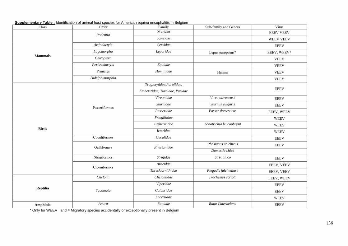
139
Supplementary Table : Identification of animal host species for American equine encephalitis in Belgium Class Order Family Sub-family and Genera Virus
Mammals
Rodentia Muridae EEEV VEEV Sciuridae WEEV VEEV
Artiodactyla Cervidae EEEV Lagomorpha Leporidae Lepus europaeus* EEEV, WEEV* Chiroptera VEEV
Perissodactyla Equidae VEEV Primates Hominidae Human VEEV
Didelphimorphia VEEV
Birds
Passeriformes
Troglotytidae,Parulidae,
Emberizidae, Turdidae, Paridae EEEV
Vireonidae Vireo olivaceus# EEEV Sturnidae Sturnus vulgaris EEEV
Passeridae Passer domesticus EEEV, WEEV Fringillidae WEEV Emberizidae Zonotrichia leucophrys# WEEV
Icteridae WEEV Cuculiformes Cuculidae EEEV
Galliformes Phasianidae Phasianus colchicus EEEV
Domestic chick Strigiformes Strigidae Strix aluco EEEV
Ciconiformes Ardeidae EEEV, VEEV
Threskiornithidae Plegadis falcinellus# EEEV, VEEV
Reptilia
Chelonii Cheloniidae Trachemys scripta EEEV, WEEV
Squamata
Viperidae EEEV Colubridae EEEV Lacertidae WEEV
Amphibia Anura Ranidae Rana Catesbeiana EEEV * Only for WEEV and # Migratory species accidentally or exceptionally present in Belgium
Annexe 2
140
Diversity and ecology survey of mosquitoes potential vectors
in Belgian equestrian farms: a threat prevention of
mosquito-borne equine arboviruses arboviruses
Accepté sous réserve de modifications mineures, Preventive Veterinary Medicine
Annexe 2
Slimane Boukraaa, *, Maria A. de La Grandiereb, Thomas. Bawina, Fara N.
Raharimalalac, Jean-Yves Zimmera, Eric Haubrugea, Etienne Thiryb, Frédéric
Francisa
a Unit of Functional and Evolutionary Entomology, Gembloux Agro-Bio Tech,
University of Liège, Passage des Déportés 2, 5030 Gembloux, Belgium.
b Veterinary virology and animal viral diseases, Faculty of Veterinary Medicine,
FARAH Center, University of Liège, Quartier Vallée 2, Avenue de Cureghem 10,
B43b, B-4000 Liège, Belgium.
c Medical Entomology Unit, Pasteur Institute, Ambatofotsikely, 101-Antananarivo,
Madagascar.
Annexe 2
141
Abstract
Emergence of West Nile Virus was recently recorded in several European countries, which can lead to
severe health problems in horse populations. Europe is also at risk of introduction of mosquito-borne
equine alphavirus from Americas. Prevention of these arboviruses requires a clear understanding of
transmission cycles, especially their vectors. To characterize mosquito fauna, their ecology and
identify potential vectors of equine arboviruses in Belgium, entomological surveys of six equestrian
farms located in the Wolloon Region were conducted during 2011-2012. The harvest of mosquitoes
was based on larval sampling (272 samples from 111 breeding sites) and monthly adults trapping
(CO2-baited traps, Mosquito Magnet Liberty Plus). Among 51493 larvae and 319 adult mosquitoes
collected, morphological identification showed the presence of 11 species: Anopheles claviger
(Meigen), An. maculipennis s.l. (Meigen), An. plumbeus (Stephens), Culex hortensis (Ficalbi), Cx.
territans (Walker), Cx. pipiens s.l. L., Cx. torrentium (Martini), Coquillettidia richiardii (Ficalbi),
Culisata annulata (Schrank), Oc. cantens (Meigen), Oc. geniculatus (Olivier). Molecular
identification of Cx. pipiens species complex allowed the detection of three molecular forms,
Pipiens (92.3%), Molestus (4.6%) and Hybrid (3.1%). Larvae of Cx. pipiens sl and Cx.
torrentium were omnipresent and the most abundant species. Water troughs, ponds and slurry (liquid
manure) were the most favorable breeding sites of mosquito larvae. Based upon behavior and
ecology of the identified mosquito species, equestrian farms seem to provide a suitable environment
and breeding sites for the proliferation of potential vectors of arboviruses and those being a real
nuisance problem for horses and neighboring inhabitants.
Keywords: Belgium; Culex; Equine arbovirus; Horse; Mosquito; West Nile Virus
Annexe 2
142
Introduction
Viruses transmitted by hematophagous arthropods (arboviruses) represent a threat for animal
and human health with the increase of their emergence outside the endemic areas.
Environment and climate change, as well as intensification of international trade could favor
the re-emergence of vector-borne diseases (Gould and Higgs, 2009). In terms of morbidity
and mortality, mosquitoes are the most dangerous animals confronting mankind (Becker et al.,
2010). Among the hundreds viruses that can infect humans and animals, several are
transmitted by mosquitoes such as some of Togaviridae (Chikungunya, Sindbis, Equine
Encephalitis alphaviruses and Ross River virus), Flaviviridae (Yellow fever, Dengue, West
Nile Fever, Japanese and St. Louis encephalitis viruses) and Bunyaviridae (Rift Valley fever)
(Goddard, 2008). Mosquitoes are considered as the main vectors of some arboviruses that
affect horses and that cause serious diseases in humans during epidemics. Eastern Equine
Encephalitis virus (EEEV) is an Alphavirus that belongs to the family Togaviridae as Western
equine encephalitis virus (WEEV) and Venezuelan equine encephalitis virus (VEEV). The
EEEV is the most pathogenic arbovirus endemic in the Americas and can be distinguished
into two strains: South American and North American, the latter being more virulent for
humans. The EEEV causes sporadic infections in the human population during the summer
months and occasionally causes epidemics (Zacks and Paessler, 2010). For the horse, clinical
signs are non-specific and last 1-2 weeks, after which the outcome is either recovery or death.
There is no available treatment and the only protection against the disease remains the vector
control and vaccination (Davis et al., 2008). This arbovirus has been isolated from 20
mosquito species including Aedes vexans (Meigen), Ae. albopictus (Skuse), Culex pipiens s.l.
L., Culiseta morsitans (Theobald), and Cx. territans Walker (all: Diptera, Culicidae) that can
be found frequently in Europe (Scott and Weaver, 1989 and Pfeffer and Dobler, 2010). In
America, the majority of mosquito vectors of EEEV have been identified as belonging to the
genus Culex (melanoconion), which has a wider host range (Arrigo, 2010).
Western equine encephalitis virus (WEEV) is found in the Western part of North America and
South America. The WEEV is a recombinant virus between Sindbis virus and EEEV (Weaver
et al., 1997). Despite the decrease in the number of cases since the 1950s, WEEV is always a
risk to human and equine populations. As for the other encephalitis alphaviruses, WEEV
causes non-specific clinical signs of fever, meningitis or encephalitis according to the immune
status and age of the infected individual.
Annexe 2
143
No therapy is available and the only way to fight is also the routine immunization of horses
and the vector control (Davis et al., 2008 and Zacks and Paessler, 2010). The WEEV has two
transmission cycles; the main transmission cycle (North America) is maintained between Cx.
tarsalis (Coquillett) (Diptera: Culicidae) and host birds. The secondary cycle of transmission
(South America) involves Ae. melanimon Dyar (Diptera: Culicidae) and host mammals and /
or rodents (Pfeffer and Dobler, 2010). Aedes melanimon acts as a bridge vector to transmit the
disease to humans and horses, as Ae. dorsalis Meigen (Diptera: Culicidae) and Ae. campestris
(Dyar & Knab) (Diptera: Culicidae). Others vector species have been tested in laboratory
conditions: Cx. quinquefasciatus (Say) (Diptera: Culicidae), Cx. pipiens, and Ae. vexans
(Pfeffer and Dobler, 2010) .
Venezuelan equine encephalitis virus (VEEV) is an important emerging and re-emerging
pathogen of horses and humans in South America. During epidemics, the lethality mortality
rate varies from 19 to 83% of horses, while for humans, the mortality is less common but
important neurological complications are frequent. Epizootic strains are opportunistic for the
choice of potential vectors. A wide variety of vectors may be involved as Psorophora
confinnis (Lynch Arribalzaga), Ps. columbiae (Dyar & Knab), Oc sollicitans (Walker), or else
Oc. taeniorhynchus (Wiedemann) (all: Diptera: Culicidae) whose was considered as the
primary vector during an epizootic (Weaver et al., 2004 and Aguilar et al., 2011). Enzootic
strains are almost exclusively transmitted by mosquitoes of the genus Culex subgenus
Melanoconion (Weaver et al., 2004). Aedes albopictus is an efficient vector of VEEE under
laboratory conditions (Aguilar et al., 2011).
West Nilevirus (WNV) is an arthropod-borne virus worldwide distributed belonging to
Flaviviridae and to Flavivirus genus. This arbovirus is maintained in an enzootic cycle with
birds that serve as amplification reservoir hosts and mosquitoes as the principal transmitting
vectors. Humans and horses are susceptible hosts and can develop clinical illness but are
considered dead-end hosts because they do not frequently produce sufficient viremia to infect
mosquitoes, and therefore, do not contribute to the transmission cycle (Campbell et al., 2002
and Hayes et al., 2005). West Nilevirus was recognized as a cause of central nervous system
infections such as meningitis, encephalitis and myelitis (Campbell et al., 2002). The clinical
signs in horses are most commonly ataxia, weakness, and changes in mental state. However,
the majority of equines are very susceptible to WNV infection, which can be responsible for
encephalomyelitis in a fraction of infected animals, and may evolve into fatal encephalitis
(Campbell et al., 2002 and Venter et al., 2009).
Annexe 2
144
Mosquitoes in the genus Culex have been widely implicated as primary vectors of WNV
(Becker et al., 2010 and Calistri et al., 2010). Despite the low vector competence of these
species to transmit the WNV, other factors such as mosquito density, biting preference and
seasonal activity makes Culex species the most important mosquito genus in WNV
transmission (Kilpatrick et al., 2005). Culex univittatus (Theobald) (Diptera: Culicidae) is
considered the primary vector in Africa (Hubalek and Halouzka, 1999) and Cx. vishnui
Theobald (Diptera: Culicidae) in Asia. In the northeast America, Cx. pipiens s.l. is regarded as
the most important vector of WNV and in the southeast the subspecies Cx. quinquefasciatus is
considered as an important vector. Culex tarsalis takes place as the primary vector in the west
American (Campbell et al., 2002 and Kilpatrick et al., 2005). In Europe, WNV has been
detected from largely ornithophilic mosquitoes especially Cx. pipiens s.l., Cx. modestus
(Ficalbi), Cx. torrentium (Martini), Coquillettidia richiardii (Ficalbi), and occasional vectors
such as Ochlerotatus cantans (Meigen) and Anopheles maculipennis s.l. (Meigen) (all:
Diptera: Culicidae) (Engler et al., 2013). Aedes albopictus is also a competent vector of WNV
and has been demonstrated experimentally that can infect horses (Bunning et al., 2002).
Since that WNV was first detected in New York in 1999, more than 26581 horses (period,
1999–2013) were affected in the United States
(http://www.aphis.usda.gov/vs/nahss/equine/wnv/wnv_distribution_maps.htm),
with many horses being euthanized because of grave prognosis indicating that several
infections were fatal (Sebastian et al., 2008). A strong resurgence of horse and human cases of
WNV was recorded in Europe in the last decade, including Hungary, Italy, southern France,
Portugal, Romania, Spain and more recently Greece (Engler et al., 2013 and Di Sabatino et
al., 2014). The main route of introduction of WNV in the regions of Europe with a temperate
climate is most likely by infected migratory birds from Africa (Hubálek and Halouzka, 1999
and Calistri et al., 2010). Moreover, WNV could be introduced in Western Europe by birds
arriving from central Europe (Koopmans et al., 2008).
West Nile virus circulation in Europe is confined to two basic types of cycles and ecosystems.
The first belongs to rural cycle where wetland birds were usually serve as amplification hosts
and ornithophilic mosquitoes such as Cx. pipiens pipiens as vectors. The second is the urban
cycle with synanthropic or domestic birds as amplifying hosts and mosquitoes feeding on both
birds and mammals, mainly Cx. p. molestus (Hubalek and Halouzka, 1999). Environmental
factors, including human activities that enhance vector population densities (irrigation, heavy
rains followed by floods, higher than usual temperatures, and formation of ecologic niches
Annexe 2
145
enabling the mass breeding of mosquitoes) allow the reemergence of this mosquito-borne
disease (Hubalek and Halouzka, 1999). An understanding of arboviral transmission cycles
begins with the correct identification of potential vectors and thorough knowledge of their
bioecology in the environment. The goals of this work were to determine diversity,
abundance, and seasonal dynamics of potential mosquito vectors for equine arboviruses in
equestrian farms in the Walloon Region of Belgium. Additionally, this study is intended to be
useful in assessing the risk that those mosquito species and their breeding sites represent for
transmission of equine arboviruses in Belgium.

Figure
Chênée
Provinc
1. Map of h
(Ch), Sprim
ces: Liège (o
horses densi
mont (Sp), L
on the right)
ity by locali
La Reid (Rd
) and Namu
ities showin
d), Gemblou
ur (on the le
ng the six sa
ux (Gx), Ma
eft).
ampling site
alone (Mn)]
es [Warsage
] in the two
e (Wg),
Annexe 2
146
Material and methods
Study sites
The present study was performed in 2011-2012, with the aim to investigate the mosquito
fauna species and their bioecology in the equestrian farms. The study was conducted in six
locations in Belgium belonging to Namur and Liège Provinces (Figure 1). Each of the six
equestrian farms selected contain a hundred horses and had two types of environments: horse
livestock in the buildings of the farms (stables) and surrounding grasslands. In addition to the
presence of many breeding larval sites in and around stables studied, the farms were also
close to forests and streams, which give humid environment and provide suitable conditions
for the proliferation of mosquito species. Localities of the equestrian farms inspected are:
Gembloux (50°34'40"N, 4°44'40"E; 33 m above sea level), Malonne (50°27'06"N, 4°48'15"E;
85 m a.s.l.), Warsage (50°43'38"N, 5°47'57"E; 223 m a.s.l.), Chênée (50°37'00"N, 5°37'58"E;
151 m a.s.l.), La Reid (50°28'51"N, 5°46'38"E; 372 m a.s.l.), and Sprimont (50°32'46"N,
5°40'16"E; 274 m a.s.l.). Weather data (precipitation and temperature) were obtained from
local meteorological stations of Royal Meteorological Institute (RMI®) and were presented by
Ombrothermic diagram for each of the localities of equestrian farms (Figure 2).
Mosquito collection
Larvae sampling
Larvae sampling is realized in all potential larval habitats reported after inspection of each
study site and its surroundings (a circle of radius of 250 ± 50 m). All sites have been
investigated 4 times/year, in 2011 (June, July, August and October) and 2012 (June, July,
August and September). Larvae were collected by using the dipper method made-up by a
metal pan (500 ml). However, the smallness of some breeding sites such as the temporal
puddles and the inaccessibility of others sites like used tires had necessitated the use of an
aquarium net 12 X 10 cm to inspect them and collect immature stages. We collected in several
places of breeding sites to obtain homogeneous samples (4-10 times). In the case of breeding
sites which are formed by tires, the sample is composed by the sum of 4-10 tires selected on
all tires placed in the same location (heap of tires). Data sheets of physical and biological
characteristics were designed for each breeding site, to collect all possible data such as: type
(artificial/natural), kind (buckets/tires/water troughs/puddle/pond/streams), sunshine
(full/partial/shadow), state water (permanent/temporary, standing/running), water depth and
vegetation situation (presence/absence of floating and/or emergent vegetation) (Figure 3).

Figure
2. Weather
data (ombr
rothermic di
iagrams) of
f each studie
ed station in
A
n 2011 and 2
Annexe 2
147
2012.

Figure
farms in
Obstacl
Waterco
behind s
3. Some ex
nspected: a,
e of cross c
ourse (in gr
stables; j, T
xamples of p
Water trou
country race
assland); g,
Tractor rut; k
positive bree
ughs; b, Pud
es (water ob
slurry (or l
k, Used tire
eding sites o
ddle behind
bstacle); e, S
liquid manu
s; l, Puddle
of mosquito
stables (farm
Septic of ho
ure); h, Pond
and horse h
o larvae in t
m); c, Tarpa
rse washing
d (in grassla
hoofprint (g
the equestria
aulin cover
g residues; f
and); i, Drai
grassland).
ans
; d,
f,
in
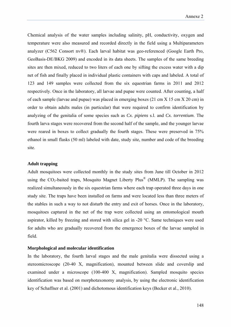
Annexe 2
148
Chemical analysis of the water samples including salinity, pH, conductivity, oxygen and
temperature were also measured and recorded directly in the field using a Multiparameters
analyzer (C562 Consort nv®). Each larval habitat was geo-referenced (Google Earth Pro,
GeoBasis-DE/BKG 2009) and encoded in its data sheets. The samples of the same breeding
sites are then mixed, reduced to two liters of each one by sifting the excess water with a dip
net of fish and finally placed in individual plastic containers with caps and labeled. A total of
123 and 149 samples were collected from the six equestrian farms in 2011 and 2012
respectively. Once in the laboratory, all larvae and pupae were counted. After counting, a half
of each sample (larvae and pupae) was placed in emerging boxes (21 cm X 15 cm X 20 cm) in
order to obtain adults males (in particular) that were required to confirm identification by
analyzing of the genitalia of some species such as Cx. pipiens s.l. and Cx. torrentium. The
fourth larva stages were recovered from the second half of the sample, and the younger larvae
were reared in boxes to collect gradually the fourth stages. These were preserved in 75%
ethanol in small flasks (50 ml) labeled with date, study site, number and code of the breeding
site.
Adult trapping
Adult mosquitoes were collected monthly in the study sites from June till October in 2012
using the CO2-baited traps, Mosquito Magnet Liberty Plus® (MMLP). The sampling was
realized simultaneously in the six equestrian farms where each trap operated three days in one
study site. The traps have been installed on farms and were located less than three meters of
the stables in such a way to not disturb the entry and exit of horses. Once in the laboratory,
mosquitoes captured in the net of the trap were collected using an entomological mouth
aspirator, killed by freezing and stored with silica gel in -20 °C. Same techniques were used
for adults who are gradually recovered from the emergence boxes of the larvae sampled in
field.
Morphological and molecular identification
In the laboratory, the fourth larval stages and the male genitalia were dissected using a
stereomicroscope (20-40 X, magnification), mounted between slide and coverslip and
examined under a microscope (100-400 X, magnification). Sampled mosquito species
identification was based on morphotaxonomy analysis, by using the electronic identification
key of Schaffner et al. (2001) and dichotomous identification keys (Becker et al., 2010).
Annexe 2
149
Because of the remarkable morphological resemblance between Cx. pipiens and Cx.
torrentium the separation at larval stage or adult females is very delicate. For this purpose,
morphological identification of these two species has been based much more on the
examination of the male genitalia, which carries most discriminating and distinguishing
characters. Regarding the species of the tribe of Aedini and their recent reclassifications, we
maintained in this article the use of the traditional names of species, with the only exception
of the elevating of Ochlerotatus to genus (Reinert, 2000). Members of the Anopheles
maculipennis complex have not been differentiated in this study and are cited as An.
maculipennis s.l. However, the molecular study was performed to identify the different forms
of Cx. pipiens complex. Total DNA from 126 individual mosquitoes (4th stage larvae),
selected from six more abundant breeding sites of Cx. pipiens complex was extracted with the
DNeasy Blood & Tissue kit (Qiagen), according to the method recommended by the
manufacturer and diluted in a final volume of 70 µl of TE. Purity and quantity of DNA then
checked with Nanodrop (NanoDrop ND-1000 Spectrophotometer, Isogen®) and finally stored
at -20 °C. A multiplex PCR assay which targeted the region of a microsatellite locus (CQ11)
was used with 60 ng of genomic DNA. Sets of primers were composed by the specific reverse
primer for Molestus: molCQ11R 5’-CCCTCCAGTAAGGTATCAAC-3’, for Pipiens:
pipCQ11R 5’-CATGTTGAGCTTCGGTGAA-3’, and the common forward primer: CQ11F2
5’-GATCCTAGCAAGCGAGAAC-3’ (Bahnck & Fonseca, 2006). Reactions were performed
in 50 μl volume containing 2 µl of 60 ng of DNA template, 5 µl of Dream Taq PCR buffer 1X
(Fermentas, St. Leon-Rot, Germany), 1 µl of 1 mM of each primer (molCQ11, pipCQ11 and
CQ11F2), 1 µl of 0.2 mM dNTP and 0.5 µl of Dream Taq (Fermentas, St. Leon-Rot,
Germany). PCR amplification conditions were 5 min at 94 °C for the initial denaturation,
followed by 40 cycles of 94 °C for 30 s, 54 °C for 30 s, 72 °C for 40 s and a final extension at
72 °C for 5 min. Aliquot of 10 μl of the amplification products were run by electrophoresis on
a 2% agarose gel stained with SYBR® Safe DNA (Invitrogen) at 90 V for 30 min, then
photographed with Gel Doc 2000 system (BioRad, USA). Selected samples of positive CQ11
PCR amplifications were purified and sent for sequencing to GATC Biotech (Germany)
(www.gatc-biotech.com). The sequences results were compared with data available in NCBI
(www.ncbi.nlm.nih.gov) and then deposit in GenBank.

Annexe 2
150
Diversity and Statistical analysis
The results obtained on the survey of Culicidae in Belgian equestrian farms are treated with
ecological indices of species diversity and similarity between different study sites. We
expressed the species richness (S), relative abundance (pi = ni / N where ni = number of the i-
th species and N is the total number of individuals in the studied station) and frequency of
occurrence (Constancy) of species in each study sites. Constancy (C) was calculated with the
index C = P × 100 / N, where P is the number of surveys (larval samples) containing the
analyzed species and N the total number of surveys. Then it was categorized into five classes
using Surge’s rule (Scherrer, 1984): Number of classes (N.C.) = 1 + (3.3 log 10 P’), where P'
is the total number of inventoried mosquitoes in the study sites. In this study, the following
groupings for species categories were proposed: 0 < C ≤ 26% accidental (A); 26% < C ≤ 54%
infrequent (I); 54% < C ≤ 80% constant (C) and 80% < C ≤ 100% for omnipresent (O)
species. In parallel, larval abundance per station and per year was classified into four
categories as low (<10 larvae present), intermediate (11-100 larvae present), high (101-500
larvae present), or very high abundance (>500 larvae present). In each study site, Shannon-
Weavers's diversity index (H') and Evenness (J’) were calculated. The first, H' = - Σpi log2 pi
(where pi the relative frequency of species i-th in the studied habitat) was used to measure and
evaluate heterogeneity of mosquito species diversity for each of the six equestrian farms.
Evenness (J’) or equitability = H' / Hmax (where Hmax = log2 S) was calculated to measure the
distribution of individuals within species irrespective of species richness. It varies from 0, if
only one species dominates, to 1, if all species show similar abundance (Silver, 2008). The
degree of similarity/dissimilarity between study site compared to mosquito species was
expressed by index (Qi) and distance (Qiδ) of Jaccard [Qi = a / (a + b + c) where a is number
of species commons to both sites; b and c are number of species absents in one of the two
sites] [Qiδ = 1 - Qi] (Silver, 2008). Jaccard’s distance and dendrogram were carried out in R,
version 3.1.0 (R Core Team 2014). The mean abundance per species was treated with an
analysis of variance (ANOVA-1) with equestrian farm stations (q = 6) and months (n = 4).
When assumptions of the one-way ANOVA (normality of populations and homoscedasticity)
were not met, Kruskall-Wallis’s test was applied. All statistical analyses were performed
using Minitab® software version 16. In all the cases, results were considered as statistically
significant when the p-value was lower than 5% (p < 0.05).
Table 1
2012.
Station
Gembloux
Malonne
Chênée
Warsage
Sprimont
La Reid
Totals
F
b
1. Numbers
Veryabund
2011
u 4 (16.0%)
e 0 (0.0%)
4 (19.0%)
e 0 (0.0%)
t 0 (0.0%)
d 4
(17.4%) 12
(9.8%)
Figure 4. Cl
between equ
and percen
y high dance
2012
3 (10.0%)
8
2 (7.7%) 2
2 (6.5%)
1 (6.7%) 7
3 (14.3%)
4 (15.4%)
7
15 (10.1)
luster analy
uestrian farm
ntages of sa
High abunda
2011 2
8 (32.0%)(1
2 (10.0%)(3
11 (52.4%) (6
7 (50.0%)(2
10 (50.0%) (3
7 (30.4%)(1
45 (36.6%) (
ysis of mosq
ms studied i
ampling of l
ance I
2012 20
5 16.5%)
8(32.0
8 30.8%)
16(80.0
2 6.5%)
5(23.8
4 26.7%)
7(50.0
8 38.1%)
9(45.0
4 15.4%)
8(34.8
31 (20.8)
53(43.1
quito fauna,
in 2011 and
larval abund
ntermediate abundance
11 2012
8 0%)
10 (33.3%
6 0%)
12 (46.2%
5 8%)
17 (54.8%
7 0%)
7 (46.7%
9 0%)
6 (28.6%
8 8%)
14 (53.8%
3 1%)
66 (44.
by Jaccard
d 2012.
dances per
Low a
2011
%) 5
(20.0%)
%) 2 (10%)
%) 1 (4.8%)
%) 0 (0.0%)
%) 1 (5.0%)
%) 4
(17.4%)
3)13
(10.6%)
’s distance
station in 2
abundance
2012
12 (40.0%)
4 (15.4%)
) 10
(32.3%)
) 3 (20.0%)
) 4 (19.0%)
4 (15.4%)
37 (24.8)
2011 and
No. Samples
2011201
2
25 30
20 26
21 31
14 15
20 21
23 26
123 149
Annexe 2
151
Results
In total, 123 and 149 larval samples were collected from the six equestrian farms in 2011 and
2012 respectively. Samples with intermediate larval abundance presented 43.1% (2011) and
44.3% (2012), followed by high abundance with 36.6% (2011) and 20.8% (2012) of all
samplings per year (Table 1).
A total of 51493 larvae and 319 adults were collected, morphological identifications showing
the presence of 11 species: 7 obtained by larval sampling and 10 by adult trapping (Table 2
and Table 3). Among the larval species identified in all equestrian farms, Cx. pipiens s.l.
(68.0% in 2011; 79.1% in 2012) and Cx. torrentium (22.4% in 2011; 15.5% in 2012) were the
most abundant species. In parallel, the two most abundant adult species were Cq. richiardii
(54.2%) and Cx. pipiens s.l./Cx. torrentium (35.7%). Morphological differentiation between
adult females of Cx. pipiens s.l. and Cx. torrentium was not possible and these two species
were referred to as Cx. pipiens s.l./Cx. Torrentium.
All specimens belonging to the An. maculipennis species complex were not identified to the
species level. However, molecular identification of Cx. pipiens species complex using
multiplex PCR based on the microsatellite locus CQ11 allowed to detect the presence of three
molecular form, Cx. pipiens form pipiens, Cx. pipiens f. molestus and Cx. pipiens f. hybrid
(heterozygotous). Among the 126 specimens selected for molecular study, only 65 PCR gave
positive results. Sixty individuals belonged to Cx. pipiens f. pipiens (92.3% of positive PCR)
only 3 to Cx. pipiens f. molestus (4.6%), and 2 to Cx. pipiens f. hybrid (3.1%). Nucleotide
sequences of 10 Cx. pipiens f. pipiens and 2 Cx. pipiens f. molestus are deposited in GenBank
under accession numbers KP666140, KP675968-KP675972, KP675976, and KP675978-
KP675980 for Pipiens form and KP675973 and KP675982 for Molestus form.
Taxonomic diversity differed among the six equestrian farms and years (2011 and 2012)
(Table 2). Richness and Shannon-Wiener diversity index were both higher in Sprimont (2011)
and Chênée (2012) where S = 7, H' = 1.53 and S = 7, H' = 1.6, respectively. These two
stations were very close (≈ 8.3 km) and similar (Qi = 100%; Qiδ = 0.0%). During larval
sampling for both years, lowest species richness was observed in Warsage (only 3 species
were identified). The lowest similarities of Jaccard were observed between all pairs of
Warsage and each of others stations (Qi < 60%) (Figure 4). Even so, highest evenness were
highlighted that in this station, the three present species were in equilibrium between them
and there was not a clear dominance in 2011 (J = 0.66) and 2012 (J = 0.86). The evenness
value in 2011 was also high in Malonne (0.71) and Chênée (0.68).
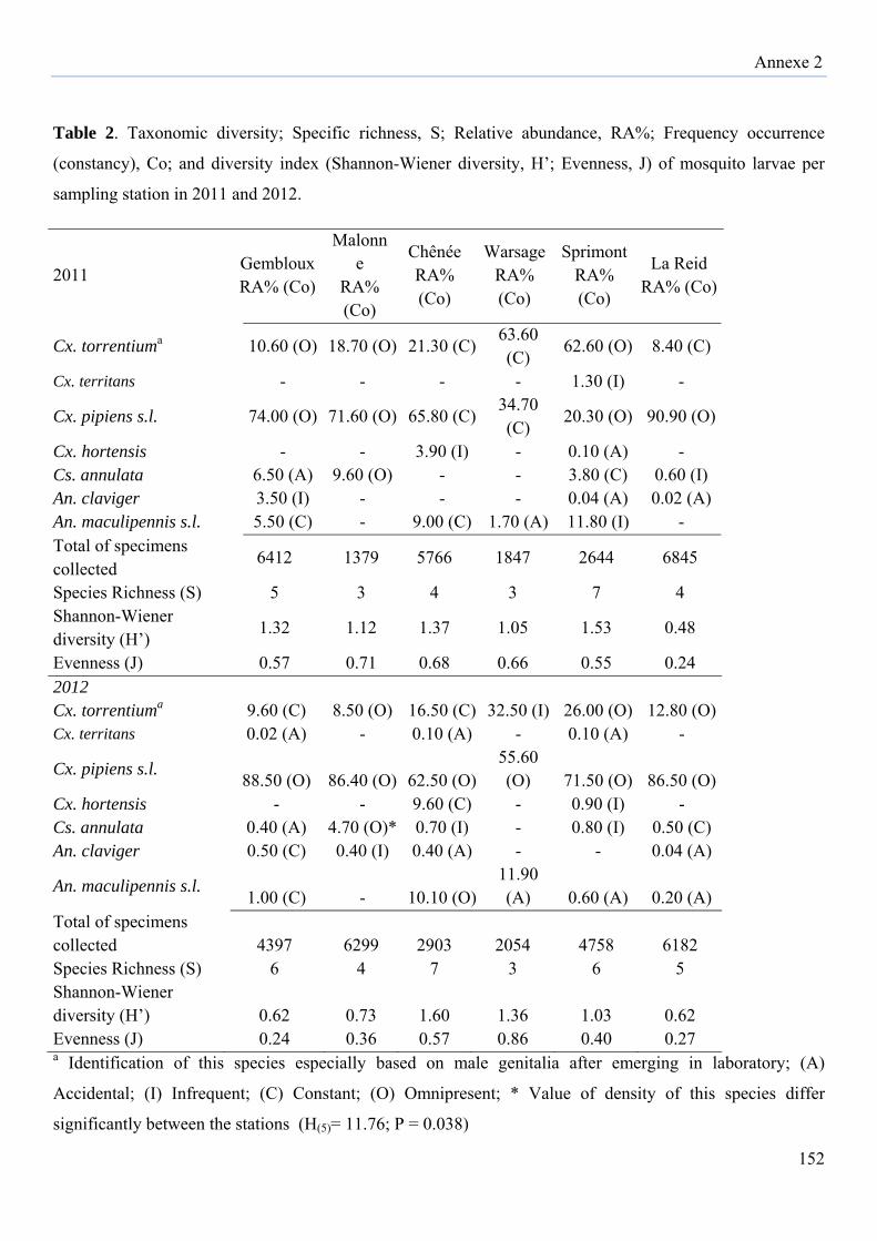
Annexe 2
152
Table 2. Taxonomic diversity; Specific richness, S; Relative abundance, RA%; Frequency occurrence
(constancy), Co; and diversity index (Shannon-Wiener diversity, H’; Evenness, J) of mosquito larvae per
sampling station in 2011 and 2012.
2011 Gembloux RA% (Co)
Malonne
RA% (Co)
Chênée RA% (Co)
Warsage RA% (Co)
Sprimont RA% (Co)
La Reid RA% (Co)
Cx. torrentiuma 10.60 (O) 18.70 (O) 21.30 (C)63.60 (C)
62.60 (O) 8.40 (C)
Cx. territans - - - - 1.30 (I) -
Cx. pipiens s.l. 74.00 (O) 71.60 (O) 65.80 (C)34.70 (C)
20.30 (O) 90.90 (O)
Cx. hortensis - - 3.90 (I) - 0.10 (A) - Cs. annulata 6.50 (A) 9.60 (O) - - 3.80 (C) 0.60 (I) An. claviger 3.50 (I) - - - 0.04 (A) 0.02 (A) An. maculipennis s.l. 5.50 (C) - 9.00 (C) 1.70 (A) 11.80 (I) - Total of specimens collected
6412 1379 5766 1847 2644 6845
Species Richness (S) 5 3 4 3 7 4 Shannon-Wiener diversity (H’)
1.32 1.12 1.37 1.05 1.53 0.48
Evenness (J) 0.57 0.71 0.68 0.66 0.55 0.24 2012 Cx. torrentiuma 9.60 (C) 8.50 (O) 16.50 (C) 32.50 (I) 26.00 (O) 12.80 (O) Cx. territans 0.02 (A) - 0.10 (A) - 0.10 (A) -
Cx. pipiens s.l. 88.50 (O) 86.40 (O) 62.50 (O)55.60 (O) 71.50 (O) 86.50 (O)
Cx. hortensis - - 9.60 (C) - 0.90 (I) - Cs. annulata 0.40 (A) 4.70 (O)* 0.70 (I) - 0.80 (I) 0.50 (C) An. claviger 0.50 (C) 0.40 (I) 0.40 (A) - - 0.04 (A)
An. maculipennis s.l. 1.00 (C) - 10.10 (O)11.90 (A) 0.60 (A) 0.20 (A)
Total of specimens collected 4397 6299 2903 2054 4758 6182 Species Richness (S) 6 4 7 3 6 5 Shannon-Wiener diversity (H’) 0.62 0.73 1.60 1.36 1.03 0.62 Evenness (J) 0.24 0.36 0.57 0.86 0.40 0.27 a Identification of this species especially based on male genitalia after emerging in laboratory; (A)
Accidental; (I) Infrequent; (C) Constant; (O) Omnipresent; * Value of density of this species differ
significantly between the stations (H(5)= 11.76; P = 0.038)
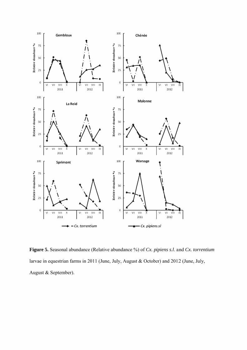
Figure
larvae in
August
5. Seasonal
n equestrian
& Septemb
l abundance
n farms in 2
ber).
e (Relative a
2011 (June,
abundance %
July, Augu
%) of Cx. p
st & Octobe
ipiens s.l. a
er) and 2012
and Cx. torr
2 (June, Jul
entium
ly,
Annexe 2
153
However, in 2012, Cx. pipiens s.l. was strongly dominant (up to 88.5% in Gembloux) and
evenness values were low in Gembloux (0.24), La Reid (0.27), Malonne (0.36), and Sprimont
(0.40) (Table 2).
Table 3 shows that some species have a high ecological plasticity and their larvae can develop
and encounter a wide range of artificial or natural/semi-natural breeding sites. There were in
particular, the larvae of Cx. pipiens s.l., Cx. torrentium, and Cs. annulata. However, other
species are more related to characteristic ecological features of some natural breeding sites
(such as An. claviger and Cx. territans). At the level of six equestrian farms studied, water
trough, ponds, and purin (liquid manure) were the most favorable habitats in development of
mosquito larvae. The highest mosquito diversity was observed in ponds (all species) and
water trough (6 species). Of all the breeding sites (111) sampled in studied stations, 100%
contained larvae of Cx. pipiens s.l., Cx. torrentium. The highest larval densities for these two
species for artificial breeding sites occurred in water troughs, puddles, and abandoned
buckets. For natural/semi-natural breeding sites, the highest larval densities occurred in ponds
and liquid of manure (slurry) (Table 4).
Figure 5 presents seasonal dynamics (relative abundance) of Cx. pipiens s.l. and Cx.
torrentium larvae in the six equestrian farms in 2011 (VI, VII, VIII and X) and 2012 (VI, VII,
VIII and IX). The relative abundance of these mosquito larvae was expressed as the mean
number of larvae in each month by the total number of larvae of the species in each station.
Seasonal dynamics of these two species showed different variations depending on stations and
months. In most stations, the relative abundance of Cx. pipiens s.l. and Cx. torrentium follows
a classic thermophilic trend. However, variations and heterogeneity of the dynamics were
recorded mainly in 2012. The calculation of Pearson correlation coefficients (PCC) shows that
there is a significant relationship between the relative abundance of Cx. pipiens s.l. larvae and
the mean temperature in Chênée in 2011. The correlation coefficient of 0.972 (p = 0.028)
reflects indeed a strong positive linear relationship between the sampled larvae and the mean
temperature measured in this station. Strong correlations were also obtained in 2011 [Chênée:
Cx. torrentium (PCC = 0.914; P = 0.086) and Warsage: Cx. torrentium (PCC = 0.937; P =
0.063)] and 2012 between larval abundance of both species and mean temperatures. In
parallel, the PCC shows a significant correlation between the relative abundance of Cx.
torrentium and rainfall in 2012 [Chênée: PCC = 0,988; P = 0.012 and La Reid: PCC = 0,976;
P = 0.024]. There were no significant relationships between relative abundance of both
species and rainfall in 2011 and they also were remained low for most stations in 2012.
Annexe 2
154
Table 3. Relative abundance (%) of adult mosquitoes collected using Mosquito Magnet
Liberty Plus® in equestrian farms studied in 2012.
*Morphological identification between females (majority of specimens) of Cx. pipiens s.l. and
Cx. torrentium was not possible, because characters that are used to separate these species are
damaged or not visible.
Species Gembloux Malonne Chênée Warsage Sprimont La Reid Cx. pipiens s.l./Cx. torrentium*
84.0 76.9 76.2 63.6 38.9 3.4
Cx. hortensis - - 4.8 - - - Cs. annulata 4.9 15.4 9.5 18.2 - 0.6 An. claviger 4.9 7.7 4.8 - - 3.4 An. maculipennis s.l. - - 4.8 9.1 - - An. plumbeus - - - 9.1 - - Oc. cantens - - - - - 1.1 Oc. geniculatus 3.7 - - - - - Cq. richiardii 2.5 - - - 61.1 91.4 Number of specimens collected
81 13 21 11 18 175
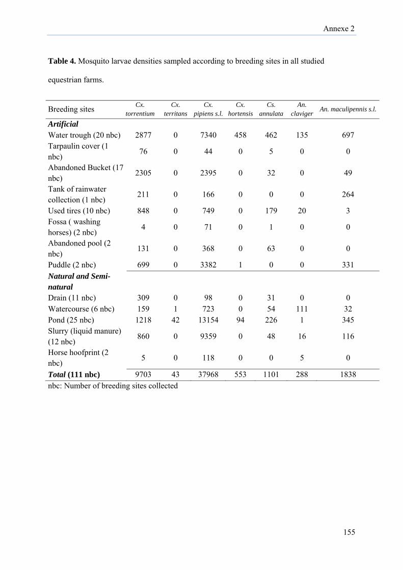
Annexe 2
155
Table 4. Mosquito larvae densities sampled according to breeding sites in all studied
equestrian farms.
Breeding sites Cx. torrentium
Cx. territans
Cx. pipiens s.l.
Cx. hortensis
Cs. annulata
An. claviger
An. maculipennis s.l.
Artificial Water trough (20 nbc) 2877 0 7340 458 462 135 697 Tarpaulin cover (1 nbc)
76 0 44 0 5 0 0
Abandoned Bucket (17 nbc)
2305 0 2395 0 32 0 49
Tank of rainwater collection (1 nbc)
211 0 166 0 0 0 264
Used tires (10 nbc) 848 0 749 0 179 20 3 Fossa ( washing horses) (2 nbc)
4 0 71 0 1 0 0
Abandoned pool (2 nbc)
131 0 368 0 63 0 0
Puddle (2 nbc) 699 0 3382 1 0 0 331 Natural and Semi-natural
Drain (11 nbc) 309 0 98 0 31 0 0 Watercourse (6 nbc) 159 1 723 0 54 111 32 Pond (25 nbc) 1218 42 13154 94 226 1 345 Slurry (liquid manure) (12 nbc)
860 0 9359 0 48 16 116
Horse hoofprint (2 nbc)
5 0 118 0 0 5 0
Total (111 nbc) 9703 43 37968 553 1101 288 1838 nbc: Number of breeding sites collected
Annexe 2
156
Discussion
Compared with 2011, the number of collected mosquitoes in the six equestrian farms studied
(Gembloux, Malonne, Chênée, Warsage, Sprimont, and La Reid) was nearly identical that in
2012. In both years, most samples were characterized with intermediate and high abundance
of larvae. Eleven species were encountered during the present study on Belgian equestrian
farms, whereas 30 confirmed species have been recorded on the entire country (Dekoninck et
al., 2011a and Versteirt et al., 2013). Only seven taxa were identified by larval
morphotaxonomic. Adult morphotaxonomic and genitalia analysis have allowed to identify
four others species which are An. plumbeus, Oc. cantens, Oc. geniculatus, and Cq. richiardii.
Romi et al. (2004) also recorded eleven species in a study on potential vectors of WNV
following an equine disease outbreak in Toscana region (Italy).
Among the identified larvae in all equestrian farms, Cx. pipiens s.l. accounted for more than
73% of the mosquitoes collected during both studied years. Sebastian et al. (2008) found
similar results and confirmed that Cx. pipiens is the most abundant species found during an
entomological investigation of WNV epidemic in a horse farm. Molecular identification of
Cx. pipiens species, allowed detecting the presence of three molecular forms (Pipiens,
Molestus, and Hybrid). Pipiens form constituted itself more than 92% of positive selected
specimens. Similar results were found by Osório et al. (2014), where they found that the
majority of specimens of Cx. pipiens s.l. were identified as Cx. pipiens f. pipiens and followed
with less importance by Cx. pipiens f. molestus, and hybrid forms. Rudolf et al. (2013) also
confirmed that the predominant species in Germany are Cx. pipiens pipiens biotype pipiens
and Cx. torrentium. In temperate areas, Cx. pipiens s.l. can be subdivided into two distinct
biotypes, which are morphologically indistinguishable but differ greatly in physiology and
behaviour. The biotype pipiens is mainly ornithophilic, anautogenous, and eurygamous,
whereas the biotype molestus is mammalophilic, autogenous, and stenogamous (Becker et al.,
2010 and Osório et al., 2014). Hybridization between the both forms can also be found in
sympatric with the two distinct biotypes pipiens and molestus (Bahnck and Fonseca, 2006).
This hybrid form has been observed in different countries in Europe (Reusken et al., 2010 and
Osório et al., 2014). The hybrids showed opportunistic feeding behaviour and therefore can
bite both birds and mammals. Its behaviour and ecological flexibility to grow in the same
habitats of pipiens and molestus forms may serve to be as important bridge vectors for the
transmission of arboviruses from infected birds to mammals, and thus complicating the
epidemiologic landscape (Reusken et al., 2010 and Osório et al., 2014).
Annexe 2
157
Many researches demonstrated that Cx. pipiens is one of principal vectors of WNV in Europe
(Hubalek and Halouzka, 1996 and Becker et al., 2010), including in Italy where it was
suspected to play a major role in the transmission of this infectious agent in horse farms
(Romi et al., 2004).
Culex torrentium was also largely abundant and omnipresent in the most equestrians farms
studied. Morphological differentiation between larvae of Cx. pipiens s.l. and Cx. torrentium
was very delicate and even impossible between females of these both species. For that reason,
we based much more on male genitalia examinations, which were considered as a good
discriminatory character between these two species (Schaffner et al., 2001 and Becker et al.,
2010). Others researches confirmed that this species is widespread and common in Europe
where it is dominant in northern areas (Rudolf et al., 2013 and Hesson et al., 2014). Despite
the ignorance of a vectorial role of Cx. torrentium relative to the transmission of WNV, its
abundance and co-dominance alongside of Cx. pipiens in central and northern Europe require
deepening taxonomic studies to avoid identification mistakes and determine the true vectors.
In parallel, it is also advisable to carry out local studies on vector competence and capacity of
Cx. torrentium for WNV and other arboviruses (Weitzel et al., 2011 and Hesson et al., 2014).
We confirmed the presence and establishment of Cx. hortensis, recently identified for the first
time in Belgium by the MODIRISK (Versteit et al., 2012). In this study, Cx. hortensis was
found in two localities (Chênée and Sprimont) with both the larval and adult stages. Larvae of
this species were sampled primarily in the water troughs in grasslands and less important at
the ponds level. Larvae of Cx. hortensis may develop in varied breeding sites with presence or
not of plants such as cemented drinking troughs, puddles in a torrent-bed, ponds sides, and
drain pipes (Schaffner et al., 2001). Culex territans was found only in the larval stage and
only in the natural/semi natural breeding sites (ponds and streams). Becker et al. (2010) noted
that preferred larval habitats of Cx. territans are permanent bodies of water such as ponds,
swamps, pools along streams, edges of lakes, or slow streams. Just like the females of Cx.
hortensis, Cx. territans feeds especially on batrachians and reptiles. Both species have never
been involved in the transmission of human pathogens (Schaffner et al., 2001). However,
recent research suggests that some snakes and amphibian species were found competent hosts
for EEEV. In addition, some snakes may represent an overwintering host and can support
long-term virus replication, especially at low temperatures (White et al., 2011). Burkett-
Cadena et al. (2008) demonstrated that Cx. territans can also feed on birds and mammals what
Annexe 2
158
constitutes the possibility that this species may be important in the transfer of arboviruses to
and from ectotherms and other host groups. The competency of Cx. territans for EEEV is
unknown to this day (White et al., 2011), but this mosquito was found to be infected by this
arbovirus (Burkett-Cadena et al., 2008). Culex territans can therefore be considered as
potential and probable vector of EEEV in the case of its overgrowth and accidental biting of
horses.
For adult trapping, Cq. richiardii was the most abundant species in equestrian farms, but this
abundance may be explained by the studied station “La Reid”, where a marsh with water
plants was present around the farm. These aquatic plants are essential for the biology of Cq.
richiardii immature stages. Becker et al. (2010) noted that breeding sites of this species may
be various permanent water bodies where larvae and pupae live submerged and obtain oxygen
from the aerenchyma of aquatic plants. In addition, the females can cause a severe nuisance to
humans and domestic animals in the surroundings of fresh waters or slightly saline marshes,
lakes, and estuaries (Becker et al., 2010). Few adult specimens of An. plumbeus were captured
in equestrian farm (only in Warsage). However, a recent data indicated a strong population
expansion of this potential vector of Plasmodium falciparum (Haemosporida: Plasmodiidae)
and WNV all over Belgium and demonstrated its adaptation to man-made artificial breeding
sites (Dekoninck et al., 2011b).
In the majority of breeding sites found in the equestrian farms studied, Cx. pipiens s.l. and Cx.
torrentium were the most frequent mosquito larvae collected, where they co-occurred in the
majority of artificial and natural/semi-natural breeding sites sampled. Higher number of
mosquito species coexisted in water troughs, used tires, ponds, and watercourses compared
with other breeding site types. Our results suggest that water troughs and ponds are numerous
and more productive habitats for Cx. pipiens s.l. and Cx. torrentium, along with abandoned
buckets, puddles, and liquid manures (slurry). Although slightly less abundant, Cs. annulata
preferred similar habitats. Very wealthy in organic matter, liquid manure (or slurry) which
flow from the dung heaps was one of the most favorable breeding sites for the development of
Cx. pipiens s.l. larvae. Elimination and cleaning of this breeding site may therefore play an
interesting role in the reduction and control of nuisance caused by Cx. pipiens s.l. None
breeding site was found inside of the studied stations for the four species An. plumbeus, Oc.
cantens, Oc. geniculatus, and Cq. richiardii that were trapped only in adult stages. Despite the
availability of favorable breeding sites for the development of Aedes/Ochlerotatus larvae, no
species of this genus were identified in the larval stage.
Annexe 2
159
In parallel, equestrian farms studied appear to contain various favourable habitats to both
pipiens and molestus forms, and therefore a high-level of hybridization outcome of
interbreeding between both forms. Further detailed researches are needed on local ecology
(breeding sites, seasonal dynamics, and feeding behaviours) of the three identified forms of
Cx. pipiens complex for understanding and assessing epidemiological risks of potential
arboviruses such as WNV and equine encephalitis viruses.
In this study, seasonal dynamics of Cx. pipiens s.l. and Cx. torrentium larvae showed different
variations based on stations and months. In most stations, larval abundance of Cx. pipiens s.l.
and Cx. torrentium follows a classic thermophilic trend and a strong relationship between this
abundance and the mean temperature measured in some equestrian farms was observed.
However, variations and heterogeneity of the dynamics were recorded mainly in 2012. While
no direct mechanism may be determined from this study, we hypothesize that human
intervention is behind the deletion and cleaning of some artificial breeding sites, which were
identified as suitable for proliferation of mosquitoes. This was the case of water troughs in
grasslands where horse breeders were informed that these breeding sites may be suitable for
the development of mosquito larvae. Several studies demonstrated that high temperatures
appeared to favor high mosquito larval abundance and shorten their development time,
especially for larvae of some Culex species (Gardner et al., 2012). This abiotic factor may
have significant implications for WNV activity and increases also the ability of Cx. pipiens as
vector to transmit this arbovirus (Dohm et al., 2002). In parallel, only larval abundance of Cx.
torrentium was influenced by the precipitation in equestrian farms of Chênée and La Reid. In
general, there were no significant relationships between larvae abundance of Cx. pipiens s.l.
and Cx. torrentium and rainfall during the study period in equestrian farms. Several studies of
WNV and their vectors have found positive associations between precipitation, human cases
and larval abundance of Culex species as vectors. Others have indicated that an excess of
rainfall may actually limit WNV incidence and vector production (Gardner et al., 2012).
Gardner et al. (2012) demonstrated that low rainfall promotes high larval abundance while
high rainfall was associated with absence of larvae. These authors assumed that high rainfall
flushed Culex larvae out of their breeding sites and prevented also adult females from laying
egg rafts until water flow slowed.
Although some of our equestrian farms studied had international activities including receiving
horses from some countries of Eastern Europe, no exotic mosquito species had been identified
in our study.
Annexe 2
160
It is also worth noting that services of the Belgian Federal Agency for the Safety of the Food
Chain (FASFC) recorded several cases of equine infectious anemia (EIA) in the Province of
Liège including horses in two of our study sites. All these cases of EIA were horses originated
mainly from Romania (FASFC, http://www.afsca.be/santeanimale/aie/#SituationBelgique).
The more recent record of new exotic species in Belgium, were Oc. japonicus (Theobald) in
Natoye (Namur) and Oc. koreicus (Edwards) in Maasmechelen (Limburg), both collected
during the national inventory MODIRISK (Versteirt et al., 2013) and Ae. albopictus (Skuse)
in a platform of imported used tires located in Vrasene (Oost-Vlaanderen) (Boukraa et al.,
2013) and also in Lucky bamboo [Dracaena braunii Engl. (Asparagales, Asparagaceae)]
shipments originating from the South coast of China (Demeulemeester et al., 2014).
Based upon their behaviour and ecology in equestrian farms, several species are potential
vectors of WNV (Cx. pipiens s.l., Cx. torrentium, An. plumbeus, Cq. richiardii, Oc.
geniculatus), Tahyna virus (Cs. annulata, Oc. cantans) and Usutu virus (Cx. torrentium, Cx.
pipiens s.l., An. maculipennis s.l.) (Schaffner et al., 2001, Becker et al., 2010 and Calzolari et
al., 2013). However, none of the identified species seemed able to transmit EEEV, WEEV and
VEEV, besides Cx. pipiens s.l. which is considered as a potential vector of EEEV and WEEV
(Pfeffer and Dobler, 2010) or Cx. territans which can be considered as a probable vector of
EEEV. In addition to arboviral diseases, it is to note that equestrian farms seem gave suitable
environments and breeding sites for the proliferation of mosquitoes, that can cause a real
nuisance problem for horses and neighboring inhabitants. Our results about the diversity of
breeding sites and the importance of these ones in the proliferation of potential vectors of
arboviruses, can be used by equine farmers to design management strategies to reduce
mosquito populations and thus eliminate nuisance sources, especially preventing the potential
transmission of arboviruses.
Annexe 2
161
Conclusion
Given the low variation in species composition of potential mosquito vectors and their
breeding sites in the studied stations, the transmission dynamics of probable mosquito-borne
viruses will likely be similar between Belgian equestrian farms. However, it is important to
note that other factors can also be involved in arboviruses incidence such as the local weather
variability, the management of equestrian farms and hygiene compliance to reduce potential
breeding sites, and also composition and distribution of avian species as reservoirs. Although
none autochthonous transmission of the four equine arboviruses (WNV, EEEV, WEEV, and
VEEV) was detected in Belgium, this country undergoes important changes just like Europe
in terms of intensification of the international trade with endemic countries of the diseases,
increasing the risk of arboviruses importation. In this regard, an intensified vigilance of
veterinary services, as well as deepened entomological surveys in order to assess the
bionomics of potential vectors in relation to potential reservoirs species and other
environmental factors are highly recommended.
Acknowledgements
We would like to thank Edgard FABRE ANGUILET and Yves BROSTAUX for advice on
the statistical analysis. We also thank landowners of the equestrian farms for allowing us to
conduct this research inside their farms and grasslands. This work was co-supported by
Wallonia-Brussels International (PhD scholarship: S. Boukraa) and Belgian Federal Public
Service (Conventions Indevireq 1.0 and 2.0: M. A. de La Grandière)
Annexe 2
162
References
Aguilar, P. V, Estrada-Franco, J.G., Navarro-Lopez, R., Ferro, C., Haddow, A.D., Weaver,
S.C., 2011. Endemic Venezuelan equine encephalitis in the Americas: hidden under the
dengue umbrella. Future Virol. 6, 721-740. doi:10.2217/fvl.11.50
Arrigo, N.C., Paige Adams, A., Watts, D.M., Newman, P.C., Weaver, S.C., 2010. Cotton rats
and house sparrows as hosts for north and south american strains of eastern equine
encephalitis virus. Emerg. Infect. Dis. 16, 1373-1380. doi:10.3201/eid1609.100459
Becker, N., Petric, D., Zgomba, M., Boase, C., Dahl, C., Madon, M., Kaiser, A., 2010.
Mosquitoes and their control. 2nd Edition, Springer Berlin Heidelberg, Berlin.
Boukraa, S., Raharimalala, F.N., Zimmer, J.-Y., Schaffner, F., Bawin, T., Haubruge, E.,
Francis, F., 2013. Reintroduction of the invasive mosquito species Aedes albopictus in
Belgium in July 2013. Parasite 20, 54. doi: 10.1051/parasite/2013054
Bunning, M.L., Bowen, R. a., Bruce Cropp, C., Sullivan, K.G., Davis, B.S., Komar, N.,
Godsey, M.S., Baker, D., Hettler, D.L., Holmes, D. a., Biggerstaff, B.J., Mitchell, C.J.,
2002. Experimental infection of horses with West Nile virus. Emerg. Infect. Dis. 8, 380-
386. doi:10.3201/eid0804.010239
Burkett-Cadena, N.D., Graham, S.P., Hassan, H.K., Guyer, C., Eubanks, M.D., Katholi, C.R.,
Unnasch, T.R., 2008. Blood feeding patterns of potential arbovirus vectors of the genus
Culex targeting ectothermic hosts. Am. J. Trop. Med. Hyg. 79, 809-815.
Calistri, P., Giovannini, A., Hubalek, Z., Ionescu, A., Monaco, F., Savini, G., Lelli, R., 2010.
Epidemiology of west nile in europe and in the mediterranean basin. Open Virol. J. 4,
29-37. doi:10.2174/1874357901004020029
Calzolari, M., Bonilauri, P., Bellini, R., Albieri, A., Defilippo, F., Tamba, M., Tassinari, M.,
Gelati, A., Cordioli, P., Angelini, P., Dottori, M., 2013. Usutu Virus Persistence and
Annexe 2
163
West Nile Virus Inactivity in the Emilia-Romagna Region (Italy) in 2011. PLoS One 8.
doi:10.1371/journal.pone.0063978
Campbell, G.L., Marfin, A.A., Lanciotti, R.S., Gubler, D.J.. 2002. West Nile virus. Lancet
Infect. Dis. 2, 519-529.
Davis, L.E., Beckham, J.D., Tyler, K.L., 2008. North American Encephalitic Arboviruses.
Neurol. Clin. 26, 727-757. doi:10.1016/j.ncl.2008.03.012
Dekoninck, W., De Keyser, R., Hendrickx, F., Kerkhof, S., Van Bortel, W., Versteirt, V.,
Grootaert, P., 2011a. Mosquito (Culicidae) voucher specimens in the RBINS collection:
remnants of a past glory or hidden treasure. Eur. Mosq. Bull. 29, 13-21.
Dekoninck, W., Hendrickx, F., Vasn Bortel, W., Versteirt, V., Coosemans, M., Damiens, D.,
Hance, T., De Clercq, E.M., Hendrickx, G., Schaffner, F., Grootaert, P., 2011. Human-
induced expanded distribution of Anopheles plumbeus, experimental vector of West Nile
virus and a potential vector of human malaria in Belgium. J. Med. Entomol. 48, 924-928.
doi:10.1603/ME10235
Demeulemeester, J., Deblauwe, I., De Witte, J., Jansen, F., Hendy, A., Madder, M., 2014.
First interception of Aedes (Stegomyia) albopictus in Lucky bamboo shipments in
Belgium. J. Eur. Mosq. Contr. Assoc. 32, 14-16.
Di Sabatino, D., Bruno, R., Sauro, F., Danzetta, M.L., Cito, F., Iannetti, S., Narcisi, V., De
Massis, F., Calistri, P., 2014. Epidemiology of West Nile Disease in Europe and in the
Mediterranean Basin from 2009 to 2013. Biomed Res. Int. 2014, 1-10.
doi:10.1155/2014/907852
Dohm, D.J., O’Guinn, M.L., Turell, M.J., 2002. Effect of environmental temperature on the
ability of Culex pipiens (Diptera: Culicidae) to transmit West Nile virus. J. Med.
Entomol. 39, 221-225.
Annexe 2
164
Engler, O., Savini, G., Papa, A., Figuerola, J., Groschup, M.H., Kampen, H., Medlock, J.,
Vaux, A., Wilson, A.J., Werner, D., Jöst, H., Goffredo, M., Capelli, G., Federici, V.,
Tonolla, M., Patocchi, N., Flacio, E., Portmann, J., Rossi-Pedruzzi, A., Mourelatos, S.,
Ruiz, S., Vázquez, A., Calzolari, M., Bonilauri, P., Dottori, M., Schaffner, F., Mathis, A.,
Johnson, N., 2013. European surveillance for West Nile virus in mosquito populations.
Int. J. Environ. Res. Public Health 10, 4869-4895. doi:10.3390/ijerph10104869
Gardner, A.M., Hamer, G.L., Hines, A.M., Newman, C.M., Walker, E.D., Ruiz, M.O., 2012.
Weather variability affects abundance of larval Culex (Diptera: Culicidae) in storm water
catch basins in suburban Chicago. J. Med. Entomol. 49, 270-276.
Goddard, J., 2008. Mosquito-Borne Diseases. Infectious Diseases and Arthropods, Humana
Press, New York.
Gould, E.A., Higgs, S., 2009. Impact of climate change and other factors on emerging
arbovirus diseases. Trans. R. Soc. Trop. Med. Hyg. 103, 109-121.
Hayes, E.B., Komar, N., Nasci, R.S., Montgomery, S.P., O’Leary, D.R., Campbell, G.L.,
2005. Epidemiology and Transmission Dynamics of West Nile Virus. Emerg. Infect. Dis.
11, 1167-1173.
Hesson, J.C., Rettich, F., Merdić, E., Vignjević, G., Östman, Ö., Schäfer, M., Schaffner, F.,
Foussadier, R., Besnard, G., Medlock, J., Scholte, E.J., Lundström, J.O., 2014. The
arbovirus vector Culex torrentium is more prevalent than Culex pipiens in northern and
central Europe. Med. Vet. Entomol. 28, 179-186. doi:10.1111/mve.12024 Hubálek, Z.,
Halouzka, J., 1999. West Nile fever--a reemerging mosquito-borne viral disease in
Europe. Emerg. Infect. Dis. 5, 643-650. doi:10.3201/eid0505.990506
Kilpatrick, a. M., Kramer, L.D., Campbell, S.R., Alleyne, E.O., Dobson, A.P., Daszak, P.,
2005. West Nile virus risk assessment and the bridge vector paradigm. Emerg. Infect. Dis. 11,
425-429. doi:10.3201/eid1103.040364
Annexe 2
165
Koopmans, M., Martina, B., Reusken, C., van Maanen, K., 2007. West Nile virus in Europe:
waiting for the start of the epidemic? In: Takken, W., Knols, B. G. J., Emerging pests
and vector-borne diseases in Europe, Vol. 1, Ecology and control of vector-borne
diseases, Wageningen Academic Pub. Wageningen.
Osório, H.C., Zé-Zé, L., Amaro, F., Nunes, A., Alves, M.J., 2014. Sympatric occurrence of
Culex pipiens (Diptera, Culicidae) biotypes pipiens, molestus and their hybrids in
Portugal, Western Europe: Feeding patterns and habitat determinants. Med. Vet.
Entomol. 28, 103-109. doi:10.1111/mve.12020
Pfeffer, M., Dobler, G., 2010. Emergence of zoonotic arboviruses by animal trade and
migration. Parasit. Vectors 3, 35. doi:10.1186/1756-3305-3-35
Reinert, J.F., 2000. New classification for the composite genus Aedes (Diptera: Culicidae:
Aedini), elevation of subgenus Ochlerotatus to generic rank, reclassification of the other
subgenera, and notes on certain subgenera and species. J. Am. Mosq. Control. Assoc. 16:
175-188.
Reusken, C.B.E.M., De Vries, a., Buijs, J., Braks, M. a H., Den Hartog, W., Scholte, E.J.,
2010. First evidence for presence of Culex pipiens biotype molestus in the Netherlands,
and of hybrid biotype pipiens and molestus in Northern Europe. J. Vector Ecol. 35, 210-
212. doi:10.1111/j.1948-7134.2010.00080.x
Rudolf, M., Czajka, C., Börstler, J., Melaun, C., Jöst, H., von Thien, H., Badusche, M.,
Becker, N., Schmidt-Chanasit, J., Krüger, A., Tannich, E., Becker, S., 2013. First
nationwide surveillance of Culex pipiens complex and Culex torrentium mosquitoes
demonstrated the presence of Culex pipiens biotype pipiens/molestus hybrids in
Germany. PLoS One 8, e71832. doi:10.1371/journal.pone.0071832
Annexe 2
166
Scherrer, B., 1984. Biostatistique. Gaëtan Morris, Québec.
Schaffner, F., Angel, G., Geoffroy, B., Hervy, J.P., Rhaiem, A., Brunhes, J., 2001. The
mosquitoes of Europe. An identification and training programme. IRD Éditions & EID
Méditerrannée, Montpellier, France.
Scott, T.W., Weaver, S.C., 1989. Eastern equine encephalomyelitis virus: epidemiology and
evolution of mosquito transmission. Adv. Virus Res. 37, 277-328.
Sebastian, M.M., Stewart, I., Williams, N.M., Poonacha, K.B., Sells, S.F., Vickers, M.L.,
Harrison, L.R., 2008. Pathological, entomological, avian and meteorological
investigation of a West nile virus epidemic in a horse farm. Transbound. Emerg. Dis. 55,
134-139. doi:10.1111/j.1865-1682.2007.01006.x
Silver, J.B., 2008. Mosquito Ecology: Field Sampling Methods. 3rd Edition, Springer,
Dordrecht.
Venter, M., Human, S., Zaayman, D., Gerdes, G.H., Williams, J., Steyl, J., Leman, P.A.,
Paweska, J.T., Setzkorn, H., Rous, G., Murray, S., Parker, R., Donnellan, C., Swanepoel,
R., 2009. Lineage 2 West Nile virus as cause of fatal neurologic disease in horses, South
Africa. Emerg. Infect. Dis. 15, 877-884. doi:10.3201/eid1506.081515
Versteirt, V., Boyer, S., Damiens, D., De Clercq, E.M., Dekoninck, W., Ducheyne, E.,
Grootaert, P., Garros, C., Hance, T., Hendrickx, G., Coosemans, M., Van Bortel, W.,
2013. Nationwide inventory of mosquito biodiversity (Diptera: Culicidae) in Belgium,
Europe. Bull. Entomol. Res. 103, 193-203. doi:10.1017/S0007485312000521
Annexe 2
167
Weaver, S.C., Ferro, C., Barrera, R., Boshell, J., Navarro, J.-C., 2004. Venezuelan equine
encephalitis. Annu. Rev. Entomol. 49, 141-174.
doi:10.1146/annurev.ento.49.061802.123422
Weaver, S.C., Kang, W., Shirako, Y., Rumenapf, T., Strauss, E.G., Strauss, J.H., 1997.
Recombinational history and molecular evolution of western equine encephalomyelitis
complex alphaviruses. J. Virol. 71, 613-623.
Weitzel, T., Braun, K., Collado, A., Becker, N., 2011. Distribution and frequency of Culex
pipiens and Culex torrentium (Culicidae) in Europe and diagnostic allozyme markers.
Eur Mosq Bull 29, 22-37.
White, G., Ottendorfer, C., Graham, S., Unnasch, T.R., 2011. Competency of reptiles and
amphibians for eastern equine encephalitis virus. Am. J. Trop. Med. Hyg. 85, 421-425.
Zacks, M.A., Paessler, S., 2010. Encephalitic alphaviruses. Vet. Microbiol. 140, 281-286.
doi:10.1016/j.vetmic.2009.08.023

![HumanLiverCellsExpressingAlbuminandMesenchymal ......bone marrow- (BM-) derived cells [20–22]. Activation of the pancreatic lineage in mice in vivo has been reported to occur in](https://static.fdocuments.fr/doc/165x107/60e09ab44a39df492a73ab90/humanlivercellsexpressingalbuminandmesenchymal-bone-marrow-bm-derived.jpg)