Epilepsy Radiology Reports Classification Using Deep ...
19
ech T Press Science Computers, Materials & Continua DOI:10.32604/cmc.2022.018742 Article Epilepsy Radiology Reports Classification Using Deep Learning Networks Sengul Bayrak 1,2 , Eylem Yucel 2, * and Hidayet Takci 3 1 Department of Computer Engineering, Halic University, Istanbul, 34445, Turkey 2 Department of Computer Engineering, Istanbul University – Cerrahpasa, Istanbul, 34320, Turkey 3 Department of Computer Engineering, Sivas Cumhuriyet University, Sivas, 58140, Turkey * Corresponding Author: Eylem Yucel. Email: [email protected] Received: 19 March 2021; Accepted: 14 June 2021 Abstract: The automatic and accurate classification of Magnetic Resonance Imaging (MRI) radiology report is essential for the analysis and interpreta- tion epilepsy and non-epilepsy. Since the majority of MRI radiology reports are unstructured, the manual information extraction is time-consuming and requires specific expertise. In this paper, a comprehensive method is proposed to classify epilepsy and non-epilepsy real brain MRI radiology text reports automatically. This method combines the Natural Language Processing tech- nique and statistical Machine Learning methods. 122 real MRI radiology text reports (97 epilepsy, 25 non-epilepsy) are studied by our proposed method which consists of the following steps: (i) for a given text report our systems first cleans HTML/XML tags, tokenize, erase punctuation, normalize text, (ii) then it converts into MRI text reports numeric sequences by using index- based word encoding, (iii) then we applied the deep learning models that are uni-directional long short-term memory (LSTM) network, bidirectional long short-term memory (BiLSTM) network and convolutional neural network (CNN) for the classifying comparison of the data, (iv) finally, we used 70% of used for training, 15% for validation,and 15% for test observations. Unlike previous methods, this study encompasses the following objectives: (a) to extract significant text features from radiologic reports of epilepsy disease; (b) to ensure successful classifying accuracy performance to enhance epilepsy data attributes. Therefore, our study is a comprehensive comparative study with the epilepsy dataset obtained from numeric sequences by using index-based word encoding method applied for the deep learning models. The traditional method is numeric sequences by using index-based word encoding which has been made for the first time in the literature, is successful feature descriptor in the epilepsy data set. The BiLSTM network has shown a promising performance regarding the accuracy rates. We show that the larger sized medical text reports can be analyzed by our proposed method. Keywords: Epilepsy; radiology text report analysis; natural language processing; feature engineering; index-based word encoding; deep learning networks-based text classification This work is licensed under a Creative Commons Attribution 4.0 International License, which permits unrestricted use, distribution, and reproduction in any medium, provided the original work is properly cited.
Transcript of Epilepsy Radiology Reports Classification Using Deep ...

echT PressScienceComputers, Materials & ContinuaDOI:10.32604/cmc.2022.018742
Article
Epilepsy Radiology Reports Classification Using Deep Learning Networks
Sengul Bayrak1,2, Eylem Yucel2,* and Hidayet Takci3
1Department of Computer Engineering, Halic University, Istanbul, 34445, Turkey2Department of Computer Engineering, Istanbul University – Cerrahpasa, Istanbul, 34320, Turkey
3Department of Computer Engineering, Sivas Cumhuriyet University, Sivas, 58140, Turkey*Corresponding Author: Eylem Yucel. Email: [email protected]
Received: 19 March 2021; Accepted: 14 June 2021
Abstract: The automatic and accurate classification of Magnetic ResonanceImaging (MRI) radiology report is essential for the analysis and interpreta-tion epilepsy and non-epilepsy. Since the majority of MRI radiology reportsare unstructured, the manual information extraction is time-consuming andrequires specific expertise. In this paper, a comprehensive method is proposedto classify epilepsy and non-epilepsy real brain MRI radiology text reportsautomatically. This method combines the Natural Language Processing tech-nique and statistical Machine Learning methods. 122 real MRI radiology textreports (97 epilepsy, 25 non-epilepsy) are studied by our proposed methodwhich consists of the following steps: (i) for a given text report our systemsfirst cleans HTML/XML tags, tokenize, erase punctuation, normalize text,(ii) then it converts into MRI text reports numeric sequences by using index-based word encoding, (iii) then we applied the deep learning models that areuni-directional long short-term memory (LSTM) network, bidirectional longshort-term memory (BiLSTM) network and convolutional neural network(CNN) for the classifying comparison of the data, (iv) finally, we used 70% ofused for training, 15% for validation, and 15% for test observations. Unlikeprevious methods, this study encompasses the following objectives: (a) toextract significant text features from radiologic reports of epilepsy disease; (b)to ensure successful classifying accuracy performance to enhance epilepsy dataattributes. Therefore, our study is a comprehensive comparative study with theepilepsy dataset obtained from numeric sequences by using index-based wordencodingmethod applied for the deep learningmodels. The traditionalmethodis numeric sequences by using index-based word encoding which has beenmade for the first time in the literature, is successful feature descriptor in theepilepsy data set. The BiLSTM network has shown a promising performanceregarding the accuracy rates.We show that the larger sized medical text reportscan be analyzed by our proposed method.
Keywords: Epilepsy; radiology text report analysis; natural languageprocessing; feature engineering; index-based word encoding; deep learningnetworks-based text classification
This work is licensed under a Creative Commons Attribution 4.0 International License,which permits unrestricted use, distribution, and reproduction in any medium, providedthe original work is properly cited.

3590 CMC, 2022, vol.70, no.2
1 Introduction
The systems used in transferring the MRI radiology reports to the electronic medical recordssystems are being updated continuously and integrated leading to potential researches and appli-cations in the area of radiology [1–3]. Because the majority of MRI radiology reports areunstructured and free form language, extracting information manually is a time-consuming, andunmanageable task. And it is prone to human error, labour intensive and requires specific exper-tise [4]. Natural Language Processing (NLP) is widely used in the analysis of unstructured textdata [2]. NLP techniques are rule-based and statistical Machine Learning (ML). The rule-basedNLP techniques are widely used in the clinical tasks, such as recordings for the incidence findingsin radiology reports and employed as string mapping using a set of predefined keywords by theexperts. The ML-based techniques learn the lexical and clinical characteristics of pre-labeled reportcontent to achieve classification [5].
In the recent literature, various studies regarding the radiology and clinical reports havebeen classified and identified. A new model to identify acute lung injury [3] using the chestX-ray report in two corpora that was modeled by the 6-gram and maximum entropy methods.The F-measure accuracy was 91%. In [6], the authors applied the rule based and conditionalrandom fields methods on the radiology and pathology reports for the hepatocellular carcinomaclassification. The F-score accuracy was obtained greater than 80%. In [7], a model was introducedto extract radiology reports BoW features by the Multinomial Naïve Bayes algorithm for thesix compartments patellar cartilage. The accuracy rate based on BoW features were obtainedby 88.76%. In [8], deep CNN-based, LSTM-based, BiLSTM-based classification models wereapplied on pulmonary nodular findings radiology reports by the word embedding features. Theclassification accuracies were based on F1-score which were 89.69%, 88.36%, 89.44%, respectively.Furthermore, the clinical notes for the smoking status and proximal femur were classified by theCNN model with 0.92 according to F1 score and 0.97 according to fracture classification in [9].In the [10], brain tumor status was investigated from MRI reports based on statistical-SVM andrule based-SVM. The brain masses (metastasis, meningioma, gliomas grade ii, gliomas grade iii,glioblastomas) were classified by the SVM modelling in the [11]. The classification accuracy wasobtained by 85%. The clinical examination unigram and bigram features were modeled by the TF-IDF and SVM algorithms in the [12]. The accuracy based on n-gram feature was 90.6%. The brainMRI ischemic stroke documents frequency matrix features were classified by the single decisiontree in the [13] by the 98% classification accuracy. EHRs notes pertaining to diabetes BoW featureswere identified by the SVM in the [14]. The clinical reports for the breast pathology were identifiedby the rule based and boosting methods in the [15] by the 97% classification accuracy rate. Thepathology reports were analyzed for the keyword extraction by the fine-tuning and deep learningapproaches in the [16]. The bidirectional encoder representations from transformers model wassuccessful model for the precision and recall values. The head computed tomography reports wereidentified by the general labeling that were combined Word2Vec word embedding, LSTM-attentionmethods in the [17] by the 97% classification accuracy rate. The pulmonary embolism reportswere classified for the presence and chronicity with the CNN and RNN attention approaches inthe [18]. The RNN precision and recall values were more successful than CNN model. Pulmonarynodules were diagnosed by the CNN and LSTM algorithms obtained the same precision, recalland F1 score values which were 91%, 90%, 90%, respectively in the [19]. However, the numberof NLP studies to classify epilepsy and non-epilepsy from radiology reports of brain MRI hasfound to be limited.

CMC, 2022, vol.70, no.2 3591
The goal of this study is to classify epilepsy patients automatically with the implementationof LSTM, BiLSTM, and CNN networks based on the real free-text brain MRI reports.
In our proposed method, firstly the 122 real MRI radiology text reports (97 epilepsy, 25non-epilepsy) are converted into the numerical sequences by using index-based word encoding.Secondly, the networks of the LSTM, BiLSTM, and CNN are created and trained by the wordembedding layer. Finally, the testing and validation text data are trained with the proposednetworks.
Contrarily the previous studies, our proposed method adopt the following contributions:1) The clever way present to information extraction from the MRI radiology reports, 2) Thesuggestion a clinical support system to classify epilepsy and non-epilepsy by automatically, 3) Itbrings together the traditional NLP technique that is index-based word encoding method, andmodern deep learning models that are LSTM, BiLSTM, and CNN for the comparing classificationaccuracies.
This paper is organized as follows: A brief description of the real MRI radiology report, textanalysis for text feature extraction are presented in Section 2. The experimental results obtainedby the proposed training approach with the deep learning RNN and CNN networks are given inSection 3. The conclusion is addressed in Section 4.
2 Materials and Methods
In this study, the performed MRI sampling is a single-center retrospective case-control study.The study protocol has been approved by the Ethics Committee of Halic University, with awaiver of informed consent. The Avicenna Hospital in Turkey stores the entire medical recordsin a clinical data warehouse on MS-SQL, which has allowed us to screen all brain MRI reportsrecorded between June 30, 2006 and March 19, 2020. The brain MRI has been examined bythe radiologists and their descriptions and findings have been stored on the MS-SQL server. Theformats of the reports are the rich text files (.rtf) which were queried data by MS-SQL serverwith the reports having the size of 5 KB in Turkish. The MRI reports selected from the databasewere inquired to determine whether they indicate an epilepsy or non-epilepsy, resulting in the factthat 97 patients are epilepsy and 25 patients are non-epilepsy. The detailed results of the adultindividuals are given in Tab. 1.
Table 1: Patient details
Class Age Woman Man
Epilepsy 18–88 59 38Non-Epilepsy 18–65 12 13
2.1 Text PreprocessingAn MRI report is a text source consisting of a sequence of words. A piece of text is a
sequence of words which can be correlated. Before the text preprocessing the LSTM, BiLSTMand CNN will first learn the correlated words and then classify the sequence data dependingon the degree of the correlations dependencies [20–29]. In this study, “Text Analytics” (TheMathworks ©) has been used, which classify texts using NLP algorithms [22]. The full-text brainMRI reading sentences have been;
3592 CMC, 2022, vol.70, no.2
• cleaned from the HTML/XML entities,• parsed into “tokens,” such as numbers, punctuations, symbols and hyphens. These tokens
have been removed from the text data.• used by the lowercase letterings• removed from connector words (e.g., “e{g}er (if)”, “ve (and)”, “i{c}in (for)”, “gibi (as)”,
etc.) which have trivial lexical meaning. Before training LSTM, BiLSTM, and CNN, it isnecessary to extract of the meaningful text the features. The number of the words in thereport of each patients are approximately 4500 before text preprocessing. After text pre-processing, the number of the words in the report of each patients are approximately 1500words which form the feature vectors obtained by using the index-based word encodingapproach.
2.2 Word EncodingWord encoding techniques [30–33] transform words into numbers and texts into number
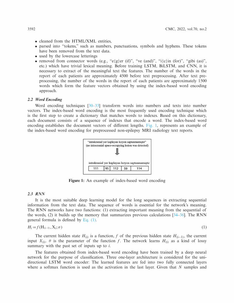
vectors. The index-based word encoding is the most frequently used encoding technique whichis the first step to create a dictionary that matches words to indexes. Based on this dictionary,each document consists of a sequence of indexes that encode a word. The index-based wordencoding establishes the document vectors of different lengths. Fig. 1, represents an example ofthe index-based word encoding for preprocessed non-epilepsy MRI radiology text reports.
Figure 1: An example of index-based word encoding
2.3 RNNIt is the most suitable deep learning model for the long sequences in extracting sequential
information from the text data. The sequence of words is essential for the network’s meaning.The RNN networks have two functions: (1) extracting important meaning from the sequential ofthe words, (2) it builds up the memory that summarizes previous calculations [34–36]. The RNNgeneral formula is defined by Eq. (1).
Hi = f (H(i−1) ,Xi;σ) (1)
The current hidden state H(i) is a function, f of the previous hidden state H(i−1), the currentinput X(i). θ is the parameter of the function f . The network learns H(i) as a kind of lossysummary with the past set of inputs up to i.
The features obtained from index-based word encoding have been trained by a deep neuralnetwork for the purpose of classification. Three one-layer architecture is considered for the uni-directional LSTM word encoder: The learned features are fed into two fully connected layerswhere a softmax function is used as the activation in the last layer. Given that N samples and

CMC, 2022, vol.70, no.2 3593
C classes let g(·) defines the encoder function in the first fully-connected layer, (yi) and (yi)respectively denote the prediction and the ground truth sample of (xi). Then, the prediction loss(Lpred) is defined by Eqs. (2) and (3) [37–41].
yi = softmax(Wg(xi+b)); (i= 1, . . . ,N) (2)
Lpred =1N
−(
N∑i=1
C∑i=1
yci log yci
)(3)
BiLSTM structure allows the network to receive both backward and forward informationabout the sequence at each step. It works in two ways, one from the past to the future and theother from the future to the past. With this approach, what sets it apart from being unidirectionalis protecting information from the future in LSTM working backwards. It can preserve informa-tion from both the past and the future at any point in time [42,43]. The LSTM and BiLSTMnetworks general structures can be seen in Figs. 2a, and 2b respectively.
Figure 2: The general structure of RNN networks (a) The LSTM network general structure [41](b) The BiLSTM network general structure [43]
2.4 CNNCNN has three layers which are convolution, activation (Rectified Lineer Unit-ReLU), and
pooling in Eqs. (4)–(6), respectively. The convolution layer activates the data features. The ReLUprovides a faster and more effective training process by equating negative values to zero andpreserving positive values. The pooling layer reduces the number of parameters that the networkmust learn. The model learns the weight and bias values in the training process and updates itcontinuously with each new training example. After learning the properties in layers, the architec-ture of a CNN switches to classification. The next layer is a fully connected layer that outputsa vector of K dimensions where K is the number of classes that the network can predict. Thelast layer of CNN architecture uses a classification layer like softmax to provide classificationoutput [37,44–47].

3594 CMC, 2022, vol.70, no.2
In this study, the dataset is 1D vector, so that the sum of the dot product and filter can bepresented as Eq. (4).
conv(I, K)x =nN∑i=1
Ki,kIx+i−1,k (4)
dim(conv(I, K))=⌊nN + 2p− f
s+ 1
⌋; s> 0
= (nN + 2p− f ); s= 0
(5)
According to Eq. (5), the floor function of x is �x�. The pooling is performed over eachchannel and therefore only affects dimensions nN and keeps nC intact. When the text data is given,the filter is shifted after a specific step with no parameters to learn, and a function is applied tothe selected items. We have Eq. (6) that is square filter is used and the parameters f and s areusually described as 2.
dim(pooling(N))=(⌊
nN + 2p− fs
+ 1⌋, nC
); s> 0
= (nN + 2p− f , nC); s= 0
(6)
The CNN algorithm is the most suitable for the repetitive data such as images, signals, audio.CNN is also applied to the repeating text data. The input is collection of sentences which isobtained word vectors. The network is based on the convolutianal filter that calculates as smallmatrices of the weights. The filter slides over the rows in the text and dot products calculatesin the linear unit between weights and words [37,47]. In this study, N = 122 MRI radiology textreport samples (97 of them are epilepsy individuals, 25 of them are non-epilepsy individuals) andC = 2 classes (epilepsy and non-epilepsy).
The evaluations have been performed on the hold-out validation. The positive class has beendefined as epilepsy, and the negative class was defined as the non-epilepsy. Three measures havebeen sensitivity, specificity, and accuracy are described as in Tab. 2.
Table 2: 2× 2 confusion matrix explanation
Condition positive Condition negative
True positive (TP) False positive (FP)False negative (FN) True negative (TN)
Sensitivity, specificity, accuracy, precision, recall, F-Measure, and Geometric Mean (G-Mean)formulations are given in Eqs. (7)–(13) [40,48,49].
Sensitivity= TPTP+FN
(7)
Specificity= TNTN +FP
(8)

CMC, 2022, vol.70, no.2 3595
Accuracy= TP+TNTP+TN +FP+FN
(9)
Precision= TPTP+FP
(10)
Recall= TPTP+FN
(11)
F-Measure= 2×Precision×RecallPrecision+Recall
(12)
G-Mean=√
TPTP+FN
× TNTN+FP
(13)
3 Experiments and Discussions
3.1 Experimental StudyIn this subsection we give the algorithm for the proposed method, which is given in Fig. 3
and the flowchart of this algorithm in Tab. 3.
Figure 3: An overview of the proposed epilepsy/non-epilepsy classification framework
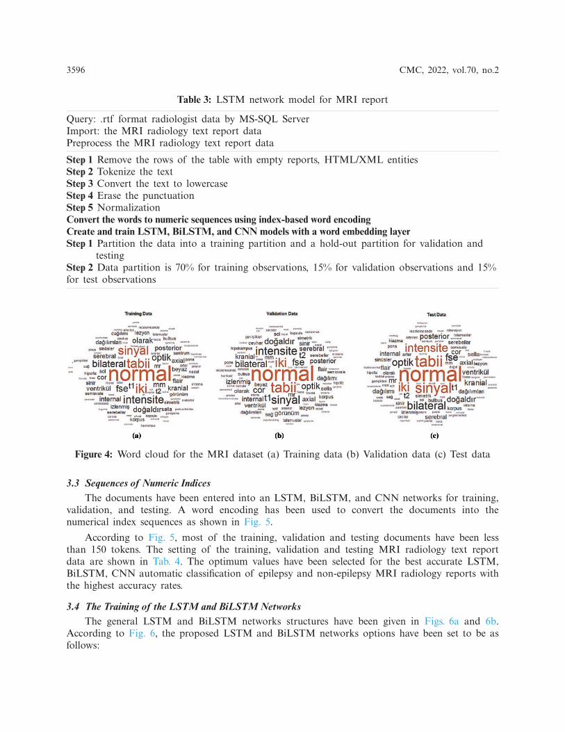
3.2 Word Cloud for Training/Validation/Testing DataThe word cloud indicates the size of the word that appears on the document data set is
obtained from MRI radiology text reports. The words have been grouped in clusters according tohow frequently they are mentioned for training, validation, and testing. The data obtained fromMRI radiology text reports have been accurately transferred to the word cloud model. The wordcloud for training is given in Fig. 4a, for validation is given in Fig. 4b and for testing is given inFig. 4c.
3596 CMC, 2022, vol.70, no.2
Table 3: LSTM network model for MRI report
Query: .rtf format radiologist data by MS-SQL ServerImport: the MRI radiology text report dataPreprocess the MRI radiology text report data
Step 1 Remove the rows of the table with empty reports, HTML/XML entitiesStep 2 Tokenize the textStep 3 Convert the text to lowercaseStep 4 Erase the punctuationStep 5 NormalizationConvert the words to numeric sequences using index-based word encodingCreate and train LSTM, BiLSTM, and CNN models with a word embedding layerStep 1 Partition the data into a training partition and a hold-out partition for validation and
testingStep 2 Data partition is 70% for training observations, 15% for validation observations and 15%for test observations
Figure 4: Word cloud for the MRI dataset (a) Training data (b) Validation data (c) Test data
3.3 Sequences of Numeric IndicesThe documents have been entered into an LSTM, BiLSTM, and CNN networks for training,
validation, and testing. A word encoding has been used to convert the documents into thenumerical index sequences as shown in Fig. 5.
According to Fig. 5, most of the training, validation and testing documents have been lessthan 150 tokens. The setting of the training, validation and testing MRI radiology text reportdata are shown in Tab. 4. The optimum values have been selected for the best accurate LSTM,BiLSTM, CNN automatic classification of epilepsy and non-epilepsy MRI radiology reports withthe highest accuracy rates.
3.4 The Training of the LSTM and BiLSTM NetworksThe general LSTM and BiLSTM networks structures have been given in Figs. 6a and 6b.
According to Fig. 6, the proposed LSTM and BiLSTM networks options have been set to be asfollows:

CMC, 2022, vol.70, no.2 3597
Figure 5: Documents into the numerical index sequences (a) Training document (b) Validationdocument (c) Test document
Table 4: Partitioning MRI radiology text report data
Data Samples Word encoding (number of words)
Training 86 1126Validation 18 677Test 18 540

3598 CMC, 2022, vol.70, no.2
Step 1: sequence input layer and input size are set to 1,
Step 2: the dimension of the word embedding layer and the word encoding layer are set to100 dimensions and 1126 real words for training, 677 words for validation, 540 words for testingare used.
Step 3: the number of hidden units is set to 180,
Step 4: the fully connected layers are used for the classification, which are softmax layer andclassification layer.
Figure 6: General LSTM and BiLSTM networks structures (a) LSTM network (b) BiLSTM layer
When the solver in the systems have been set to ‘adam’, the data are trained for 100 epochs,the gradient thresholds have been set to 1 and the initial learning rates have been set to 0.0001and 0.001 for the LSTM and the BiLSTM networks, respectively. By default, the training networkmodels have been used as a Graphics Processing Unit (GPU). The same parameters have beenused for testing and validation which used for the training of LSTM and BiLSTM networks.In order to make a prediction of epilepsy and non-epilepsy, the preprocessed test and validation

CMC, 2022, vol.70, no.2 3599
data are used. The training process for the LSTM network has been given in the Fig. 7a. Theconfusion matrices of the LSTM network have been given in the Fig. 7b.
Figure 7: The experimental results for the proposed LSTM network (a) The LSTM networktraining process (b) Confusion matrices of the LSTM network
According to Fig. 7b, the accuracy rates are the proportion of the labels that the accuracyrates for training, validation and testing are 97.67%, 94.44% and 66.67%, respectively.
The training process for the BiLSTM has been given in the Fig. 8a. The confusion matricesof the BiLSTM network have been given in the Fig. 8b.
According to Fig. 8b, the accuracy rates are the proportion of the labels that the accuracyrates for training, validation and testing are 100%, 88.89% and 88.89%, respectively.

3600 CMC, 2022, vol.70, no.2
Figure 8: The experimental results for the proposed BiLSTM network (a) The BiLSTM networktraining process (b) Confusion matrices of the BiLSTM network
3.5 The Training of the CNNIn this study, 1-D CNN model were constructed for the MRI text reports classification has
been as in Fig. 9.
To train the CNN, the input size value has been set to 1. The mini-batch size has been used15 with 150 epochs. 1-D CNN has been trained with the scattering sequences. The scatteringsequences have been set 1-by-3, where 3 is the number of time steps and 5 is the number of scatterpaths. To use this in a 1-D convolutional network, transfer and reshape the scattering sequencesby 3-by-1-by-1-by-to training samples. The results of the loss, test loss, and accuracy have beenobtained 36.85%, 51.94%, and 77.90%, respectively, at the end of 770th epoch. The training loss

CMC, 2022, vol.70, no.2 3601
and validation loss plot for our proposed 1-D CNN have been shown in Fig. 10. According toFig. 10, as the training loss decreases, the validation loss increases.
Figure 9: Single-layer CNN for the MRI text reports classification
Figure 10: The training loss and validation loss plot for proposed 1-D CNN
3.6 Statistical AnalysisIt was carried out by calculating the hypothesis test for the p-value. The p-value, as it is
useful to interpret the null hypothesis statistical test results is considered less than the level ofsignificance (usually 0.05) and it is statistically significant. In this study, the test analysis ofvariance (ANOVA) was used. When the p-value is lower the significance, level rejects the nullhypothesis and alternative hypothesis of the data is supported. The only way ANOVA designwas used to detect differences in results, accuracy rate, sensitivity, specificity, precision, recall,F-measure, and G-Mean [49]. Each statistical analysis was used for the p-value which was 0.05(significance level). The columns of the matrix represent 7-performance metrics (accuracy rate,sensitivity, specificity, precision, recall, F-measure, and G-Mean) for the LSTM, BiLSTM andCNN models. Fig. 11 showed the 7 performance evaluation metrics. In Fig. 11, modelling epilepticMRG text reports with the LSTM, BiLSTM, and CNN algorithms presented that were efficientmodels for classification. The p-value was obtained as 2.70521e-06 indicates that the models werenot the same.
According to Fig. 11, the statistical testing rejected the null hypothesis on the proposedmodels which were LSTM, BiLSTM, and CNN. All the results showed that the proposed methods

3602 CMC, 2022, vol.70, no.2
had significantly improved the performance as p-values in all cases were our statistical thresholdof 0.05.
Figure 11: One-way ANOVA test for the proposed models
3.7 The Evaluation of the Experimental ResultsFig. 12 presents the elapsed time for the LSTM, BiLSTM, and CNN. According to Fig. 12,
the CNN is the most time-consuming algorithm for the training process and the LSTM networkis the least time-consuming algorithm for the training process.
0
100
200
300
400
LSTM BiLSTM CNN
Figure 12: The elapsed time for the proposed networks
Significant Artificial Intelligence (AI) applications have been developed for the various fields.One of the notable developments with AI is NLP applications. The NLP methods process med-ical text data obtained from various sources. Meaningful information could be extracted aboutunstructured data including EHRs/EMRs, clinical notes and clinical records, and others, physiciannote, nurse note, triage note, radiology note, patient schedule, discharge summary, laboratoryrecord, incident report, imaging report. The pathology report, patient narrative, drug label andinformation provided by the patient, text message, online discussion, state license list, and medicalpublication allow disease follow-up [50–52]. Although there are various studies with the MRIreports in the literature, there are almost no studies on epilepsy patients. In the literature, themodels were developed with text-based medical data were given in Tab. 5.

CMC, 2022, vol.70, no.2 3603
Table 5: Classification accuracy comparison for the other text reports
Models Data Accuracy (%)
6-gram + maximum entropy [3] Chest x-ray report in two corpora 91.00Rule based + conditional random fields [6] Radiology and pathology reports 80.00Multinominal Naïve Bayes + BoW [7] Radiology reports 88.76Word embedding model + CNN [8] Radiology reports 89.69SVM [9] MRI reports 91.60CNN [10] Clinical notes 97.00SVM [11] MRI reports 85.00Unigram and bigram + TF-IDF + SVM [12] Radiology reports 90.60Frequency matrix + single decision tree [13] MRI reports 98.00BoW + SVM [14] EHRS notes 94.70Rule based + boosting [15] Pathology reports 97.00
The results in Tab. 6 were implemented by Matlab, and Phyton. The classification accuracyrates were obtained from LSTM, BiLSTM, and CNN networks applied to the epilepsy MRIdataset. The LSTM, BiLSTM, and CNN networks classification sensitivities, specificities, andaccuracy rates results for the training were presented on Tab. 6. The BiLSTM training networkcan protect input data by operating it in two ways, from past to future and from future topast, by using it bi-directionally. Therefore, in this study, BiLSTM modeling provided the bestaccuracy for epileptic and non-epileptic classification compared to LSTM and CNN models. Also,both RNN algorithms have more successful classification accuracy than the CNN algorithm. Inliterature, very limited number of studies exist on classification modellings of the MRI radiologyreports belonging with the epileptic individuals. In this study, three different mathematical modelswere developed comparatively for the MRI report data of individuals with epilepsy, written by aradiologist and diagnosed by a neurologist with epilepsy. Classification of MRI radiology reportsby processing them with the LSTM, BiLSTM and CNN algorithms are completely new approach.
Table 6: Evaluation of the proposed methods
Proposed method Sensitivity (%) Specificity (%) Accuracy (%)
LSTM network 97.05 100 97.67BiLSTM network 100 100 100CNN 81.08 58.33 77.90
This study has some limitations. Initially, the algorithm was developed using MRI radiologyreports of healthy individuals, 97 with epilepsy and 25 non-epilepsy. MRG report of individuals inthe data set is increasing day by day. However, we are constantly working to adapt our algorithmto the growth of the data set. Thus, in the future it can help physicians in the faster andmore effective identification of epilepsy more information will be inferred. Secondly, the modelshave been made with LSTM, BiLSTM and CNN, which are among the deep learning models.While BiLSTM has been given the most successful classification score during the training process,the classification accuracy has been obtained as 88.89% for the testing and validation processes.There is a need for new models where test and validation performance can be increased. Thirdly,

3604 CMC, 2022, vol.70, no.2
classification of epilepsy, more effective modeling should be developed by adding data set such asEEG and laboratory results as well as MRI reports to the data set.
4 Conclusion
Transferring MRI radiology reports to electronic health record systems, which are constantlyupdated, integrated, and shared data, has led to the potential for advancement in radiologyresearch and practice. Because the majority of MRI radiology reports are unstructured andfree-form language, extracting information manually is time-consuming, often unmanageable, andprone to human error, so it requires special expertise. This study was developed as a decisionsupport system to assist medical professionals in the diagnosis of epilepsy.
The main contribution of this study, to the real MRI reports of individuals suffering fromepilepsy in Turkey by applying the traditional methods of numeric sequences by using index-basedword encoding have been obtained data sets precedent with epilepsy. Modeling of the epilepsydataset with the LSTM, BiLSTM, and CNN models, which are popular deep learning models,are presented comparatively. When this study is compared to other works [6–19], no study likethis study was found for the actual MRI data set of patients with the epilepsy. Among ourproposed methods, due to the BiLSTM network’s ability to operate both backward and forwardinformation, it has been proven to give the best classification result. The following objectives canbe considered as the future works: Firstly, the size of the samples can be increased. Secondly,classification of epilepsy, to make fine judgment, more effective modeling can be developed byadding data set such as EEG and laboratory results as well as MRI reports to the data set.Thirdly, our proposed LSTM, BiLSTM, and CNN networks can be used for the other medicaltext reports datasets. Fourthly, the features can be extracted from the other NLP techniques suchas n-grams, BoW. Finally, the new classification and mathematical modeling will be applied tocompare with our proposed models.
Acknowledgement: The authors are grateful to deputy chief physician Dr. Lutfi Hocaoglu ofAvicenna Hospital in Turkey for their support and understanding during this research.
Funding Statement: The authors received no specific funding for this study.
Conflicts of Interest: The authors declare that they have no conflicts of interest to report regardingthe present study.
References[1] T. Rüber, B. David and C. E. Elger, “MRI in epilepsy: Clinical standard and evolution,” Current
Opinion in Neurology, vol. 31, no. 2, pp. 223–231, 2018.[2] M. Woolfe, D. Prime, L. Gillinder, D. Rowlands, S. O’keefe et al., “Automatic detection of the epilep-
togenic zone: An application of the fingerprint of epilepsy,” Journal Neuroscience Methods, vol. 325,no. 2019, pp. 1–20, 2019.
[3] I. Solti, C. R. Cooke, F. Xia and M. M. Wurfel, “Automated classification of radiology reports foracute lung injury: Comparison of keyword and machine learning based natural language processingapproaches,” in Proc. IEEE Int. Conf. Bioinformatics and Biomedicine Workshop, BIBMW , Washington,United States, pp. 314–319, 2009.
[4] K. Krabbe, P. Gideon, P. Wagn, U. Hansen, C. Thomsen et al., “MR diffusion imaging of humanintracranial tumours,” Neuroradiology, vol. 39, no. 7, pp. 483–489, 1997.
CMC, 2022, vol.70, no.2 3605
[5] J. M. Provenzale, S. Mukundan and D. P. Barboriak, “Diffusion-weighted and perfusion MR imagingfor brain tumor characterization and assessment of treatment response,” Radiology, vol. 239, no. 3, pp.632–649, 2006.
[6] L. Chen, L. Song, Y. Shao, D. Li and K. Ding, “Using natural language processing to extract clinicallyuseful information from Chinese electronic medical records,” International Journalof Medical Informatics,vol. 124, pp. 6–12, 2019.
[7] L. Chen, R. Shah, T. Link, M. Bucknor, S. Majumdar et al., “Text sentiment analysis for cartilageabnormalities detection from radiology reports,” Osteoarthritis Cartilage, vol. 7, no. 1, pp. 400–401,2019.
[8] J. Yuan, H. Zhu and A. Tahmasebi, “Classification of pulmonary nodular findings based on charac-terization of change using radiology reports,” in Proc. Joint Summits Translational Science Proc., AMIA,Bethesda, USA, pp. 285–294, 2019.
[9] L. T. E. Cheng, J. Zheng, G. K. Savova and B. J. Erickson, “Discerning tumor status from unstructuredMRI reports-completeness of information in existing reports and utility of automated natural languageprocessing,” Journal of Digital Imaging, vol. 23, no. 2, pp. 119–132, 2010.
[10] Y. Wang, S. Sohn, S. Liu, F. Shen, L. Wang et al., “A clinical text classification paradigm using weaksupervision and deep representation,” BMCMedical Informatics and Decision Making, vol. 19, no. 1, pp.1–13, 2019.
[11] E. I. Zacharaki, S. Wang, S. Chawla, D. S. Yoo, R. Wolf et al., “Classification of brain tumor type andgrade using MRI texture and shape in a machine learning scheme,” Magnetic Resonance in Medicine,vol. 62, no. 6, pp. 1609–1618, 2009.
[12] P. H. Chen, H. Zafar, M. Galperin-Aizenberg and T. Cook, “Integrating natural language processingand machine learning algorithms to categorize oncologic response in radiology reports,” Journal ofDigital Imaging, vol. 31, no. 2, pp. 178–184, 2018.
[13] C. Kim, V. Zhu, J. Obeid and L. Lenert, “Natural language processing and machine learning algorithmto identify brain MRI reports with acute ischemic stroke,” PLOS One, vol. 14, no. 2, pp. 1–13, 2019.
[14] A. Wright, A. B. McCoy, S. Henkin, A. Kale and D. F. Sittig, “Use of a support vector machine forcategorizing free-text notes: Assessment of accuracy across two institutions,” Journal of the AmericanMedical Informatics Association, vol. 20, pp. 887–890, 2013.
[15] A. Yala, R. Barzilay, L. Salama, M. Griffin, G. Sollender et al., “Using machine learning to parsebreast pathology reports,” Breast Cancer Research and Treatment, vol. 161, no. 2, pp. 203–211, 2017.
[16] Y. Kim, J. H. Lee, S. Choi, J. M. Lee, J. H. Kim et al., “Validation of deep learning natural languageprocessing algorithm for keyword extraction from pathology reports in electronic health records,”Scientific Reports, vol. 10, no. 1, pp. 1–9, 2020.
[17] Y. Barash, G. Guralnik, N. Tau, S. Soffer, T. Levy et al., “Comparison of deep learning models fornatural language processing-based classification of non-English head CT reports,” Neuroradiology, vol.62, no. 10, pp. 1247–1256, 2020.
[18] B. Imon, Y. Ling, M. C. Chen, S. A. Hasan, C. P. Langlotz et al., “Comparative effectiveness ofconvolutional neural network (CNN) and recurrent neural network (RNN) architectures for radiologytext report classification,” Artificial Intelligence in Medicine, vol. 97, pp. 79–88, 2019.
[19] Y. Jianbo, H. Zhu and A. Tahmasebi, “Classification of pulmonary nodular findings based on charac-terization of change using radiology reports,” AMIA Summits on Translational Science Proc., vol. 2019,pp. 285–294, 2019.
[20] M. Berry and J. Kogan, “Text mining: Applications and theory,” in Text Extraction, Classification, andClustering, 1st ed., United Kingdom: John Wiley & Sons Press, pp. 1–35, 2010.
[21] H. Takci and T. Gungor, “A high performance centroid-based classification approach for languageidentification,” Pattern Recognition Letters, vol. 33, no. 16, pp. 2077–2084, 2012.
[22] X. Zhang, W. Lu, F. Li, X. Peng and R. Zhang, “Deep feature fusion model for sentence semanticmatching,” Computers, Materials & Continua, vol. 61, no. 2, pp. 601–616, 2019.

3606 CMC, 2022, vol.70, no.2
[23] G. Mujtaba, L. Shuib, N. Idris, W. L. Hoo, R. G. Raj et al., “Clinical text classification research trends:Systematic literature review and open issues,” Expert Systems with Applications, vol. 116, pp. 494–520,2019.
[24] J. T. Oliva, H. D. Lee, N. Spolaôr, W. S. R. Takaki, C. S. R. Coy et al., “A computational systembased on ontologies to automate the mapping process of medical reports into structured databases,”Expert Systems with Applications, vol. 115, pp. 37–56, 2019.
[25] S. Albahli, A. Algsham, S. Aeraj, M. Alsaeed, M. Alrashed et al., “Covid-19 public sentiment insights:A text mining approach to the gulf countries,” Computers, Materials & Continua, vol. 67, no. 2, pp.1613–1627, 2021.
[26] W. L. Martinez and A. R. Martinez, “Introduction to exploratory data analysis,” in Exploratory DataAnalysis with MATLAB, 3rd ed., London: Taylor & Francis Group, pp. 3–27, 2017.
[27] S. P. Conlon, B. J. Reithel, M. W. Aiken and A. I. Shirani, “A natural language processing based groupdecision support system,” Decision Support Systems, vol. 12, no. 3, pp. 181–188, 1994.
[28] B. M. Decker, C. E. Hill, S. N. Baldassano and P. Khankhanian, “Can antiepileptic efficacy andepilepsy variables be studied from electronic health records? A review of current approaches,” Seizure,vol. 85, pp. 138–144, 2021.
[29] C. I. Chesñevar, M. Sabaté-Carrové and A. G. Maguitman, “An argument-based decision supportsystem for assessing natural language usage on the basis of the web corpus,” International Journal ofIntelligent Systems, vol. 21, no. 11, pp. 1151–1180, 2006.
[30] A. E. Omolara, A. Jantan, O. I. Abiodun and H. E. Poston, “A novel approach for the adaptation ofhoney encryption to support natural language message,” in Proc. of the Int. Multi Conf. of Engineers andComputer Scientists, Hong Kong, pp. 1–7, 2018.
[31] S. Mankad, H. S. Han, J. Goh and S. Gavirneni, “Understanding online hotel reviews throughautomated text analysis,” Service Science, vol. 8, no. 2, pp. 124–138, 2016.
[32] D. D. Wickens, “Some characteristics of word encoding,” Memory & Cognition, vol. 1, pp. 485–490,1973.
[33] M. Khayyat, L. Lam and C. Y. Suen, “Arabic handwritten word spotting using language models,” inProc.: 13th Int. Conf. on Frontiers in Handwriting Recognition, Bari, pp. 1–7, 2012.
[34] K. Shaukat, S. Luo, V. Varadharajan, I. A. Hameed and M. Xu, “A survey on machine learningtechniques for cyber security in the last decade,” IEEE Access, vol. 8, pp. 222310–222354, 2020.
[35] X. Wei, L. Zhou, Z. Zhang, Z. Chen and Y. Zhou, “Early prediction of epileptic seizures using along-term recurrent convolutional network,” Journal of Neuroscience Methods, vol. 32, pp. 1–10, 2019.
[36] H. Eskandari, M. Imani and M. P. Moghaddam, “Convolutional and recurrent neural network basedmodel for short-term load forecasting,” Electric Power Systems Research, vol. 195, no. 107173, pp. 1–14,2021.
[37] B. Zhong, X. Xing, P. Love, X. Wang and H. Luo, “Convolutional neural network: Deep learning-based classification of building quality problems,” Advanced Engineering Informatics, vol. 40, pp. 46–57,2019.
[38] C. Zhang, K. Qiao, L. Wang, L. Tong, G. Hu et al., “A visual encoding model based on deep neuralnetworks and transfer learning for brain activity measured by functional magnetic resonance imaging,”Journal of Neuroscience Methods, vol. 325, pp. 1–9, 2019.
[39] R. Samli, S. Senan, E. Yucel and Z. Orman, “Some generalized global stability criteria for delayedcohen–Grossberg neural networks of neutral-type,” Neural Networks, vol. 116, pp. 198–207, 2019.
[40] I. Soltesz and K. Staley, “Validating models of epilepsy,” in ComputationalNeuroscience in Epilepsy, 1st
ed., USA: Academic Press, pp. 3–17, 2008.[41] S. Hochreiter and J. Schmidhuber, “Long short-term memory,” Neural Computation, vol. 9, no. 8, pp.
1735–1780, 1997.[42] K. Shaukat, S. Luo, V. Varadharajan, I. A. Hameed, S. Chen et al., “Performance comparison and
current challenges of using machine learning techniques in cybersecurity,” Energies, vol. 13, no. 2509,pp. 1–27, 2020.
CMC, 2022, vol.70, no.2 3607
[43] A. Graves and J. Schmidhuber, “Framewise phoneme classification with bidirectional LSTM and otherneural network architectures,” Neural Networks, vol. 18, no. 5–6, pp. 602–610, 2005.
[44] V. Sorin, Y. Barash, E. Konen and E. Klang, “Deep learning for natural language processing inradiology—Fundamentals and a systematic review,” Journal of the American College of Radiology, vol.17, no. 5, pp. 639–648, 2020.
[45] K. S. Dar, A. B. Shafat and M. U. Hassan, “An efficient stop word elimination algorithm for urdulanguage,” in 14th Int. Conf. on Electrical Engineering/Electronics, Computer, Telecommunications andInformation Technology, Phuket, Thailand, pp. 911–914, 2017.
[46] S. Kiranyaz, O. Avci, O. Abdeljaber, T. Ince, M. Gabbouj et al., “1D convolutional neural networksand applications: A survey,” Mechanical Systems and Signal Processing, vol. 151, no. 107398, pp. 1–21,2021.
[47] P. Li and K. Mao, “Knowledge-oriented convolutional neural network for causal relation extractionfrom natural language texts,” Expert Systems with Applications, vol. 115, pp. 512–523, 2019.
[48] S. Bayrak, E. Yucel and H. Takci, “Classification of extracranial and intracranial EEG signals byusing finite impulse response filter through ensemble learning,” in Proc. of the Twenty Seventh SignalsProcessing and Communications Applications Conf., Sivas, Turkey, pp. 1–4, 2019.
[49] T. M. Alam, K. Shaukat, I. A. Hameed, S. Luo, M. U. Sarwar et al., “An investigation of credit carddefault prediction in the imbalanced datasets,” IEEE Access, vol. 8, pp. 201173–201198, 2020.
[50] Z. Shen, “Natural language processing (NLP) applications in patient care: A systematic analysis,”Quarterly Review of Business Disciplines, vol. 7, no. 3, pp. 223–244, 2020.
[51] K. Shaukat, M. U. Hassan, N. Masood and A. B. Shafat, “Stop words elimination in urdu languageusing finite state automaton,” International Journal of Asian Language Processing, vol. 27, no. 1, pp.21–32, 2017.
[52] K. Shaukat, F. Iqbal, T. M. Alam, G. K. Aujla, L. Devnath et al., “The impact of artificial intelligenceand robotics on the future employment opportunities,” Trends in Computer Science and InformationTechnology, vol. 5, no. 1, pp. 050–054, 2020.