ARTICLE Broad and potent neutralizing human antibodies to ...
Jc~r~lo~
Biomedical Science
Original Paper
J Biomed Sci 2004;11:117-126 DOk 10.1159/000075294
III Received: September 2, 2003 Accepted: September 26, 2003
Early Detection of Antibodies against Various Structural Proteins of the SARS-Associated Coronavirus in SARS Patients
Ho-Sheng Wu a-c Yueh -Chun Hsieh c Ih-Jen Su b T ing -Hs iang Lin b
Shu -Chun Chiu b Yu-Fen Hsu b J ih -Hu i Lin b Me i -Ch ing W a n g b
J e o u - Y u a n Chen d Pei -Wen Hsiao e Geen-Dong Chang f
A n d r e w H.-J. W a n g c Hs ien-Wei T ing c C h i h - M i n g Choug
Chang-Jen Huang c
aGraduate Institute of Life Sciences, National Defense Medical Center, bDivision of Laboratory Research and Development, Center for Disease Control, Department of Health, Institutes of c Biological Chemistry, dBiomedical Sciences, and eBioAgricultural Sciences, Academia Sinica, fGraduate Institute of Biochemical Sciences, National Taiwan University, and gDepartment of Biochemistry, Taipei Medical University, Taipei, Taiwan, ROC
Key Words SARS-CoV infection • Western blotting • Recombinant proteins. Antibody response
Abstract Severe acute respiratory syndrome (SARS), a new dis- ease with symptoms similar to those of atypical pneu- monia, raised a global alert in March 2003. Because of its relatively high transmissibility and mortality upon infec- tion, probable SARS patients were quarantined and treated with special and intensive care. Therefore, in- stant and accurate laboratory confirmation of SARS- associated coronavirus (SARS-CoV) infection has be- come a worldwide interest. For this need, we purified recombinant proteins including the nucleocapsid (N), en- velope (E), membrane (M), and truncated forms of the spike protein ($1-$7) of SARS-CoV in Escherichia coil The six proteins N, E, M, $2, $5, and $6 were used for Western blotting (WB) to detect various immunoglobulin classes in 90 serum samples from 54 probable SARS patients. The results indicated that N was recognized in most of the sera. In some cases, $6 could be recognized
as early as 2 or 3 days after illness onset, while $5 was recognized at a later stage. Furthermore, the result of recombinant-protein-based WB showed a 90% agree- ment with that of the whole-virus-based immunofluores- cence assay. Combining WB with existing RT-PCR, the laboratory confirmation for SARS-CoV infection was greatly enhanced by 24.1%, from 48.1% (RT-PCR alone) to 72.2%. Finally, our results show that IgA antibodies against SARS-CoV can be detected within 1 week after illness onset in a few SARS patients.
Copyright@ 2004 National Science Council, ROC and S. Karger AG, Basel
Introduction
Severe acute respiratory syndrome (SARS) is a newly emerged infectious disease. The causative agent of SARS has been identified to be a new type of coronavirus, name- ly SARS-associated coronavirus (SARS-CoV) [5, 8, 14]. Shortly after the 1 st reported case in Guangdong, south- ern China, in late 2002 [2], outbreaks of SARS spread to Vietnam, Hong Kong, Singapore, Canada, and other areas, including Taiwan. As of June 30, 2003, 8,447 prob-
KARG E R Fax+41 61 306 12 34 E-Mail karger@karger, ch www. karger.com
@ 2004 National Science Council, ROC S. Karger AG, Basel 1021-7770/04/0111-0117521.00/0 Accessible online at: www.karger.ccm/jbs
Dr. Chang-Jen Huang Institute of Biological Chemistry, Academia Sinica No. 128 Academia Rd., Sec. 2 "raipei 115, Taiwan (ROC) Tel. +886 2 27855696, Fax +886 2 27889759, E-Mail [email protected]

able SARS cases including 811 deaths had been reported to the World Health Organization (WHO) from 32 coun- tries or regions worldwide. The 1 st case of SARS in Tai- wan, a 54-year-old male who returned from a business trip to Guangdong, was diagnosed and reported in early March 2003 [16]. Subsequently, a large outbreak took place in late April partly due to an unrecognized SARS index patient who made multiple visits to different hospi- tals and exposed many patients, visitors, and health care workers [9], resulting in 678 probable cases and 84 deaths in Taiwan by the end of June [3].
The genome of the SARS-CoV is positive-sense, single- stranded RNA with sequences distantly related to all known coronaviruses that infect humans and animals [ 1 I, 15]. Like other known coronaviruses, SARS-CoV is an enveloped virus containing three envelope proteins, namely membrane (M), envelope (E), and spike (S) pro- teins. The nucleocapsid (N) protein, together with the viral RNA genome, presumably tbrms a helical core located inside the viral envelope. Based on the structure of all known coronaviruses, the M protein, a transmem- brane glycoprotein, is the most abundant protein in the virus particle, while the E protein is present in a minute amount in the viral envelope. In some coronaviruses, the S protein has been shown to bind to cellular receptors on the host cell surface. Generally, the immune response against the S protein is believed to prevent viral attach- ment and entry into host ceils.
At the very beginning of the SARS outbreak, the case definition for SARS was merely based on clinical history and symptoms. However, after confirmation that SARS- CoV was the causative agent, laboratory methods, includ- ing viral isolation, nucleic acid tests, and serological tests, were quickly developed and were accepted as one of the categories for a SARS diagnosis [ 17]. Even though the RT- PCR or the real-time PCR is currently the most sensitive technique for detecting SARS-CoV, its positive rate for probable SARS cases is around 38% according to data of the Center for Disease Control of Taiwan (CDC-Taiwan). This is likely because specimens might not be obtained at an optimal time point during the course of the SARS-CoV infection, or specimens might be taken improperly, espe- cially for nasopharyngeal aspirates and throat swabs. Thus, it was suggested that multiple and repeated collec- tions of nasopharyngeal, throat, and fecal samples be tak- en in order to increase the positive rate determined by RT-PCR. In addition to nucleic acid tests, serodiagnosis of SARS is also important. Given that SARS is a new dis- ease in humans, antibodies against SARS-CoV should not be detected in people who have not been exposed to the
virus; an antibody occurring in sera would be a useful index for SARS infection. It was reported that IgG against SARS-CoV was detectable in convalescent sera using SARS-CoV-infected Vero cells as antigens in an indirect immunofluorescence assay (IFA) [ 13].
To reduce the risk of infection, bacterially expressed recombinant SARS-CoV proteins instead of whole virus were used as antigens in the Western blotting (WB) assay. The recombinant proteins including the N, E, M, and var- ious truncated forms of the S protein were tested to see whether they reacted with the sera of probable SARS patients collected during the acute and convalescent stages of the illness. Because humoral immunity displays differ- ent isotypes of antibodies, including IgA, IgG, and IgM, during the infection, we thus evaluated whether these anti- bodies could be used as diagnostic indices for SARS infec- tion. The results of WB analyses were summarized and compared with those of IFA and RT-PCR. Overall, the results between the recombinant protein-based WB and whole-virus-based IFA showed a high correlation.
Materials and Methods
Construction of Expression Plasmids The coding regions for the SARS-CoV proteins N, M, and E and
fragments of S ($1, $2, $3, $4, $5, $6, and $7) protein were amplified by 30 cycles of PCR using reverse-transcribed RNA extracted from the Urbani strain of SARS-CoV (GenBank accession No. AY278741). The Urbani strain with 29,727 nucteotides in length was kindly provided by the Centers for Disease Control and Prevention (CDC-US; Atlanta, Ga., USA). Sequences of the primers used in the PCR are listed in table 1, the amplicons from which carried either BamHI/Sall or BamHI/HindiII restriction sites. The size of the nucleotide sequence of N was 1,269 bp, those of M 666, E 231, S1 429, $2 353, $3 560, $4 725, $5 630, $6 690, and $7 663 bp. The amplified products were purified from the gel and cloned into the pQE30 expression vector (Qiagen, Studio City, Calif., USA). The resulting constructs were transformed into Escherichia coli JM109 cells (Invitrogen, Carlsbad, Calif., USA). All sequences of' inserts were verified using an ABI 3730 DNA analyzer (Applied Biosystems, Tokyo, Japan).
Expression and Purification of Recombinant Proteins These proteins were expressed in E. coIi JM109. A colony of E.
coli cells was separately inoculated into LB broth in the presence of 100 gg/ml ampicillin, and the culture was grown overnight at 37 ° C, until its optical density reached 1.2 at 600 nm. To induce expression of these recombinant proteins, isopropyl-13-D-thiogalactopyranoside (IPTG) was added to a final concentration of 1.0 raM, and the incu- bation was continued for 4 h. All recombinant proteins accumulated in the bacteria as inclusion bodies. The cells were harvested by cen- trifugation and used for the preparation of inclusion bodies. Briefly, cells from 5 mI of culture were resuspended in 1 mt of phosphate- buffered saline (PBS), pH 7, and sonication was performed three times in an ice bath at 10-second intervals to disrupt the cells. After
i 18 J Biomed Sci 2004;11:11%I26 Wu et al.

Table 1. Primers used for amplification of various DNA fragments of SARS-CoV
N SA-NF a 5'-CTGGATCCATGTCTGATAATGGACCCCAT-3' SA-NR b 5'-GCGTCGACTTATGCCTGAGTTGAATCAGC- 3'
BamHI 1,269 Sa/I
M SA-MF 5'-CTGGATCCATGGCAGACAACGGTACT- 3' SA-MR 5'-GCGTCGACCTGTACTAGCAAAGCAAT-3'
BamHI 666 SalI
E SA-EF SA-ER
5'-CTGGATCCATGTACTCATTCGTTTCGGAA- 3' 5'-GCAAGCTTTTAGACCAGAAGATCAGGAAC- 3'
BamHI 231 tIindIII
S1 SA-SF 1 5'-CTGGATCCATGTTTATTTTCTTATTATTT-3' SA-SR1 5'-GCAAGCTTGGGTTTAGAAACAGCAAAGAA- 3'
BamHI 429 HindIII
$2 SA-SF2 5'-CTGGATCCATGGGTACACAGACACAT-3' SA-SR2 5"-GCAAGCTTGTAGTTGGCTTTAAATAG- 3'
BamHI 353 HindIII
$3 SA-SF3 5'-CTGGATCCATGCTCAAGTATGATGAA-3' SA-SR3 5'-CTAAGCTTGCCATGTCTAAGATACCT- 3'
BamHI 560 HindllI
$4 SA-SF4 5'-CTGGATCCATGAGGCCCTTTGAGAGA-3' SA-SR4 5'-GCAAGCTTGAGTAAGCAATI'GAACTA-3'
BamHI 725 HindIII
$5 SA-SF5 5<CTGGATCCATGTCTTTAGGTGCTGAT-3' SA-SR5 5'-GCAAGCTTGAACCTATATGCCATTTG- 3"
BamHI 630 HindIII
$6 SA-SF6 5'-CTGGATCCATGGCATATAGGTTCAAT-3' SA-SR6 5'-GCAAGCTTGCC.&ATAACGACATCACA- 3'
BamHI 690 HindItI
$7 SA-SF7 5'-CTGGATCCATGTCCTTCCCACAAGCA-3' SA-SR7 5'-GCAAGCTTTTATGTGT~%ATGTAATTTGACACC-3'
BamHl 663 HindIII
a F denotes tbrward primer. b R denotes reverse primer.
centrifugation at 13,000 rpm for 5 rain, the pellet was resuspended in an Eppendorf vial containing 1.5°/0 sarcosine and 10 mM Tris-HC1 buffer, pH 7.0, vortexed at room temperature for 1 h, until the lysate appeared clear, and recentrifuged at 13,000 rpm for 5 min. The supernatant was collected, and BD TALON TM metal affinity resins (BD Biosciences, San Jose, Calif., USA) were then added. The vials were mildly agitated at 4 °C overnight. The resins were centrifuged and washed twice with 10 mM Tris-HC1 buffer containing 1 M NaC1. These proteins were examined by 12% SDS-PAGE. The proteins were either stained with Coomassie blue or were transferred to a polyvinylidene difluoride membrane (PVDF Immobilon P, pore size 0.45 gin; Millipore, Bedford, Mass., USA) to determine the recombi- nant proteins. To eliminate the few bactel-ial contaminants present in the inclusion bodies, these recombinant proteins were eluted with 50-200 mM imidazole solution according to the manufacturer's instructions.
Human Sera According to WHO criteria, a person presenting after November
1, 2002, with a history of high fever (> 38 ° C), coughing, or breathing difficulty and having resided in or traveled to an area with recent local transmission of SARS during the 10 days prior to onset of symp- toms was classified as a suspected case. A suspect case with radio-
graphic evidence of infiltrates consistent with pneumonia or respira- tory distress syndrome on a chest X-ray was considered a probable case. In this study, 90 human serum samples from 54 patients, fulfill- ing WHO criteria for probable SARS cases, were selected from hospi- talized patients in northern Taiwan. Of these 18 male and 38 female cases, 36 had paired sera, and 18 had only single serum collected either at the acute stage or in the convalescent stage of the illness. All serum samples had been analyzed for SARS-CoV by RT-PCR, among which 48 were positive and 42 negative. The primers used for the RT-PCR were synthesized according to CDC-US recommenda- tions [8]. The handling of specimens, such as collecting, taking ali- quots of, or diluting specimens, and the practice of nucleic acid extraction or the RT-PCR assay were performed in BSL-2 (biosafety level 2) laboratories. Laboratory workers were required to wear pro- tective equipment, including disposable gloves, laboratory coats, and an N-95 mask.
Horseradish Peroxidase (HRP) Conjugated Antihuman IgA, IgG, and IgM HRP-conjugated goat antihuman immunoglobulins, IgA (c~
chain), IgG (3' chain), and IgM (g chain), were purchased from Savyon Diagnostics (Ashdod, Israel). These products were purified with affinity columns.
Antibody Response to SARS-CoV Infection J Biomed Sci 2004; 11:117-126 119
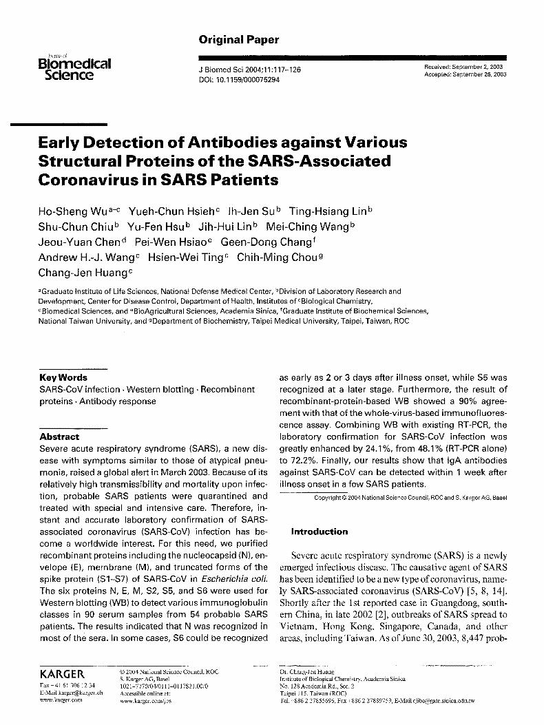
Fig. 1. Partial genomic structure of SARS- CoV. A Localization of known genes encod- ing the proteins S (spike glycoprotein), E (small-envelope protein), M (membrane gly- coprotein), and N (nucleocapsid) and other putative open reading frames, X 1 -X5. B The S, E, M, and N proteins contained 1,255, 76, 22I, and 422 amino acid residues, respec- tively. Seven consecutive truncated forms of S protein, designated S1-$7, are indicated with an enlarged scale.
Immunofluorescence Assay Veto E6 cells were grown in MEM containing 10% fetal bovine
serum at 35°C. At a density of 80%, the cells were infected with SARS-CoV (106/ml). After cytopathic effects appeared, the cells were washed with 0.025% trypsin and spotted onto slides. These slides were placed in a closed heating container, until they completely dried, then the slides were fixed in acetone for 15 rain. Ten microlit- ers of diluted serum, starting from 1:100, was placed onto each well of the slide and incubated at 37 °C for 30 rain. After washing twice with PBS for 5 rain each, 10 gl of l:100-diluted specific antihuman gamma globulins labeled with FITC (Zymed Laboratories, South San Francisco, Calif., USA) was added to each well and incubated at 37 °C for 30 rain. After washing twice with PBS, the slides were observed under a fluorescence microscope. Culture of the virus and preparation of viral antigens for IFA were conducted in a BSL-3 labo- ratory. After being fixed with acetone, the IFA slides were examined in a BSL-2 laboratory.
Western Blotting Equal amounts of purified recombinant proteins were mixed,
subjected to SDS-PAGE, and transferred to PVDF membranes. The membranes were blocked with 5% skim milk (Difco, Sparks, Md., USA) in PBS for 2 h at room temperature and then were sliced into strips (0.5 x 8 cm). Therefore, the loading in each strip was theoreti- cally equal. Serum samples were 1:500 diluted with 5% skim milk. Two milliliters of the diluted serum was added to each strip and incu- bated overnight at 4°C. On the following day, these strips were washed with PBS containing 0.2% Tween 20 three times for 10 min each and incubated at room temperature for 2 h with 2 ml of 1: 1,000- diluted goat antihuman IgG, IgA, and IgM HRP conjugate (Savyon Diagnostics) separately. After washing as described above, the strips were incubated in ECL solution (Perkin-Elmer Life Sciences, Boston,
Mass., USA) for 1 rain. The strips were then dried and exposed to X-ray film to visualize the reaction.
Statistics Sensitivity [number of true positives/(number of true positives +
number of false negatives)] and specificity [number &true negatives/ (number of true negatives + number of false positives)] were calcu- lated as previously described [1 ]. The antibody response of different immunoglobulin classes to SARS-CoV recombinant proteins was plotted according to the optical density of the WBs which were scanned and quantified using TotalLab software (Nonlinear Dynam- ics, Newcastle upon Tyne, UK). The value of each band was normal- ized wkh the control serum. Results were subjected to Sigma Plot for curve plotting and pair-to-pair t-test. For all statistical analyses p < 0.05 was considered significant,
Resul ts
Expression and Purification oJT~ecombinant SA RS-Co V Proteins The encoding regions for the full-length E, M, and N
proteins and the truncated forms of S proteins of SARS- CoV are marked in figure 1. For easy handling, the 1,255 amino acid long open reading frame of the S protein was split into seven consecutive truncated forms of polypep- tides, designated S 1-$7. After induction with IPTG, most of the proteins were synthesized and were present in inclusion bodies. They were resolved in buffer containing
120 J Biomed Sci 2004;I 1:117-126 Wu et al.

1.5 % sarcosine and were purified by metal affinity resins. Samples of the bacterially expressed SARS-CoV N, M, E, $2, $5, and $6 proteins were analyzed and are shown in figure 2. The molecular masses of N, E, $2, $5, and $6 were 46, 10, 14, 23, and 25 kD, respectively, as expected. However, the apparent molecular mass of recombinant M protein in gel, at 35 kD, was larger than the calculated size (approximately 25 kD), possibly due to the high content of hydrophobic amino acid residues (49%, 109/221) in the M protein. The recombinant proteins of S1, $3, $4, and $7 were poorly expressed in E. coli; therefore, they were not used in the study (data not shown). A
w
WB Analysis for Detection of Antibodies to SARS-CoV The recombinant proteins were then pooled and tested
to see whether they reacted with the antibodies in the serum samples of probable SARS patients by WB analy- sis. Among them, 14 positive results in our WB and RT- PCR analyses, which also represent various stages of the illness, were selected and are presented in figure 3. Serum samples of these patients were collected from the 2nd through the 33rd days after the onset of illness and were labeled F-1854-D2, for example, to indicate female pa- tient 1854's day 2 serum. Two serum samples from healthy people were used as negative controls for SARS- CoV-specific antibodies (fig. 3, lanes 1 and 16). As shown in figure 3, the N protein could be recognized by IgA, IgG, or IgM (fig. 3A, lanes 4-15; fig. 3B, lanes 8-11, 13-16; fig. 3C, lanes 7, 8, 13-15). However, the S ($5 or $6) and M proteins were recognized by IgA or IgG, but were bare- ly recognized by IgM (fig. 3A, lanes 2, 3, 11-15; fig. 3B, lanes 3-5, 8-15; fig. 3C, lanes 5, 7, 11). Two recombinant proteins, E and $2, were not recognized by IgA, IgG, or IgM in any serum of SARS patients. A protein band with an arrowhead might represent degraded products of M protein or contaminated unknown proteins copurified with the recombinant M protein. Since a specific IgM was found to weakly react with M and S antigens and since positive cases for IgM were all included in that of IgA or IgG, IgM was not further analyzed in the following stud- ies. Taken together, our results demonstrated that recom- binant N, M, $5, and $6 proteins could be used as diag- nostic markers for SARS-CoV infection.
Response Profile of Various Immunoglobulins to Recombinant Proteins In order to elucidate more clearly the antibody re-
sponse to various antigens of SARS-CoV, line diagrams were drawn according to the WB results (fig. 3) by Sigma Plot version 8.0 and were normalized with the data of 2
kD N
9 7 - -
6 6 -
4 3 -
3 0 -
20.1 -
t4 .4 -
x M E S2 S5 S6
o
O
Fig. 2. Recombinant proteins N, M, E, $2, $5, and $6 produced inE. coli. The proteins were purified with affinity resins and stained with Coomassie blue after SDS-PAGE on a 12% polyacrylamide gel. Lanes 1-6 show individual purified proteins. Lane 7 shows the six pooled proteins used for WB mlalysis.
healthy volunteers. However, it must be emphasized that this antibody response profile represents pooled data selected from t4 probable SARS patients, not the kinetic change of 1 individual. As shown in figure 4, N protein possessed major antigenicity to various antibodies (tgA, IgG, and IgM). Noticeably, the antibody response of IgA to N protein was initiated as early as day 2 or 3 after illness onset, with a sevenfold increase which progressive- ly increased to 15-fold within 1 month (fig. 4A). The increased multiples of antibodies to N protein were signif- icantly higher than those of M proteins and S proteins according to the paired t-test (p < 0.01 and p < 0.05, respectively). The responses of IgM and IgG to N protein were similar to that of IgA. However, they appeared on days 10 and 16, respectively, after the onset of illness. The M protein could be detected by IgA or IgG. The antibody response to M protein was much lower than that to N pro- tein (fig. 4A-C). Interestingly, few patients had an IgA antibody response to spike proteins in the early stage (days 2-3), but most other patients had a similar response in the convalescent stage (days 16-21 after illness onset).
Relative Sensitivity and Specificity of lgA and IgG Responses to SARS-CoV Infection Table 2 summarizes the results of the 48 RT-PCR-pos-
itive serum samples of probable SARS cases by WB, in which N-related antigens (N, N + M, N + M + S, and N + S) showed a positive rate of 54.2% (26 of 48) for detection
Antibody Response to SARS-CoV Infection J Biomed Sci 2004;11:117-126 121

A
IgA
1
¢~ ¢O ~ ¢~1 04 ̧ CO CO
,, ,, u. u. ~ u,. ~ ~ ,, u.
2 3 4 5 6 7 8 9 10 11 t2 14 15 16
o
t 3
|
ii:ili:ii;;i! ̧
=E _=
17
~ N
'~ .... M
~i s6 $5
B 1
IgG~
C 1
igM
3 4 5 6 7 8 9 10 11 12 13 14 15
J
16 17 18
~ N
i
S5
: i i i ;!i~ii!!~i;!!! ~
2 3 4 5 6 7 8 9 10 11 12 13 14 15 16 17
~ N
M
s6 $ 5
Fig. 3. Detection of various subclasses of immunoglobulins against SARS-CoV in sera from 14 probable SARS cases and 2 normal volunteers by WB analysis. The serum sample from each SARS patient was labeled 17-1854-D2 (lane 2), for example, to indicate that the serum was collected from female patient 1854 on day 2 after illness onset. Similarly, F-MCW-Ctrl (lane 1) demonstrated that the sernm was collected from healthy female MCW. The input antigens were either stained with a 0.2 % amido black solution (lane 17) or used to detect various subclasses of immunoglobulins, such as IgA (A), IgG (B), and IgM (C), in the sera of SARS patients (lanes 2-15).
122 .l Biomed Sci 2004; 11:117-126 Wu et al.
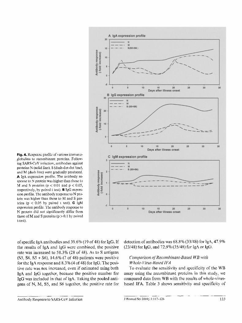
Fig. 4. Response profile of various immuno- globulins to recombinant proteins. Follow- ing SARS-CoV infection, antibodies against proteins N (solid line), S (dash-dot-dot line), and M (dash line) were gradually produced. A I g A expression profile. The antibody re- sponse to N protein was higher than those to M and S proteins (p < 0.01 and p < 0.05, respectively, by paired t test). B IgG expres- sion profile. The antibody response to N pro- tein was higher than those to M and S pro- teins (p < 0.05 by paired t test). C IgM expression profile. The antibody response to N protein did not significantly differ from those of M and S proteins (p > 0.1 by paired t-test).
of specific IgA antibodies and 39.6% (19 of 48) for IgG. If the results of IgA and IgG were combined, the positive rate was increased to 58.3% (28 of 48). As to S antigens ($5, $6, $5 + $6), 14.6% (7 of 48) patients were positive for the IgA response and 8.3% (4 of 48) for IgG. The posi- tive rate was not increased, even if estimated using both IgA and IgG together, because the positive number for IgG was included in that of IgA. Taking the pooled anti- gens of N, M, $5, and $6 together, the positive rate for
detection of antibodies was 68.8% (33/48) for IgA, 47.9% (23/48) for IgG, and 72.9% (35/48) for IgA or IgG.
Comparison of Recombinant-Based WB with Whole- Virus-Based IFA To evaluate the sensitivity and specificity of the WB
assay using the recombinant proteins in this study, we compared data from WB with the results of whole-virus- based IFA. Table 3 shows sensitivity and specificity of
Antibody Response to SARS-CoV Infection J Biomed Sci 2004; 11:117-126 123
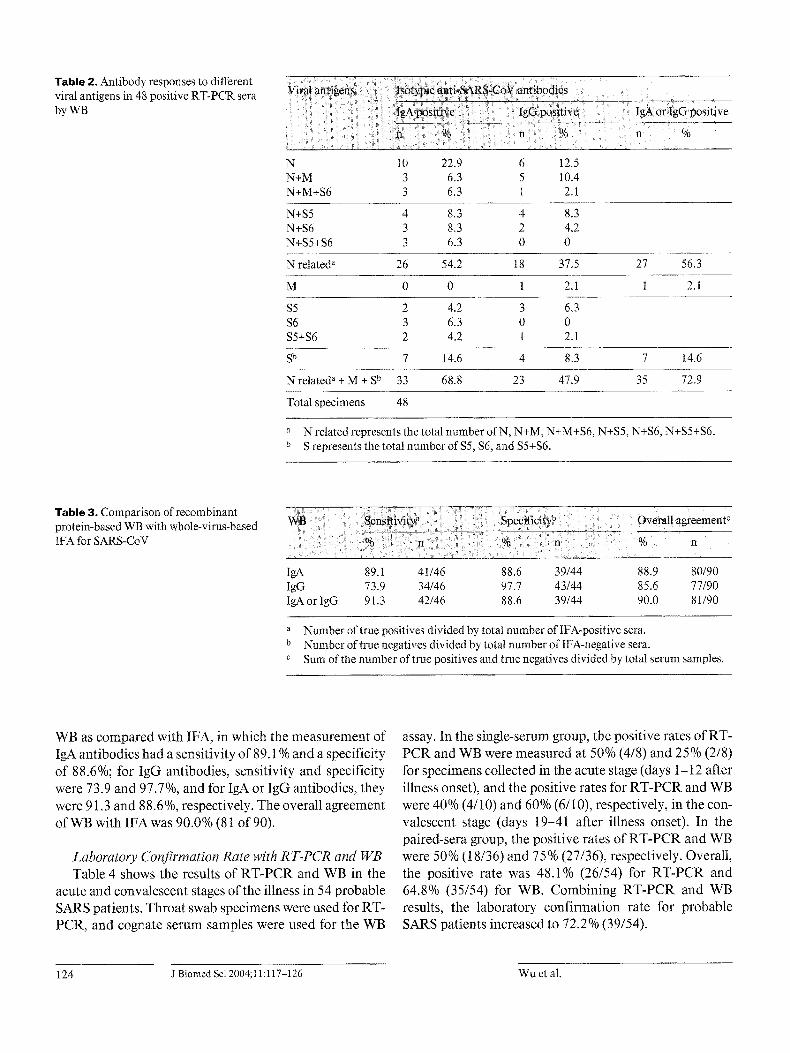
T a b l e 2 . Antibody responses to different viral antigens in 48 positive RT-PCR sera by WB
N 10 22.9 6 12.5 N+M 3 6.3 5 10.4 N+M+S6 3 6.3 1 2.1
N+S5 4 8.3 4 8.3 N+S6 3 8.3 2 4.2 N+$5+$6 3 6.3 0 0
N related a 26 54.2 18 37.5 27 56.3
M 0 0 1 2.t 1 2.1
$5 2 4.2 3 6.3 $6 3 6.3 0 0 $5+$6 2 4.2 1 2.1
S b 7 14.6 4 8.3 7 14.6
Nrelated a + M + S u 33 68.8 23 47.9 35 72.9
Total specimens 48
a N related represents the total number of N, N+M, N+M+S6, N+S5, N+S6, N+$5+$6. b S represents the total number of S5, $6, and S5+$6.
T a b l e 3. Comparison of recombinant protein-based WB with whole-virus-based IFA for SARS-CoV
! agreement c
n
IgA 89.1 41/46 88.6 39/44 88.9 80/90 IgG 73.9 34/46 97.7 43/44 85.6 77/90 IgA or IgG 91.3 42/46 88.6 39/44 90.0 81/90
a Number of true positives divided by total number of IFA-positive sera. u Number of true negatives divided by total number of IFA-negative sera. c Sum of the number of true positives and true negatives divided by total serum samples.
WB as compared with IFA, in which the measurement of IgA antibodies had a sensitivity of 89.1% and a specificity of 88.6%; for IgG antibodies, sensitivity and specificity were 73.9 and 97.7%, and for IgA or IgG antibodies, they were 91.3 and 88.6 %, respectively. The overall agreement of WB with IFA was 90.0% (81 of 90).
Laboratory Confirmation Rate with RT-PCR and WB Table 4 shows the results of RT-PCR and WB in the
acute and convalescent stages of the illness in 54 probable SARS patients. Throat swab specimens were used for RT- PCR, and cognate serum samples were used for the WB
assay, tn the single-serum group, the positive rates of RT- PCR and WB were measured at 500/o (4/8) and 25% (2/8) for specimens collected in the acute stage (days 1-12 after illness onset), and the positive rates for RT-PCR and WB were 400/0 (4/10) and 600/0 (6/10), respectively, in the con- valescent stage (days 19-41 after illness onset). In the paired-sera group, the positive rates of RT-PCR and WB were 50% (18/36) and 75% (27/36), respectively. Overall, the positive rate was 48.1°/0 (26/54) for RT-PCR and 64.8% (35/54) for WB. Combining RT-PCR and WB results, the laboratory confirmation rate for probable SARS patients increased to 72.2% (39/54).
124 J Biomed Sci 2004; 11:117-126 Wu et al.

Table 4. Laboratory-confirmed rate with RT-PCR and WB in probable SARS cases
Single serum acute stage 8 50.0 4/8 25.0 2/8 50.0 4/8 convalescent stage 10 40.0 4/10 60.0 6/t0 70.0 7/10
Paired sera acute/convalescent stages 36 50.0 18/36 75 27/36 77.8 28/36
Total 54 48.1 26/54 64.8 35/54 72.2 39/54
a Throat swabs were used for the RT-PCR assay.
Discuss ion
A WB assay using recombinant proteins as antigens was developed to detect SARS-CoV-specific antibodies. To evaluate whether the WB could be used, sensitivities and specificities were measured and compared with those of IFA. As shown in table 4, the recombinant-protein- based WB analysis correlated very well with the whole- virus-based IFA. These findings indicate that the recom- binant proteins of N, M, $5, and $6 may be useful as anti- gens to detect specific antibodies against SARS-CoV. Moreover, large amounts of recombinant proteins can easily be obtained in an E. coli system which offer several advantages over the antigens prepared from virus-in- fected cells, such as decreasing the risk of infection and ease of performing the assay with standardization.
As shown in table 2, N-related antigens showed a prin- cipal antigenicity of 58.3% for SARS-CoV in RT-PCR- positive patients, and S antigens contributed 14.6% to the overall positive rate of 72.9%. These data are related to those of Krokhin et al. [7] who conducted a WB assay using convalescent sera of SARS patients to detect pro- teins in the supernatants of cells containing SARS-CoV and identified the predominant protein N and minute quantities of protein S. However, we could not detect antibodies against E protein, and only a few SARS patients (19.2%, 10/54) had antibodies against M protein. These findings are not correlated with the results obtained by Elia et aI. [6] who reported that antibodies against M protein of a canine coronavirus were consistently detected in seropositive dogs.
In terms of the reactivity of different immunoglobulin classes, we found that specific IgA antibodies already existed 2 or 3 days after illness onset in some cases; how- ever, at the same time, IgM antibodies could not be detected in the same patients (fig. 3). The weak signal of
IgM might have resulted from the characteristics of IgM itself, since the affinity of IgM to antigens is relatively low as compared with that of IgG or IgA. Therefore, the detectable sensitivity of IgM to SARS-CoV proteins is limited. The human respiratory tract has been proposed to be the major site of SARS-CoV infection in humans, and it is totally covered with mucosal surfaces. Not only do cellular immune responses result from mucosat infec- tion by intracellular pathogens, but also secretory IgAs are produced to function as another important defense mech- anism against invasion of deeper tissues by these patho- gens [18]. Therefore, the synthesis of specific IgA anti- bodies in the early stage of a SARS-CoV infection is rea- sonable, and this phenomenon was also observed in other studies related to coronaviruses [4, I0, 12]. For example, Loa et al. [10] reported that the specific IgA antibodies against a turkey coronavirus were initially detected in serum 1 week after oral inoculation; these gradually increased and reached a peak at week 4 and finally declined at weeks 6-9. Furthermore, we noted that all three classes of immunoglobulins reacted with N and S antigens, but the antibody response of IgM to S antigen was much lower than that of IgA or IgG. Whether this phenomenon is significant for the immunological role of IgA, IgG, or IgM in SARS patients requires more detailed investigation.
Table 4 illustrates that RT-PCR was more sensitive than WB during the acute phase of SARS-CoV infection. However, WB was more sensitive than RT-PCR, if sera were collected in the convalescent phase of the illness. In general, the combined use of RT-PCR and WB raised the positive rate from 48.1 to 72.2% for probable SARS patients. Taken together, our data indicate that the use of recombinant protein-based WB along with RT-PCR can increase the rate of confirmation of suspected SARS patients.
Antibody Response to SARS-CoV Infection J Biomed Sci 2004; 11:117-126 125
Although most of the antibodies to S ($5 or $6) protein were detected during the later stage of the disease, 2 of 44 serum samples collected during the acute phase had anti- bodies against $6 on days 2 and 3 after illness onset (fig. 4). Since the data were pooled from many patients, we do not know whether this pattern was truly significant or merely due to variations among different individuals. Nonetheless, it is still valuable to elucidate the trend of antibody responses in different classes of immunoglobu- lins against various SARS-CoV antigens during the course of the infection.
In summary, our data suggest that WB using recombi- nant proteins N, M, $5, and $6 as antigens to detect var- ious classes of immunoglobulins might provide an alter-
native method for the early detection of SARS-CoV infec- tion. In addition, detection of specific IgA antibodies at the very early stage of the illness may be valuable for monoclonal antibodies or vaccine development to combat the SARS-CoV infection.
Acknowledgements
We thank Dr. Jyh-Yuan Yang for providing the Urbani strain of the SARS-CoV RNA, Dr. Shu-Ying Li for providing the specific goat an t ihuman IgG, IgA, and IgM HRP conjugate, and all members of the SARS Laboratory Diagnostic Team of CDC-Taiwan for their excellent performance during the SARS outbreak in Taiwan.
References
1 Btittner J. Evaluation of the diagnostic value of laboratory investigations. J Clin Chem Clin Biochem 15:1-12;1977.
2 CDC SARS Investigative Team, Fleischauer AT. Outbreak of severe acute respiratory syn- drome - worldwide, 2003. Mor Mortal Wkly Rep 52:226-228;2003.
3 Cumulative number of SARS probable cases in Taiwan, SARS Online Information Center, Center for Disease Control, Taiwan [Accessed July 2, 2003]. Available from: URL: http:/ /www,cdc.gov.tw/sarsen.
4 de Arriba ML, Carvajal A, Pozo J, Rubio P. Mucosal and systemic isotype-specific anti- body responses and protection in conventional pigs exposed to virulent or attenuated porcine epidemic diarrhoea virus. Vet Immunol Immu- nopathol 85:85-97;2002.
5 Drosten C, Gunther S, Preiser W, van der Werf S, Brodt HR, Becker S, Rabenau H, Panning M, Kolesnikova L, Fouchier RA, et al. Identifi- cation of a novel coronavirus in patients with severe acute respiratory syndrome. N Engl J Med 348:1967-1976;2003.
6 Elia G, Fiermonte G, Pratelti A, Martetla V, Camero M, Cirone F, Buonavoglia C. Recom- binant M protein-based ELISA test for detec- tion of antibodies to canine coronavirus. J Vi- rol Methods 109:139-142;2003.
7 Kmkhin O, Li Y, Andonov A, Feldman H, Flick R, Jones S, Stroeher U, Bastien N, Dasuri KV, Cheng K, et al. Mass spectrometric charac- terization of proteins from the SARS virus: A preliminary report. Mol Cell Proteomics 2: 346-356;2003.
8 Ksiazek TG, Erdman D, Goldsmith CS, Zaki SR, Peret T, Emery S, Tong S, Urbani C, Co- mer JA, Lim W, et al. A novel coronavirus asso- ciated with severe acute respirator), syndrome. N Engl J Med 348:1953-966;2003.
9 Lee ML, Chen C J, Su I J, Chen KT, Yeh CC, King CC, Chang HL, Wu YC, Ho MS, Jiang DD, et al. Severe acute respiratory syndrome - Taiwan, 2003. Morb Mortal Wkly Rep 52:461- 466;2003.
10 Loa CC, Lin TL, Wu CC, Bryan T, Hooper T, Schrader D. Specific mucosal IgA immunity in turkey poults infected with turkey coronavirus. Vet Immunol Immunopatho188:57-64;2002.
11 Marm MA, Jones S J, Astell CR, Holt RA, Brooks-Wilson A, Butterfield YS, Khattra J, Asano JK, Barber SA, Chan SY, et al. The Genome sequence of the SARS-associated co- ronavims. Science 300:1399-I 404;2003.
12 Naslund K, Traven M, Larsson B, Silvan A, Linde N. Capture ELISA systems for the detec- tion of bovine eoronavirus-specific IgA and IgM antibodies in milk and serum. Vet Micro- bio172:183-206;2002.
13 Peiris JS, Chu CM, Cheng VC, Chan KS, Hung IF, Pooh LL, Law KI, Tang BS, Hon TY, Chan CS, et al. Clinical progression and viral load in a community outbreak of coronavirus-associat- ed SARS pneumonia: A prospective study. Lancet 361:1767-1772;2003.
14 Peiris JS, Lai ST, Poon LL, Guan Y, Yam LY, Lim W, NichoIls J, Yee WK, Yan WW, Cheung MT, et al. Coronavirus as a possible cause of severe acute respiratory syndrome. Lancet 361: 1319-325;2003.
15 Rota PA, Oberste MS, Monroe SS, Nix WA, Campagnoli R, Icenogle JP, Penaranda S, Ban- kamp B, Maher K, Chert MH, et al. Character- ization of a novel coronavirus associated with severe acute respiratory syndrome. Science 300:1394-1399;2003.
16 Twu S J, (?hen TJ, Chen C J, Olsen S J, Lee LT, Fisk T, Hsu KH, Chang SC, Chen KT, Chiang IH, et al. Control measures for severe Acute respiratory syndrome (SARS) in Taiwan. Emerg Infect Dis 9:718-720;2003.
17 Use of laboratory methods for SARS diagnosis, WHO 2003 [Accessed July 2, 2003]. Available from: URL: http://www.who.int/csr/sars/lab- methods/en/print.html.
18 van GinkeI FW, Nguyen HH, McGhee JR. Vaccines for mucosal immunity to combat emerging infectious diseases. Emerg Infect Dis 6:123-132;2000.
126 J Biomed Sci 2004; 11:117-126 Wu et al.


![3-1-2 Propoal Toward an Alliance against CRD at Country le ... · Microsoft PowerPoint - 3-1-2___Propoal Toward an Alliance against CRD at Country le.ppt [Read-Only] Author: deslooverep](https://static.fdocuments.fr/doc/165x107/5f178903f7f2db234851b63d/3-1-2-propoal-toward-an-alliance-against-crd-at-country-le-microsoft-powerpoint.jpg)