DOCTORAT DE L'UNIVERSITÉ DE TOULOUSE · m. mohamed ghoul, universitÉ lorraine, président m. ali...
210
En vue de l'obtention du DOCTORAT DE L'UNIVERSITÉ DE TOULOUSE Délivré par : Institut National Polytechnique de Toulouse (Toulouse INP) Discipline ou spécialité : Sciences des Agroressources Présentée et soutenue par : Mme SARA OBEID le lundi 12 novembre 2018 Titre : Unité de recherche : Ecole doctorale : Ecodesign process for microalgae fractionation: use of supercritical CO2, membrane technology and low frequency ultrasounds Sciences de la Matière (SDM) Laboratoire de Chimie Agro-Industrielle (L.C.A.) Directeur(s) de Thèse : M. PIERRE YVES PONTALIER M. ALI ISMAIL Rapporteurs : M. KARIM ALLAF, UNIVERSITE DE LA ROCHELLE M. LUC MARCHAL, UNIVERSITE DE NANTES Membre(s) du jury : M. MOHAMED GHOUL, UNIVERSITÉ LORRAINE, Président M. ALI ISMAIL, UNIVERSITE LIBANAISE, Membre M. HOSNI TAKACHE, UNIVERSITE LIBANAISE, Membre Mme HELENE GREIGE-GERGES, UNIVERSITE LIBANAISE, Membre M. PIERRE YVES PONTALIER, INP TOULOUSE, Membre M. YOUSSEF EL RAYESS, UNIVERSITE SAINT-ESPRIT DE KASLIK, Membre
Transcript of DOCTORAT DE L'UNIVERSITÉ DE TOULOUSE · m. mohamed ghoul, universitÉ lorraine, président m. ali...
En vue de l'obtention du
DOCTORAT DE L'UNIVERSITÉ DE TOULOUSEDélivré par :
Institut National Polytechnique de Toulouse (Toulouse INP)Discipline ou spécialité :
Sciences des Agroressources
Présentée et soutenue par :Mme SARA OBEID
le lundi 12 novembre 2018
Titre :
Unité de recherche :
Ecole doctorale :
Ecodesign process for microalgae fractionation: use of supercritical CO2,membrane technology and low frequency ultrasounds
Sciences de la Matière (SDM)
Laboratoire de Chimie Agro-Industrielle (L.C.A.)Directeur(s) de Thèse :
M. PIERRE YVES PONTALIERM. ALI ISMAIL
Rapporteurs :M. KARIM ALLAF, UNIVERSITE DE LA ROCHELLE
M. LUC MARCHAL, UNIVERSITE DE NANTES
Membre(s) du jury :M. MOHAMED GHOUL, UNIVERSITÉ LORRAINE, Président
M. ALI ISMAIL, UNIVERSITE LIBANAISE, MembreM. HOSNI TAKACHE, UNIVERSITE LIBANAISE, Membre
Mme HELENE GREIGE-GERGES, UNIVERSITE LIBANAISE, MembreM. PIERRE YVES PONTALIER, INP TOULOUSE, Membre
M. YOUSSEF EL RAYESS, UNIVERSITE SAINT-ESPRIT DE KASLIK, Membre

2
Dedication
This thesis is dedicated to my husband, mother, father, brothers and sisters. You have been my
constant support. I owe you the world for that.
Mama, I just want you to be proud and happy…
شكرا طيبا مبارك
Acknowledgment
3
Acknowledgment
First of all, I want to acknowledge my kind director Mr. Pierre-Yves Pontalier, for his enthusiasm,
guidance, and unrelenting support throughout this Ph.D. His observation and comment helped me
to establish the overall direction of the research and to move forward with investigation in depth.
He has consistently gone beyond his duties to fight my worries and concerns and has worked
magically to instill great confidence in the way I worked during my thesis. Thank you for all the
advices, ideas, moral support. Your wealth of knowledge in the field of algorefinery is truly
inspiring. Thank you for giving me the opportunity to grow in this field of research.
I would like to thank my director and co-director Mr. Ali Ismail and Mr. Hosni Takache, for their
limitless help during my research and study at the Lebanese University. The enthusiasm they have
for research was very contagious and motivational for me. I appreciate all their contributions of
time and ideas in aim to make my experience productive and stimulating. This thesis would not
have been possible without your extraordinary support. I owe you lots of gratitude and appreciation
for showing me how to be a real scientist, a good supervisor and a kind person.
I also want to thank my committee members, Mr. Mohamed Ghoul, Mr. Karim Allaf, Mr. Luc
Marchal, Mrs. Helene Greige-Gerges and Mr. Youssef El Rayess. Thank you for investing time
and providing interesting remarks and feedback. I am thankful that you have accepted to be on my
committee.
Special mention goes to Mrs. Laurie Barthe and Mrs. Imane Bnehamed at LGC, my work with
you has been an amazing experience, thanks for helping me enormously with the ultrasound
assisted extraction and for your time and encouragement.
I extend my sincere gratitude to the administrative and technical staff members of LCA. I am
grateful also for your hospitality and for always making me feel so welcome. I had a great stay in
France, thanks to you.
All my lab friends at the LCA made it a pleasant place to work. In particular, I would like to thank
my office-mates. I would like to thank also my colleagues, Bouchra Sayed Ahmad (my daily
Acknowledgment
4
coffee-break partner with the special ‘galette’), Esaïe Kouassi (the humorous one), Douaa, Pablo,
Jesus, Nicholas, Remi et Marian. Assad Mogni thanks for the friendship and beautiful memories.
My time in Toulouse was amazing thanks to my friend Alaa Hamdar. I am indebted to you and
you were always so helpful in numerous ways. Thank you for your unconditional friendship
throughout these years.
Much gratitude goes to Rayan and Fleurine, your solid unwavering support, friendship, sense of
humor and your constant encouragement was more valuable than you could ever imagine. I love
you so much sisters.
I am extremely grateful to my mom, your love, prayers, patience, caring and sacrifices. I have
made it this far because of you my all, thank you for offering me encouragement and support in
all my accomplishments and for preparing me to face the challenges with faith and humility. Dad,
thank you for believing in me and being always by my side. I love you so much!
I also want to thank my brother Mohammad, whom I can't force myself to stop loving. Thank you
for your love, for believing in me and being there for practical and emotional support in all those
things of life. I know I always have you to count on when times are rough.
Thank you my beloved sisters: Taghrid “nixi”, Aya “ouways”, Lara “Monti”, and my little
Lebanese karate champion Walid “walti”.
Finally, a good husband makes a good wife. My love Bassem, I enjoy spending each second with
you. You are not just my husband, you are my rock and my very best friend. You are my best
treasure. I appreciate whatever you do for me. I love you much. Techy!
***
Abstract
5
Abstract
The development of an integrated and simplified biorefinery process able to produce a wide range
of multiple molecules of interest is crucial for the commercialization of microalgae. The objective
of this study was to simplify the downstream processes of microalgae valorization by optimizing
the selectivity of extraction while respecting the principles of ecodesign. On the one hand, using
dry microalgae, supercritical CO2 extraction was carried out for selective recovery of neutral lipids
without cell disruption pretreatment. Results showed that up to 97% of neutral lipids from
Chlorella vulgaris were extracted. Neutral lipids from Nannochloropsis oculata represented most
of the extracts (83%), whereas the proportion of glycolipids and phospholipids did not exceed
12.1% and 5.3%, respectively. Following the lipophilic recovery via supercritical extraction, 67 %
of total proteins were released using double stage aqueous extraction. Moreover, up to 84% of the
extracted pigments free proteins were obtained using semi-frontal ultrafiltration. On the other
hand, a green ultrasound assisted extraction process using water as solvent was performed on wet
microalgae for proteins recovery at low frequency (12 kHz). Results of Chlorella vulgaris showed
that almost all the proteins content (97%) were released in the aqueous medium allowing a high
selective extraction while microalgae cells remained intact. In contrary, total proteins content were
co-extracted with impurities from totally disintegrated Spirulina sp. Thus, for proteins
clarification, quasi-frontal ultrafiltration process was carried out leading to 67% of total proteins
obtained in the permeate. Furthermore, up to 95% of phycocyanin were retained with other
impurities like residues and pigments. Since selective extraction of all the molecules of interest in
microalgae biomass remains a challenge, additional research must be conducted to alleviate the
downstream process.
Keywords: Microalgae, fractionation, extraction, biorefinery, supercritical CO2, ultrasound.
Résumé
6
Résumé
Le développement d'un procédé intégré et simplifié de bioraffinerie capable de produire une large
gamme de molécules d'intérêt est crucial pour la commercialisation des microalgues. L'objectif de
cette étude était de simplifier le processus de valorisation des microalgues en optimisant la
sélectivité de l'extraction tout en respectant les principes de l'écoconception. Dans un premier
temps, les travaux se sont focalisés sur la récupération sélective de lipides neutres sans
désintégration préalable des cellules par extraction au CO2 supercritique de microalgues
lyophilisées. Les résultats ont montré que près de 97% des lipides neutres de Chlorella vulgaris
ont été extraits. Pour Nannochloropsis oculata, les lipides neutres représentaient la majorité de
l’extrait (83%), tandis que la proportion de glycolipides et de phospholipides ne dépassait pas
12,1% et 5,3% respectivement. Après la récupération de la fraction lipophile par extraction
supercritique, 67% des protéines totales ont été libérées par une double extraction aqueuse. En
outre, plus que 84% des protéines, exemptes de pigments ont été obtenues, en utilisant
l’ultrafiltration semi-frontale. Ensuite, en utilisant des microalgues humides, un procédé
d'extraction assistée par ultrasons à basse fréquence a été étudié pour la récupération des protéines.
Les résultats sur Chlorella vulgaris ont montré que presque toute la teneur en protéines (97%) était
libérée dans la phase aqueuse, permettant une extraction hautement sélective tout en conservant
l’intégrité cellulaire. En revanche, la totalité des protéines a été co-extraite avec des impuretés à
partir de cellules de Spirulina sp. entièrement désintégrées. Ainsi, pour la clarification des
protéines, un procédé d'ultrafiltration semi-frontale a conduit à 67% des protéines totales obtenues
dans le perméat. En outre, jusqu'à 95% de phycocyanine ont été retenus avec d'autres impuretés
comme les résidus et les pigments. Les essais montrent l’intérêt de l’extraction sélective des
molécules, mais que l’efficacité du procédé dépend de la structure des cellules. Des travaux
complémentaires devraient encore être réalisés pour mieux comprendre les mécanismes impliqués
dans la libération des molécules.
Mots clés: Microalgues, fractionnement, extraction, algoraffinerie, CO2 supercritique, ultrasons.
Table of content
7
Table of content
Dedication ...................................................................................................................................... 2
Acknowledgment ........................................................................................................................... 3
Abstract .......................................................................................................................................... 5
Résumé ........................................................................................................................................... 6
Table of content ............................................................................................................................. 7
List of abbreviations ................................................................................................................... 11
General introduction .................................................................................................................. 13
Chapter 1: state of the art on microalgal biomass: algorefinery defies ................................. 17
1.1. Generalities on microalgae .............................................................................................. 17
1.2. Diversity of groups and species ....................................................................................... 18
1.2.1. Green algae (Chlorophyceae) ...................................................................................... 19
1.2.2. Cyanobacteria or blue-green algae (Cyanophyceae) ................................................... 19
1.2.3. Coccoid (Eustigmatophyceae) ..................................................................................... 20
1.3. Microalgal cell wall .......................................................................................................... 21
1.4. Microalgae biochemical composition ............................................................................. 22
1.4.1. Pigments ...................................................................................................................... 22
1.4.2. Proteins ........................................................................................................................ 24
1.4.3. Lipids ........................................................................................................................... 27
1.4.4. Sugars .......................................................................................................................... 28
1.5. Microalgae cultivation ..................................................................................................... 31
1.5.1. Open ponds culture ...................................................................................................... 31
1.5.2. Closed culture or tubular photobioreactor ................................................................... 32
1.6. Microalgae harvesting...................................................................................................... 33
1.6.1. Centrifugation .............................................................................................................. 33
1.6.2. Flocculation ................................................................................................................. 34
1.6.3. Sedimentation .............................................................................................................. 34
1.6.4. Filtration ...................................................................................................................... 34
1.6.5. Flotation ....................................................................................................................... 34
1.7. Effects of the medium on the growth and biochemical composition of microalgae ... 35
1.7.1. Carbon ......................................................................................................................... 36
1.7.2. Nitrogen ....................................................................................................................... 37
Table of content
8
1.7.3. Phosphorus................................................................................................................... 37
1.7.4. Sulfur ........................................................................................................................... 38
1.7.5. Salts ............................................................................................................................. 38
1.8. Extraction processes for component recovery ............................................................... 39
1.8.1. Extraction using organic solvents ................................................................................ 39
1.8.2. Cell disintegrating techniques ..................................................................................... 40
1.9. Supercritical extraction ................................................................................................... 46
1.9.1. Influence of extraction parameters .............................................................................. 48
1.9.2. Supercritical CO2 and microalgae: benefits and drawbacks ........................................ 50
1.10. Membrane filtration ....................................................................................................... 51
1.10.1 Limitations .................................................................................................................. 52
1.10.2. Influence of operating conditions .............................................................................. 55
1.10.3. Membrane efficiency ................................................................................................. 56
1.11. Algorefinery: advantages and difficulties .................................................................... 56
1.12. Conclusion and thesis action plan ................................................................................. 57
Chapter 2: Materials and methods ............................................................................................ 61
2.1. Microalgal biomass .......................................................................................................... 61
2.2. Biomass quantification ..................................................................................................... 62
2.3. Extraction methods .......................................................................................................... 62
2.3.1. Total lipids content: Conventional extraction according to modified Bligh and Dyer
method ................................................................................................................................... 62
2.3.2. Supercritical CO2 extraction ........................................................................................ 63
2.3.3. Ultrasound ................................................................................................................... 65
2.3.4. Total pigments extraction ............................................................................................ 67
2.4. Purification technique: ultrafiltration ............................................................................ 67
2.5. Analytical techniques ....................................................................................................... 69
2.5.1. Proteins analysis .......................................................................................................... 69
2.5.2. SDS-PAGE Analysis ................................................................................................... 70
2.5.3. Size exclusion chromatography ................................................................................... 70
2.5.4. Lipids analysis ............................................................................................................. 71
2.5.5. Pigment analysis .......................................................................................................... 72
References .................................................................................................................................... 75
Chapter 3: Fractional extraction ............................................................................................... 88
3.1. Introduction ...................................................................................................................... 88
Table of content
9
3.2. Supercritical carbon dioxide extraction and fractionation of lipids from freeze-dried
microalgae Nannochloropsis oculata and Chlorella vulgaris ............................................... 89
3.2.1. Introduction ................................................................................................................. 89
3.2.2. Materials and methods ................................................................................................. 91
3.2.3. Results and discussion ................................................................................................. 94
3.2.4. Conclusion ................................................................................................................. 104
3.2.5. References ................................................................................................................. 104
3.3. Supercritical fluid extraction and solid phase fractionation of pigments from
Chlorella vulgaris and Nannochloropsis oculata ................................................................. 107
3.3.1. Introduction ............................................................................................................... 108
3.3.2. Materials and methods ............................................................................................... 109
3.3.3. Results ....................................................................................................................... 114
3.3.4. Conclusion ................................................................................................................. 121
3.3.5. References ................................................................................................................. 122
3.4. Extraction and purification of soluble proteins from Nannochloropsis oculata by pH-
shifting and ultrafiltration .................................................................................................... 124
3.4.1. Introduction ............................................................................................................... 125
3.4.2. Materials and methods ............................................................................................... 126
3.4.3. Results and discussion ............................................................................................... 130
3.4.4. Conclusion ................................................................................................................. 138
3.4.5. References ................................................................................................................. 138
3.5. Chapter conclusion ......................................................................................................... 141
Chapter 4: Extracts purification ............................................................................................. 142
4.1. Introduction .................................................................................................................... 142
4.2. Proteins and pigments extraction from microalgae Spirulina sp. assisted by
ultrasound irradiation: effect of variable low power and pre-treatment conditioning .. 144
4.2.1. Introduction ............................................................................................................... 145
4.2.2. Material and methods ................................................................................................ 146
4.2.3. Results and Discussion .............................................................................................. 148
4.2.4. Conclusion ................................................................................................................. 152
4.2.5. References ................................................................................................................. 153
4.3. Clarification of non-pigmented proteins from Spirulina sp. using ultrasound assisted
extraction coupled with ultrafiltration ................................................................................ 155
4.3.1. Introduction ............................................................................................................... 155
4.3.2. Materials and Methods .............................................................................................. 157
4.3.3. Results and discussions ............................................................................................. 162
Table of content
10
4.3.4. Conclusion ................................................................................................................. 169
4.3.5. References ................................................................................................................. 169
4.4. Selectivity of low-frequency ultrasound-assisted extraction for proteins recovery
from the microalga Chlorella vulgaris ................................................................................. 173
4.4.1. Introduction ............................................................................................................... 173
4.4.2. Methods ..................................................................................................................... 175
4.4.3. Results and discussion ............................................................................................... 178
4.4.4. Conclusion ................................................................................................................. 183
4.4.5. References ................................................................................................................. 183
4.5. Chapter conclusion ......................................................................................................... 187
General Conclusionta ............................................................................................................... 188
Scientific production ................................................................................................................. 191
Résumé générale en français .................................................................................................... 193
Chapitre 1: Etat de l'art........................................................................................................ 193
Chapitre 2 : Méthodes et matériels ...................................................................................... 198
Chapitre 3 : Extraction fractionnée ..................................................................................... 200
Publication # 1 ..................................................................................................................... 200
Publication # 2 ..................................................................................................................... 201
Publication # 3 ..................................................................................................................... 202
Chapitre 4 : Purification des extraits .................................................................................. 203
Publication # 4 ..................................................................................................................... 203
Publication # 5 ..................................................................................................................... 204
Publication # 6 ..................................................................................................................... 205
Conclusion générale .............................................................................................................. 207
List of abbreviations
11
List of abbreviations
APC Allophycocyanin
BSA Bovine serum albumin
β-car β-carotenes
CPC C-phycocyanin
Chl a Chlorophyll a
Chl b Chlorophyll b
Cpi Concentration of component i in the permeate,
Cri Concentration of component i in the retentate
DW Dry weight
DAG Diacylglycerol
EPS Exopolysaccharides
FFA Free fatty acids
GL Glycolipids
Lut Lutein
MAG Monoacylglycerol
MWCO Molecular weight cut-off
NL Neutral lipids
NTP Nitrogen-to-protein factor
OD Optical density
PEF Pulsed electric fields
P Partition-coefficient
PPL Phospholipids
List of abbreviations
12
PE Phycoerythrin
PES Polyethersulfone
PC Phosphatidyl choline
PG Phosphatidyl glycerol
PBR Photobioreactor
PE Phosphatidyl ethanolamine
Rf Retention factor in%.
ScCO2 Supercritial carbon dioxide
SCF Supercritical fluids
SFE Supercritical fluid extraction
SPE Solid Phase Extraction
TN Total nitrogen
TAG Triacylglycerol
TMP Trans-membrane pressure
UF Ultrafiltration
VCR Volume concentration rate
WHO World Health Organization
General introduction
13
General introduction
To date, more than 20,000 microalgae species have been identified. These organisms afford
considerable reservoir of interesting biomolecules like lipids, proteins, pigments, polysaccharides,
vitamins and so on [1]–[5]. Recently, scientists, researchers and also entrepreneurs have begun to
recognize the potential of this biomass mainly as promising source of sustainable energy. Thus,
microalgae have been investigated for the production of a number of different biofuels including
biodiesel, bio‐oil, bio‐syngas, and bio‐hydrogen [6]. Besides, development in photobioreactor
design, biomass harvesting, drying and extraction processes have begun to lower the overall cost
of the biofuel production from microalgae [6]. However, the production dedicated to bioenergy is
still uncompetitive in the market. Hence, scientists found that, considering all the benefits this
biomass can provide, it is inconvenient to limit the outputs of the microalgae to bioenergy
purposes, and this biomass should be optimally exploited into a variety of valuable co-products
that could be used in several fields such as pharmaceuticals, nutraceuticals, chemicals, cosmetics,
biomaterials, feed and food [2][4][6][7]. In this view, the collaboration between research and
private sector commenced to play an important role and a variety of valuable chains started to be
developed and established in aim to apply the algorefinery concept by valorizing different co-
products from the algae biomass fractions. Scientists are convinced that a sequence of unit
operations to achieve the whole fractionation of microalgae would make the industrial sector
profitable [7]–[9].
The dominating species of microalgae in commercial production includes Nannochloropsis,
Chlorella and many other green microalgae which are known to have rigid cell wall [9] [10]–[12].
However, the extraction of valuable components outside this rigid wall requires a specific
pretreatment for maximum recovery yield [13]–[18]. Various chemical, physical, physico-
chemical, and biological methods have been applied for lipids, proteins and pigments recovery
[19][16][17][18][19][23][24][25]. Pre-treatments such as high pressure homogenizers, bead mills,
enzymatic or chemical treatments, thermal or osmotic shocks (repeated freezing/thawing) are
frequently applied allowing a dramatic cell disintegration, while many other components
(undesirable) are also co-released [18] [19] [23] [24] [25]. Thus, in absence of a selective
extraction, this could lead to complex extracts of hydrophilic and hydrophobic components.
General introduction
14
Consequently, additional costly stages in downstream processing for phase separation and
compounds purification are mandatory complicating then the downstream processes [26].
Particular challenge in this regard is that the cell integrity has to be maintained while high
extraction yield is needed. Thus, since cell wall plays an important role as barrier to hinder the
passage of inner compounds, it could be well considered for its importance on extraction
selectivity.
The challenge is not only in the selective separation of targeted components to simplify the
downstream processing stages, but also in taking into consideration the environmental aspect of
the applied extraction procedures. In fact, extraction of lipids from microalgal biomass is generally
achieved using chemical solvents like hexane, methanol and other flammable and toxic solvents
[27]. For that, this technique is currently being phased out for their environmental, health and
safety impacts.
Recently, supercritical carbon dioxide (ScCO2) extraction [28] has received attention as an
alternative to solvent extraction of microalgal lipids. Similarly, ultrasound-assisted extraction
(UAE) as an environmental friendly mechanical method, is able to be universally applied to any
type of microalgae cell for valuable component like proteins [29]. However in major cases, prior
to ScCO2 extraction, mechanical pretreatment was carried out and high amount of organic co-
solvent was used to increase lipids recovery yield all-in sharply decreasing selective extraction.
Also, many scientists investigated the addition of toxic solvent during ultrasonic treatment for
proteins release resulting in high extraction yield with low extraction selectivity [30]. Furthermore,
all UAE studies conducted for microalgae components release (like proteins) have been performed
with frequencies comprised between 20 and 100 kHz [31], frequencies known as hazardous and
dangerous for human health. However, to our best of knowledge, UAE treatments for microalgal
proteins extraction at audible frequencies (12 kHz) have never been reported in the literature.
Concerning the purification/concentration techniques, membrane processes, particularly
ultrafiltration, have begun to find new applications in the field of marine bioresources [15]
[16][33][34]. Actually, limited studies have investigated the use of membrane technology to purify
components from microalgae [33]–[38]. Ultrafiltration technology is considered as an efficient
technique with relatively moderate capital and operating costs [32]. Contrary to other separation
techniques, membrane processes are isothermal and involve no phase change or chemical reagents
General introduction
15
[39] [40], [41]. These separation techniques are particularly suited to the purification of water-
soluble biomolecules without neither thermal denaturation nor chemical solvents use.
Hence, in this context, this study proposes to evaluate the selective extraction of neutral lipids,
proteins and pigments from Chlorella vulgaris, Nannochloropsis oculata and Spirulina sp.
First, after reducing the water content below 2%, an extraction with ScCO2 without any previous
cell wall disintegration has been performed on dry biomass to selectively recover reserve lipids
(neutral lipids), proteins and pigments from C. vulgaris and N. oculata.
Second, in the absence of any drying treatment, a sequence of purification operations implemented
on humid biomass of C. vulgaris and Spirulina sp. has been applied in order to obtain separated
enriched fractions of biomolecules like proteins and pigments. New innovative parameters (low
power, low frequency) to achieve some intensification and optimization of UAE techniques have
been investigated. Furthermore, water as green solvent, to comply with the criteria of the green-
chemistry concept, has been evaluated for proteins selective extraction and high recovery yield.
Ultrafiltration has been used as concentration/purification technique.
Our challenge is to effectively separate each component with minimum process stages (enhancing
selective extraction) and minor environmental footprint. This study should be harmonious with
industrial realities. This challenge represents a part of the global algorefinery concern and could
contribute to achieve this goal.
***
The manuscript is composed of four chapters accessorized with six publications (published or
ready to be submitted) that reveal the obtained results:
Chapter one presents a general idea on microalgae, their composition and extraction processing
for component recovery. This literature review is followed by a discussion of the algorefinery
challenges and concludes on the implemented plan of our work.
Chapter two is composed of materials and methods applied during this research.
Chapter three is composed of three articles which investigated the impact of bypassing the cell
disruption step on the extraction selectivity and the recovery yield. It also deals with the concept
of fractional extraction.
General introduction
16
Chapter four contains three publications that deal with the extracts purification and the role of cell
wall characteristic on extraction selectivity.
Finally a general conclusion recapitalizes the main results and opens new perspectives in the
algorefinery concept.
***
17
Chapter 1: state of the art on microalgal biomass: algorefinery
defies
1.1. Generalities on microalgae
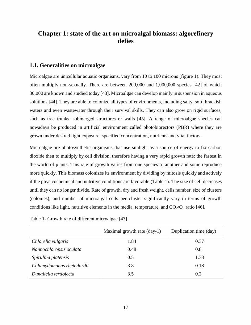
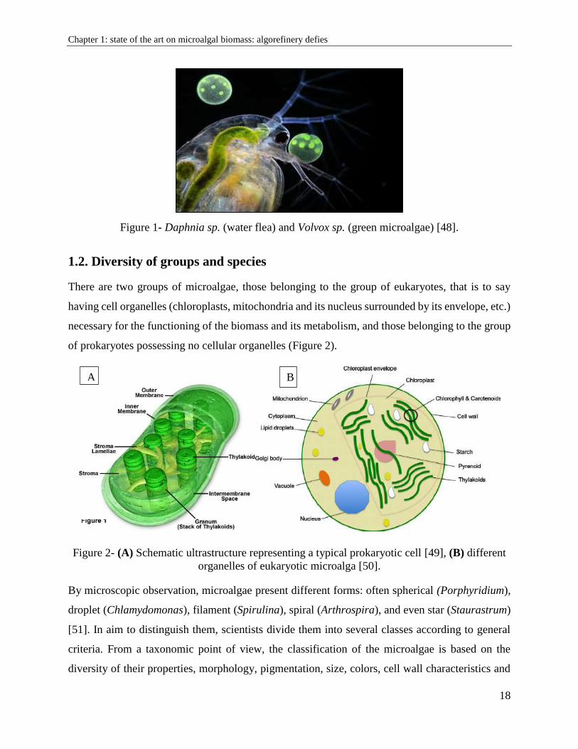
Microalgae are unicellular aquatic organisms, vary from 10 to 100 microns (figure 1). They most
often multiply non-sexually. There are between 200,000 and 1,000,000 species [42] of which
30,000 are known and studied today [43]. Microalgae can develop mainly in suspension in aqueous
solutions [44]. They are able to colonize all types of environments, including salty, soft, brackish
waters and even wastewater through their survival skills. They can also grow on rigid surfaces,
such as tree trunks, submerged structures or walls [45]. A range of microalgae species can
nowadays be produced in artificial environment called photobiorectors (PBR) where they are
grown under desired light exposure, specified concentration, nutrients and vital factors.
Microalgae are photosynthetic organisms that use sunlight as a source of energy to fix carbon
dioxide then to multiply by cell division, therefore having a very rapid growth rate: the fastest in
the world of plants. This rate of growth varies from one species to another and some reproduce
more quickly. This biomass colonizes its environment by dividing by mitosis quickly and actively
if the physicochemical and nutritive conditions are favorable (Table 1). The size of cell decreases
until they can no longer divide. Rate of growth, dry and fresh weight, cells number, size of clusters
(colonies), and number of microalgal cells per cluster significantly vary in terms of growth
conditions like light, nutritive elements in the media, temperature, and CO2/O2 ratio [46].
Table 1- Growth rate of different microalgae [47]
Maximal growth rate (day-1) Duplication time (day)
Chlorella vulgaris 1.84 0.37
Nannochloropsis oculata 0.48 0.8
Spirulina platensis 0.5 1.38
Chlamydomonas rheindardii 3.8 0.18
Dunaliella tertiolecta 3.5 0.2
Chapter 1: state of the art on microalgal biomass: algorefinery defies
18
Figure 1- Daphnia sp. (water flea) and Volvox sp. (green microalgae) [48].
1.2. Diversity of groups and species
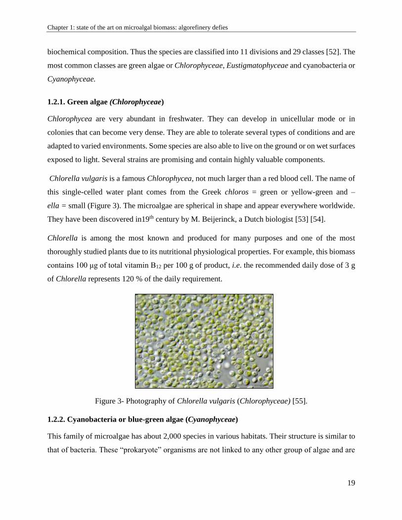
There are two groups of microalgae, those belonging to the group of eukaryotes, that is to say
having cell organelles (chloroplasts, mitochondria and its nucleus surrounded by its envelope, etc.)
necessary for the functioning of the biomass and its metabolism, and those belonging to the group
of prokaryotes possessing no cellular organelles (Figure 2).
Figure 2- (A) Schematic ultrastructure representing a typical prokaryotic cell [49], (B) different
organelles of eukaryotic microalga [50].
By microscopic observation, microalgae present different forms: often spherical (Porphyridium),
droplet (Chlamydomonas), filament (Spirulina), spiral (Arthrospira), and even star (Staurastrum)
[51]. In aim to distinguish them, scientists divide them into several classes according to general
criteria. From a taxonomic point of view, the classification of the microalgae is based on the
diversity of their properties, morphology, pigmentation, size, colors, cell wall characteristics and
A B
Chapter 1: state of the art on microalgal biomass: algorefinery defies
19
biochemical composition. Thus the species are classified into 11 divisions and 29 classes [52]. The
most common classes are green algae or Chlorophyceae, Eustigmatophyceae and cyanobacteria or
Cyanophyceae.
1.2.1. Green algae (Chlorophyceae)
Chlorophycea are very abundant in freshwater. They can develop in unicellular mode or in
colonies that can become very dense. They are able to tolerate several types of conditions and are
adapted to varied environments. Some species are also able to live on the ground or on wet surfaces
exposed to light. Several strains are promising and contain highly valuable components.
Chlorella vulgaris is a famous Chlorophycea, not much larger than a red blood cell. The name of
this single-celled water plant comes from the Greek chloros = green or yellow-green and –
ella = small (Figure 3). The microalgae are spherical in shape and appear everywhere worldwide.
They have been discovered in19th century by M. Beijerinck, a Dutch biologist [53] [54].
Chlorella is among the most known and produced for many purposes and one of the most
thoroughly studied plants due to its nutritional physiological properties. For example, this biomass
contains 100 μg of total vitamin B12 per 100 g of product, i.e. the recommended daily dose of 3 g
of Chlorella represents 120 % of the daily requirement.
Figure 3- Photography of Chlorella vulgaris (Chlorophyceae) [55].
1.2.2. Cyanobacteria or blue-green algae (Cyanophyceae)
This family of microalgae has about 2,000 species in various habitats. Their structure is similar to
that of bacteria. These “prokaryote” organisms are not linked to any other group of algae and are
Chapter 1: state of the art on microalgal biomass: algorefinery defies
20
able to absorb and fix nitrogen directly from the atmosphere. They essentially require only four
vital sources: water, light, nitrogen and CO2. They can be found in aquatic or terrestrial habitats.
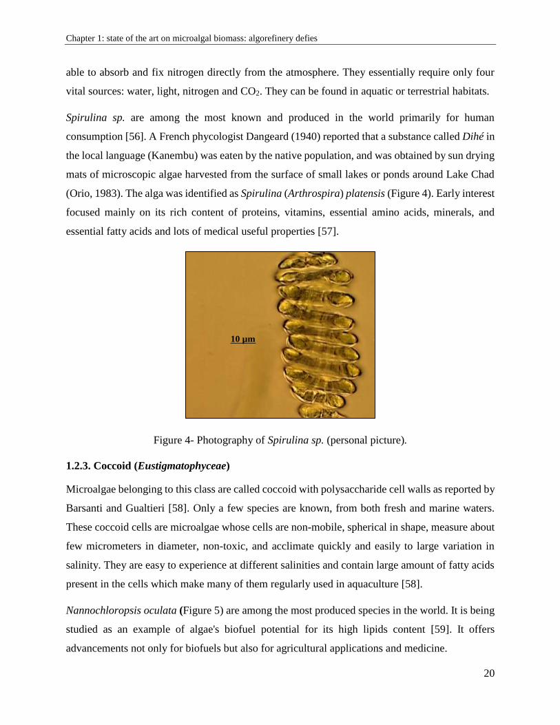
Spirulina sp. are among the most known and produced in the world primarily for human
consumption [56]. A French phycologist Dangeard (1940) reported that a substance called Dihé in
the local language (Kanembu) was eaten by the native population, and was obtained by sun drying
mats of microscopic algae harvested from the surface of small lakes or ponds around Lake Chad
(Orio, 1983). The alga was identified as Spirulina (Arthrospira) platensis (Figure 4). Early interest
focused mainly on its rich content of proteins, vitamins, essential amino acids, minerals, and
essential fatty acids and lots of medical useful properties [57].
Figure 4- Photography of Spirulina sp. (personal picture).
1.2.3. Coccoid (Eustigmatophyceae)
Microalgae belonging to this class are called coccoid with polysaccharide cell walls as reported by
Barsanti and Gualtieri [58]. Only a few species are known, from both fresh and marine waters.
These coccoid cells are microalgae whose cells are non-mobile, spherical in shape, measure about
few micrometers in diameter, non-toxic, and acclimate quickly and easily to large variation in
salinity. They are easy to experience at different salinities and contain large amount of fatty acids
present in the cells which make many of them regularly used in aquaculture [58].
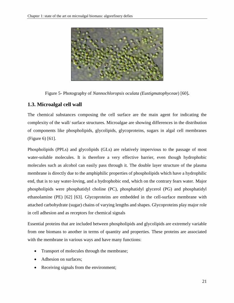
Nannochloropsis oculata (Figure 5) are among the most produced species in the world. It is being
studied as an example of algae's biofuel potential for its high lipids content [59]. It offers
advancements not only for biofuels but also for agricultural applications and medicine.
10 µm
Chapter 1: state of the art on microalgal biomass: algorefinery defies
21
Figure 5- Photography of Nannochloropsis oculata (Eustigmatophyceae) [60].
1.3. Microalgal cell wall
The chemical substances composing the cell surface are the main agent for indicating the
complexity of the wall/ surface structures. Microalgae are showing differences in the distribution
of components like phospholipids, glycolipids, glycoproteins, sugars in algal cell membranes
(Figure 6) [61].
Phospholipids (PPLs) and glycolipids (GLs) are relatively impervious to the passage of most
water-soluble molecules. It is therefore a very effective barrier, even though hydrophobic
molecules such as alcohol can easily pass through it. The double layer structure of the plasma
membrane is directly due to the amphiphilic properties of phospholipids which have a hydrophilic
end, that is to say water-loving, and a hydrophobic end, which on the contrary fears water. Major
phospholipids were phosphatidyl choline (PC), phosphatidyl glycerol (PG) and phosphatidyl
ethanolamine (PE) [62] [63]. Glycoproteins are embedded in the cell-surface membrane with
attached carbohydrate (sugar) chains of varying lengths and shapes. Glycoproteins play major role
in cell adhesion and as receptors for chemical signals
Essential proteins that are included between phospholipids and glycolipids are extremely variable
from one biomass to another in terms of quantity and properties. These proteins are associated
with the membrane in various ways and have many functions:
Transport of molecules through the membrane;
Adhesion on surfaces;
Receiving signals from the environment;
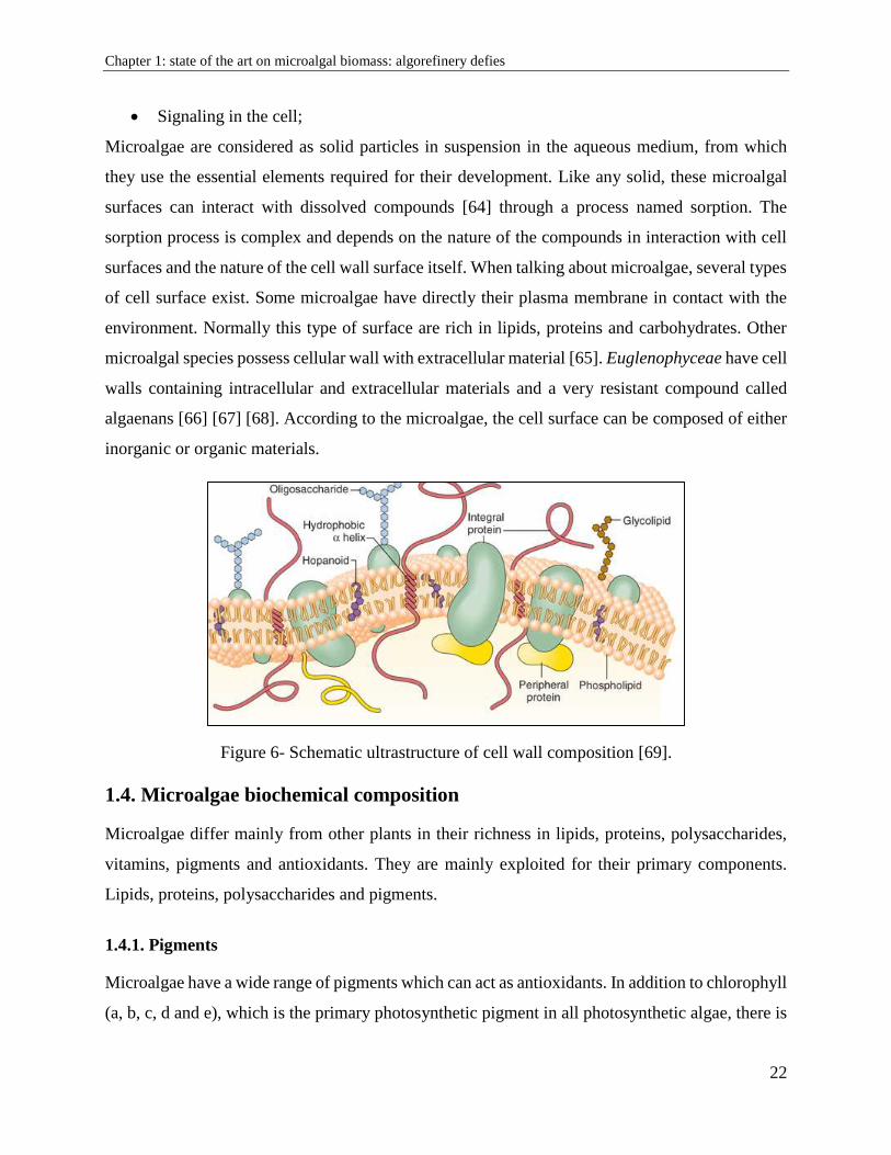
Chapter 1: state of the art on microalgal biomass: algorefinery defies
22
Signaling in the cell;
Microalgae are considered as solid particles in suspension in the aqueous medium, from which
they use the essential elements required for their development. Like any solid, these microalgal
surfaces can interact with dissolved compounds [64] through a process named sorption. The
sorption process is complex and depends on the nature of the compounds in interaction with cell
surfaces and the nature of the cell wall surface itself. When talking about microalgae, several types
of cell surface exist. Some microalgae have directly their plasma membrane in contact with the
environment. Normally this type of surface are rich in lipids, proteins and carbohydrates. Other
microalgal species possess cellular wall with extracellular material [65]. Euglenophyceae have cell
walls containing intracellular and extracellular materials and a very resistant compound called
algaenans [66] [67] [68]. According to the microalgae, the cell surface can be composed of either
inorganic or organic materials.
Figure 6- Schematic ultrastructure of cell wall composition [69].
1.4. Microalgae biochemical composition
Microalgae differ mainly from other plants in their richness in lipids, proteins, polysaccharides,
vitamins, pigments and antioxidants. They are mainly exploited for their primary components.
Lipids, proteins, polysaccharides and pigments.
1.4.1. Pigments
Microalgae have a wide range of pigments which can act as antioxidants. In addition to chlorophyll
(a, b, c, d and e), which is the primary photosynthetic pigment in all photosynthetic algae, there is
Chapter 1: state of the art on microalgal biomass: algorefinery defies
23
a range of additional yellow-orange lipid pigments as carotenoids. They serve key roles in
microalgae in absorbing light energy for use in photosynthesis and protecting chlorophyll from
photo-damage [70]. They split into two classes: xanthophyll (which contain oxygen)
and carotenes (which are purely hydrocarbons and contain no oxygen) (Figure 7).
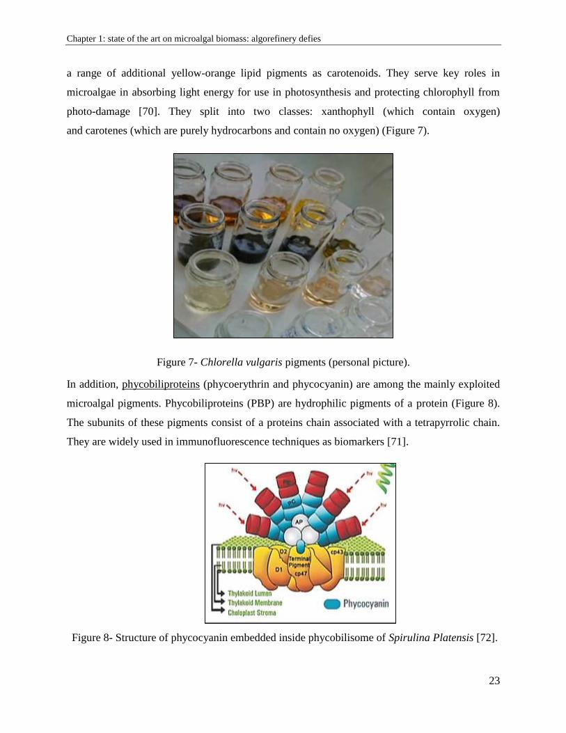
Figure 7- Chlorella vulgaris pigments (personal picture).
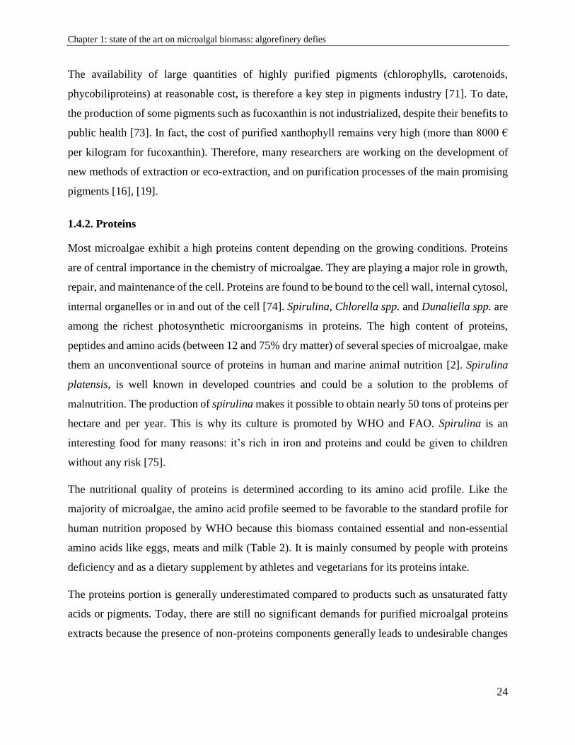
In addition, phycobiliproteins (phycoerythrin and phycocyanin) are among the mainly exploited
microalgal pigments. Phycobiliproteins (PBP) are hydrophilic pigments of a protein (Figure 8).
The subunits of these pigments consist of a proteins chain associated with a tetrapyrrolic chain.
They are widely used in immunofluorescence techniques as biomarkers [71].
Figure 8- Structure of phycocyanin embedded inside phycobilisome of Spirulina Platensis [72].
Chapter 1: state of the art on microalgal biomass: algorefinery defies
24
The availability of large quantities of highly purified pigments (chlorophylls, carotenoids,
phycobiliproteins) at reasonable cost, is therefore a key step in pigments industry [71]. To date,
the production of some pigments such as fucoxanthin is not industrialized, despite their benefits to
public health [73]. In fact, the cost of purified xanthophyll remains very high (more than 8000 €
per kilogram for fucoxanthin). Therefore, many researchers are working on the development of
new methods of extraction or eco-extraction, and on purification processes of the main promising
pigments [16], [19].
1.4.2. Proteins
Most microalgae exhibit a high proteins content depending on the growing conditions. Proteins
are of central importance in the chemistry of microalgae. They are playing a major role in growth,
repair, and maintenance of the cell. Proteins are found to be bound to the cell wall, internal cytosol,
internal organelles or in and out of the cell [74]. Spirulina, Chlorella spp. and Dunaliella spp. are
among the richest photosynthetic microorganisms in proteins. The high content of proteins,
peptides and amino acids (between 12 and 75% dry matter) of several species of microalgae, make
them an unconventional source of proteins in human and marine animal nutrition [2]. Spirulina
platensis, is well known in developed countries and could be a solution to the problems of
malnutrition. The production of spirulina makes it possible to obtain nearly 50 tons of proteins per
hectare and per year. This is why its culture is promoted by WHO and FAO. Spirulina is an
interesting food for many reasons: it’s rich in iron and proteins and could be given to children
without any risk [75].
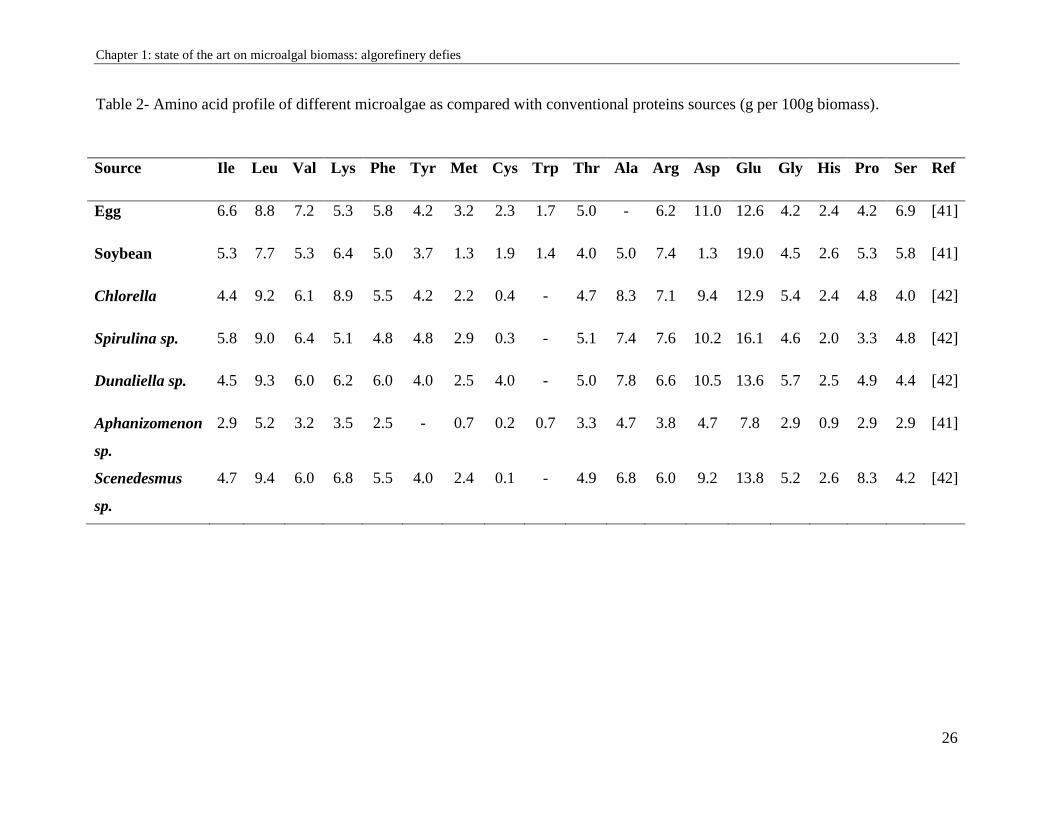
The nutritional quality of proteins is determined according to its amino acid profile. Like the
majority of microalgae, the amino acid profile seemed to be favorable to the standard profile for
human nutrition proposed by WHO because this biomass contained essential and non-essential
amino acids like eggs, meats and milk (Table 2). It is mainly consumed by people with proteins
deficiency and as a dietary supplement by athletes and vegetarians for its proteins intake.
The proteins portion is generally underestimated compared to products such as unsaturated fatty
acids or pigments. Today, there are still no significant demands for purified microalgal proteins
extracts because the presence of non-proteins components generally leads to undesirable changes
Chapter 1: state of the art on microalgal biomass: algorefinery defies
25
in the color or taste of proteins. Nevertheless, efforts are now turning towards the impressive
potential activities of microalgae proteins.
Chapter 1: state of the art on microalgal biomass: algorefinery defies
26
Table 2- Amino acid profile of different microalgae as compared with conventional proteins sources (g per 100g biomass).
Source Ile Leu Val Lys Phe Tyr Met Cys Trp Thr Ala Arg Asp Glu Gly His Pro Ser Ref
Egg 6.6 8.8 7.2 5.3 5.8 4.2 3.2 2.3 1.7 5.0 - 6.2 11.0 12.6 4.2 2.4 4.2 6.9 [41]
Soybean 5.3 7.7 5.3 6.4 5.0 3.7 1.3 1.9 1.4 4.0 5.0 7.4 1.3 19.0 4.5 2.6 5.3 5.8 [41]
Chlorella 4.4 9.2 6.1 8.9 5.5 4.2 2.2 0.4 - 4.7 8.3 7.1 9.4 12.9 5.4 2.4 4.8 4.0 [42]
Spirulina sp. 5.8 9.0 6.4 5.1 4.8 4.8 2.9 0.3 - 5.1 7.4 7.6 10.2 16.1 4.6 2.0 3.3 4.8 [42]
Dunaliella sp. 4.5 9.3 6.0 6.2 6.0 4.0 2.5 4.0 - 5.0 7.8 6.6 10.5 13.6 5.7 2.5 4.9 4.4 [42]
Aphanizomenon
sp.
2.9 5.2 3.2 3.5 2.5 - 0.7 0.2 0.7 3.3 4.7 3.8 4.7 7.8 2.9 0.9 2.9 2.9 [41]
Scenedesmus
sp.
4.7 9.4 6.0 6.8 5.5 4.0 2.4 0.1 - 4.9 6.8 6.0 9.2 13.8 5.2 2.6 8.3 4.2 [42]
Chapter 1: state of the art on microalgal biomass: algorefinery defies
27
1.4.3. Lipids
Microalgae can accumulate more than 80% of their dry weight in lipids [76]. Marine lipids can be
classified according to two different categories based on their polarity: on the one hand, neutral
lipids (NL) including monoacylglycerol (MAG), diacylglycerol (DAG), triacylglycerol (TAG) and
free fatty acids (FFA). On the other hand, polar lipids divided into two groups: glycolipids (GLs)
and phospholipids (PLs).
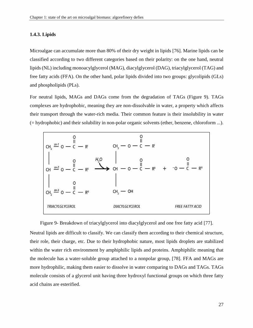
For neutral lipids, MAGs and DAGs come from the degradation of TAGs (Figure 9). TAGs
complexes are hydrophobic, meaning they are non-dissolvable in water, a property which affects
their transport through the water-rich media. Their common feature is their insolubility in water
(= hydrophobic) and their solubility in non-polar organic solvents (ether, benzene, chloroform ...).
Figure 9- Breakdown of triacylglycerol into diacylglycerol and one free fatty acid [77].
Neutral lipids are difficult to classify. We can classify them according to their chemical structure,
their role, their charge, etc. Due to their hydrophobic nature, most lipids droplets are stabilized
within the water rich environment by amphiphilic lipids and proteins. Amphiphilic meaning that
the molecule has a water-soluble group attached to a nonpolar group, [78]. FFA and MAGs are
more hydrophilic, making them easier to dissolve in water comparing to DAGs and TAGs. TAGs
molecule consists of a glycerol unit having three hydroxyl functional groups on which three fatty
acid chains are esterified.
Chapter 1: state of the art on microalgal biomass: algorefinery defies
28
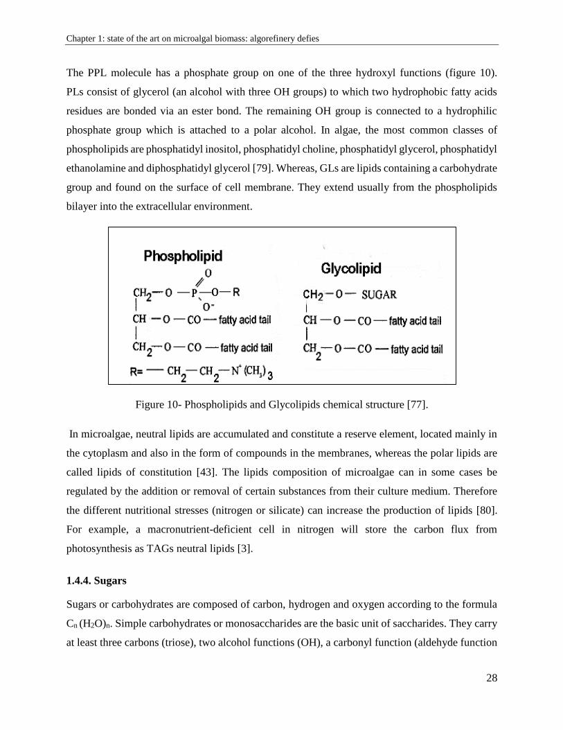
The PPL molecule has a phosphate group on one of the three hydroxyl functions (figure 10).
PLs consist of glycerol (an alcohol with three OH groups) to which two hydrophobic fatty acids
residues are bonded via an ester bond. The remaining OH group is connected to a hydrophilic
phosphate group which is attached to a polar alcohol. In algae, the most common classes of
phospholipids are phosphatidyl inositol, phosphatidyl choline, phosphatidyl glycerol, phosphatidyl
ethanolamine and diphosphatidyl glycerol [79]. Whereas, GLs are lipids containing a carbohydrate
group and found on the surface of cell membrane. They extend usually from the phospholipids
bilayer into the extracellular environment.
Figure 10- Phospholipids and Glycolipids chemical structure [77].
In microalgae, neutral lipids are accumulated and constitute a reserve element, located mainly in
the cytoplasm and also in the form of compounds in the membranes, whereas the polar lipids are
called lipids of constitution [43]. The lipids composition of microalgae can in some cases be
regulated by the addition or removal of certain substances from their culture medium. Therefore
the different nutritional stresses (nitrogen or silicate) can increase the production of lipids [80].
For example, a macronutrient-deficient cell in nitrogen will store the carbon flux from
photosynthesis as TAGs neutral lipids [3].
1.4.4. Sugars
Sugars or carbohydrates are composed of carbon, hydrogen and oxygen according to the formula
Cn (H2O)n. Simple carbohydrates or monosaccharides are the basic unit of saccharides. They carry
at least three carbons (triose), two alcohol functions (OH), a carbonyl function (aldehyde function
Chapter 1: state of the art on microalgal biomass: algorefinery defies
29
(-CHO) or ketone (-C = O)), and sometimes an amine (-NH2) or carboxylic function.
Polysaccharides are either holosides composed solely of monosaccharides, or glycosides
composed of monosaccharides and a non-osidic part called aglycone. Polysaccharides can have
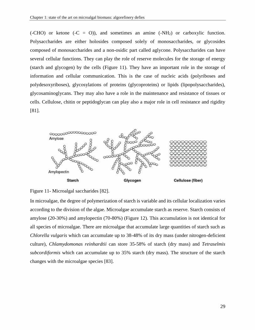
several cellular functions. They can play the role of reserve molecules for the storage of energy
(starch and glycogen) by the cells (Figure 11). They have an important role in the storage of
information and cellular communication. This is the case of nucleic acids (polyriboses and
polydesoxyriboses), glycosylations of proteins (glycoproteins) or lipids (lipopolysaccharides),
glycosaminoglycans. They may also have a role in the maintenance and resistance of tissues or
cells. Cellulose, chitin or peptidoglycan can play also a major role in cell resistance and rigidity
[81].
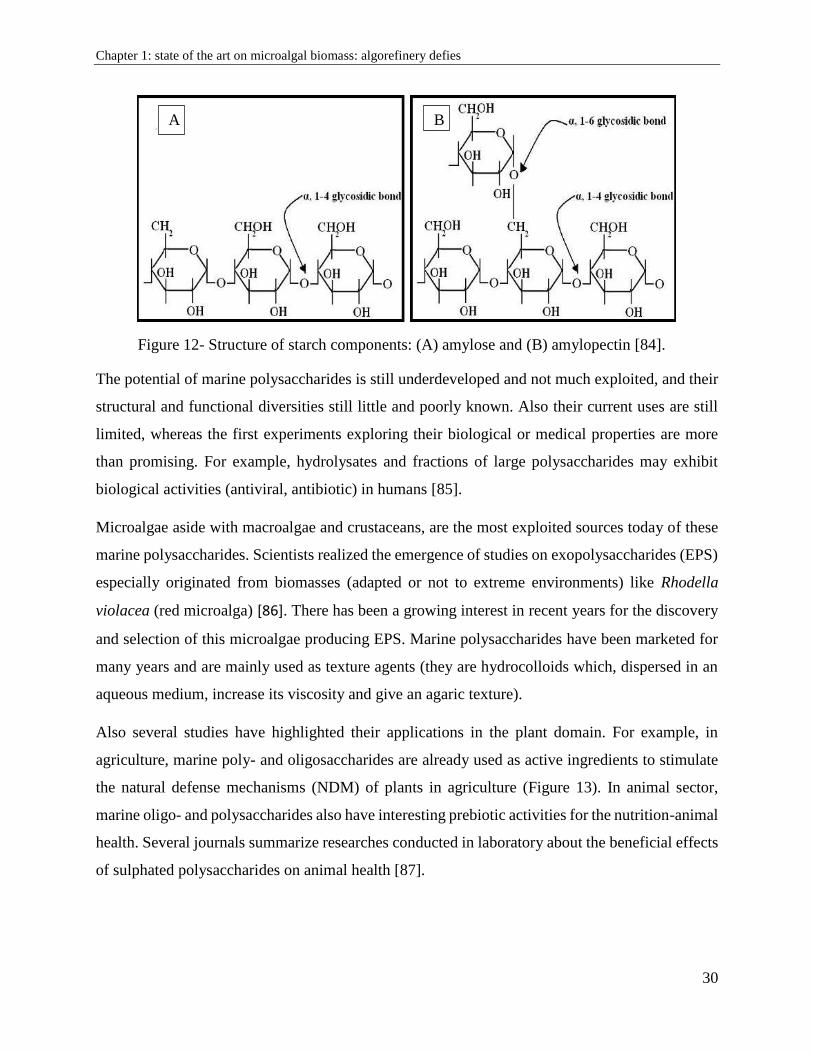
Figure 11- Microalgal saccharides [82].
In microalgae, the degree of polymerization of starch is variable and its cellular localization varies
according to the division of the algae. Microalgae accumulate starch as reserve. Starch consists of
amylose (20-30%) and amylopectin (70-80%) (Figure 12). This accumulation is not identical for
all species of microalgae. There are microalgae that accumulate large quantities of starch such as
Chlorella vulgaris which can accumulate up to 38-48% of its dry mass (under nitrogen-deficient
culture), Chlamydomonas reinhardtii can store 35-58% of starch (dry mass) and Tetraselmis
subcordiformis which can accumulate up to 35% starch (dry mass). The structure of the starch
changes with the microalgae species [83].
Chapter 1: state of the art on microalgal biomass: algorefinery defies
30
Figure 12- Structure of starch components: (A) amylose and (B) amylopectin [84].
The potential of marine polysaccharides is still underdeveloped and not much exploited, and their
structural and functional diversities still little and poorly known. Also their current uses are still
limited, whereas the first experiments exploring their biological or medical properties are more
than promising. For example, hydrolysates and fractions of large polysaccharides may exhibit
biological activities (antiviral, antibiotic) in humans [85].
Microalgae aside with macroalgae and crustaceans, are the most exploited sources today of these
marine polysaccharides. Scientists realized the emergence of studies on exopolysaccharides (EPS)
especially originated from biomasses (adapted or not to extreme environments) like Rhodella
violacea (red microalga) [86]. There has been a growing interest in recent years for the discovery
and selection of this microalgae producing EPS. Marine polysaccharides have been marketed for
many years and are mainly used as texture agents (they are hydrocolloids which, dispersed in an
aqueous medium, increase its viscosity and give an agaric texture).
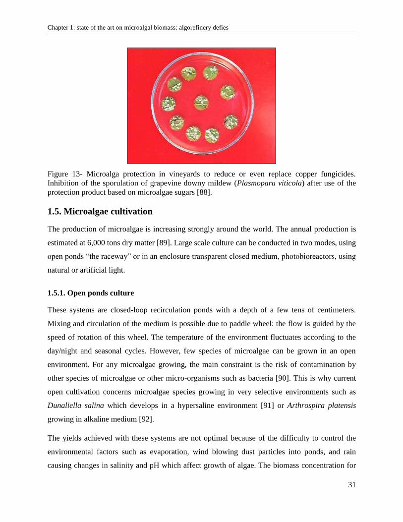
Also several studies have highlighted their applications in the plant domain. For example, in
agriculture, marine poly- and oligosaccharides are already used as active ingredients to stimulate
the natural defense mechanisms (NDM) of plants in agriculture (Figure 13). In animal sector,
marine oligo- and polysaccharides also have interesting prebiotic activities for the nutrition-animal
health. Several journals summarize researches conducted in laboratory about the beneficial effects
of sulphated polysaccharides on animal health [87].
A B
Chapter 1: state of the art on microalgal biomass: algorefinery defies
31
Figure 13- Microalga protection in vineyards to reduce or even replace copper fungicides. Inhibition of the sporulation of grapevine downy mildew (Plasmopara viticola) after use of the
protection product based on microalgae sugars [88].
1.5. Microalgae cultivation
The production of microalgae is increasing strongly around the world. The annual production is
estimated at 6,000 tons dry matter [89]. Large scale culture can be conducted in two modes, using
open ponds “the raceway” or in an enclosure transparent closed medium, photobioreactors, using
natural or artificial light.
1.5.1. Open ponds culture
These systems are closed-loop recirculation ponds with a depth of a few tens of centimeters.
Mixing and circulation of the medium is possible due to paddle wheel: the flow is guided by the
speed of rotation of this wheel. The temperature of the environment fluctuates according to the
day/night and seasonal cycles. However, few species of microalgae can be grown in an open
environment. For any microalgae growing, the main constraint is the risk of contamination by
other species of microalgae or other micro-organisms such as bacteria [90]. This is why current
open cultivation concerns microalgae species growing in very selective environments such as
Dunaliella salina which develops in a hypersaline environment [91] or Arthrospira platensis
growing in alkaline medium [92].
The yields achieved with these systems are not optimal because of the difficulty to control the
environmental factors such as evaporation, wind blowing dust particles into ponds, and rain
causing changes in salinity and pH which affect growth of algae. The biomass concentration for
Chapter 1: state of the art on microalgal biomass: algorefinery defies
32
this type of culture is generally low because the agitation of the medium is low and the existence
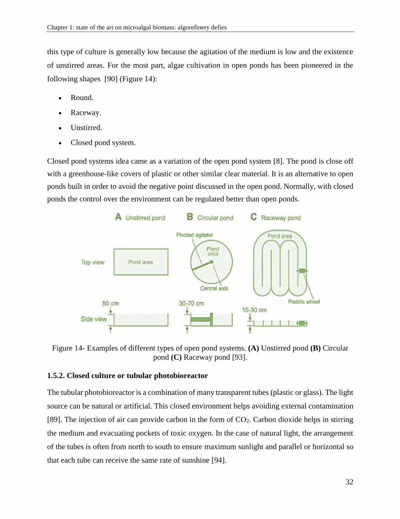
of unstirred areas. For the most part, algae cultivation in open ponds has been pioneered in the
following shapes [90] (Figure 14):
Round.
Raceway.
Unstirred.
Closed pond system.
Closed pond systems idea came as a variation of the open pond system [8]. The pond is close off
with a greenhouse-like covers of plastic or other similar clear material. It is an alternative to open
ponds built in order to avoid the negative point discussed in the open pond. Normally, with closed
ponds the control over the environment can be regulated better than open ponds.
Figure 14- Examples of different types of open pond systems. (A) Unstirred pond (B) Circular
pond (C) Raceway pond [93].
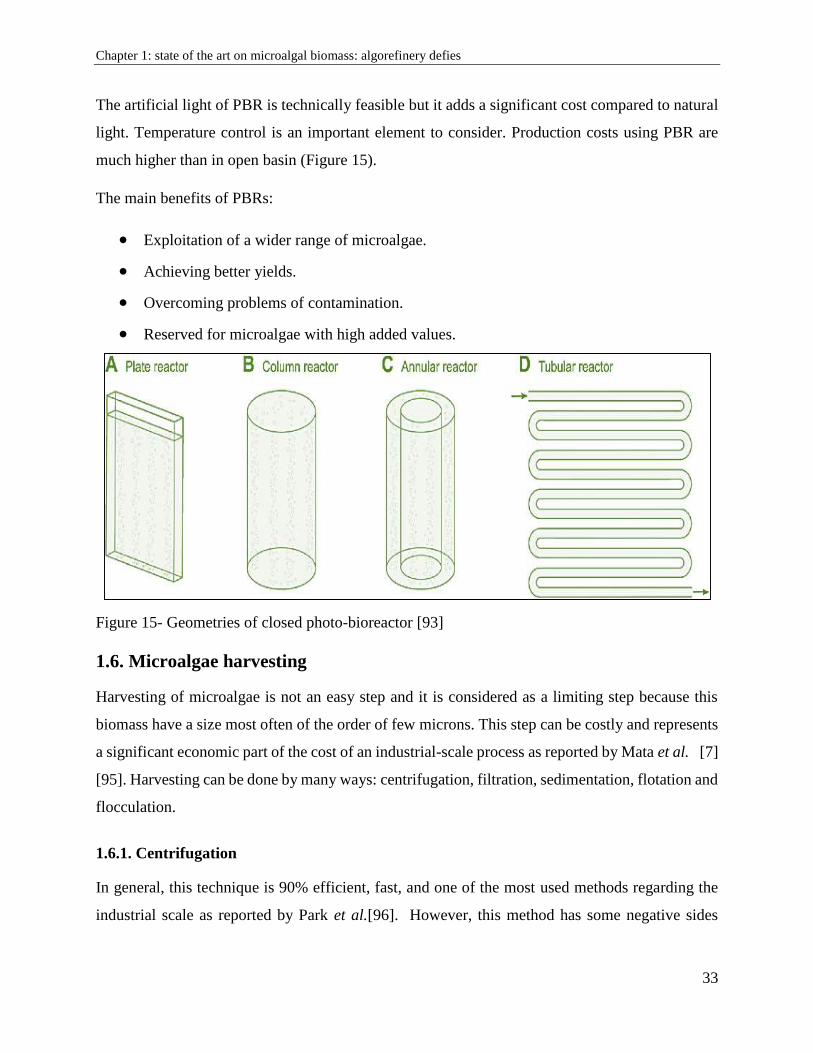
1.5.2. Closed culture or tubular photobioreactor
The tubular photobioreactor is a combination of many transparent tubes (plastic or glass). The light
source can be natural or artificial. This closed environment helps avoiding external contamination
[89]. The injection of air can provide carbon in the form of CO2. Carbon dioxide helps in stirring
the medium and evacuating pockets of toxic oxygen. In the case of natural light, the arrangement
of the tubes is often from north to south to ensure maximum sunlight and parallel or horizontal so
that each tube can receive the same rate of sunshine [94].
Chapter 1: state of the art on microalgal biomass: algorefinery defies
33
The artificial light of PBR is technically feasible but it adds a significant cost compared to natural
light. Temperature control is an important element to consider. Production costs using PBR are
much higher than in open basin (Figure 15).
The main benefits of PBRs:
Exploitation of a wider range of microalgae.
Achieving better yields.
Overcoming problems of contamination.
Reserved for microalgae with high added values.
Figure 15- Geometries of closed photo-bioreactor [93]
1.6. Microalgae harvesting
Harvesting of microalgae is not an easy step and it is considered as a limiting step because this
biomass have a size most often of the order of few microns. This step can be costly and represents
a significant economic part of the cost of an industrial-scale process as reported by Mata et al. [7]
[95]. Harvesting can be done by many ways: centrifugation, filtration, sedimentation, flotation and
flocculation.
1.6.1. Centrifugation
In general, this technique is 90% efficient, fast, and one of the most used methods regarding the
industrial scale as reported by Park et al.[96]. However, this method has some negative sides
Chapter 1: state of the art on microalgal biomass: algorefinery defies
34
which could be summarized by the fact of being expensive in energy which makes it difficult to
achieve economically [97] and could exposure cells to damage due to its centripetal force [95].
1.6.2. Flocculation
It is a process in which the microalgae in solution join together to form aggregates called "flocs"
[98]. When flocculation is combined with filtration, the efficiency is significantly increased [97].
For example, a study conducted by Gerde et al. [99] to flocculate cells
of Scenedesmus spp., Chlamydomonas reinhardtii, and Schizochytrium limacinum using
aluminum sulfate (Al2 (SO4)3) and two cationic starches as flocculants, showed that cationic
starches provide an efficient and ecologically friendly way to harvest microalgae.
A flocculation efficiency >98 % was achieved at both pilot-scale by Ndikubwimana et al. [100]
using a simple, effective, economic, and environmentally friendly bio-flocculant “broth of Bacillus
licheniformis”.
1.6.3. Sedimentation
Microalgae of large size and high density can be harvested by sedimentation such as Spirulina sp.
The sedimentation rate can be significantly improved by the addition of flocculants as reported by
several authors [95] [98] [101] [99].
1.6.4. Filtration
In this technique, the microalgal suspension passes through filtering membranes to retain only the
cells. There are different forms of filtration: microfiltration, vacuum filtration, pressure filtration,
ultrafiltration and tangential or frontal flow filtration. The choice of the molecular weight cut-
off (MWCO) of the membrane depends on the size of the species to be harvested [102].
1.6.5. Flotation
A process in which gas bubbles are bound to microalgal cells allowing them to float on the surface.
Flotation system for microalgae harvesting is an effective method for small size cells such as
Nannochloropsis oculata [97] [95]. In the dissolved air flotation process, the air bubbles are
reduced in size from 10 to 100 μm [95]. The air bubbles pass through the medium, adhere to each
other, bind to the particles and thus increase their buoyancy, and create "flocs" on the surface
Chapter 1: state of the art on microalgal biomass: algorefinery defies
35
where a "compacting" zone is formed [96]. In the dispersed air flotation process, air injection and
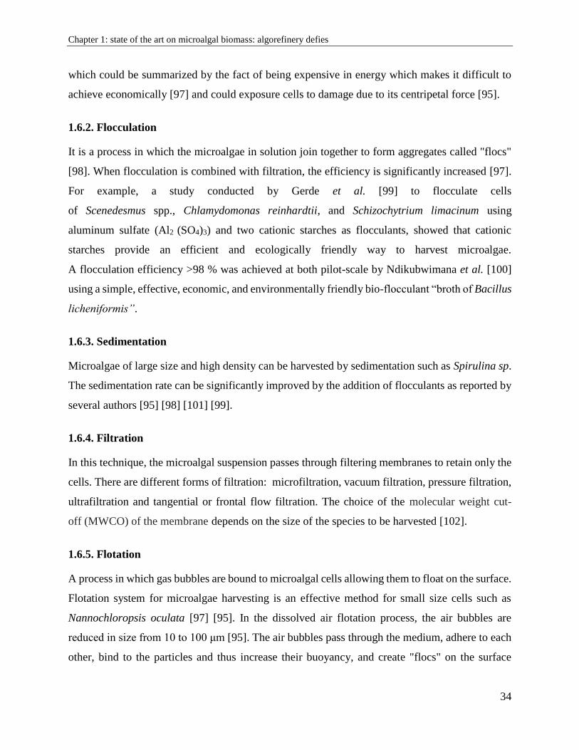
strong agitation form 700 to1500 μm bubbles inside the medium. These bubbles will "react" with
the negative charges of the microalgae cells [95] (Figure 16).
Figure 16- A laboratory model showing electro-coagulation–flocculation of microalgae [103].
The technique can be improved with the addition of surfactants that will bring a positive charge to
the environment [97]. For example, Kurniawati et al. investigated the harvesting of Chlorella
vulgaris and Scenedesmus obliquus using natural bio surfactant saponin as the collector and
chitosan as the flocculent [104].
1.7. Effects of the medium on the growth and biochemical composition of
microalgae
The nutritional requirements of microalgae are similar to those of higher plants [105]. The culture
medium must satisfy the needs in major elements (or macro-elements) C, H, N, O, P, S and micro-
elements called traceable elements. Table 3 indicates the major constituent elements of microalgae.
Microalgae (like most cells) are composed mainly of carbohydrate proteins and lipids. The culture
medium must be able to support the needs of the organisms, however, cells could be adapted in
order to continue their development under certain conditions of stress or deficiency.
Chapter 1: state of the art on microalgal biomass: algorefinery defies
36
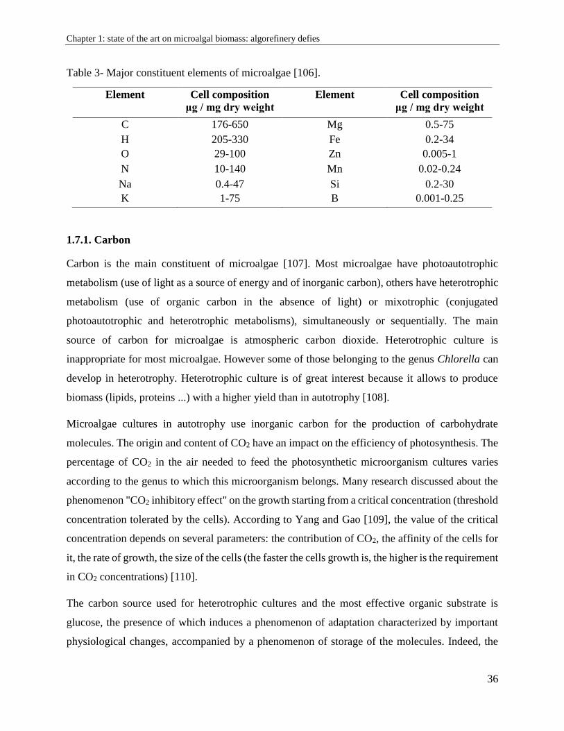
Table 3- Major constituent elements of microalgae [106].
Element Cell composition
μg / mg dry weight
Element Cell composition
μg / mg dry weight
C 176-650 Mg 0.5-75
H 205-330 Fe 0.2-34
O 29-100 Zn 0.005-1
N 10-140 Mn 0.02-0.24
Na 0.4-47 Si 0.2-30
K 1-75 B 0.001-0.25
1.7.1. Carbon
Carbon is the main constituent of microalgae [107]. Most microalgae have photoautotrophic
metabolism (use of light as a source of energy and of inorganic carbon), others have heterotrophic
metabolism (use of organic carbon in the absence of light) or mixotrophic (conjugated
photoautotrophic and heterotrophic metabolisms), simultaneously or sequentially. The main
source of carbon for microalgae is atmospheric carbon dioxide. Heterotrophic culture is
inappropriate for most microalgae. However some of those belonging to the genus Chlorella can
develop in heterotrophy. Heterotrophic culture is of great interest because it allows to produce
biomass (lipids, proteins ...) with a higher yield than in autotrophy [108].
Microalgae cultures in autotrophy use inorganic carbon for the production of carbohydrate
molecules. The origin and content of CO2 have an impact on the efficiency of photosynthesis. The
percentage of CO2 in the air needed to feed the photosynthetic microorganism cultures varies
according to the genus to which this microorganism belongs. Many research discussed about the
phenomenon "CO2 inhibitory effect" on the growth starting from a critical concentration (threshold
concentration tolerated by the cells). According to Yang and Gao [109], the value of the critical
concentration depends on several parameters: the contribution of CO2, the affinity of the cells for
it, the rate of growth, the size of the cells (the faster the cells growth is, the higher is the requirement
in CO2 concentrations) [110].
The carbon source used for heterotrophic cultures and the most effective organic substrate is
glucose, the presence of which induces a phenomenon of adaptation characterized by important
physiological changes, accompanied by a phenomenon of storage of the molecules. Indeed, the
Chapter 1: state of the art on microalgal biomass: algorefinery defies
37
cells increase the intracellular synthesis of polysaccharides, lipids, proteins, chlorophylls, RNA
and vitamins, which leads to an increase in their volume according to Perez-Garcia et al. [111].
1.7.2. Nitrogen
For the development of microalgae, nitrogen is essential. Indeed, it is a constituent element of the
amino acids that make up the proteins. The nitrogen sources are mainly mineral (NH4+, NO3
- and
NO2-), but there are also organic sources such as urea and amino acids.
When microalgae culture medium is deficient in nitrogen, the growth rate of microalgae decreases.
Cells enter the stationary phase and accumulate lipids (triacylglycerol) and / or carbohydrates, to
the detriment of proteins and pigments [112] [113]. This phenomenon induces an increase in cell
volume [113]. Nitrogen deficiency is responsible for a quantitative and qualitative change in the
production of molecules by microalgae. Microalgae belonging to the genus Chlorella accumulate
neutral lipids in response to nitrogen substrate stress as reported by Lin et al. [114]. Nitrogen
deficiency is known as an effective tool for the production of algal lipids. In addition, researches
show a connection between the cellular lipids accumulation (observed in nitrogen deficiency) and
autophagy. In the green alga Chlamydomonas reinhardtii, Perez-Perez et al. [115] identified that
autophagy was active in this alga in stress conditions, including nitrogen deficiency, oxidative
stress, or the presence of mis-folded proteins in the endoplasmic reticulum.
Other than autophagy and lipids accumulation, nitrogen deficiency can also result in a change of
pigmentation of microalgae. To protect against nitrogen deficiency and maintain an available
source of nitrogen, microalgae catabolize their constitutive pigments and in particular the
supernumerary pigments of collecting antennae such as phycoerythrin [113].
However, the concentration of certain pigments increases during a nitrogen deficiency. This is the
case of the intracellular beta-carotene of the microalga Dunaliella salina [116]. The same
phenomenon has been described, in Eustigmatos cf. polyphem for which the intracellular
concentration of beta-carotene increases by a factor of 3 following a nitrogen deficiency.
1.7.3. Phosphorus
Phosphorus is not widespread in nature. It is mainly found in the form of phosphate salts (PO43-).
Phosphorus is an indispensable element for the microalgae growth. It participates in many
Chapter 1: state of the art on microalgal biomass: algorefinery defies
38
metabolic processes, notably through energy exchanges (ATP), in the formation of nucleic acids
and phospholipids [117]. Like nitrogen, phosphorus deficiency causes a decrease in cell growth.
In Chlorella vulgaris, the same deficiency causes a specific decrease in the amount of intracellular
inorganic polyphosphate [118]. The affinity of microorganisms for the substrate (phosphate) varies
depending on the species. It is 4-5 μM for the genus Chlorella as reported by Martinez Sancho et
al. [119].
1.7.4. Sulfur
Sulfur is also important for the development of organisms. It is used in certain amino acids, such
as cysteine and methionine. It is used by photosynthetic organisms mainly in the form of sulphate
(SO42-) as reported by Prescott et al., [117]. Sulfur deficiency leads to a decrease in growth by a
factor of 2 to 2.5 in Chlorella vulgaris and Chlorella sorokiniana, and leads to an increase in starch
reserves.
1.7.5. Salts
Some microalgae are adapted to fresh water, others to marine environments and some are
halophiles. Marine microalgae can adapt to media with salt concentrations (NaCl) of 10% to 50%
(w/v) depending on the microalgae.
Beyond these threshold values, cell growth is slowed down or even halted as cited by Eggert et al.
[120]. When the salt concentration becomes too important for the marine microalgae, osmotic
stress is created which induces changes in its metabolism. Excess salt has no lethal effect on the
cells of marine microalgae. Microalgae of the genus Scenedesmus under salt stress conditions
accumulate lipids (triacylglycerols) and carotenoids. The morphology of the microalgae is also
modified and the cells round out. All of this adaptation is at the expense of cell growth as reported
by Vidyashankar et al. [121]. During osmotic stress, some microalgae produce an exo-
polysaccharide that forms a mucilage around the cells. This carbohydrate capsule limits ion
exchange and protects cells.
Chapter 1: state of the art on microalgal biomass: algorefinery defies
39
1.8. Extraction processes for component recovery
The potential of microalgae is now well established. They arouse the interest and hope of
researchers. The general processing scheme involves the step of production / cultivation of micro-
algal biomass, harvesting / concentration and extraction of components.
The components extraction stage remains a critical step for which significant efforts must be made.
Given the size and nature of the components (hydrophilic or hydrophobic), many of the techniques
used in the field are more or less effective.
The valuable compounds of microalgae are stored within the cells that can be protected by a thick
wall. Their extraction can be done using specific organic solvents and often requires a step to break
the cell walls to make them accessible. To do this, several treatments are possible: grinding,
ultrasound, microwaves, osmotic shocks, enzymatic lysis, etc. The choice of the bursting technique
depends mainly on the cellular characteristics and the dry matter content of the algal biomass
[122].
1.8.1. Extraction using organic solvents
The lipids and pigments can be generally extracted with a water-immiscible organic solvent such
as n-hexane, chloroform, petroleum ether, or a mixture of solvents on the basis of the method
developed by Bligh and Dyer [123]. This general principle of dissolution is often expressed by the
expression "the like dissolves in the like".
The extraction can also be optimized by an intensification of the temperature and pressure
conditions thus making it possible to increase the solvating power of the solvent used [95].
The ideal extraction solvent for marine lipids should have a low boiling point to facilitate its
removal. For example, chloroform/methanol (1/2: v/v) is the most commonly used solvents
mixture for extracting lipids from living tissue. This procedure is known as the Bligh and Dyer
method (Bligh and Dyer, 1959), which today constitutes the reference method in the literature. The
Soxhlet extraction technique is also a widely used method [124].
There is, moreover, a process for extracting lipids from fresh biocompatible microalgae which
makes it possible to keep the cells alive during the extraction step. In this case, the algae are
brought into contact with an organic solvent, n-decane, and proceed to a liquid/liquid extraction
Chapter 1: state of the art on microalgal biomass: algorefinery defies
40
operation, followed by phase separation. The treated algae are then put back into cultivation, with
in some cases survival rates close to 100%. This process is called "milking" [125]. However, this
procedure has limitations because it requires the use of large volume of toxic solvent for human
health.
Currently, "green" solvents constitute a potential way of substitution of n-hexane from renewable
raw materials. They have the advantage of offering an alternative to fossil resources and can be
produced from biomass such as wood, starch, vegetable oils, fruits or even aromatic plants. They
are classified according to 3 categories, esters, alcohols and terpenes. Some of these have already
been shown to be effective for oilseed oil extraction [126]. However, very little work has been
done on the extraction of lipids from microalgae using these solvents. Zbinden et al. [127] has
used ethyl acetate as an alternative for the extraction of lipids from Ankistrodesmus falcatus.
1.8.2. Cell disintegrating techniques
The efficiency of the extraction of microalgal valuable components like lipids, proteins, pigments,
polysaccharides is closely linked to the level of cells disintegration. When the intact cells are
disintegrated, the intracellular molecules are released from the cell structures and then diffuse into
the surrounding medium. The methods of disintegrating cells are classified according to their
mechanical or non-mechanical mode of action (Figure 17).
Mechanical and physical methods include bead mill, ultrasound, autoclave, freeze-drying and
microwaves, while non-mechanical methods involve cell lysis by enzymes, acidic or basic
treatments, or osmotic shock. These different techniques can be used to assist the solvent extraction
or can constitute a pretreatment before the actual extraction step.
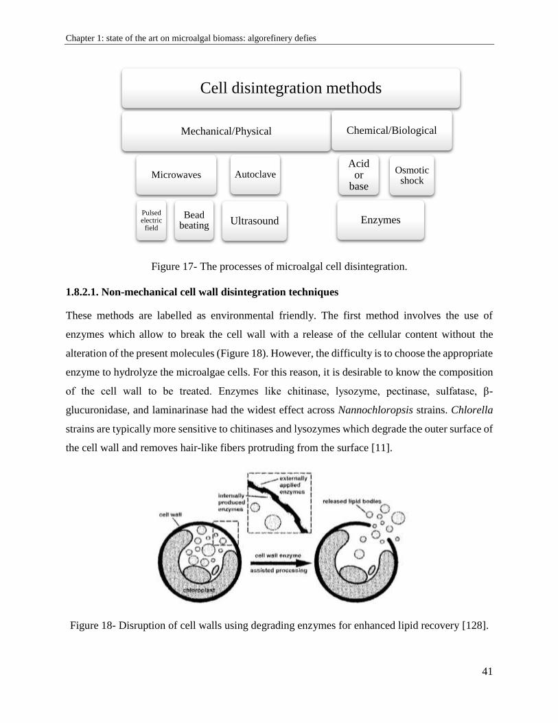
Chapter 1: state of the art on microalgal biomass: algorefinery defies
41
Figure 17- The processes of microalgal cell disintegration.
1.8.2.1. Non-mechanical cell wall disintegration techniques
These methods are labelled as environmental friendly. The first method involves the use of
enzymes which allow to break the cell wall with a release of the cellular content without the
alteration of the present molecules (Figure 18). However, the difficulty is to choose the appropriate
enzyme to hydrolyze the microalgae cells. For this reason, it is desirable to know the composition
of the cell wall to be treated. Enzymes like chitinase, lysozyme, pectinase, sulfatase, β-
glucuronidase, and laminarinase had the widest effect across Nannochloropsis strains. Chlorella
strains are typically more sensitive to chitinases and lysozymes which degrade the outer surface of
the cell wall and removes hair-like fibers protruding from the surface [11].
Figure 18- Disruption of cell walls using degrading enzymes for enhanced lipid recovery [128].
Cell disintegration methods
Mechanical/Physical
Microwaves
Pulsed electric
field
Beadbeating
Autoclave
Ultrasound
Chemical/Biological
Acid or
base
Enzymes
Osmotic shock
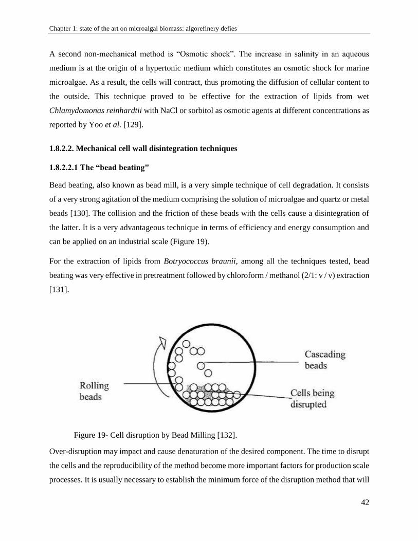
Chapter 1: state of the art on microalgal biomass: algorefinery defies
42
A second non-mechanical method is “Osmotic shock”. The increase in salinity in an aqueous
medium is at the origin of a hypertonic medium which constitutes an osmotic shock for marine
microalgae. As a result, the cells will contract, thus promoting the diffusion of cellular content to
the outside. This technique proved to be effective for the extraction of lipids from wet
Chlamydomonas reinhardtii with NaCl or sorbitol as osmotic agents at different concentrations as
reported by Yoo et al. [129].
1.8.2.2. Mechanical cell wall disintegration techniques
1.8.2.2.1 The “bead beating"
Bead beating, also known as bead mill, is a very simple technique of cell degradation. It consists
of a very strong agitation of the medium comprising the solution of microalgae and quartz or metal
beads [130]. The collision and the friction of these beads with the cells cause a disintegration of
the latter. It is a very advantageous technique in terms of efficiency and energy consumption and
can be applied on an industrial scale (Figure 19).
For the extraction of lipids from Botryococcus braunii, among all the techniques tested, bead
beating was very effective in pretreatment followed by chloroform / methanol (2/1: v / v) extraction
[131].
Figure 19- Cell disruption by Bead Milling [132].
Over-disruption may impact and cause denaturation of the desired component. The time to disrupt
the cells and the reproducibility of the method become more important factors for production scale
processes. It is usually necessary to establish the minimum force of the disruption method that will
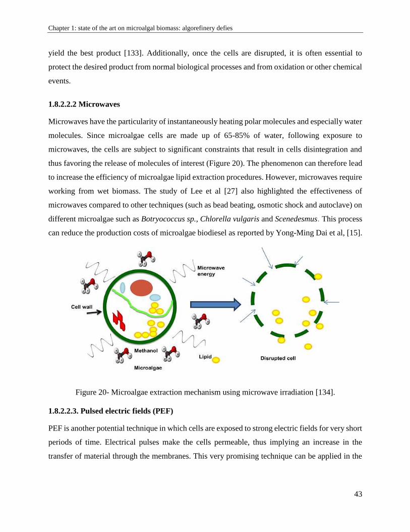
Chapter 1: state of the art on microalgal biomass: algorefinery defies
43
yield the best product [133]. Additionally, once the cells are disrupted, it is often essential to
protect the desired product from normal biological processes and from oxidation or other chemical
events.
1.8.2.2.2 Microwaves
Microwaves have the particularity of instantaneously heating polar molecules and especially water
molecules. Since microalgae cells are made up of 65-85% of water, following exposure to
microwaves, the cells are subject to significant constraints that result in cells disintegration and
thus favoring the release of molecules of interest (Figure 20). The phenomenon can therefore lead
to increase the efficiency of microalgae lipid extraction procedures. However, microwaves require
working from wet biomass. The study of Lee et al [27] also highlighted the effectiveness of
microwaves compared to other techniques (such as bead beating, osmotic shock and autoclave) on
different microalgae such as Botryococcus sp., Chlorella vulgaris and Scenedesmus. This process
can reduce the production costs of microalgae biodiesel as reported by Yong-Ming Dai et al, [15].
Figure 20- Microalgae extraction mechanism using microwave irradiation [134].
1.8.2.2.3. Pulsed electric fields (PEF)
PEF is another potential technique in which cells are exposed to strong electric fields for very short
periods of time. Electrical pulses make the cells permeable, thus implying an increase in the
transfer of material through the membranes. This very promising technique can be applied in the
Chapter 1: state of the art on microalgal biomass: algorefinery defies
44
pretreatment of microalgal biomass before solvent extraction. Zbinden et al., [127] showed the
effectiveness of PEF on lipid extraction of the microalgae Ankistrodesmus falcatus
1.8.2.2.4. Ultrasound Assisted Extraction (UAE)
Ultrasound Assisted Extraction has the advantage of being a technique of disintegration at low
temperature, thus leading to a decrease of the thermal denaturation of the sensitive compounds.
Ultrasound is easily transposable at industrial scale and can operate continuously [135].
When low frequency ultrasound is applied in a liquid, a cavitation phenomenon occurs, the
formation of tiny bubbles within the liquid medium, which is responsible for cell damage [136].
When these bubbles reach resonance size, they collapse releasing mechanical energy in the form
of shock waves equivalent to several thousand atmospheres of pressure. The shock waves disrupt
cells present in suspension. As ultrasound breaks the cell wall mechanically by cavitation shear
forces, and facilitates the transfer of inner components like lipids, proteins, pigments and
carbohydrates from the cell into the solvent (Figure 21). In ultrasonic applied field, a frequency
range between 20-25 to 600 kHz (mega Sonics) is commonly used for cell disruption and
separation of particles from biomass as reported by Pablo Juliano et al., Chemat et al., [31], [137].
The duration of ultrasound needed depends on the cell type, the sample size and the cell
concentration. In microalgal field, Jaeschke et al., [138] find ultrasound as an alternative
technology to extract carotenoids and lipids from Heterochlorella luteoviridisultra. Prabakaram
and Ravindram [139] carried out a study on three species of microalgae on which different methods
of disintegration were tested (autoclave, bead beating, microwaves, osmotic shock and ultrasound)
where ultrasound appears to be the technique of choice to obtain the best lipid yield.
In 2016, Ferreira et al [141], studied the effect of low frequency acoustic ultrasound on microalgae
Chlorella vulgaris, Nannochloropsis oculata and Scenedesmus obliquus for carbohydrates,
proteins, lipids and pigments valorization. But the study did not exclude the use of toxic and
harmful organic and high volatile solvent like n-hexane, chloroform, 2-butanol, isopropanol,
ethanol and methanol.
As reported by Oohashi et al. [142], it is generally accepted that humans cannot perceive sounds
in the frequency range above 20 kHz, and the use of such "inaudible" high-frequency components
may significantly affect the brain activity of listeners. High noise levels require hearing protection
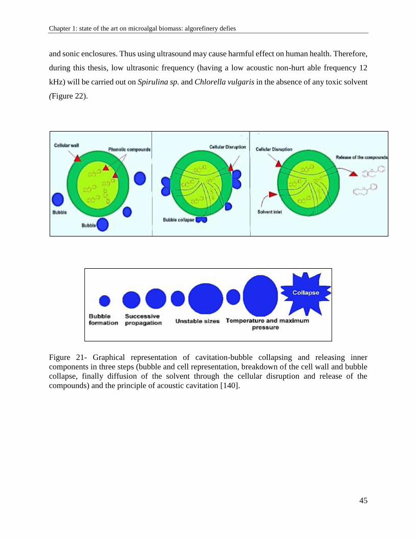
Chapter 1: state of the art on microalgal biomass: algorefinery defies
45
and sonic enclosures. Thus using ultrasound may cause harmful effect on human health. Therefore,
during this thesis, low ultrasonic frequency (having a low acoustic non-hurt able frequency 12
kHz) will be carried out on Spirulina sp. and Chlorella vulgaris in the absence of any toxic solvent
(Figure 22).
Figure 21- Graphical representation of cavitation-bubble collapsing and releasing inner
components in three steps (bubble and cell representation, breakdown of the cell wall and bubble
collapse, finally diffusion of the solvent through the cellular disruption and release of the
compounds) and the principle of acoustic cavitation [140].
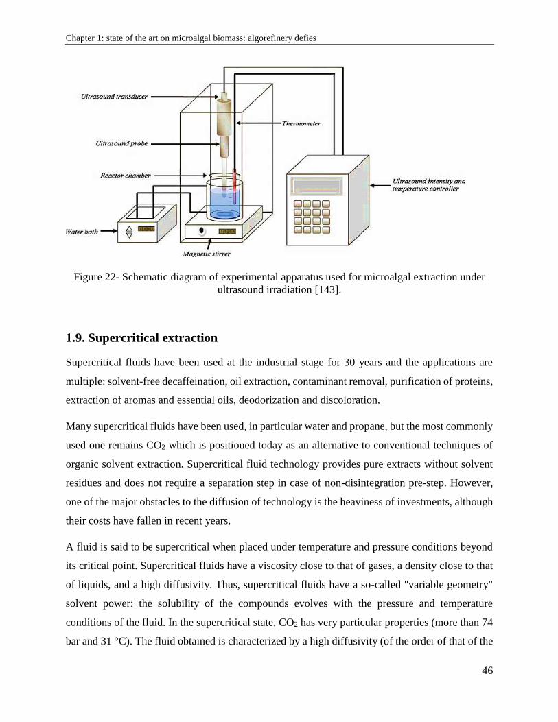
Chapter 1: state of the art on microalgal biomass: algorefinery defies
46
Figure 22- Schematic diagram of experimental apparatus used for microalgal extraction under
ultrasound irradiation [143].
1.9. Supercritical extraction
Supercritical fluids have been used at the industrial stage for 30 years and the applications are
multiple: solvent-free decaffeination, oil extraction, contaminant removal, purification of proteins,
extraction of aromas and essential oils, deodorization and discoloration.
Many supercritical fluids have been used, in particular water and propane, but the most commonly
used one remains CO2 which is positioned today as an alternative to conventional techniques of
organic solvent extraction. Supercritical fluid technology provides pure extracts without solvent
residues and does not require a separation step in case of non-disintegration pre-step. However,
one of the major obstacles to the diffusion of technology is the heaviness of investments, although
their costs have fallen in recent years.
A fluid is said to be supercritical when placed under temperature and pressure conditions beyond
its critical point. Supercritical fluids have a viscosity close to that of gases, a density close to that
of liquids, and a high diffusivity. Thus, supercritical fluids have a so-called "variable geometry"
solvent power: the solubility of the compounds evolves with the pressure and temperature
conditions of the fluid. In the supercritical state, CO2 has very particular properties (more than 74
bar and 31 °C). The fluid obtained is characterized by a high diffusivity (of the order of that of the
Chapter 1: state of the art on microalgal biomass: algorefinery defies
47
gases), which gives it a good diffusivity, and a high density which endows it with a large transport
and extraction capacity.
A supercritical CO2 (ScCO2) extraction process operates in closed circuit. It comprises
pressurizing elements (pumps) and temperature (exchangers) in order to bring the solvent above
its critical point [144]. The biomass to be treated is placed in an extractor crossed by the
supercritical CO2 flow. The fluid is charged with the extracted compound, then it is expanded,
passes into the gaseous phase and separates from the extracted compound. The latter is collected
in a separator.
The soluble molecules in ScCO2, and therefore extractable, are nonpolar such as aromatic
compounds, many pigments, neutral lipids, TAG, DAG, and MAG. Non-soluble molecules in
supercritical CO2 are polar like proteins, polysaccharides, glycolipids, phospholipids, sugars,
minerals. The ScCO2 extraction is based on the CO2 solvent power which can be modulated
according to the applied pressure and temperature conditions. It allows selective extraction without
denaturing sensitive molecules due to moderate temperature conditions. The products obtained do
not contain solvent residues, which is a major regulatory advantage. Moreover, the use of co-
solvents modifies the solvent power of ScCO2 and allows other extraction possibilities [145].
The benefits of ScCO2 are:
It is a natural product, abundant in nature and available.
It is nontoxic, inert, odorless, colorless and tasteless.
Its use does not alter products, and does not generate polluting residues.
It is inexpensive which makes the technique attractive and viable.
It has disinfecting properties vis-à-vis viruses and bacteria, although it is not considered
as a sterilization technique.
It has low critical coordinates (Tc, Pc).
The use of other supercritical fluids is also limited because of their high critical
temperatures and pressures such as water [146]. Methanol is a very good supercritical
solvent but its critical temperature and pressure are relatively high, making it unsuitable
for the treatment of thermo labile compounds. In addition, it is highly corrosive in the
presence of oxygen [146][147]. Another alternative fluid is nitric oxide, N2O. The
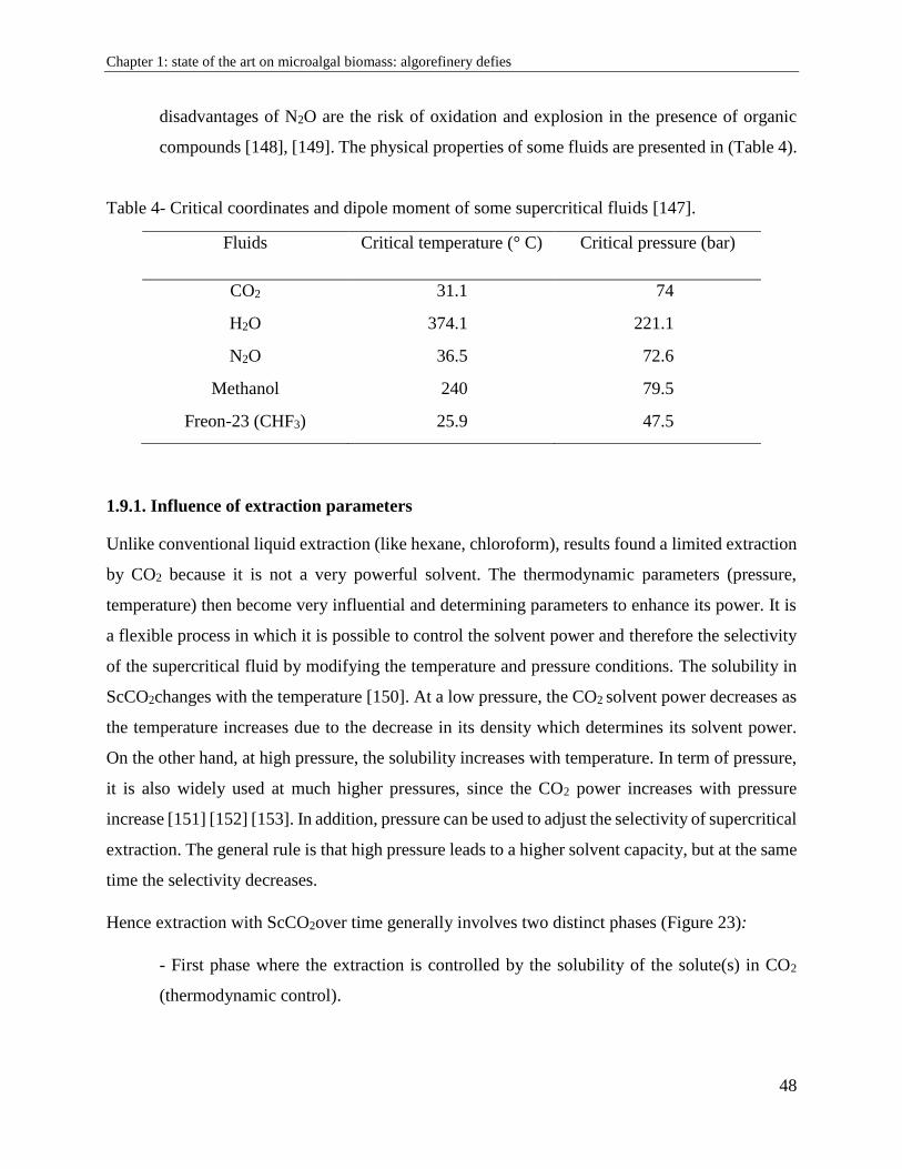
Chapter 1: state of the art on microalgal biomass: algorefinery defies
48
disadvantages of N2O are the risk of oxidation and explosion in the presence of organic
compounds [148], [149]. The physical properties of some fluids are presented in (Table 4).
Table 4- Critical coordinates and dipole moment of some supercritical fluids [147].
Fluids Critical temperature (° C) Critical pressure (bar)
CO2 31.1 74
H2O 374.1 221.1
N2O 36.5 72.6
Methanol 240 79.5
Freon-23 (CHF3) 25.9 47.5
1.9.1. Influence of extraction parameters
Unlike conventional liquid extraction (like hexane, chloroform), results found a limited extraction
by CO2 because it is not a very powerful solvent. The thermodynamic parameters (pressure,
temperature) then become very influential and determining parameters to enhance its power. It is
a flexible process in which it is possible to control the solvent power and therefore the selectivity
of the supercritical fluid by modifying the temperature and pressure conditions. The solubility in
ScCO2changes with the temperature [150]. At a low pressure, the CO2 solvent power decreases as
the temperature increases due to the decrease in its density which determines its solvent power.
On the other hand, at high pressure, the solubility increases with temperature. In term of pressure,
it is also widely used at much higher pressures, since the CO2 power increases with pressure
increase [151] [152] [153]. In addition, pressure can be used to adjust the selectivity of supercritical
extraction. The general rule is that high pressure leads to a higher solvent capacity, but at the same
time the selectivity decreases.
Hence extraction with ScCO2over time generally involves two distinct phases (Figure 23):
- First phase where the extraction is controlled by the solubility of the solute(s) in CO2
(thermodynamic control).
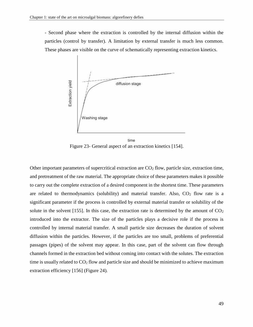
Chapter 1: state of the art on microalgal biomass: algorefinery defies
49
- Second phase where the extraction is controlled by the internal diffusion within the
particles (control by transfer). A limitation by external transfer is much less common.
These phases are visible on the curve of schematically representing extraction kinetics.
Figure 23- General aspect of an extraction kinetics [154].
Other important parameters of supercritical extraction are CO2 flow, particle size, extraction time,
and pretreatment of the raw material. The appropriate choice of these parameters makes it possible
to carry out the complete extraction of a desired component in the shortest time. These parameters
are related to thermodynamics (solubility) and material transfer. Also, CO2 flow rate is a
significant parameter if the process is controlled by external material transfer or solubility of the
solute in the solvent [155]. In this case, the extraction rate is determined by the amount of CO2
introduced into the extractor. The size of the particles plays a decisive role if the process is
controlled by internal material transfer. A small particle size decreases the duration of solvent
diffusion within the particles. However, if the particles are too small, problems of preferential
passages (pipes) of the solvent may appear. In this case, part of the solvent can flow through
channels formed in the extraction bed without coming into contact with the solutes. The extraction
time is usually related to CO2 flow and particle size and should be minimized to achieve maximum
extraction efficiency [156] (Figure 24).
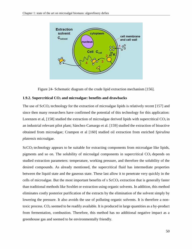
Chapter 1: state of the art on microalgal biomass: algorefinery defies
50
Figure 24- Schematic diagram of the crude lipid extraction mechanism [156].
1.9.2. Supercritical CO2 and microalgae: benefits and drawbacks
The use of ScCO2 technology for the extraction of microalgae lipids is relatively recent [157] and
since then many researchers have confirmed the potential of this technology for this application:
Lorenzen et al, [158] studied the extraction of microalgae derived lipids with supercritical CO2 in
an industrial relevant pilot plant; Sánchez-Camargo et al. [159] studied the extraction of bioactive
obtained from microalgae; Crampon et al [160] studied oil extraction from enriched Spirulina
platensis microalgae.
ScCO2 technology appears to be suitable for extracting components from microalgae like lipids,
pigments and so on. The solubility of microalgal components in supercritical CO2 depends on
studied extraction parameters: temperature, working pressure, and therefore the solubility of the
desired compounds. As already mentioned, the supercritical fluid has intermediate properties
between the liquid state and the gaseous state. These last allow it to penetrate very quickly in the
cells of microalgae. But the most important benefits of s ScCO2 extraction that is generally faster
than traditional methods like Soxhlet or extraction using organic solvents. In addition, this method
eliminates costly posterior purification of the extracts by the elimination of the solvent simply by
lowering the pressure. It also avoids the use of polluting organic solvents. It is therefore a non-
toxic process. CO2 seemed to be readily available. It is produced in large quantities as a by-product
from fermentation, combustion. Therefore, this method has no additional negative impact as a
greenhouse gas and seemed to be environmentally friendly.
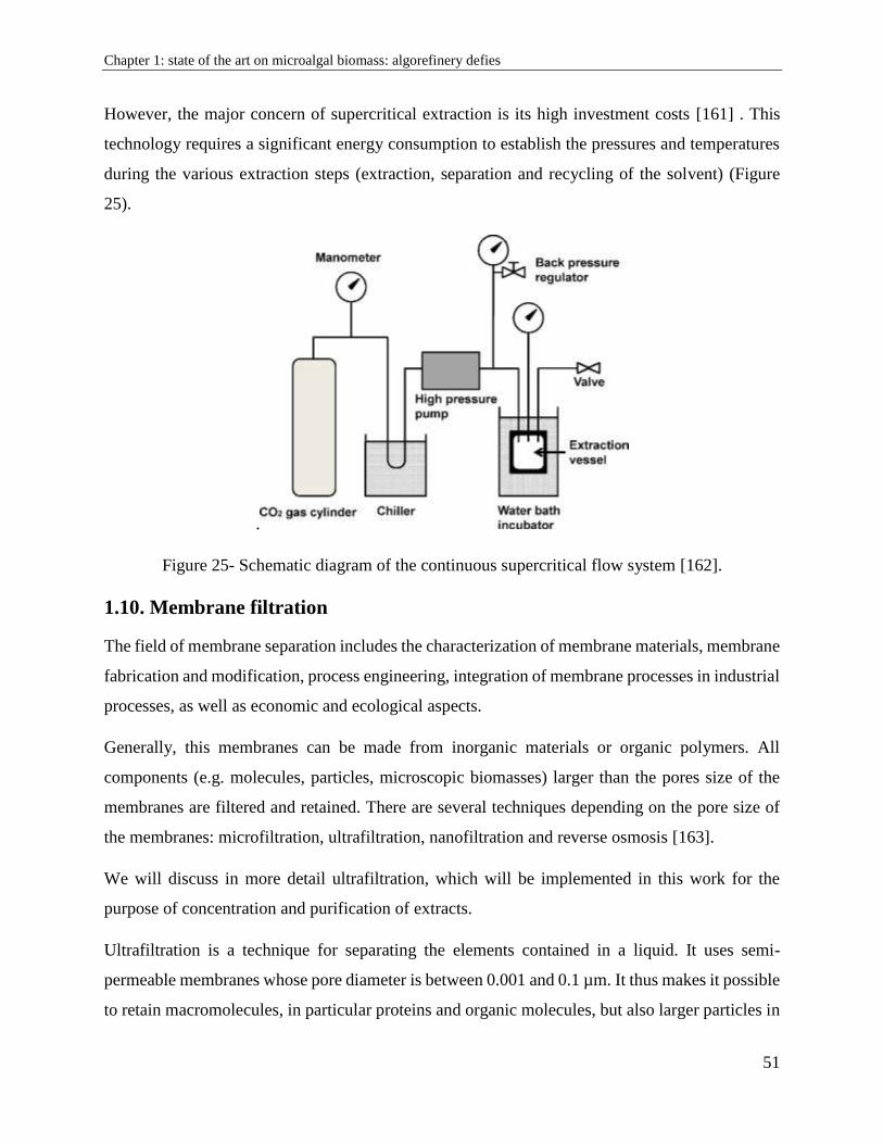
Chapter 1: state of the art on microalgal biomass: algorefinery defies
51
However, the major concern of supercritical extraction is its high investment costs [161] . This
technology requires a significant energy consumption to establish the pressures and temperatures
during the various extraction steps (extraction, separation and recycling of the solvent) (Figure
25).
.
Figure 25- Schematic diagram of the continuous supercritical flow system [162].
1.10. Membrane filtration
The field of membrane separation includes the characterization of membrane materials, membrane
fabrication and modification, process engineering, integration of membrane processes in industrial
processes, as well as economic and ecological aspects.
Generally, this membranes can be made from inorganic materials or organic polymers. All
components (e.g. molecules, particles, microscopic biomasses) larger than the pores size of the
membranes are filtered and retained. There are several techniques depending on the pore size of
the membranes: microfiltration, ultrafiltration, nanofiltration and reverse osmosis [163].
We will discuss in more detail ultrafiltration, which will be implemented in this work for the
purpose of concentration and purification of extracts.
Ultrafiltration is a technique for separating the elements contained in a liquid. It uses semi-
permeable membranes whose pore diameter is between 0.001 and 0.1 µm. It thus makes it possible
to retain macromolecules, in particular proteins and organic molecules, but also larger particles in
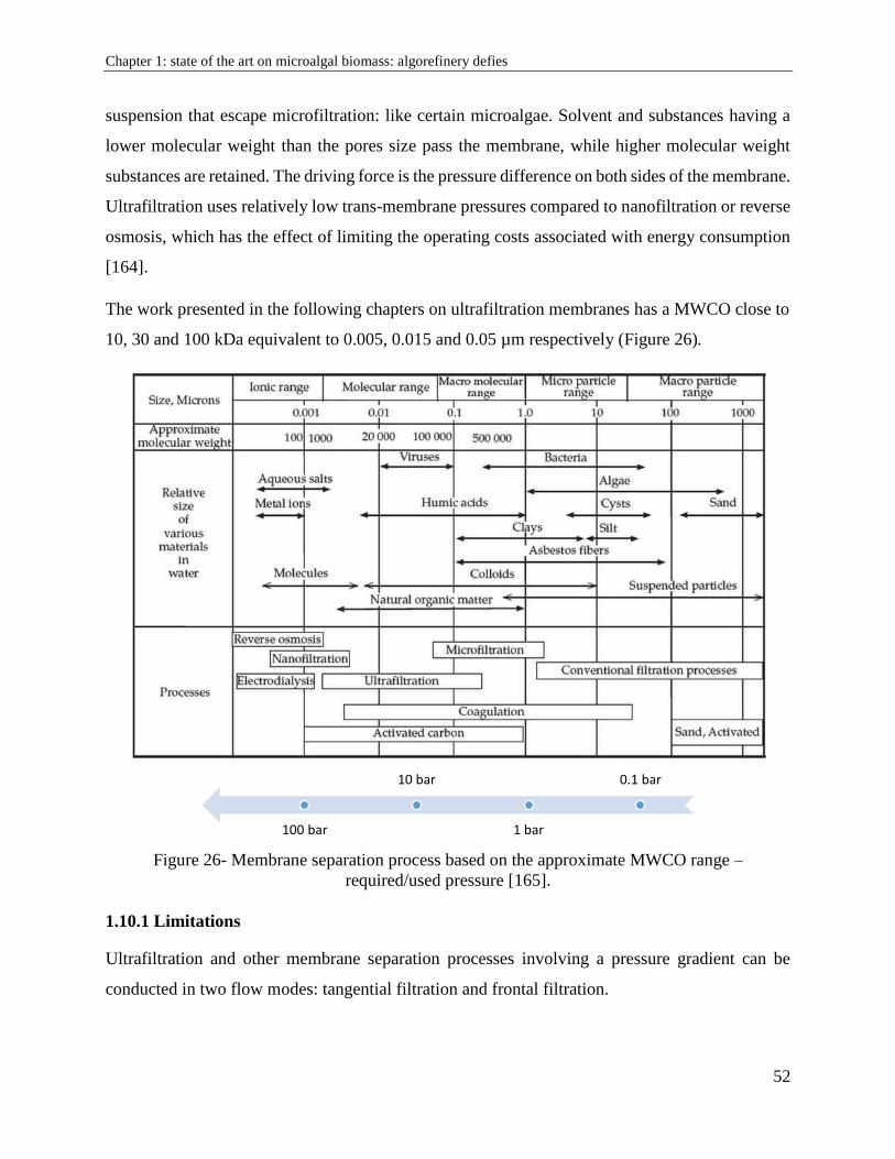
Chapter 1: state of the art on microalgal biomass: algorefinery defies
52
suspension that escape microfiltration: like certain microalgae. Solvent and substances having a
lower molecular weight than the pores size pass the membrane, while higher molecular weight
substances are retained. The driving force is the pressure difference on both sides of the membrane.
Ultrafiltration uses relatively low trans-membrane pressures compared to nanofiltration or reverse
osmosis, which has the effect of limiting the operating costs associated with energy consumption
[164].
The work presented in the following chapters on ultrafiltration membranes has a MWCO close to
10, 30 and 100 kDa equivalent to 0.005, 0.015 and 0.05 µm respectively (Figure 26).
Figure 26- Membrane separation process based on the approximate MWCO range –
required/used pressure [165].
1.10.1 Limitations
Ultrafiltration and other membrane separation processes involving a pressure gradient can be
conducted in two flow modes: tangential filtration and frontal filtration.
0.1 bar
1 bar
10 bar
100 bar
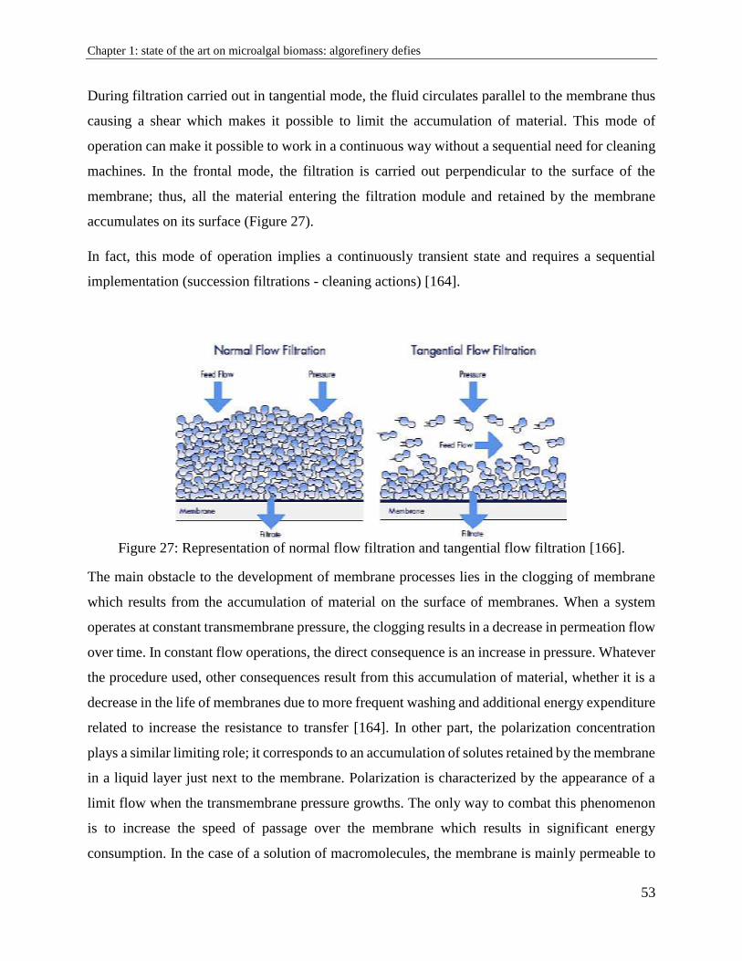
Chapter 1: state of the art on microalgal biomass: algorefinery defies
53
During filtration carried out in tangential mode, the fluid circulates parallel to the membrane thus
causing a shear which makes it possible to limit the accumulation of material. This mode of
operation can make it possible to work in a continuous way without a sequential need for cleaning
machines. In the frontal mode, the filtration is carried out perpendicular to the surface of the
membrane; thus, all the material entering the filtration module and retained by the membrane
accumulates on its surface (Figure 27).
In fact, this mode of operation implies a continuously transient state and requires a sequential
implementation (succession filtrations - cleaning actions) [164].
Figure 27: Representation of normal flow filtration and tangential flow filtration [166].
The main obstacle to the development of membrane processes lies in the clogging of membrane
which results from the accumulation of material on the surface of membranes. When a system
operates at constant transmembrane pressure, the clogging results in a decrease in permeation flow
over time. In constant flow operations, the direct consequence is an increase in pressure. Whatever
the procedure used, other consequences result from this accumulation of material, whether it is a
decrease in the life of membranes due to more frequent washing and additional energy expenditure
related to increase the resistance to transfer [164]. In other part, the polarization concentration
plays a similar limiting role; it corresponds to an accumulation of solutes retained by the membrane
in a liquid layer just next to the membrane. Polarization is characterized by the appearance of a
limit flow when the transmembrane pressure growths. The only way to combat this phenomenon
is to increase the speed of passage over the membrane which results in significant energy
consumption. In the case of a solution of macromolecules, the membrane is mainly permeable to
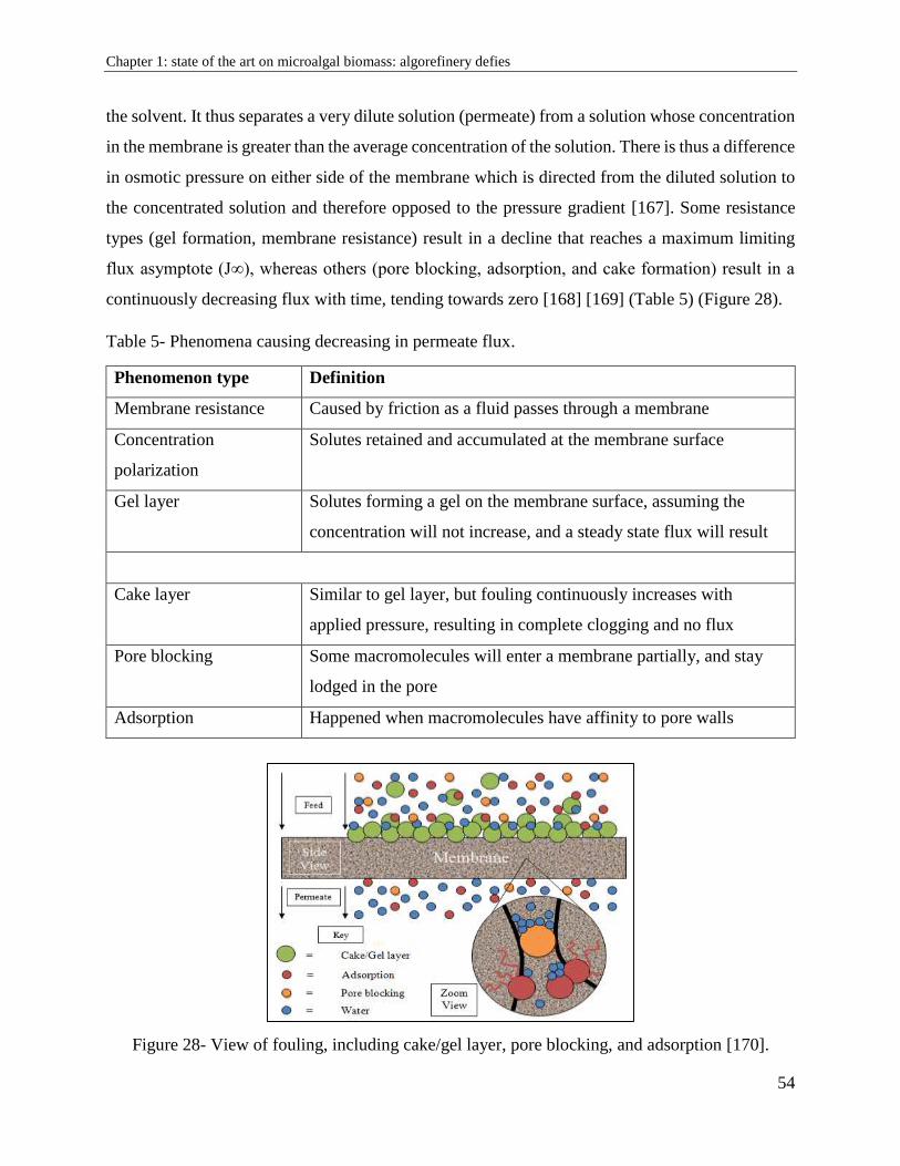
Chapter 1: state of the art on microalgal biomass: algorefinery defies
54
the solvent. It thus separates a very dilute solution (permeate) from a solution whose concentration
in the membrane is greater than the average concentration of the solution. There is thus a difference
in osmotic pressure on either side of the membrane which is directed from the diluted solution to
the concentrated solution and therefore opposed to the pressure gradient [167]. Some resistance
types (gel formation, membrane resistance) result in a decline that reaches a maximum limiting
flux asymptote (J∞), whereas others (pore blocking, adsorption, and cake formation) result in a
continuously decreasing flux with time, tending towards zero [168] [169] (Table 5) (Figure 28).
Table 5- Phenomena causing decreasing in permeate flux.
Phenomenon type Definition
Membrane resistance Caused by friction as a fluid passes through a membrane
Concentration
polarization
Solutes retained and accumulated at the membrane surface
Gel layer Solutes forming a gel on the membrane surface, assuming the
concentration will not increase, and a steady state flux will result
Cake layer Similar to gel layer, but fouling continuously increases with
applied pressure, resulting in complete clogging and no flux
Pore blocking Some macromolecules will enter a membrane partially, and stay
lodged in the pore
Adsorption Happened when macromolecules have affinity to pore walls
Figure 28- View of fouling, including cake/gel layer, pore blocking, and adsorption [170].
Chapter 1: state of the art on microalgal biomass: algorefinery defies
55
1.10.2. Influence of operating conditions
The role of physico-chemical parameters (pH, temperature, ionic strength, composition of the
supernatant...) has a great importance on the clogging of the membranes. The transfer of solute
and solvent is also a function of the operating conditions: hydraulic regime, transmembrane
pressure, temperature, solute concentration [171].
1.10.2.1. Flow velocity
The increase of tangential velocity reduces the thickness of the polarization layer. It also decreases
the deposition of large suspended particles that are more easily entrained. Finally, it generates
shear forces in the vicinity of the membrane. All this makes it possible to increase the flow of
permeate. However, if the clogging takes place inside the pores or in the form of compact thin
layers, the effect of the speed on the flow can become very limited [171].
1.10.2.2 Transmembrane pressure
This parameter ensures the filtration of the solvent. Its increase favors the accumulation of solutes
and suspended particles in the vicinity of the membrane. This can increase polarization and
clogging. In general, the increase in transmembrane pressure increases the flow of permeate, but
sometimes there may be a decrease in flux depending on the composition of the solution [171].
1.10.2.3. Concentration
The concentration of the solution affects the polarization and clogging of the membrane. Its
increase leads to an increase in the concentration at the membrane and favors the phenomena
involved in clogging [171].
1.10.2.4. Temperature
A rise in temperature improves the ultrafiltration flows by decreasing the viscosity of the solution
and increasing the diffusion coefficient of the solutes [171].
Chapter 1: state of the art on microalgal biomass: algorefinery defies
56
1.10.3. Membrane efficiency
The passage of a solute through the membrane is characterized by the rejection rate (R) and
calculated as follows:
𝑅 = [1 −𝐶𝑝𝑖
𝐶𝑟𝑖] ∗ 100
Cpi: concentration of component i in the permeate,
Cri: concentration of component i in the retentate,
R: retention factor in%.
In the case of an ideal retention, R = 100.
The volume concentration rate (VCR) represents the ratio between the feed rate and the retentate
flow rate. It is calculated according to Shukla et al, [172]:
𝑉𝐶𝑅 = 𝑉𝑜𝑙𝑢𝑚𝑒 𝑜𝑓 𝑓𝑒𝑒𝑑
𝑉𝑜𝑙𝑢𝑚𝑒 𝑜𝑓 𝑟𝑒𝑡𝑒𝑛𝑡𝑎𝑡𝑒
1.11. Algorefinery: advantages and difficulties
Algorefinery opens up a considerable field of biotechnology and enables the production of a very
wide range of products, but it is still far from guaranteeing mass production with acceptable
economic profitability.
Algal biomass represents a considerable potential in the world and have already been identified
for their ability to produce valuable products for food, feed, for the benefit of chemistry, medicine,
agriculture and as energy and biofuel source. The algorefinery is now at the stage of research and
development in aim to achieve sufficient ecologic/economic profitability [173].
The application of biotechnologies to microalgae enables to "over-accumulate" a precise element
in microalgae for the benefits of humankind. The selected species then becomes a real "factory"
that produces what the man has ordered. The selected microalgae then have yield per hectare much
higher than those of terrestrial plant species. A first step is to select the strains most likely to
naturally produce oils, proteins, pigments and other components of interest, and optimize their
metabolism to improve their performance.
Chapter 1: state of the art on microalgal biomass: algorefinery defies
57
The transformation of microalgae has advantages over terrestrial biomass:
Oil, proteins, pigments, sugar yields per hectare could be much higher than those of crops.
For example, at the laboratory stage, oil yields per hectare were 20 to 30 times higher than
that for terrestrial oilseeds.
Cultivated areas do not need to be arable land and therefore do not compete with
agricultural land.
The growth of lipids algae requires large amounts of CO2, which opens up prospects for
the recycling of CO2 emitted by factories or thermal power stations.
However, several difficulties arise for mass production:
The cultivation requires special conditions of sunshine and water availability, which limits
the possible locations.
It is also needy in nutrients (nitrates and phosphates) and in carbon dioxide.
Algorefinery downstream (pre-treatment - extraction – purification) consumes a lot of
energy [173].
Various coupling schemes for extraction techniques, pretreatment and separation are reported in
the literature in order to obtain extracts rich in primary components (lipids, proteins, pigments).
The downstream process requires a sequence of unit operations starting from pre-treating the
biomass by a chemical/ mechanical disintegration of the microalgal cell wall. This allows an easier
intracellular access of the extraction solvent, and contributes in enhancing the recovery yield of
the dedicated biomolecules. As known, disintegration methods are preferred due to the
acceleration of mechanism, but these methods are energy demanding and overheat the molecules,
which require cooling along the process to avoid denaturation or thermal degradation and then
generate additional cost [29].
After the step of pre-treatment and extraction comes the purification stage, which is challenging
step for many scientist at lab and at industrial scale.
1.12. Conclusion and thesis action plan
The state of art established in this chapter has shown that microalgae represented an important
renewable source for proteins, pigments and lipids.
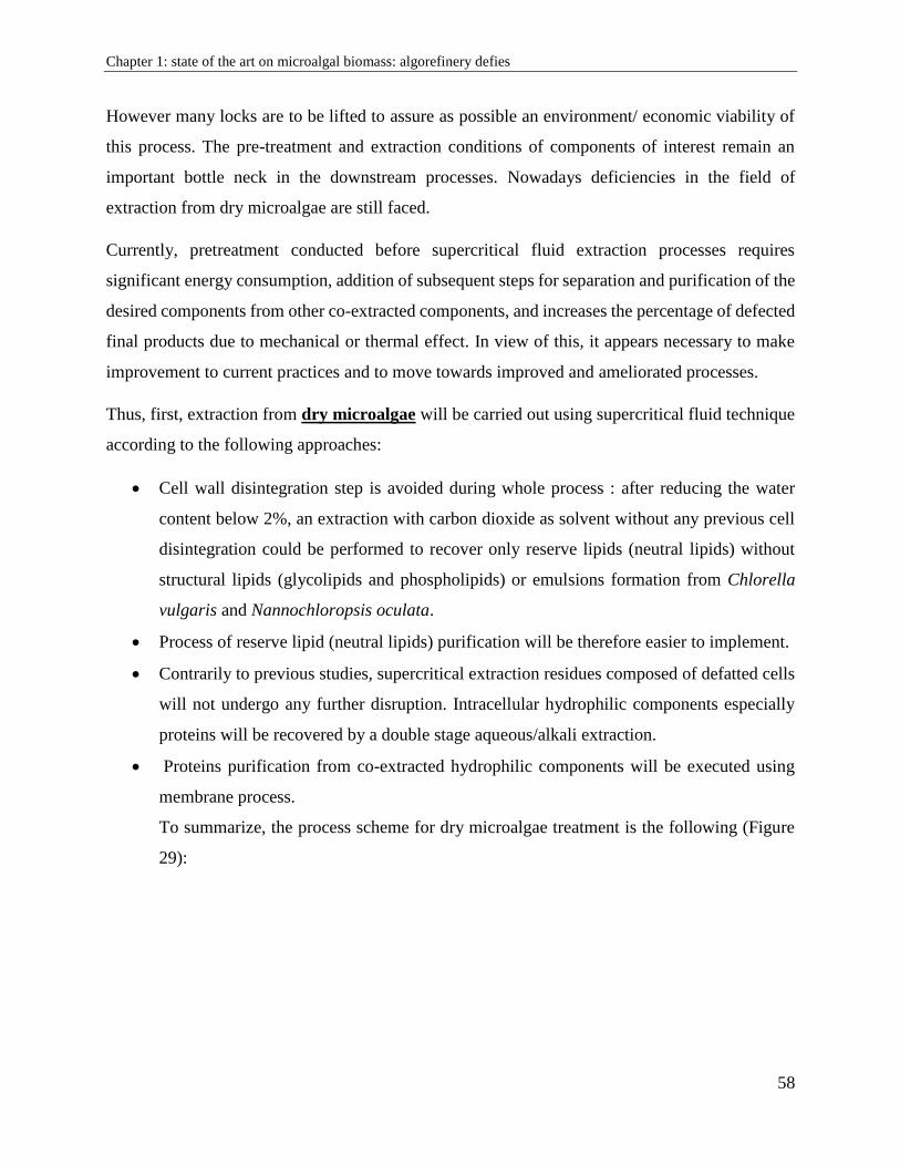
Chapter 1: state of the art on microalgal biomass: algorefinery defies
58
However many locks are to be lifted to assure as possible an environment/ economic viability of
this process. The pre-treatment and extraction conditions of components of interest remain an
important bottle neck in the downstream processes. Nowadays deficiencies in the field of
extraction from dry microalgae are still faced.
Currently, pretreatment conducted before supercritical fluid extraction processes requires
significant energy consumption, addition of subsequent steps for separation and purification of the
desired components from other co-extracted components, and increases the percentage of defected
final products due to mechanical or thermal effect. In view of this, it appears necessary to make
improvement to current practices and to move towards improved and ameliorated processes.
Thus, first, extraction from dry microalgae will be carried out using supercritical fluid technique
according to the following approaches:
Cell wall disintegration step is avoided during whole process : after reducing the water
content below 2%, an extraction with carbon dioxide as solvent without any previous cell
disintegration could be performed to recover only reserve lipids (neutral lipids) without
structural lipids (glycolipids and phospholipids) or emulsions formation from Chlorella
vulgaris and Nannochloropsis oculata.
Process of reserve lipid (neutral lipids) purification will be therefore easier to implement.
Contrarily to previous studies, supercritical extraction residues composed of defatted cells
will not undergo any further disruption. Intracellular hydrophilic components especially
proteins will be recovered by a double stage aqueous/alkali extraction.
Proteins purification from co-extracted hydrophilic components will be executed using
membrane process.
To summarize, the process scheme for dry microalgae treatment is the following (Figure
29):
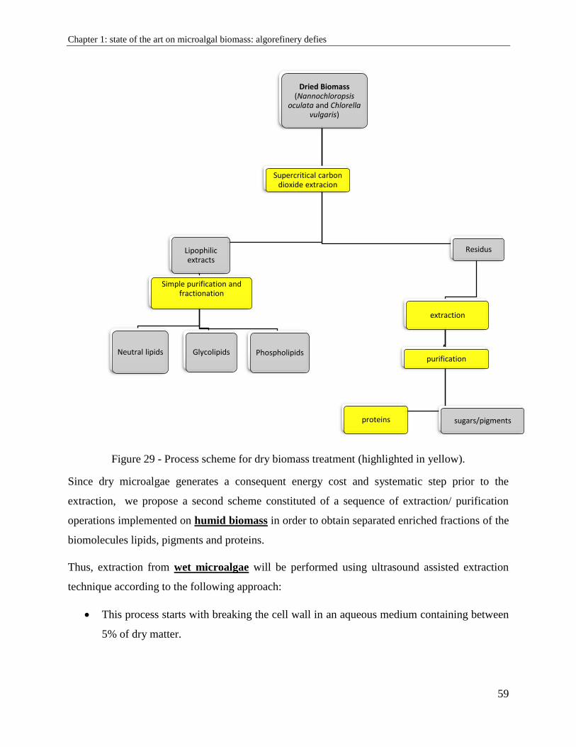
Chapter 1: state of the art on microalgal biomass: algorefinery defies
59
Figure 29 - Process scheme for dry biomass treatment (highlighted in yellow).
Since dry microalgae generates a consequent energy cost and systematic step prior to the
extraction, we propose a second scheme constituted of a sequence of extraction/ purification
operations implemented on humid biomass in order to obtain separated enriched fractions of the
biomolecules lipids, pigments and proteins.
Thus, extraction from wet microalgae will be performed using ultrasound assisted extraction
technique according to the following approach:
This process starts with breaking the cell wall in an aqueous medium containing between
5% of dry matter.
Dried Biomass (Nannochloropsis
oculata and Chlorella vulgaris)
Lipophilic extracts
Simple purification and fractionation
Neutral lipids Glycolipids Phospholipids
Residus
extraction
proteins
purification
sugars/pigments
Supercritical carbon dioxide extracion
Chapter 1: state of the art on microalgal biomass: algorefinery defies
60
New innovative parameters (low power, low frequency) will be used to achieve some
intensification and optimization of ultrasound assisted extraction technique
An emulsified mixture mainly composed of lipids, proteins, pigments will undergo variant
purification techniques to obtain specific enriched fractions
Also, understanding the degree of cell wall resistance will be equivalent to the “selective
extraction” to obtain specific enriched compound.
To summarize, our second process scheme for humid microalgae is the following (Figure 30):
Figure 30 - Process scheme for the wet biomass treatment (highlighted in yellow).
Wet Biomass(Spirulina sp. and Chlorella
vulgaris)
Liquid phase
separation process
Hydrophobic components
separation process
neutral lipids Glycolipids Phospholipids
hydrophilic components
Separation process
proteins carbohydrates
Pellets
cell wall disintegration
optimized
Chapter 2: Materials and methods
61
Chapter 2: Materials and methods
2.1. Microalgal biomass
Experiments were carried out on three species of microalgae; each of them have morphological
characteristics (constitution of membranes and walls) and different lipids content: Chlorella
vulgaris, Nannochloropsis oculata and Spirulina sp.
Chlorella vulgaris and Nannochloropsis oculata
The strains used in the framework of this thesis were provided by Alpha Biotech (Assérac, France).
Chlorella vulgaris (strain SAG 211-19) was cultivated under stressful conditions in Sueoka media
under conditions of high N nutrient concentration to favor proteins accumulation. Whereas
Nannochloropsis oculata was cultured in modified Conway media under conditions of N nutrient
deficiency to favor the accumulation of reserve lipids. This microalga has a predisposition to easily
store lipids and more particularly neutral lipids; therefore, it is considered a promising microalga
for bioenergy application. The biomasses were harvested, centrifuged to obtain an “algal paste” at
20-24% dry matter and then frozen at -20°C. The frozen paste of crude microalgae was freeze-
dried in a Fisher Bioblock Scientific Alpha 2–4 LD Plus device (Illkirch, France). During freeze
drying, the pressure was reduced to 0.01 bar, and the temperature decreased to -80°C for 48 h to
give a completely dry biomass.
Spirulina sp.
The experiments were performed on fresh filamentous Spirulina sp. provided in culture media at
a concentration of 5% (dry matter) and provided from Alg&You company (Toulouse, France).
Fractions of the culture has been frozen at -18º C then freeze-dried by a Fisher Bioblock Scientific
Alpha 2-4 LD Plus device (Illkirch, France). After reducing pressure to 0.010 bar, the temperature
further decreased to -80°C and freeze drying was conducted under vacuum for 48 h to give dry
Spirulina sp. biomass.
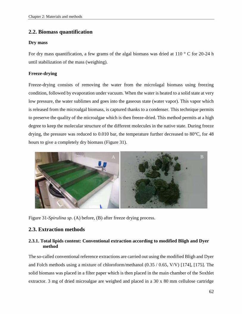
Chapter 2: Materials and methods
62
2.2. Biomass quantification
Dry mass
For dry mass quantification, a few grams of the algal biomass was dried at 110 ° C for 20-24 h
until stabilization of the mass (weighing).
Freeze-drying
Freeze-drying consists of removing the water from the microlagal biomass using freezing
condition, followed by evaporation under vacuum. When the water is heated to a solid state at very
low pressure, the water sublimes and goes into the gaseous state (water vapor). This vapor which
is released from the microalgal biomass, is captured thanks to a condenser. This technique permits
to preserve the quality of the microalgae which is then freeze-dried. This method permits at a high
degree to keep the molecular structure of the different molecules in the native state. During freeze
drying, the pressure was reduced to 0.010 bar, the temperature further decreased to 80°C, for 48
hours to give a completely dry biomass (Figure 31).
Figure 31-Spirulina sp. (A) before, (B) after freeze drying process.
2.3. Extraction methods
2.3.1. Total lipids content: Conventional extraction according to modified Bligh and Dyer
method
The so-called conventional reference extractions are carried out using the modified Bligh and Dyer
and Folch methods using a mixture of chloroform/methanol (0.35 / 0.65, V/V) [174], [175]. The
solid biomass was placed in a filter paper which is then placed in the main chamber of the Soxhlet
extractor. 3 mg of dried microalgae are weighed and placed in a 30 x 80 mm cellulose cartridge
A B
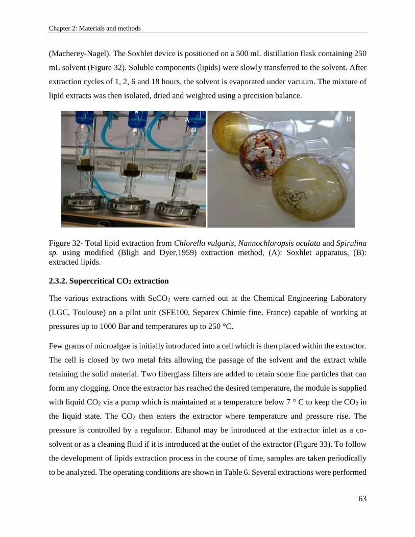
Chapter 2: Materials and methods
63
(Macherey-Nagel). The Soxhlet device is positioned on a 500 mL distillation flask containing 250
mL solvent (Figure 32). Soluble components (lipids) were slowly transferred to the solvent. After
extraction cycles of 1, 2, 6 and 18 hours, the solvent is evaporated under vacuum. The mixture of
lipid extracts was then isolated, dried and weighted using a precision balance.
Figure 32- Total lipid extraction from Chlorella vulgaris, Nannochloropsis oculata and Spirulina
sp. using modified (Bligh and Dyer,1959) extraction method, (A): Soxhlet apparatus, (B):
extracted lipids.
2.3.2. Supercritical CO2 extraction
The various extractions with ScCO2 were carried out at the Chemical Engineering Laboratory
(LGC, Toulouse) on a pilot unit (SFE100, Separex Chimie fine, France) capable of working at
pressures up to 1000 Bar and temperatures up to 250 °C.
Few grams of microalgae is initially introduced into a cell which is then placed within the extractor.
The cell is closed by two metal frits allowing the passage of the solvent and the extract while
retaining the solid material. Two fiberglass filters are added to retain some fine particles that can
form any clogging. Once the extractor has reached the desired temperature, the module is supplied
with liquid CO2 via a pump which is maintained at a temperature below 7 ° C to keep the CO2 in
the liquid state. The CO2 then enters the extractor where temperature and pressure rise. The
pressure is controlled by a regulator. Ethanol may be introduced at the extractor inlet as a co-
solvent or as a cleaning fluid if it is introduced at the outlet of the extractor (Figure 33). To follow
the development of lipids extraction process in the course of time, samples are taken periodically
to be analyzed. The operating conditions are shown in Table 6. Several extractions were performed
A B
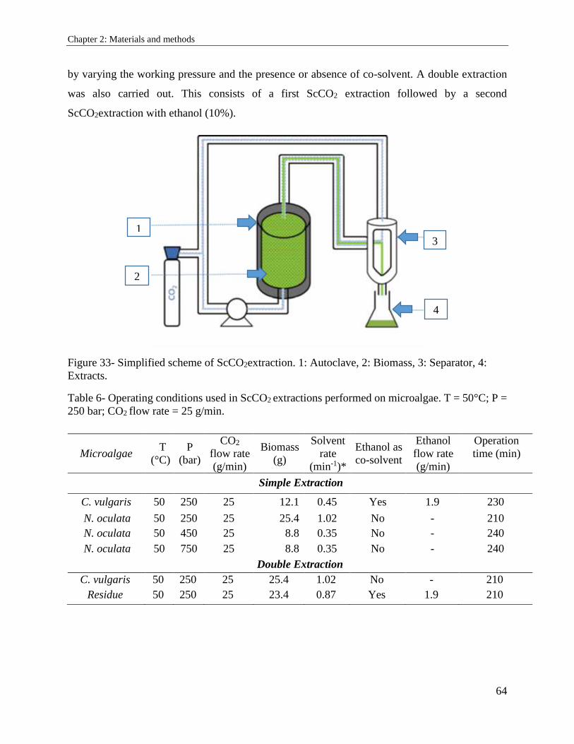
Chapter 2: Materials and methods
64
by varying the working pressure and the presence or absence of co-solvent. A double extraction
was also carried out. This consists of a first ScCO2 extraction followed by a second
ScCO2extraction with ethanol (10%).
Figure 33- Simplified scheme of ScCO2extraction. 1: Autoclave, 2: Biomass, 3: Separator, 4:
Extracts.
Table 6- Operating conditions used in ScCO2 extractions performed on microalgae. T = 50°C; P =
250 bar; CO2 flow rate = 25 g/min.
Microalgae T
(°C)
P
(bar)
CO2
flow rate
(g/min)
Biomass
(g)
Solvent
rate
(min-1)*
Ethanol as
co-solvent
Ethanol
flow rate
(g/min)
Operation
time (min)
Simple Extraction
C. vulgaris 50 250 25 12.1 0.45 Yes 1.9 230
N. oculata 50 250 25 25.4 1.02 No - 210
N. oculata 50 450 25 8.8 0.35 No - 240
N. oculata 50 750 25 8.8 0.35 No - 240
Double Extraction
C. vulgaris 50 250 25 25.4 1.02 No - 210
Residue 50 250 25 23.4 0.87 Yes 1.9 210
1
2
3
4
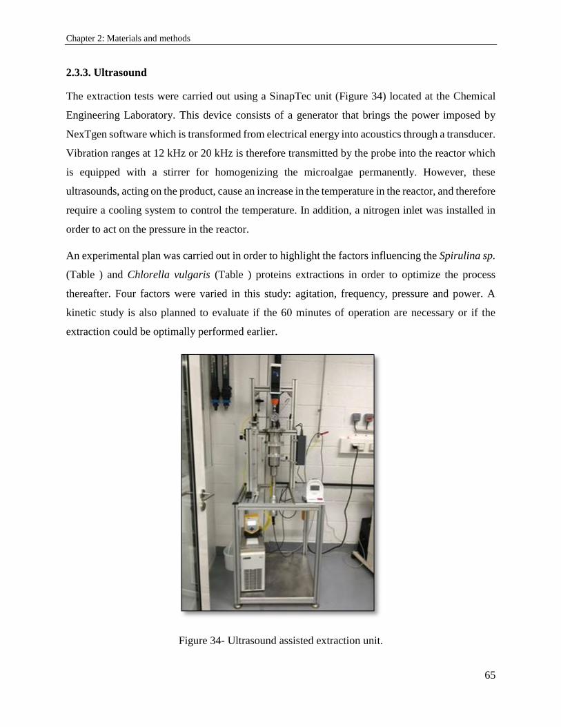
Chapter 2: Materials and methods
65
2.3.3. Ultrasound
The extraction tests were carried out using a SinapTec unit (Figure 34) located at the Chemical
Engineering Laboratory. This device consists of a generator that brings the power imposed by
NexTgen software which is transformed from electrical energy into acoustics through a transducer.
Vibration ranges at 12 kHz or 20 kHz is therefore transmitted by the probe into the reactor which
is equipped with a stirrer for homogenizing the microalgae permanently. However, these
ultrasounds, acting on the product, cause an increase in the temperature in the reactor, and therefore
require a cooling system to control the temperature. In addition, a nitrogen inlet was installed in
order to act on the pressure in the reactor.
An experimental plan was carried out in order to highlight the factors influencing the Spirulina sp.
(Table ) and Chlorella vulgaris (Table ) proteins extractions in order to optimize the process
thereafter. Four factors were varied in this study: agitation, frequency, pressure and power. A
kinetic study is also planned to evaluate if the 60 minutes of operation are necessary or if the
extraction could be optimally performed earlier.
Figure 34- Ultrasound assisted extraction unit.
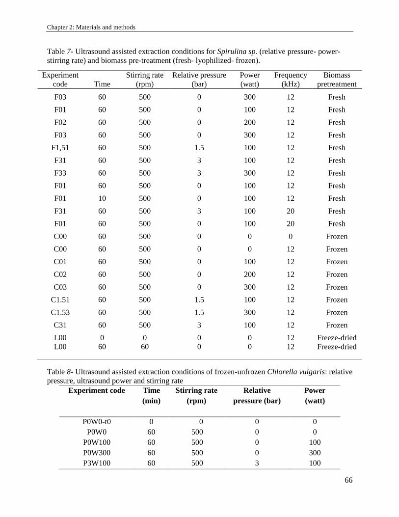
Chapter 2: Materials and methods
66
Table 7- Ultrasound assisted extraction conditions for Spirulina sp. (relative pressure- power-
stirring rate) and biomass pre-treatment (fresh- lyophilized- frozen).
Experiment
code Time
Stirring rate
(rpm)
Relative pressure
(bar)
Power
(watt)
Frequency
(kHz)
Biomass
pretreatment
F03 60 500 0 300 12 Fresh
F01 60 500 0 100 12 Fresh
F02 60 500 0 200 12 Fresh
F03 60 500 0 300 12 Fresh
F1,51 60 500 1.5 100 12 Fresh
F31 60 500 3 100 12 Fresh
F33 60 500 3 300 12 Fresh
F01 60 500 0 100 12 Fresh
F01 10 500 0 100 12 Fresh
F31 60 500 3 100 20 Fresh
F01 60 500 0 100 20 Fresh
C00 60 500 0 0 0 Frozen
C00 60 500 0 0 12 Frozen
C01 60 500 0 100 12 Frozen
C02 60 500 0 200 12 Frozen
C03 60 500 0 300 12 Frozen
C1.51 60 500 1.5 100 12 Frozen
C1.53 60 500 1.5 300 12 Frozen
C31 60 500 3 100 12 Frozen
L00 0 0 0 0 12 Freeze-dried
L00 60 60 0 0 12 Freeze-dried
Table 8- Ultrasound assisted extraction conditions of frozen-unfrozen Chlorella vulgaris: relative
pressure, ultrasound power and stirring rate
Experiment code Time
(min)
Stirring rate
(rpm)
Relative
pressure (bar)
Power
(watt)
P0W0-t0 0 0 0 0
P0W0 60 500 0 0
P0W100 60 500 0 100
P0W300 60 500 0 300
P3W100 60 500 3 100
Chapter 2: Materials and methods
67
2.3.4. Total pigments extraction
There are different types of pigments in Chlorella vulgaris, Nannochloropsis oculata and Spirulina
sp. used strains. These can be classified into two categories: chlorophylls and carotenoids. We did
focus on certain pigments in particular: chlorophylls a and b, lutein and β-carotene. Extraction was
carried out in cold conditions to protect them from heat denaturation. One gram of microalga was
mixed with specific solvent during 24 min at 10 ºC with continuous gentle stirring. Ethanol was
used as extraction solvent for chlorophylls a and b, whereas β-carotene and lutein were extracted
using ethanol. In aim to inhibit any photo degradation during extraction, glass tubes were covered
with aluminum paper. Supernatants were taken for analysis after centrifugation under 20,000g for
15 min at 5ºC.
2.4. Purification technique: ultrafiltration
The membranes used on this pilot unit are polysulfone flat membranes the cutoff threshold is given
at 10- 30- 100 kDa). The membranes were abundantly rinsed with ultrapure water, a crucial step
necessary before any use. These membranes are supplied in the form of A4 sheets and then cut
manually to fit into the filtration cell (30.2 cm2 filtration area). . The pilot unit consists of a 200
mL filtration cell (Amicon® Stirred Cells) housing flat membranes diameter corresponding to a
useful area of 30.2 cm2 (Figures 35 and 36). This filtration unit offers a small filter surface which
makes it possible to study the filtration of relatively small quantities (from 50 mL). In addition, a
stirring over the membrane was also applied to minimize concentration polarization effect due to
the accumulation of macromolecules at the membrane surface. Experiments were carried out under
a trans-membrane pressure from 0.5 to 4 bar.
The remaining suspension is removed gently and the cell is rinsed with ultrapure water with the
greatest care. This is repeated three times to ensure complete elimination of the compounds present
in the disperse phase.
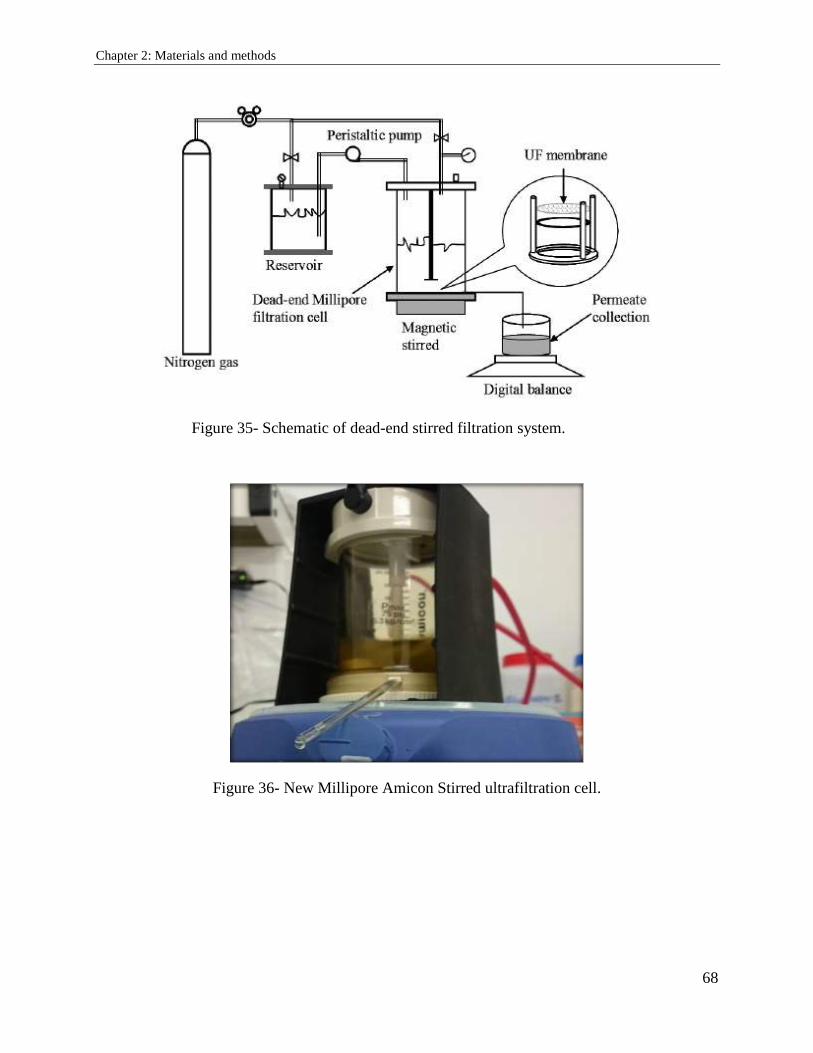
Chapter 2: Materials and methods
68
Figure 35- Schematic of dead-end stirred filtration system.
Figure 36- New Millipore Amicon Stirred ultrafiltration cell.
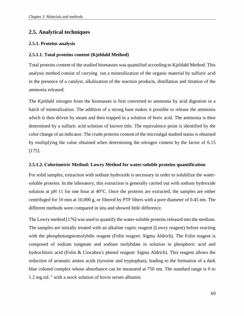
Chapter 2: Materials and methods
69
2.5. Analytical techniques
2.5.1. Proteins analysis
2.5.1.1. Total proteins content (Kjeldahl Method)
Total proteins content of the studied biomasses was quantified according to Kjeldahl Method. This
analysis method consist of carrying out a mineralization of the organic material by sulfuric acid
in the presence of a catalyst, alkalization of the reaction products, distillation and titration of the
ammonia released.
The Kjeldahl nitrogen from the biomasses is first converted to ammonia by acid digestion in a
batch of mineralization. The addition of a strong base makes it possible to release the ammonia
which is then driven by steam and then trapped in a solution of boric acid. The ammonia is then
determined by a sulfuric acid solution of known title. The equivalence point is identified by the
color change of an indicator. The crude proteins content of the microalgal studied stains is obtained
by multiplying the value obtained when determining the nitrogen content by the factor of 6.15
[175].
2.5.1.2. Colorimetric Method: Lowry Method for water-soluble proteins quantification
For solid samples, extraction with sodium hydroxide is necessary in order to solubilize the water-
soluble proteins. In the laboratory, this extraction is generally carried out with sodium hydroxide
solution at pH 11 for one hour at 40°C. Once the proteins are extracted, the samples are either
centrifuged for 10 min at 10,000 g, or filtered by PTF filters with a pore diameter of 0.45 nm. The
different methods were compared in situ and showed little difference.
The Lowry method [176] was used to quantify the water-soluble proteins released into the medium.
The samples are initially treated with an alkaline cupric reagent (Lowry reagent) before reacting
with the phosphotungstomolybdic reagent (Folin reagent: Sigma Aldrich). The Folin reagent is
composed of sodium tungstate and sodium molybdate in solution in phosphoric acid and
hydrochloric acid (Folin & Ciocalteu's phenol reagent: Sigma Aldrich). This reagent allows the
reduction of aromatic amino acids (tyrosine and tryptophan), leading to the formation of a dark
blue colored complex whose absorbance can be measured at 750 nm. The standard range is 0 to
1.2 mg.mL-1 with a stock solution of bovin serum albumin
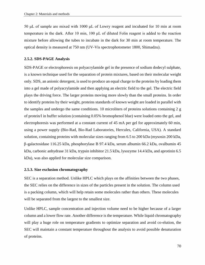
Chapter 2: Materials and methods
70
50 μL of sample are mixed with 1000 μL of Lowry reagent and incubated for 10 min at room
temperature in the dark. After 10 min, 100 μL of diluted Folin reagent is added to the reaction
mixture before allowing the tubes to incubate in the dark for 30 min at room temperature. The
optical density is measured at 750 nm (UV-Vis spectrophotometer 1800, Shimadzu).
2.5.2. SDS-PAGE Analysis
SDS-PAGE or electrophoresis on polyacrylamide gel in the presence of sodium dodecyl sulphate,
is a known technique used for the separation of protein mixtures, based on their molecular weight
only. SDS, an anionic detergent, is used to produce an equal charge to the proteins by loading them
into a gel made of polyacrylamide and then applying an electric field to the gel. The electric field
plays the driving force. The larger proteins moving more slowly than the small proteins. In order
to identify proteins by their weight, proteins standards of known weight are loaded in parallel with
the samples and undergo the same conditions. 10 microliters of proteins solutions containing 2 g
of protein/l in buffer solution (containing 0.05% bromophenol blue) were loaded onto the gel, and
electrophoresis was performed at a constant current of 45 mA per gel for approximately 60 min,
using a power supply (Bio-Rad, Bio-Rad Laboratories, Hercules, California, USA). A standard
solution, containing proteins with molecular sizes ranging from 6.5 to 200 kDa (mysosin 200 kDa,
-galactosidase 116.25 kDa, phosphorylase B 97.4 kDa, serum albumin 66.2 kDa, ovalbumin 45
kDa, carbonic anhydrase 31 kDa, trypsin inhibitor 21.5 kDa, lysozyme 14.4 kDa, and aprotinin 6.5
kDa), was also applied for molecular size comparison.
2.5.3. Size exclusion chromatography
SEC is a separation method. Unlike HPLC which plays on the affinities between the two phases,
the SEC relies on the difference in sizes of the particles present in the solution. The column used
is a packing column, which will help retain some molecules rather than others. These molecules
will be separated from the largest to the smallest size.
Unlike HPLC, sample concentration and injection volume need to be higher because of a larger
column and a lower flow rate. Another difference is the temperature. While liquid chromatography
will play a huge role on temperature gradients to optimize separation and avoid co-elution, the
SEC will maintain a constant temperature throughout the analysis to avoid possible denaturation
of proteins.
Chapter 2: Materials and methods
71
Chromatographic conditions
The tests were carried out on the same installation as for the determination of the pigments. The
difference comes from the column and conditions used:
Column: SunFire Waters
Solvent: 100% phosphate buffer
Flow rate: 0.7 mL / min
Temperature: 30 ° C
Duration: 30 min
Wavelength: 220 nm, 280 nm, 620 nm
2.5.4. Lipids analysis
Fractionation of lipids by solid phase extraction (SPE)
Fractionation of lipid extracts is a particularly important step in identifying the biomass content in
neutral lipids, glycolipids and phospholipids on the first hand, and validating the supercritical
extraction efficiency and selectivity on the other hand . Solid phase extraction (SPE) described by
Juaneda and Rocqueline [177] (Figure 37) is a technique used because of its speed and efficiency.
At the beginning, it is necessary to condition the cartridge by percolating a volume of chloroform
in order to activate the cartridges. The cartridge should not dry before depositing the lipids extract.
The principle necessitates to depose the lipids extract on the stationary phase slowly before starting
the fractionation.
To separate the lipid fractions, passing a specific eluent solvent for each fraction is the key step.
Elution of the neutral lipids, glycolipids and phospholipids is carried out with a suitable volume
of chloroform, methanol and acetone.
The compounds thus eluted were taken for dosage. It was necessary to dry out the fractions under
cold nitrogen before weighing.
Chapter 2: Materials and methods
72
Figure 37- Solid Phase Extraction (SPE) technique showing the partitioning of lipids to three
classes neutral lipids, glycolipids and phospholipid.
Green microalgae usually contained neutral lipids (non-polar) and glycolipids and phospholipids
(polar lipids) at numerous mg per gram of biomass. The proposed SPE extraction method would
be useful for the analysis of different lipids classes originally presented in the two microalgae and
in the ScCO2 extracts.
Partitioning of lipids to three classes neutral lipids, glycolipids and phospholipid was possible by
eluting them with 10 ml of chloroform, acetone and methanol respectively, using SPE silica
cartridges Supelco (500 mg, Sigma-Aldrich). Once eluted and freeze-dried under liquid nitrogen,
fractions were measured gravimetrically and expressed in mg/g dry weigh.
2.5.5. Pigment analysis
Total pigments quantification
Total pigment (chlorophylls a and b, β-carotenes and lutein) content was measured according to
the general formula developed by Bidigare [178] and mentioned value in (table 9):
C(mg/L) =OD∗1000
α∗L Where:
OD is optical density read at the specific wavelength in a given solvent
α is the specific extinction coefficient (L g-1cm-1)
L is the optical path length (cuvettes) in cm which is 1 cm
Chapter 2: Materials and methods
73
Table 9- Extinction coefficients α of the chlorophylls pigments (a, b) and carotenoids (β-
carotene, lutein) and maximal absorption peaks.
wavelength
(nm)
specific extinction coefficient α
(L g-1cm-1)
Solvent References
Chlorophyll a 665 77,9 Methanol [179] [180]
Chlorophyll b 642 44,5 Methanol [179] [180]
β-carotene 453 262 Ethanol [180] [181]
Luteins 445 255 Ethanol [180] [181]
Preparation of the standards
The pure standard pigments: chlorophylls pigments (a, b) and carotenoids (β-carotene, lutein)
necessary for the identification of the compounds in the extract were purchased commercially from
Sigma-Aldrich. Standard solutions have been prepared and operative solutions were made by
diluting standard solutions with methanol.
Pigments standards usually supplied in powder form, are solubilized in acetone to form a storage
solution and kept at -20°C. The mother solution for producing the standards is obtained by diluting
the storage solution in methanol. The stock solutions of each pigment was 5 mg/L. The standard
solutions have a concentration between 0.5 and 5 mg/L.
High Performance Liquid Chromatography for pigments analysis
High Performance Liquid Chromatography (HPLC) is more frequently used as an analytical
separation technique based on the hydrophobicity of the molecules of a compound or a mixture of
compounds. The sample to be analyzed is eluted a mobile phase in a column filled with a stationary
phase composed of very fine solid grains. The flow of eluent is provided by a high pressure pump.
In the column, the various pigments in the sample are separated from each other because of their
various affinities with respect to both phases - stationary and mobile (Table 10). At the exit of the
column the compounds are detected using a detector. In our studies, UV detectors based on the
measurement of the wavelength of the compounds were used.
The HPLC system used for this study consists of:
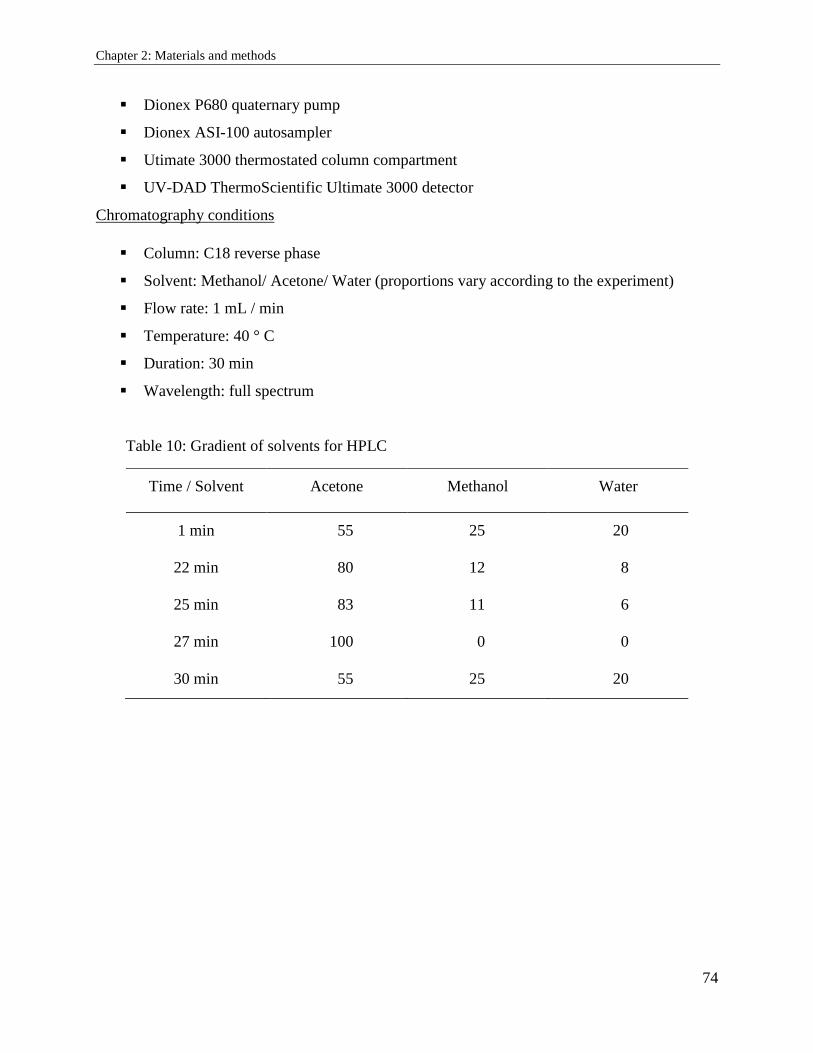
Chapter 2: Materials and methods
74
Dionex P680 quaternary pump
Dionex ASI-100 autosampler
Utimate 3000 thermostated column compartment
UV-DAD ThermoScientific Ultimate 3000 detector
Chromatography conditions
Column: C18 reverse phase
Solvent: Methanol/ Acetone/ Water (proportions vary according to the experiment)
Flow rate: 1 mL / min
Temperature: 40 ° C
Duration: 30 min
Wavelength: full spectrum
Table 10: Gradient of solvents for HPLC
Time / Solvent Acetone Methanol Water
1 min 55 25 20
22 min 80 12 8
25 min 83 11 6
27 min 100 0 0
30 min 55 25 20
References
75
References
[1] R. Chaiklahan et al., “Separation and purification of phycocyanin from Spirulina sp. using
a membrane process,” Bioresource Technology, vol. 102, no. 14, pp. 7159–7164, Jul. 2011.
[2] S. Buono, A. L. Langellotti, A. Martello, F. Rinna, and V. Fogliano, “Functional ingredients
from microalgae,” Food Funct., vol. 5, no. 8, pp. 1669–1685, Jul. 2014.
[3] Q. Hu et al., “Microalgal triacylglycerols as feedstocks for biofuel production: perspectives
and advances,” The Plant Journal, vol. 54, no. 4, pp. 621–639.
[4] P. Spolaore, C. Joannis-Cassan, E. Duran, and A. Isambert, “Commercial applications of
microalgae,” Journal of Bioscience and Bioengineering, vol. 101, no. 2, pp. 87–96, Feb.
2006.
[5] E. W. Becker, “Micro-algae as a source of protein,” Biotechnology Advances, vol. 25, no.
2, pp. 207–210, Mar. 2007.
[6] Y. Li, M. Horsman, N. Wu, C. Q. Lan, and N. Dubois‐Calero, “Biofuels from Microalgae,”
Biotechnology Progress, vol. 24, no. 4, pp. 815–820, Jul. 2008.
[7] T. M. Mata, A. A. Martins, and N. S. Caetano, “Microalgae for biodiesel production and
other applications: A review,” Renewable and Sustainable Energy Reviews, vol. 14, no. 1,
pp. 217–232, Jan. 2010.
[8] M. A. Borowitzka, “High-value products from microalgae—their development and
commercialisation,” J Appl Phycol, vol. 25, no. 3, pp. 743–756, Jun. 2013.
[9] M. Vigani et al., “Food and feed products from micro-algae: Market opportunities and
challenges for the EU,” Trends in Food Science & Technology, vol. 42, no. 1, pp. 81–92,
Mar. 2015.
[10] I. Priyadarshani and B. Rath, “Commercial and industrial applications of micro algae – A
review,” p. 12, 2012.
[11] H. G. Gerken, B. Donohoe, and E. P. Knoshaug, “Enzymatic cell wall degradation of
Chlorella vulgaris and other microalgae for biofuels production,” Planta, vol. 237, no. 1,
pp. 239–253, Jan. 2013.
[12] C. Safi et al., “Aqueous extraction of proteins from microalgae: Effect of different cell
disruption methods,” Algal Research, vol. 3, pp. 61–65, Jan. 2014.
[13] P. D. Patil, K. P. R. Dandamudi, J. Wang, Q. Deng, and S. Deng, “Extraction of bio-oils
from algae with supercritical carbon dioxide and co-solvents,” The Journal of Supercritical
Fluids, vol. 135, pp. 60–68, May 2018.
[14] C.-H. Cheng, T.-B. Du, H.-C. Pi, S.-M. Jang, Y.-H. Lin, and H.-T. Lee, “Comparative study
of lipid extraction from microalgae by organic solvent and supercritical CO2,” Bioresource
Technology, vol. 102, no. 21, pp. 10151–10153, Nov. 2011.
[15] Y.-M. Dai, K.-T. Chen, and C.-C. Chen, “Study of the microwave lipid extraction from
microalgae for biodiesel production,” Chemical Engineering Journal, vol. 250, pp. 267–
273, Aug. 2014.
References
76
[16] C. Juin et al., “Microwave-Assisted Extraction of Phycobiliproteins from Porphyridium
purpureum,” Appl Biochem Biotechnol, vol. 175, no. 1, pp. 1–15, Jan. 2015.
[17] R. D. P. Rodrigues, F. C. de Castro, R. S. de Santiago-Aguiar, and M. V. P. Rocha,
“Ultrasound-assisted extraction of phycobiliproteins from Spirulina (Arthrospira) platensis
using protic ionic liquids as solvent,” Algal Research, vol. 31, pp. 454–462, Apr. 2018.
[18] Nobre et al., “A biorefinery from Nannochloropsis sp. microalga – Extraction of oils and
pigments. Production of biohydrogen from the leftover biomass,” Bioresource Technology,
vol. 135, pp. 128–136, May 2013.
[19] V. Pasquet et al., “Study on the microalgal pigments extraction process: Performance of
microwave assisted extraction,” Process Biochemistry, vol. 46, no. 1, pp. 59–67, Jan. 2011.
[20] D.-Y. Kim et al., “Cell-wall disruption and lipid/astaxanthin extraction from microalgae:
Chlorella and Haematococcus,” Bioresource Technology, vol. 199, pp. 300–310, Jan. 2016.
[21] A. Molino et al., “Extraction of astaxanthin from microalga Haematococcus pluvialis in red
phase by using generally recognized as safe solvents and accelerated extraction,” Journal
of Biotechnology, vol. 283, pp. 51–61, Oct. 2018.
[22] M. M. Eldalatony et al., “Pretreatment of microalgal biomass for enhanced
recovery/extraction of reducing sugars and proteins,” Bioprocess Biosyst Eng, vol. 39, no.
1, pp. 95–103, Jan. 2016.
[23] R. Seetharam and S. K. Sharma, Purification and Analysis of Recombinant Proteins. CRC
Press, 1991.
[24] J. Doucha and K. Lívanský, “Influence of processing parameters on disintegration of
<Emphasis Type="Italic">Chlorella</Emphasis> cells in various types of homogenizers,”
Appl Microbiol Biotechnol, vol. 81, no. 3, p. 431, Dec. 2008.
[25] Y. W. Sari, M. E. Bruins, and J. P. M. Sanders, “Enzyme assisted protein extraction from
rapeseed, soybean, and microalgae meals,” Industrial Crops and Products, vol. 43, pp. 78–
83, May 2013.
[26] L. Brennan and P. Owende, “Biofuels from microalgae—A review of technologies for
production, processing, and extractions of biofuels and co-products,” Renewable and
Sustainable Energy Reviews, vol. 14, no. 2, pp. 557–577, Feb. 2010.
[27] J.-Y. Lee, C. Yoo, S.-Y. Jun, C.-Y. Ahn, and H.-M. Oh, “Comparison of several methods
for effective lipid extraction from microalgae,” Bioresource Technology, vol. 101, no. 1,
Supplement, pp. S75–S77, Jan. 2010.
[28] S. P. Nalawade, F. Picchioni, and L. P. B. M. Janssen, “Supercritical carbon dioxide as a
green solvent for processing polymer melts: Processing aspects and applications,” Progress
in Polymer Science, vol. 31, no. 1, pp. 19–43, Jan. 2006.
[29] J. Kim et al., “Methods of downstream processing for the production of biodiesel from
microalgae,” Biotechnology Advances, vol. 31, no. 6, pp. 862–876, Nov. 2013.
[30] Z. Duan, X. Tan, K. Dai, H. Gu, and H. Yang, “Evaluation on H2O2-aided ultrasonic
pretreatment for cell disruption of Chlorella pyrenoidosa,” Asia-Pacific Journal of Chemical
Engineering, vol. 12, no. 3, pp. 502–510.
References
77
[31] F. Chemat, N. Rombaut, A.-G. Sicaire, A. Meullemiestre, A.-S. Fabiano-Tixier, and M.
Abert-Vian, “Ultrasound assisted extraction of food and natural products. Mechanisms,
techniques, combinations, protocols and applications. A review,” Ultrasonics
Sonochemistry, vol. 34, pp. 540–560, Jan. 2017.
[32] bilad et al., “Membrane technology in microalgae cultivation and harvesting: A review,”
Biotechnology Advances, vol. 32, no. 7, pp. 1283–1300, Nov. 2014.
[33] Gerardo et al., “Integration of membrane technology in microalgae biorefineries,” Journal
of Membrane Science, vol. 464, pp. 86–99, Aug. 2014.
[34] Zhang et al., “Harvesting algal biomass for biofuels using ultrafiltration membranes,”
Bioresource Technology, vol. 101, no. 14, pp. 5297–5304, Jul. 2010.
[35] Marcati et al., “Extraction and fractionation of polysaccharides and B-phycoerythrin from
the microalga Porphyridium cruentum by membrane technology,” Algal Research, vol. 5,
pp. 258–263, Jul. 2014.
[36] Patel et al., “Separation and fractionation of exopolysaccharides from Porphyridium
cruentum,” Bioresource Technology, vol. 145, pp. 345–350, Oct. 2013.
[37] Pugh et al., “Isolation of Three High Molecular Weight Polysaccharide Preparations with
Potent Immunostimulatory Activity from Spirulina platensis , Aphanizomenon flos-aquae
and Chlorella pyrenoidosa | Request PDF,” ResearchGate. [Online]. Available:
https://www.researchgate.net/publication/11624110_Isolation_of_Three_High_Molecular
_Weight_Polysaccharide_Preparations_with_Potent_Immunostimulatory_Activity_from_
Spirulina_platensis_Aphanizomenon_flos-aquae_and_Chlorella_pyrenoidosa. [Accessed:
22-Aug-2018].
[38] Ba, Fatou, Ursu, Alina Violetta, Laroche, Celine, and Djelveh, Gholamreza,
“Haematococcus pluvialis soluble proteins: Extraction, characterization,
concentration/fractionation and emulsifying properties,” Bioresource Technology, vol. 200,
pp. 147–152, Jan. 2016.
[39] A.-V. Ursu, A. Marcati, T. Sayd, V. Sante-Lhoutellier, G. Djelveh, and P. Michaud,
“Extraction, fractionation and functional properties of proteins from the microalgae
Chlorella vulgaris,” Bioresource Technology, vol. 157, pp. 134–139, Apr. 2014.
[40] Soni et al., “Extraction, purification and characterization of phycocyanin from Oscillatoria
quadripunctulata—Isolated from the rocky shores of Bet-Dwarka, Gujarat, India,” Process
Biochemistry, vol. 41, no. 9, pp. 2017–2023, Sep. 2006.
[41] A. G. Waghmare, M. K. Salve, J. G. LeBlanc, and S. S. Arya, “Concentration and
characterization of microalgae proteins from Chlorella pyrenoidosa,” Bioresour.
Bioprocess., vol. 3, no. 1, p. 16, Mar. 2016.
[42] J. Heo, D.-H. Cho, R. Ramanan, H.-M. Oh, and H.-S. Kim, “PhotoBiobox: A tablet sized,
low-cost, high throughput photobioreactor for microalgal screening and culture
optimization for growth, lipid content and CO2 sequestration,” Biochemical Engineering
Journal, vol. 103, pp. 193–197, Nov. 2015.
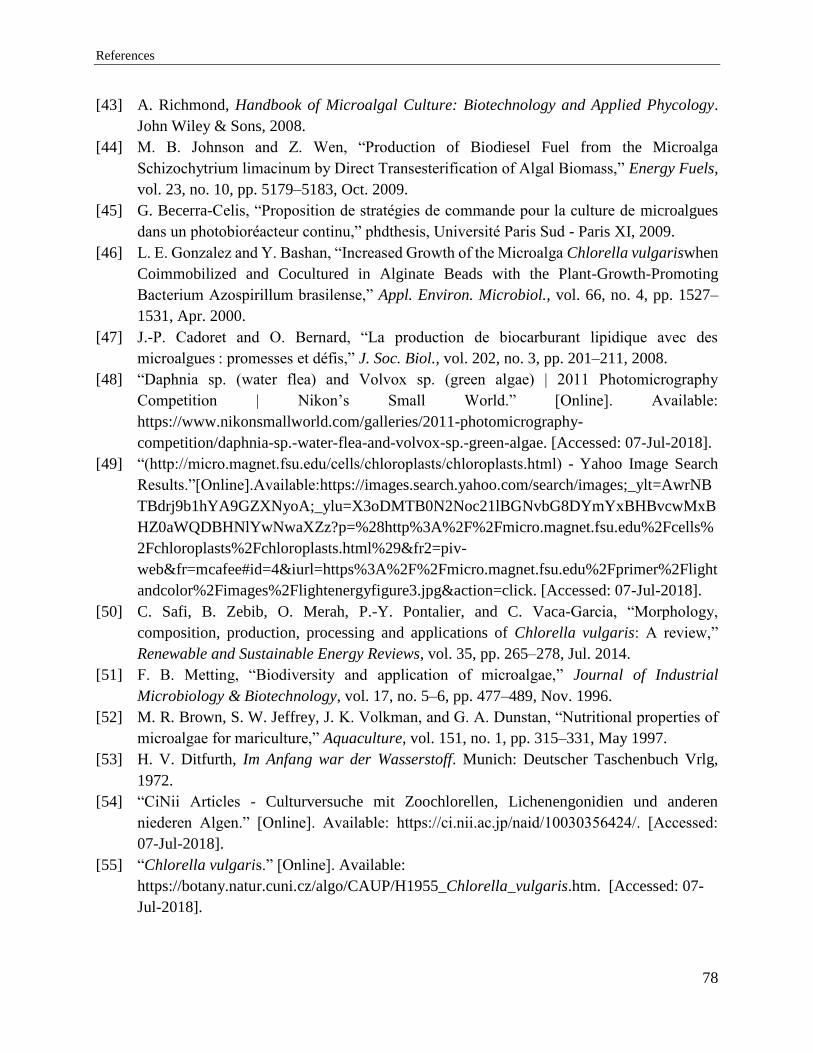
References
78
[43] A. Richmond, Handbook of Microalgal Culture: Biotechnology and Applied Phycology.
John Wiley & Sons, 2008.
[44] M. B. Johnson and Z. Wen, “Production of Biodiesel Fuel from the Microalga
Schizochytrium limacinum by Direct Transesterification of Algal Biomass,” Energy Fuels,
vol. 23, no. 10, pp. 5179–5183, Oct. 2009.
[45] G. Becerra-Celis, “Proposition de stratégies de commande pour la culture de microalgues
dans un photobioréacteur continu,” phdthesis, Université Paris Sud - Paris XI, 2009.
[46] L. E. Gonzalez and Y. Bashan, “Increased Growth of the Microalga Chlorella vulgariswhen
Coimmobilized and Cocultured in Alginate Beads with the Plant-Growth-Promoting
Bacterium Azospirillum brasilense,” Appl. Environ. Microbiol., vol. 66, no. 4, pp. 1527–
1531, Apr. 2000.
[47] J.-P. Cadoret and O. Bernard, “La production de biocarburant lipidique avec des
microalgues : promesses et défis,” J. Soc. Biol., vol. 202, no. 3, pp. 201–211, 2008.
[48] “Daphnia sp. (water flea) and Volvox sp. (green algae) | 2011 Photomicrography
Competition | Nikon’s Small World.” [Online]. Available:
https://www.nikonsmallworld.com/galleries/2011-photomicrography-
competition/daphnia-sp.-water-flea-and-volvox-sp.-green-algae. [Accessed: 07-Jul-2018].
[49] “(http://micro.magnet.fsu.edu/cells/chloroplasts/chloroplasts.html) - Yahoo Image Search
Results.”[Online].Available:https://images.search.yahoo.com/search/images;_ylt=AwrNB
TBdrj9b1hYA9GZXNyoA;_ylu=X3oDMTB0N2Noc21lBGNvbG8DYmYxBHBvcwMxB
HZ0aWQDBHNlYwNwaXZz?p=%28http%3A%2F%2Fmicro.magnet.fsu.edu%2Fcells%
2Fchloroplasts%2Fchloroplasts.html%29&fr2=piv-
web&fr=mcafee#id=4&iurl=https%3A%2F%2Fmicro.magnet.fsu.edu%2Fprimer%2Flight
andcolor%2Fimages%2Flightenergyfigure3.jpg&action=click. [Accessed: 07-Jul-2018].
[50] C. Safi, B. Zebib, O. Merah, P.-Y. Pontalier, and C. Vaca-Garcia, “Morphology,
composition, production, processing and applications of Chlorella vulgaris: A review,”
Renewable and Sustainable Energy Reviews, vol. 35, pp. 265–278, Jul. 2014.
[51] F. B. Metting, “Biodiversity and application of microalgae,” Journal of Industrial
Microbiology & Biotechnology, vol. 17, no. 5–6, pp. 477–489, Nov. 1996.
[52] M. R. Brown, S. W. Jeffrey, J. K. Volkman, and G. A. Dunstan, “Nutritional properties of
microalgae for mariculture,” Aquaculture, vol. 151, no. 1, pp. 315–331, May 1997.
[53] H. V. Ditfurth, Im Anfang war der Wasserstoff. Munich: Deutscher Taschenbuch Vrlg,
1972.
[54] “CiNii Articles - Culturversuche mit Zoochlorellen, Lichenengonidien und anderen
niederen Algen.” [Online]. Available: https://ci.nii.ac.jp/naid/10030356424/. [Accessed:
07-Jul-2018].
[55] “Chlorella vulgaris.” [Online]. Available:
https://botany.natur.cuni.cz/algo/CAUP/H1955_Chlorella_vulgaris.htm. [Accessed: 07-
Jul-2018].
References
79
[56] O. Pulz and W. Gross, “Valuable products from biotechnology of microalgae,” Appl
Microbiol Biotechnol, vol. 65, no. 6, pp. 635–648, Nov. 2004.
[57] M. Soheili and K. Khosravi-Darani, “The Potential Health Benefits of Algae and Micro
Algae in Medicine: A Review on Spirulina platensis,” Nov-2011. [Online]. Available:
https://www.ingentaconnect.com/content/ben/cnf/2011/00000007/00000004/art00007.
[Accessed: 07-Jul-2018].
[58] T. S. Abu-Rezq, L. Al-Musallam, J. Al-Shimmari, and P. Dias, “Optimum production
conditions for different high-quality marine algae,” Hydrobiologia, vol. 403, no. 0, pp. 97–
107, May 1999.
[59] B. Tamburic, M. Szabó, N.-A. T. Tran, A. W. D. Larkum, D. J. Suggett, and P. J. Ralph,
“Action spectra of oxygen production and chlorophyll a fluorescence in the green microalga
Nannochloropsis oculata,” Bioresource Technology, vol. 169, pp. 320–327, Oct. 2014.
[60] “Phycokey-Nannochloropsisimages.”[Online].Available:
http://cfb.unh.edu/phycokey/Choices/Eustigmatophyceae/nannochloropsis/Nannochloropsi
s_Image_page.html. [Accessed: 07-Jul-2018].
[61] J. D. Wehr, “Algae: Anatomy, Biochemistry, and Biotechnology by Barsanti, L. &
Gualtieri, P.,” Journal of Phycology, vol. 43, no. 2, pp. 412–414.
[62] G. A. Thompson, “Lipids and membrane function in green algae,” Biochimica et Biophysica
Acta (BBA) - Lipids and Lipid Metabolism, vol. 1302, no. 1, pp. 17–45, Jul. 1996.
[63] V. M. Dembitsky, O. A. Rozentsvet, and E. E. Pechenkina, “Glycolipids, phospholipids and
fatty acids of brown algae species,” Phytochemistry, vol. 29, no. 11, pp. 3417–3421, Jan.
1990.
[64] P. Sánchez-Marín, J. Santos-Echeandía, M. Nieto-Cid, X. A. Álvarez-Salgado, and R.
Beiras, “Effect of dissolved organic matter (DOM) of contrasting origins on Cu and Pb
speciation and toxicity to Paracentrotus lividus larvae,” Aquatic Toxicology, vol. 96, no. 2,
pp. 90–102, Jan. 2010.
[65] V. Evangelista, A. M. Frassanito, V. Passarelli, L. Barsanti, and P. Gualtieri,
“Microspectroscopy of the Photosynthetic Compartment of Algae,” Photochemistry and
Photobiology, vol. 82, no. 4, pp. 1039–1046.
[66] F. Gelin, I. Boogers, A. A. M. Noordeloos, J. S. S. Damste, R. Riegman, and J. W. De
Leeuw, “Resistant biomacromolecules in marine microalgae of the classes
Eustigmatophyceae and Chlorophyceae: Geochemical implications,” Organic
Geochemistry, vol. 26, no. 11, pp. 659–675, Jul. 1997.
[67] F. Gelin, J. K. Volkman, C. Largeau, S. Derenne, J. S. Sinninghe Damsté, and J. W. De
Leeuw, “Distribution of aliphatic, nonhydrolyzable biopolymers in marine microalgae,”
Organic Geochemistry, vol. 30, no. 2, pp. 147–159, Mar. 1999.
[68] F. Gelin, I. Boogers, A. A. M. Noordeloos, J. S. S. Damsté, P. G. Hatcher, and J. W. de
Leeuw, “Novel, resistant microalgal polyethers: An important sink of organic carbon in the
marine environment?,” Geochimica et Cosmochimica Acta, vol. 60, no. 7, pp. 1275–1280,
Apr. 1996.
References
80
[69]“f37_plasma_membrane_st.jpg(800×427).”[Online].Available:http://usmlefasttrack.com/wp-
content/uploads/2014/01/f3-7_plasma_membrane_st.jpg. [Accessed: 07-Jul-2018].
[70] G. A. Armstrong and J. E. Hearst, “Carotenoids 2: Genetics and molecular biology of
carotenoid pigment biosynthesis.,” The FASEB Journal, vol. 10, no. 2, pp. 228–237, Feb.
1996.
[71] L. Picot, “Lutter contre les cancers grâce aux pigments de microalgues,” Biofutur, Dec.
2014.
[72] “Nutraculture Phycocyanin Natural Anti-Cancer Supplement for Cancer Prevention &
Cancer Cure.” [Online]. Available: http://www.nutraculture.com/phycocyanin.html.
[Accessed: 15-Sep-2018].
[73] A. C. Guedes, H. M. Amaro, and F. X. Malcata, “Microalgae as Sources of Carotenoids,”
Marine Drugs, vol. 9, no. 4, pp. 625–644, Apr. 2011.
[74] F. J. Barba, N. Grimi, and E. Vorobiev, “New Approaches for the Use of Non-conventional
Cell Disruption Technologies to Extract Potential Food Additives and Nutraceuticals from
Microalgae,” Food Eng Rev, vol. 7, no. 1, pp. 45–62, Mar. 2015.
[75] A. Belay, Y. Ota, K. Miyakawa, and H. Shimamatsu, “Current knowledge on potential
health benefits of Spirulina,” J Appl Phycol, vol. 5, no. 2, pp. 235–241, Apr. 1993.
[76] Z.-Y. Liu, G.-C. Wang, and B.-C. Zhou, “Effect of iron on growth and lipid accumulation
in Chlorella vulgaris,” Bioresource Technology, vol. 99, no. 11, pp. 4717–4722, Jul. 2008.
[77] “Digestion of lipids.” [Online]. Available: http://www.1life63.com/en/omega-in-your-
body-digestion-of-lipids/digestion-of-lipids. [Accessed: 14-Jul-2018].
[78] V. L. W. Go, The pancreas: biology, pathobiology, and disease. Raven Pr, 1993.
[79] A. Ben‐Amotz, T. G. Tornabene, and W. H. Thomas, “Chemical Profile of Selected Species
of Microalgae with Emphasis on Lipids1,” Journal of Phycology, vol. 21, no. 1, pp. 72–81.
[80] F. Pernet, R. Tremblay, E. Demers, and M. Roussy, “Variation of lipid class and fatty acid
composition of Chaetoceros muelleri and Isochrysis sp. grown in a semicontinuous system,”
Aquaculture, vol. 221, no. 1, pp. 393–406, May 2003.
[81] J.-M. Kornprobst, Substances naturelles d’origine marine: chimiodiversité,
pharmacodiversité, biotechnologies, vol. 1. Editions Tec & Doc, 2005.
[82] “byjus.com/chemistry/importance-of-polysaccharides/ - Recherche Google.” [Online].
Available: https://www.google.fr/search?hl=fr&q=byjus.com/chemistry/importance-of-
polysaccharides/. [Accessed: 14-Jul-2018].
[83] C. Yao, J. Ai, X. Cao, S. Xue, and W. Zhang, “Enhancing starch production of a marine
green microalga Tetraselmis subcordiformis through nutrient limitation,” Bioresource
Technology, vol. 118, pp. 438–444, Aug. 2012.
[84] “Action of Alpha- Amylase on starch,” Biochemistry for Medics - Clinical Cases, 09-Aug-
2014. .
[85] S. Corinne and C.-J. Sylvia, “Les polysaccharides marins et leurs applications dans le
domaine de la santé,” Techniques de l’Ingénieur, pp. 1–20, 2014.
References
81
[86] A. Villay, Production en photobioréacteurs et caractérisation structurale d’un
exopolysaccharide produit par une microalgue rouge, Rhodella violacea : application à
l’obtention d’actifs antiparasitaires. Clermont-Ferrand 2, 2013.
[87] “Prebiotics in animal feed market global industry analysis, size, share, growth, trends and
forecast,”Issuu.[Online].Available:https://issuu.com/amyjames1/docs/prebiotics_in_anima
l_feed_market_-__5481ffd0709757. [Accessed: 14-Jul-2018].
[88] “Organic plant protection product from microalgae against fungal infestation - Fraunhofer
IGB,” Fraunhofer Institute for Interfacial Engineering and Biotechnology. [Online].
Available:https://www.igb.fraunhofer.de/en/research/competences/environmental-
biotechnology/microalgae/use-of-microalgae-in-organic-farming/organic-plant-protection-
product-from-microalgae-against-fungal-.html. [Accessed: 14-Jul-2018].
[89] O. Pulz, “Photobioreactors: production systems for phototrophic microorganisms,” Appl
Microbiol Biotechnol, vol. 57, no. 3, pp. 287–293, Oct. 2001.
[90] O. Jorquera, A. Kiperstok, E. A. Sales, M. Embiruçu, and M. L. Ghirardi, “Comparative
energy life-cycle analyses of microalgal biomass production in open ponds and
photobioreactors,” Bioresource Technology, vol. 101, no. 4, pp. 1406–1413, Feb. 2010.
[91] A. Besson and P. Guiraud, “High-pH-induced flocculation–flotation of the hypersaline
microalga Dunaliella salina,” Bioresource Technology, vol. 147, pp. 464–470, Nov. 2013.
[92] H. Volkmann, U. Imianovsky, J. L. B. Oliveira, and E. S. Sant’Anna, “Cultivation of
Arthrospira (spirulina) platensis in desalinator wastewater and salinated synthetic medium:
protein content and amino-acid profile,” Brazilian Journal of Microbiology, vol. 39, no. 1,
pp. 98–101, Mar. 2008.
[93] “Algae Biotechnology – Green Cell-Factories on the Rise: Ingenta Connect.” [Online].
Available:https://www.ingentaconnect.com/content/ben/cbiot/2015/00000004/00000004/a
rt00003. [Accessed: 14-Jul-2018].
[94] A. Sánchez Mirón, A. Contreras Gómez, F. Garcı́a Camacho, E. Molina Grima, and Y.
Chisti, “Comparative evaluation of compact photobioreactors for large-scale monoculture
of microalgae,” Journal of Biotechnology, vol. 70, no. 1, pp. 249–270, Apr. 1999.
[95] C.-Y. Chen, K.-L. Yeh, R. Aisyah, D.-J. Lee, and J.-S. Chang, “Cultivation, photobioreactor
design and harvesting of microalgae for biodiesel production: A critical review,”
Bioresource Technology, vol. 102, no. 1, pp. 71–81, Jan. 2011.
[96] J. B. K. Park, R. J. Craggs, and A. N. Shilton, “Recycling algae to improve species control
and harvest efficiency from a high rate algal pond,” Water Research, vol. 45, no. 20, pp.
6637–6649, Dec. 2011.
[97] I. Rawat, R. Ranjith Kumar, T. Mutanda, and F. Bux, “Dual role of microalgae:
Phycoremediation of domestic wastewater and biomass production for sustainable biofuels
production,” Applied Energy, vol. 88, no. 10, pp. 3411–3424, Oct. 2011.
[98] N. Uduman, Y. Qi, M. K. Danquah, G. M. Forde, and A. Hoadley, “Dewatering of
microalgal cultures: A major bottleneck to algae-based fuels,” Journal of Renewable and
Sustainable Energy, vol. 2, no. 1, p. 012701, Jan. 2010.
References
82
[99] J. A. Gerde, L. Yao, J. Lio, Z. Wen, and T. Wang, “Microalgae flocculation: Impact of
flocculant type, algae species and cell concentration,” Algal Research, vol. 3, pp. 30–35,
Jan. 2014.
[100] T. Ndikubwimana et al., “Harvesting of freshwater microalgae with microbial bioflocculant:
a pilot-scale study,” Biotechnology for Biofuels, vol. 9, p. 47, Feb. 2016.
[101] S. Salim, R. Bosma, M. H. Vermuë, and R. H. Wijffels, “Harvesting of microalgae by bio-
flocculation,” J Appl Phycol, vol. 23, no. 5, pp. 849–855, Oct. 2011.
[102] M. K. Danquah, L. Ang, N. Uduman, N. Moheimani, and G. M. Forde, “Dewatering of
microalgal culture for biodiesel production: exploring polymer flocculation and tangential
flow filtration,” Journal of Chemical Technology & Biotechnology, vol. 84, no. 7, pp. 1078–
1083.
[103] N. Mallick, S. K. Bagchi, S. Koley, and A. K. Singh, “Progress and Challenges in
Microalgal Biodiesel Production,” Front. Microbiol., vol. 7, 2016.
[104] H. A. Kurniawati, S. Ismadji, and J. C. Liu, “Microalgae harvesting by flotation using
natural saponin and chitosan,” Bioresource Technology, vol. 166, pp. 429–434, Aug. 2014.
[105] E. W. Becker, Microalgae: Biotechnology and Microbiology. Cambridge University Press,
1994.
[106] B. Sialve and J.-P. Steyer, “Les microalgues, promesses et défis,” Innovations
Agronomiques, vol. 26, pp. 25–39, 2013.
[107] S. Van Den Hende, H. Vervaeren, and N. Boon, “Flue gas compounds and microalgae: (Bio-
)chemical interactions leading to biotechnological opportunities,” Biotechnology Advances,
vol. 30, no. 6, pp. 1405–1424, Nov. 2012.
[108] F. Bumbak, S. Cook, V. Zachleder, S. Hauser, and K. Kovar, “Best practices in
heterotrophic high-cell-density microalgal processes: achievements, potential and possible
limitations,” Appl Microbiol Biotechnol, vol. 91, no. 1, p. 31, Jul. 2011.
[109] Y. Yang and K. Gao, “Effects of CO2 concentrations on the freshwater microalgae,
Chlamydomonas reinhardtii, Chlorella pyrenoidosaand Scenedesmus obliquus
(Chlorophyta),” Journal of Applied Phycology, vol. 15, no. 5, pp. 379–389, Sep. 2003.
[110] R. A. Andersen, Algal Culturing Techniques. Elsevier, 2005.
[111] O. Perez-Garcia, F. M. E. Escalante, L. E. de-Bashan, and Y. Bashan, “Heterotrophic
cultures of microalgae: Metabolism and potential products,” Water Research, vol. 45, no.
1, pp. 11–36, Jan. 2011.
[112] S.-H. Ho, C.-Y. Chen, and J.-S. Chang, “Effect of light intensity and nitrogen starvation on
CO2 fixation and lipid/carbohydrate production of an indigenous microalga Scenedesmus
obliquus CNW-N,” Bioresource Technology, vol. 113, pp. 244–252, Jun. 2012.
[113] A. F. da Silva, S. O. Lourenço, and R. M. Chaloub, “Effects of nitrogen starvation on the
photosynthetic physiology of a tropical marine microalga Rhodomonas sp.
(Cryptophyceae),” Aquatic Botany, vol. 91, no. 4, pp. 291–297, Nov. 2009.
References
83
[114] I.-P. Lin, P.-L. Jiang, C.-S. Chen, and J. T. C. Tzen, “A unique caleosin serving as the major
integral protein in oil bodies isolated from Chlorella sp. cells cultured with limited
nitrogen,” Plant Physiology and Biochemistry, vol. 61, pp. 80–87, Dec. 2012.
[115] M. E. Pérez-Pérez and J. L. Crespo, “Autophagy in the model alga Chlamydomonas
reinhardtii,” Autophagy, vol. 6, no. 4, pp. 562–563, May 2010.
[116] P. P. Lamers, M. Janssen, R. C. H. De Vos, R. J. Bino, and R. H. Wijffels, “Carotenoid and
fatty acid metabolism in nitrogen-starved Dunaliella salina, a unicellular green microalga,”
Journal of Biotechnology, vol. 162, no. 1, pp. 21–27, Nov. 2012.
[117] L. M. Prescott, J. P. Harley, D. A. Klein, C. M. Bacq-Calberg, and J. Dusart, “Microbiologie
(De Boeck Université),” Applied Clay Science, vol. 38, pp. 97–103, 2003.
[118] J.-P. Hernandez, L. E. de-Bashan, and Y. Bashan, “Starvation enhances phosphorus removal
from wastewater by the microalga Chlorella spp. co-immobilized with Azospirillum
brasilense,” Enzyme and Microbial Technology, vol. 38, no. 1, pp. 190–198, Jan. 2006.
[119] M. Martínez Sancho, J. M. Jiménez Castillo, and F. El Yousfi, “Influence of phosphorus
concentration on the growth kinetics and stoichiometry of the microalga Scenedesmus
obliquus,” Process Biochemistry, vol. 32, no. 8, pp. 657–664, Nov. 1997.
[120] A. Eggert, U. Nitschke, J. A. West, D. Michalik, and U. Karsten, “Acclimation of the
intertidal red alga Bangiopsis subsimplex (Stylonematophyceae) to salinity changes,”
Journal of Experimental Marine Biology and Ecology, vol. 343, no. 2, pp. 176–186, May
2007.
[121] S. Vidyashankar, K. Deviprasad, V. S. Chauhan, G. A. Ravishankar, and R. Sarada,
“Selection and evaluation of CO2 tolerant indigenous microalga Scenedesmus dimorphus
for unsaturated fatty acid rich lipid production under different culture conditions,”
Bioresource Technology, vol. 144, pp. 28–37, Sep. 2013.
[122] Y.-H. Kim et al., “Ultrasound-assisted extraction of lipids from Chlorella vulgaris using
[Bmim][MeSO4],” Biomass and Bioenergy, vol. 56, pp. 99–103, Sep. 2013.
[123] E. G. Bligh and W. J. Dyer, “A Rapid Method of Total Lipid Extraction and Purification,”
Can. J. Biochem. Physiol., vol. 37, no. 8, pp. 911–917, Aug. 1959.
[124] A. Ranjan, C. Patil, and V. S. Moholkar, “Mechanistic Assessment of Microalgal Lipid
Extraction,” Ind. Eng. Chem. Res., vol. 49, no. 6, pp. 2979–2985, Mar. 2010.
[125] M. A. Hejazi and R. H. Wijffels, “Milking of microalgae,” Trends in Biotechnology, vol.
22, no. 4, pp. 189–194, Apr. 2004.
[126] M. Virot, V. Tomao, C. Ginies, and F. Chemat, “Total Lipid Extraction of Food Using
<Emphasis Type="Italic">d</Emphasis>-Limonene as an Alternative to <Emphasis
Type="Italic">n</Emphasis>-Hexane,” Chroma, vol. 68, no. 3–4, pp. 311–313, Aug. 2008.
[127] M. D. A. Zbinden et al., “Pulsed electric field (PEF) as an intensification pretreatment for
greener solvent lipid extraction from microalgae,” Biotechnology and Bioengineering, vol.
110, no. 6, pp. 1605–1615.
[128] E. P. Knoshaug, B. S. Donohoe, h. gerken, l. laurens, and s. r. v. wychen, “Disruption of
cell walls for enhanced lipid recovery,” US20130171721A1, 04-Jul-2013.
References
84
[129] G. Yoo, W.-K. Park, C. W. Kim, Y.-E. Choi, and J.-W. Yang, “Direct lipid extraction from
wet Chlamydomonas reinhardtii biomass using osmotic shock,” Bioresource Technology,
vol. 123, pp. 717–722, Nov. 2012.
[130] P. Mercer and R. E. Armenta, “Developments in oil extraction from microalgae,” European
Journal of Lipid Science and Technology, vol. 113, no. 5, pp. 539–547.
[131] “Effects of harvesting method and growth stage on the flocculation of the green alga
Botryococcus braunii.” [Online]. Available: https://insights.ovid.com/letters-applied-
microbiology/letsam/1998/07/000/effects-harvesting-method-growth-stage/4/00043785.
[Accessed: 12-Aug-2018].
[132] U. T. Shrestha, “CELL DISRUPTION,” My Scientific Blog - Research and Articles, 14-
Sep-2012. .
[133] E. Günerken, E. D’Hondt, M. H. M. Eppink, L. Garcia-Gonzalez, K. Elst, and R. H.
Wijffels, “Cell disruption for microalgae biorefineries,” Biotechnology Advances, vol. 33,
no. 2, pp. 243–260, Mar. 2015.
[134] S. Wahidin, A. Idris, and S. R. M. Shaleh, “Rapid biodiesel production using wet microalgae
via microwave irradiation,” Energy Conversion and Management, vol. 84, pp. 227–233,
Aug. 2014.
[135] G. H. Harrison, E. K. Balcer-Kubiczek, and H. A. Eddy, “Potentiation of Chemotherapy by
Low-level Ultrasound,” International Journal of Radiation Biology, vol. 59, no. 6, pp.
1453–1466, Jan. 1991.
[136] X. Wu, E. M. Joyce, and T. J. Mason, “Evaluation of the mechanisms of the effect of
ultrasound on Microcystis aeruginosa at different ultrasonic frequencies,” Water Research,
vol. 46, no. 9, pp. 2851–2858, Jun. 2012.
[137] P. Juliano, M. A. Augustin, X.-Q. Xu, R. Mawson, and K. Knoerzer, “Advances in high
frequency ultrasound separation of particulates from biomass,” Ultrasonics Sonochemistry,
vol. 35, pp. 577–590, Mar. 2017.
[138] D. P. Jaeschke, R. Rech, L. D. F. Marczak, and G. D. Mercali, “Ultrasound as an alternative
technology to extract carotenoids and lipids from Heterochlorella luteoviridis,” Bioresource
Technology, vol. 224, pp. 753–757, Jan. 2017.
[139] P. Prabakaran and A. D. Ravindran, “A comparative study on effective cell disruption
methods for lipid extraction from microalgae,” Letters in Applied Microbiology, vol. 53, no.
2, pp. 150–154, Aug. 2011.
[140] N. Medina-Torres, T. Ayora-Talavera, H. Espinosa-Andrews, A. Sánchez-Contreras, and
N. Pacheco, “Ultrasound Assisted Extraction for the Recovery of Phenolic Compounds from
Vegetable Sources,” Agronomy, vol. 7, no. 3, p. 47, Jul. 2017.
[141] A. F. Ferreira, A. P. S. Dias, C. M. Silva, and M. Costa, “Effect of low frequency ultrasound
on microalgae solvent extraction: Analysis of products, energy consumption and
emissions,” Algal Research, vol. 14, pp. 9–16, Mar. 2016.
References
85
[142] T. Oohashi et al., “The role of biological system other than auditory air-conduction in the
emergence of the hypersonic effect,” Brain Research, vol. 1073–1074, pp. 339–347, Feb.
2006.
[143] M. A. A. Gasmalla, R. Yang, and X. Hua, “Extraction of rebaudioside-A by sonication from
Stevia rebaudiana Bertoni leaf and decolorization of the extract by polymers,” J Food Sci
Technol, vol. 52, no. 9, pp. 5946–5953, Sep. 2015.
[144] Y. Ahn et al., “Review of supercritical CO2 power cycle technology and current status of
research and development,” Nuclear Engineering and Technology, vol. 47, no. 6, pp. 647–
661, Oct. 2015.
[145] K. C. Zancan, M. O. M. Marques, A. J. Petenate, and M. A. A. Meireles, “Extraction of
ginger (Zingiber officinale Roscoe) oleoresin with CO2 and co-solvents: a study of the
antioxidant action of the extracts,” The Journal of Supercritical Fluids, vol. 24, no. 1, pp.
57–76, Sep. 2002.
[146] L. Wang and C. L. Weller, “Recent advances in extraction of nutraceuticals from plants,”
Trends in Food Science & Technology, vol. 17, no. 6, pp. 300–312, Jun. 2006.
[147] T. S. Reighard and S. V. Olesik, “Bridging the Gap Between Supercritical Fluid Extraction
and Liquid Extraction Techniques: Alternative Approaches to the Extraction of Solid and
Liquid Environmental Matrices,” Critical Reviews in Analytical Chemistry, vol. 26, no. 2–
3, pp. 61–99, Sep. 1996.
[148] S. Poh, R. Hernandez, M. Inagaki, and P. G. Jessop, “Oxidation of Phosphines by
Supercritical Nitrous Oxide,” Org. Lett., vol. 1, no. 4, pp. 583–586, Aug. 1999.
[149] Q. Lang and C. M. Wai, “Supercritical fluid extraction in herbal and natural product studies
— a practical review,” Talanta, vol. 53, no. 4, pp. 771–782, Jan. 2001.
[150] J. Chrastil, “Solubility of solids and liquids in supercritical gases,” The Journal of Physical
Chemistry, vol. 86, no. 15, pp. 3016–3021, Jul. 1982.
[151] M. A. A. Meireles, “Supercritical extraction from solid: process design data (2001–2003),”
Current Opinion in Solid State and Materials Science, vol. 7, no. 4, pp. 321–330, Aug. 2003.
[152] A. Cháfer, T. Fornari, A. Berna, E. Ibañez, and G. Reglero, “Solubility of solid carnosic
acid in supercritical CO2 with ethanol as a co-solvent,” The Journal of Supercritical Fluids,
vol. 34, no. 3, pp. 323–329, Jul. 2005.
[153] J. Shi, C. Yi, S. J. Xue, Y. Jiang, Y. Ma, and D. Li, “Effects of modifiers on the profile of
lycopene extracted from tomato skins by supercritical CO2,” Journal of Food Engineering,
vol. 93, no. 4, pp. 431–436, Aug. 2009.
[154] C.-H. Chan, R. Yusoff, and G.-C. Ngoh, “Modeling and kinetics study of conventional and
assisted batch solvent extraction,” Chemical Engineering Research and Design, vol. 92, no.
6, pp. 1169–1186, Jun. 2014.
[155] S. Knaust, M. Andersson, N. Rogeman, K. Hjort, G. Amberg, and L. Klintberg, “Influence
of flow rate, temperature and pressure on multiphase flows of supercritical carbon dioxide
and water using multivariate partial least square regression,” J. Micromech. Microeng., vol.
25, no. 10, p. 105001, 2015.
References
86
[156] R. Halim, T. W. T. Rupasinghe, D. L. Tull, and P. A. Webley, “Modelling the kinetics of
lipid extraction from wet microalgal concentrate: A novel perspective on a classical
process,” Chemical Engineering Journal, vol. 242, pp. 234–253, Apr. 2014.
[157] P. Subra and P. Boissinot, “Supercritical fluid extraction from a brown alga by stagewise
pressure increase,” Journal of Chromatography A, vol. 543, pp. 413–424, Jan. 1991.
[158] J. Lorenzen et al., “Extraction of microalgae derived lipids with supercritical carbon dioxide
in an industrial relevant pilot plant,” Bioprocess Biosyst Eng, vol. 40, no. 6, pp. 911–918,
2017.
[159] Green Extraction Techniques: Principles, Advances and Applications. Elsevier, 2017.
[160] C. Crampon et al., “Oil extraction from enriched Spirulina platensis microalgae using
supercritical carbon dioxide,” The Journal of Supercritical Fluids, vol. 119, pp. 289–296,
Jan. 2017.
[161] P. T. V. Rosa and M. A. A. Meireles, “Rapid estimation of the manufacturing cost of extracts
obtained by supercritical fluid extraction,” Journal of Food Engineering, vol. 67, no. 1, pp.
235–240, Mar. 2005.
[162] P. L. Lau, R. W. K. Allen, and P. Styring, “Continuous-flow Heck synthesis of 4-
methoxybiphenyl and methyl 4-methoxycinnamate in supercritical carbon dioxide
expanded solvent solutions,” Beilstein J Org Chem, vol. 9, pp. 2886–2897, Dec. 2013.
[163] P. Vandezande, L. E. M. Gevers, and I. F. J. Vankelecom, “Solvent resistant nanofiltration:
separating on a molecular level,” Chem. Soc. Rev., vol. 37, no. 2, pp. 365–405, Jan. 2008.
[164] K. J. Howe and M. M. Clark, “Fouling of Microfiltration and Ultrafiltration Membranes by
Natural Waters,” Environ. Sci. Technol., vol. 36, no. 16, pp. 3571–3576, Aug. 2002.
[165] “(10) Investigation of the Performance of an Anaerobic Membrane Bioreactor in the
Treatment of Mixed Municipal Sludge Under Ambient, Mesophilic and Thermophilic
Operating Conditions,” ResearchGate. [Online]. Available:
https://www.researchgate.net/publication/294918573_Investigation_of_the_Performance_
of_an_Anaerobic_Membrane_Bioreactor_in_the_Treatment_of_Mixed_Municipal_Sludg
e_Under_Ambient_Mesophilic_and_Thermophilic_Operating_Conditions. [Accessed: 12-
Aug-2018].
[166] “(10) Optimization of Polymer Enhanced Diafiltration system by studying copper removal
from aqueous solutions using LambdaCarrageenan,” ResearchGate. [Online]. Available:
https://www.researchgate.net/publication/242239371_Optimization_of_Polymer_Enhance
d_Diafiltration_system_by_studying_copper_removal_from_aqueous_solutions_using_La
mbdaCarrageenan/figures?lo=1. [Accessed: 12-Aug-2018].
[167] F. Bashtani, S. Ayatollahi, A. Habibi, and M. Masihi, “Permeability reduction of
membranes during particulate suspension flow; analytical micro model of size exclusion
mechanism,” Journal of Membrane Science, vol. 435, pp. 155–164, May 2013.
[168] M.-J. Luján-Facundo, J.-A. Mendoza-Roca, B. Cuartas-Uribe, and S. Álvarez-Blanco,
“Membrane fouling in whey processing and subsequent cleaning with ultrasounds for a
References
87
more sustainable process,” Journal of Cleaner Production, vol. 143, pp. 804–813, Feb.
2017.
[169] “Microfiltration and Ultrafiltration: Principles and Applications,” CRC Press, 09-Jul-1996.
[Online]. Available: https://www.crcpress.com/Microfiltration-and-Ultrafiltration-
Principles-and-Applications/Zeman/p/book/9780824797355. [Accessed: 12-Aug-2018].
[170] “Membrane Fouling & Flux Decline - Harvesting Microalgae with Ultrafiltration
Membranes.” [Online]. Available: https://sites.google.com/site/algaeultrafiltration/current-
issues/fouling-and-flux-optimisation. [Accessed: 12-Aug-2018].
[171] “(10) Whey ultrafiltration: Effect of operating parameters on flux and rejection,”
ResearchGate.[Online].Available:https://www.researchgate.net/publication/259186803_W
hey_ultrafiltration_Effect_of_operating_parameters_on_flux_and_rejection. [Accessed:
12-Aug-2018].
[172] R. Shukla, M. Cheryan, and R. E. DeVor, “Solvent Extraction of Zein from Dry-Milled
Corn,” Cereal Chemistry, vol. 77, no. 6, pp. 724–730, Nov. 2000.
[173] N. Pragya, K. K. Pandey, and P. K. Sahoo, “A review on harvesting, oil extraction and
biofuels production technologies from microalgae,” Renewable and Sustainable Energy
Reviews, vol. 24, pp. 159–171, Aug. 2013.
[174] K. Ramluckan, K. G. Moodley, and F. Bux, “An evaluation of the efficacy of using selected
solvents for the extraction of lipids from algal biomass by the soxhlet extraction method,”
Fuel, vol. 116, pp. 103–108, Jan. 2014.
[175] J. Jeon et al., “New mixture composition of organic solvents for efficient extraction of lipids
from Chlorella vulgaris,” Biomass and Bioenergy, vol. 59, pp. 279–284, Dec. 2013.
[176] O. H. Lowry, A. L. Rosbrough, and R. J. Randall, “Lowry assay protocol,” J. Biol. Chem.,
Lond, vol. 193, p. 265, 1951.
[177] P. Juaneda and G. Rocquelin, “Rapid and convenient separation of phospholipids and
nonphosphorus lipids from rat heart using silica cartridges,” Lipids, vol. 20, no. 1, pp. 40–
41.
[178] R. R. Bidigare, “Analysis of Algal Chlorophylls and Carotenoids,” in Marine Particles:
Analysis and Characterization, American Geophysical Union (AGU), 2013, pp. 119–123.
[179] O. Holm-Hansen and B. Riemann, “Chlorophyll a Determination: Improvements in
Methodology,” Oikos, vol. 30, no. 3, pp. 438–447, 1978.
[180] V. Brotas and M.-R. Plante-Cuny, “Identification and quantification of chlorophyll and
carotenoid pigments in marine sediments. A protocol for HPLC analysis,” Oceanologica
acta. Paris, vol. 19, no. 6, pp. 623–634, 1996.
[181] T. W. Goodwin, “Biosynthesis of Carotenoids,” in The Biochemistry of the Carotenoids,
Springer, Dordrecht, 1980, pp. 33–76.
Chapter 3: Fractional extraction
88
Chapter 3: Fractional extraction
3.1. Introduction
Several studies reported that the extraction of microalgal valuable components such as neutral
lipids, proteins and pigments outside the rigid cell wall requires a specific treatment in order to
facilitate their recovery and to obtain a high yield. However, low selectivity of extraction was
obtained due to cell disintegration and as result, further purification steps were necessary
complicating then the downstream processes. Thus, this chapter is composed of three publications
which inspect whether it is possible to bypass the cell disruption step before carrying out the
extraction processing on specific components, allowing then a high selectivity of recovery.
Numerous studies reported high neutral lipids extraction yield using ScCO2 from disintegrated
biomasses. Nevertheless, the methods used are nonselective, leading to complex extracts of
hydrophilic and hydrophobic components. For that, we investigated the extraction of neutral lipids
from Chlorella vulgaris and Nannochloropsis oculata using ScCO2 under various parameters and
without cell disruption (article 1). The selectivity of NL extraction was evaluated by analyzing the
composition of microalgal lipids extracts all long the process using solid phase extraction (SPE)
technique.
In addition, scare studies reported the use of solid phase extraction technique for the quantification
or the fractionation of several specific pigments in the same time, in aim to understand their
extraction kinetics by ScCO2. Hence, the extraction kinetics of four pigments: chlorophylls a and
b, lutein, β-carotene, from C. vulgaris and N. oculata by ScCO2 has been studied using solid phase
extraction technique (article 2).
Finally, the semi-defatted Nannochloropsis oculata residues (obtained after ScCO2 extraction)
underwent a double stage extraction to study the possibility of a high proteins extraction yield
without damaging the properties and the integrity of the cell wall (article 3). Extraction was
achieved also on defatted and non-defatted biomasses to understand the effect of defatting on
proteins release. Once extraction is done, it was necessary to establish a process for proteins
fractionation/concentration. Hence, dead-end ultrafiltration process was investigated.
Chapter 3: Fractional extraction
89
3.2. Supercritical carbon dioxide extraction and fractionation of lipids from
freeze-dried microalgae Nannochloropsis oculata and Chlorella vulgaris
Published article: Algal Research 34 (2018) 49–56
Sara Obeida,b,c,⁎, Nicholas Beaufilsa, Séverine Camyd, Hosni Takacheb,c, Ali Ismailb,c, Pierre-
Yves Pontaliera
a Laboratoire de Chimie Agro-industrielle (LCA), Université de Toulouse, INRA, INPT, 4 allée
Emile Monso, 31030 Toulouse cedex 4, France
b Département des Sciences et Technologies Alimentaires, Faculté des Sciences Agronomiques,
Université Libanaise, Dekwaneh, Lebanon.
c Platforme de Recherches et d'Analyses en Sciences de l'Environnement (PRASE), Ecole
Doctorale des Sciences et Technologies, Université Libanaise, Hadath, Lebanon.
d Laboratoire de Génie Chimique (LGC), INP-ENSIACET, Université de Toulouse, 31030
Toulouse, France
Abstract
This study deals with the selective extraction of neutral lipids from microalgae. We investigated
the consequences of bypassing cell-wall disintegration before supercritical carbon dioxide
extraction. Different operating parameters (use of co-solvent, pressure, and time) were tested on
freeze-dried Chlorella vulgaris and Nannochloropsis oculata. The solid phase extraction
technique (SPE) was used throughout the extraction process to assess variations in the yield of
liberated neutral lipids, glycolipids, and phospholipids. Under operating conditions, 97% of neutral
lipids were extracted from C. vulgaris using ethanol (10% v/v) as co-solvent. Neutral lipids from
N. oculata represented most of the extracts (83%), whereas the proportion of glycolipids and
phospholipids did not exceed 12.1% and 5.3%, respectively. Microscopic observation showed that
cell wall integrity was maintained during the extraction process.
Keywords: Supercritical carbon dioxide extraction; Solid phase extraction; Neutral lipids;
Chlorella vulgaris; Nannochloropsis oculata.
3.2.1. Introduction
Microalgal Lipids can generally be divided into two large classes based on the polarity of the
molecular head group: polar and neutral lipids. Polar lipids can be categorized into phospholipids
(PPL) and glycolipids (GL). They are essential structural components of cell membranes and
Chapter 3: Fractional extraction
90
organelle membranes that contribute to their flexibility, fluidity and selective permeability.
Neutral lipids (NL) consist of acylglycerol and free fatty acids and are used for energy storage
[1]. They are present as droplets in the chloroplast matrix and in the cytoplasm. Their extraction
from the cellular matrix into the surrounding medium can be diminished by the rigidity of cell
wall. Indeed, the microalgal lipids recovery involves several steps including cultivation,
harvesting, drying, and lipid extraction [2] [3]. Based on life cycle assessment studies, the oil
extraction process consumes 90% of the energy, which signifies a need to improve the economic
viability of the process [4].
Lipid extraction from microalgae is generally carried out using organic solvent. This is because
chemical solvent has high selectivity and solubility towards lipids. However, such processes,
which give rise to high lipids yields, cannot be used at industrial scale due to the high toxicity of
organic solvents on human and environment [5].
In extraction using chemical solvent, diffusion is always the limiting factor in the overall
mechanism [6]. However, this factor becomes more critical in microalgae as the cell wall may
prohibits solvent from diffusing into the inner cell for lipid extraction. Therefore, mechanical
pretreatments are generally introduced to enhance solvent diffusion efficiency and consequently,
to improve microalgae lipid recovery rate. However, the methods used are nonselective and
generally accompanied by cell-wall destruction, leading to complex extracts of hydrophilic and
hydrophobic components. Such treatments require additional costly stages in downstream
processing for phase separation and compound purification. For example the presence of
phospholipids in lipid extracts implies an additional degumming step to remove them either for
food, health and energy applications [3].
Supercritical carbon dioxide (ScCO2) extraction of microalgal lipids has received attention as an
alternative to organic solvent extraction and has been shown to be an ideal method for extracting
certain lipids [7]. Several studies reported high extraction yield using ScCO2 [8] [9] [10]. However,
from these works, it appears that the microalgae pretreatment is highly significant to perform an
efficient extraction with ScCO2. In major cases, prior to supercritical extraction, mechanical
pretreatment was carried out and high amount of organic co-solvent was used for increasing lipid
extraction yield all-in sharply decreasing selective NL extraction. Patil et al. [9] subjected
Nannochloropsis biomass to microwave pretreatment prior to supercritical extraction and added
Chapter 3: Fractional extraction
91
an azeotropic mixture (hexane/ethanol) as co-solvent to enhance ScCO2 affinity towards NL,
giving a low selective extraction. Increasing the co-extraction of polar lipids necessitates therefore
further purification steps. Cheng et al. [10] studied NL extraction from freeze-dried Pavlova sp.
subjected to cell disruption prior to extraction. The highest extraction yield of 98.7% was observed
using ScCO2 with bead-beating as pretreatment. However, low selectivity of lipid extraction was
obtained due to cell disintegration and as result, further purification steps were necessary.
Recently, Crampon et al. [11] reported the use of ScCO2 at industrial scale for the extraction of NL
from Spirulina platensis without cell disruption. However S. platensis is known to have a
vulnerable cell wall and did not require a severe cell disruption contrary to lipid rich microalgae
having a robust cell wall like C. vulgaris and N. oculata [12]. The aim of this study was to
investigate the extraction of NL from C. vulgaris and N. oculata using ScCO2 without cell
disruption. On the one hand, we study the effect of ScCO2 key parameters on the extraction yield:
temperature, pressure and addition of polar modifier (ethanol 10% v/v). Such addition of polar
modifier allows to understand the effect of polar lipid co-extraction on NL extraction yield. On the
second hand, the selectivity of NL extraction was evaluated by analyzing the composition of
microalgal lipid extracts all long the process using solid phase extraction (SPE) technique. To our
knowledge, similar study has never been discussed in the literature.
3.2.2. Materials and methods
3.2.2.1. Microalgae biomass
C. vulgaris and N. oculata used in this study were provided freeze dried by Alpha Biotech
(Assérac, France). These two species were used because they possess an extra rigid membrane,
due to their growth under stressful conditions [12]. N. oculata was cultivated in modified Conway
media under conditions of N nutrient deficiency to favor the accumulation of lipids, whereas the
microalga C. vulgaris was cultivated in Sueoka media under conditions of high N nutrient
concentration to favor proteins accumulation. Biomass have been harvested during the exponential
growth phase and concentrated by centrifugation (20–24 % dry weight). The frozen paste of crude
microalgae was freeze-dried in a Fisher Bioblock Scientific Alpha 2–4 LD Plus device (Illkirch,
France). During freeze drying, the pressure was reduced to 0.01 bar, and the temperature decreased
to −80 °C for 48 h to give a completely dry biomass. After freeze drying process, freeze-dried
aggregates were slightly spread with a laboratory spatula. The mean diameter of particles was
Chapter 3: Fractional extraction
92
measured using Mastersizer 2000 granulometer (Malvern Instruments Ltd.) showing a value
around 200 μm.
3.2.2.2. Total lipids content
The term “total lipids” represents the amount of all extractable lipids initially present in the algal
biomass. This amount served as a reference throughout the study to evaluate the effectiveness of
lipid extraction using ScCO2. Total extractable lipids were measured after extraction in a Soxhlet
apparatus using a slightly modified method based on the two methods developed by Blight and
Dyer (1959) and Folch (1957), using a mixture of polar and non-polar solvents:
chloroform/methanol (0.35/0.65, V/V) [13] [14] [15]. The microalgal biomass (3 g) was placed in
a cartridge filter paper in 200 mL solvent. The Soxhlet extractor was operated for multiple cycles
for 18 h to ensure the extraction of all extractable lipids.
3.2.2.3. ScCO2 extraction
Extraction was carried out using a pilot unit developed by the company Separex (Nancy, France).
The pilot was composed of a 25 cm 3 autoclave (Fig. 1). This unit could be operated at pressures
up to 1000 bar and a temperature up to 250 °C. At the beginning of the experiment, the extractor
was filled with determined mass of freeze-dried microalgae powder (Table 1) and the module was
supplied with liquid CO2 through a pump which was maintained at a temperature below 7°C to
keep the CO2 in the liquid state. Ethanol was introduced into the extractor either as a co-solvent or
a cleaning fluid.
Several extractions were performed by varying the working pressure and solvent polarity (addition
of ethanol 10% v/v). All supercritical extractions were carried out under constant temperature of
50 °C. A simple extraction consisted of one step of ScCO2 extraction with or without ethanol as
co-solvent (10% v/v), whereas a double extraction consisted of first performing ScCO2 extraction
followed by a second ScCO2 extraction with co-solvent (Table 1).
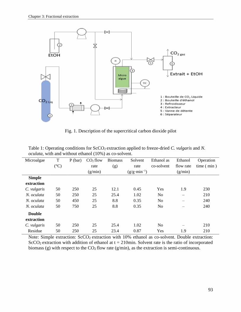
Chapter 3: Fractional extraction
93
Fig. 1. Description of the supercritical carbon dioxide pilot
Table 1: Operating conditions for ScCO2 extraction applied to freeze-dried C. vulgaris and N.
oculata, with and without ethanol (10%) as co-solvent.
Note: Simple extraction: ScCO2 extraction with 10% ethanol as co-solvent. Double extraction:
ScCO2 extraction with addition of ethanol at t = 210min. Solvent rate is the ratio of incorporated
biomass (g) with respect to the CO2 flow rate (g/min), as the extraction is semi-continuous.
Microalgae T
(°C)
P (bar) CO2 flow
rate
(g/min)
Biomass
(g)
Solvent
rate
(g/g·min−1)
Ethanol as
co-solvent
Ethanol
flow rate
(g/min)
Operation
time ( min )
Simple
extraction
C. vulgaris 50 250 25 12.1 0.45 Yes 1.9 230
N. oculata 50 250 25 25.4 1.02 No – 210
N. oculata 50 450 25 8.8 0.35 No – 240
N. oculata
Double
extraction
50 750 25 8.8 0.35 No – 240
C. vulgaris 50 250 25 25.4 1.02 No – 210
Residue 50 250 25 23.4 0.87 Yes 1.9 210
Chapter 3: Fractional extraction
94
3.2.2.4. Fractionation of neutral and polar lipids
All lipids extracted from N. oculata and C. vulgaris, either by ScCO2 or Soxhlet, were fractionated
into neutral and polar lipids using the solid-phase extraction method (SPE) described by Juaneda
and Rocquelin [16]. The cartridges were conditioned by slowly adding 5 mL chloroform followed
by 5 mL methanol, reaching the top of the gel to ensure good rinsing. A sample of 25 mg lipids
was applied to the top of the silica cartridges Supelco (500 mg, Sigma-Aldrich). The NL fraction
was eluted with 10 mL chloroform, whereas the GL and PPL fractions were eluted with 10 mL
acetone and methanol, respectively. Once eluted and freeze-dried under liquid nitrogen, every
fraction was measured gravimetrically. The contents of neutral and polar lipids are expressed in
g/g dry biomass.
3.2.2.5. Microscopic observation
Microscopically qualitative approach was done to characterize any cell disintegration of the
biomass. Observation at high magnifications (×1000) was performed before and directly after
ScCO2 treatments. A small quantity of cell suspension was placed on a specific plate (Nikon SMZ
1500). The images were captured under a constant exposure and illumination by Nikon eclipse
E600 camera.
3.2.2.6. Statistical analysis
Results for microalgae characterization in terms of lipid content were based on 3 replicates for 3
experiments (n = 9). All supercritical extraction data were presented as mean values ± standard
deviations of three experiments. Statistically significant differences (p < 0.05) between the means
were evaluated using one-way analysis of variance (ANOVA) and the Tukey's test on XLTAT
software version 2018.1.
3.2.3. Results and discussion
3.2.3.1. Microalgae characterization
The lipids composition of the studied microalgae was defined according to the values following
Soxhlet extraction [17]. The maximal lipids extraction yield was obtained after 18 h: 0.399 ± 0.016
and 0.244 ± 0.010 g/g dry weigh for N. oculata and C. vulgaris, respectively (Table 2).
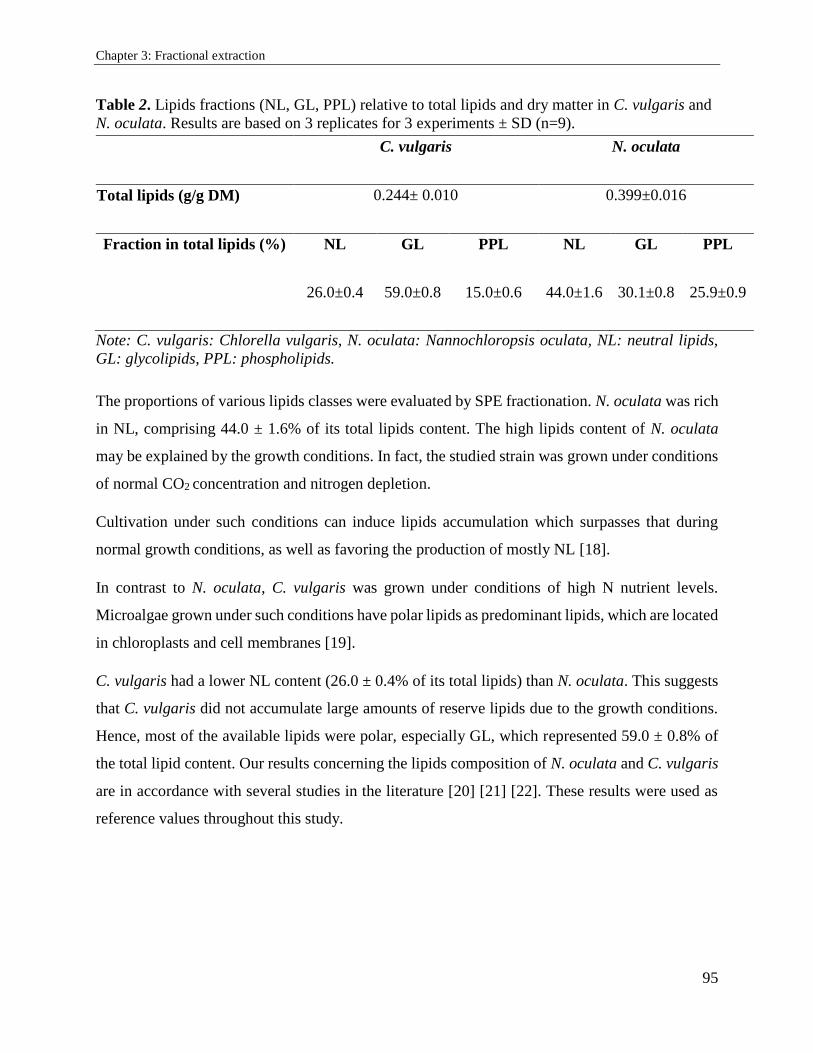
Chapter 3: Fractional extraction
95
Table 2. Lipids fractions (NL, GL, PPL) relative to total lipids and dry matter in C. vulgaris and
N. oculata. Results are based on 3 replicates for 3 experiments ± SD (n=9).
Note: C. vulgaris: Chlorella vulgaris, N. oculata: Nannochloropsis oculata, NL: neutral lipids,
GL: glycolipids, PPL: phospholipids.
The proportions of various lipids classes were evaluated by SPE fractionation. N. oculata was rich
in NL, comprising 44.0 ± 1.6% of its total lipids content. The high lipids content of N. oculata
may be explained by the growth conditions. In fact, the studied strain was grown under conditions
of normal CO2 concentration and nitrogen depletion.
Cultivation under such conditions can induce lipids accumulation which surpasses that during
normal growth conditions, as well as favoring the production of mostly NL [18].
In contrast to N. oculata, C. vulgaris was grown under conditions of high N nutrient levels.
Microalgae grown under such conditions have polar lipids as predominant lipids, which are located
in chloroplasts and cell membranes [19].
C. vulgaris had a lower NL content (26.0 ± 0.4% of its total lipids) than N. oculata. This suggests
that C. vulgaris did not accumulate large amounts of reserve lipids due to the growth conditions.
Hence, most of the available lipids were polar, especially GL, which represented 59.0 ± 0.8% of
the total lipid content. Our results concerning the lipids composition of N. oculata and C. vulgaris
are in accordance with several studies in the literature [20] [21] [22]. These results were used as
reference values throughout this study.
C. vulgaris N. oculata
Total lipids (g/g DM) 0.244± 0.010 0.399±0.016
Fraction in total lipids (%) NL GL PPL NL GL PPL
26.0±0.4 59.0±0.8 15.0±0.6 44.0±1.6 30.1±0.8 25.9±0.9
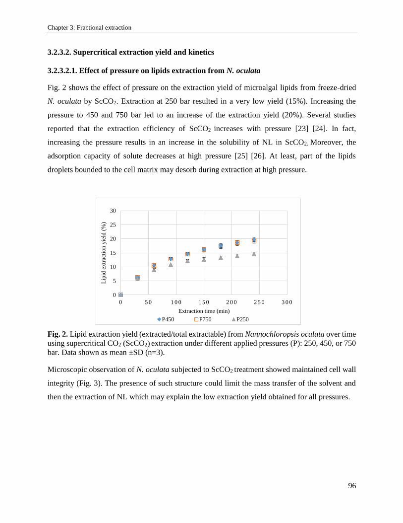
Chapter 3: Fractional extraction
96
3.2.3.2. Supercritical extraction yield and kinetics
3.2.3.2.1. Effect of pressure on lipids extraction from N. oculata
Fig. 2 shows the effect of pressure on the extraction yield of microalgal lipids from freeze-dried
N. oculata by ScCO2. Extraction at 250 bar resulted in a very low yield (15%). Increasing the
pressure to 450 and 750 bar led to an increase of the extraction yield (20%). Several studies
reported that the extraction efficiency of ScCO2 increases with pressure [23] [24]. In fact,
increasing the pressure results in an increase in the solubility of NL in ScCO2. Moreover, the
adsorption capacity of solute decreases at high pressure [25] [26]. At least, part of the lipids
droplets bounded to the cell matrix may desorb during extraction at high pressure.
Fig. 2. Lipid extraction yield (extracted/total extractable) from Nannochloropsis oculata over time
using supercritical CO2 (ScCO2) extraction under different applied pressures (P): 250, 450, or 750
bar. Data shown as mean ±SD (n=3).
Microscopic observation of N. oculata subjected to ScCO2 treatment showed maintained cell wall
integrity (Fig. 3). The presence of such structure could limit the mass transfer of the solvent and
then the extraction of NL which may explain the low extraction yield obtained for all pressures.
0
5
10
15
20
25
30
0 5 0 1 0 0 1 5 0 2 0 0 2 5 0 3 0 0
Lip
id e
xtr
acti
on
yie
ld (
%)
Extraction time (min)
P450 P750 P250
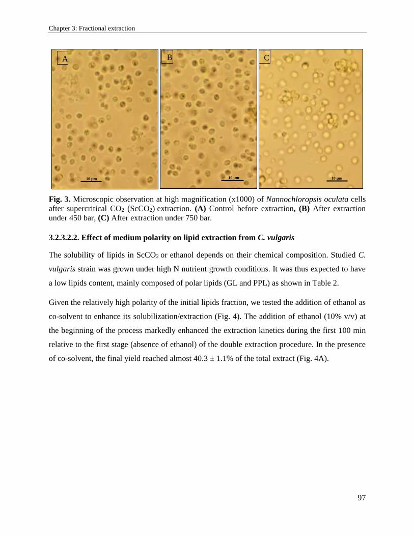
Chapter 3: Fractional extraction
97
Fig. 3. Microscopic observation at high magnification (x1000) of Nannochloropsis oculata cells
after supercritical CO2 (ScCO2) extraction. (A) Control before extraction, (B) After extraction
under 450 bar, (C) After extraction under 750 bar.
3.2.3.2.2. Effect of medium polarity on lipid extraction from C. vulgaris
The solubility of lipids in ScCO2 or ethanol depends on their chemical composition. Studied C.
vulgaris strain was grown under high N nutrient growth conditions. It was thus expected to have
a low lipids content, mainly composed of polar lipids (GL and PPL) as shown in Table 2.
Given the relatively high polarity of the initial lipids fraction, we tested the addition of ethanol as
co-solvent to enhance its solubilization/extraction (Fig. 4). The addition of ethanol (10% v/v) at
the beginning of the process markedly enhanced the extraction kinetics during the first 100 min
relative to the first stage (absence of ethanol) of the double extraction procedure. In the presence
of co-solvent, the final yield reached almost 40.3 ± 1.1% of the total extract (Fig. 4A).
B A C
10 µm 10 µm 10 µm
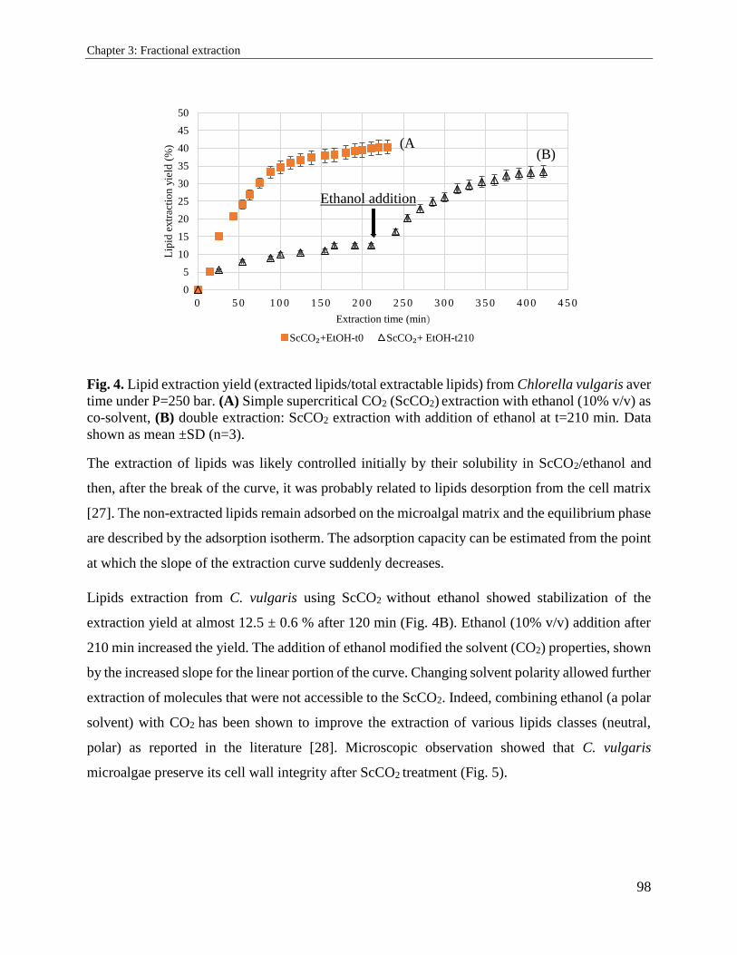
Chapter 3: Fractional extraction
98
Fig. 4. Lipid extraction yield (extracted lipids/total extractable lipids) from Chlorella vulgaris aver
time under P=250 bar. (A) Simple supercritical CO2 (ScCO2) extraction with ethanol (10% v/v) as
co-solvent, (B) double extraction: ScCO2 extraction with addition of ethanol at t=210 min. Data
shown as mean ±SD (n=3).
The extraction of lipids was likely controlled initially by their solubility in ScCO2/ethanol and
then, after the break of the curve, it was probably related to lipids desorption from the cell matrix
[27]. The non-extracted lipids remain adsorbed on the microalgal matrix and the equilibrium phase
are described by the adsorption isotherm. The adsorption capacity can be estimated from the point
at which the slope of the extraction curve suddenly decreases.
Lipids extraction from C. vulgaris using ScCO2 without ethanol showed stabilization of the
extraction yield at almost 12.5 ± 0.6 % after 120 min (Fig. 4B). Ethanol (10% v/v) addition after
210 min increased the yield. The addition of ethanol modified the solvent (CO2) properties, shown
by the increased slope for the linear portion of the curve. Changing solvent polarity allowed further
extraction of molecules that were not accessible to the ScCO2. Indeed, combining ethanol (a polar
solvent) with CO2 has been shown to improve the extraction of various lipids classes (neutral,
polar) as reported in the literature [28]. Microscopic observation showed that C. vulgaris
microalgae preserve its cell wall integrity after ScCO2 treatment (Fig. 5).
0
5
10
15
20
25
30
35
40
45
50
0 5 0 1 0 0 1 5 0 2 0 0 2 5 0 3 0 0 3 5 0 4 0 0 4 5 0
Lip
id e
xtr
acti
on
yie
ld (
%)
Extraction time (min)
ScCO₂+EtOH-t0 ScCO₂+ EtOH-t210
(A
) (B)
Ethanol addition
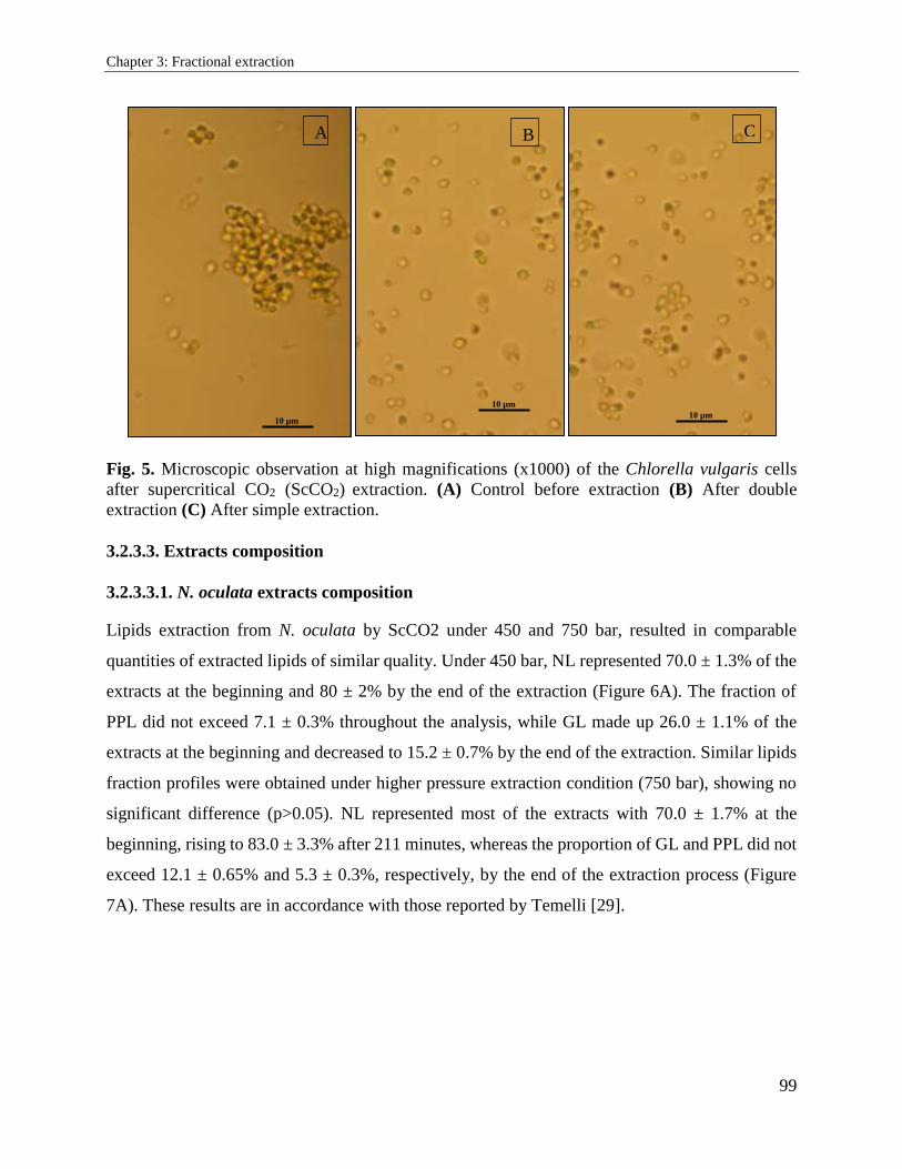
Chapter 3: Fractional extraction
99
Fig. 5. Microscopic observation at high magnifications (x1000) of the Chlorella vulgaris cells
after supercritical CO2 (ScCO2) extraction. (A) Control before extraction (B) After double
extraction (C) After simple extraction.
3.2.3.3. Extracts composition
3.2.3.3.1. N. oculata extracts composition
Lipids extraction from N. oculata by ScCO2 under 450 and 750 bar, resulted in comparable
quantities of extracted lipids of similar quality. Under 450 bar, NL represented 70.0 ± 1.3% of the
extracts at the beginning and 80 ± 2% by the end of the extraction (Figure 6A). The fraction of
PPL did not exceed 7.1 ± 0.3% throughout the analysis, while GL made up 26.0 ± 1.1% of the
extracts at the beginning and decreased to 15.2 ± 0.7% by the end of the extraction. Similar lipids
fraction profiles were obtained under higher pressure extraction condition (750 bar), showing no
significant difference (p>0.05). NL represented most of the extracts with 70.0 ± 1.7% at the
beginning, rising to 83.0 ± 3.3% after 211 minutes, whereas the proportion of GL and PPL did not
exceed 12.1 ± 0.65% and 5.3 ± 0.3%, respectively, by the end of the extraction process (Figure
7A). These results are in accordance with those reported by Temelli [29].
A B C
10 µm
10 µm
10 µm
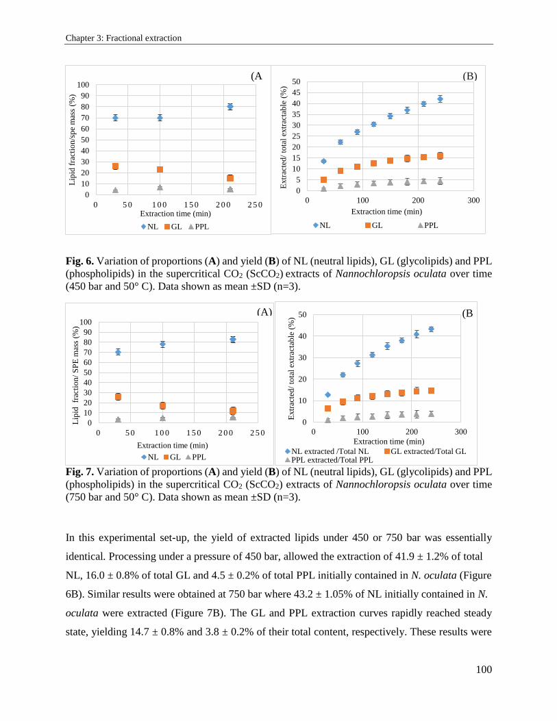
Chapter 3: Fractional extraction
100
Fig. 6. Variation of proportions (A) and yield (B) of NL (neutral lipids), GL (glycolipids) and PPL
(phospholipids) in the supercritical CO2 (ScCO2) extracts of Nannochloropsis oculata over time
(450 bar and 50° C). Data shown as mean ±SD (n=3).
Fig. 7. Variation of proportions (A) and yield (B) of NL (neutral lipids), GL (glycolipids) and PPL
(phospholipids) in the supercritical CO2 (ScCO2) extracts of Nannochloropsis oculata over time
(750 bar and 50° C). Data shown as mean ±SD (n=3).
In this experimental set-up, the yield of extracted lipids under 450 or 750 bar was essentially
identical. Processing under a pressure of 450 bar, allowed the extraction of 41.9 ± 1.2% of total
NL, 16.0 ± 0.8% of total GL and 4.5 ± 0.2% of total PPL initially contained in N. oculata (Figure
6B). Similar results were obtained at 750 bar where 43.2 ± 1.05% of NL initially contained in N.
oculata were extracted (Figure 7B). The GL and PPL extraction curves rapidly reached steady
state, yielding 14.7 ± 0.8% and 3.8 ± 0.2% of their total content, respectively. These results were
0
10
20
30
40
50
60
70
80
90
100
0 5 0 1 0 0 1 5 0 2 0 0 2 5 0
Lip
id f
ract
ion
/sp
e m
ass
(%)
Extraction time (min)
NL GL PPL
0
5
10
15
20
25
30
35
40
45
50
0 100 200 300
Extr
acte
d/
tota
l ex
trac
tab
le (
%)
Extraction time (min)
NL GL PPL
0
10
20
30
40
50
60
70
80
90
100
0 5 0 1 0 0 1 5 0 2 0 0 2 5 0
Lip
id fr
acti
on
/ S
PE
mas
s (%
)
Extraction time (min)
NL GL PPL
0
10
20
30
40
50
0 100 200 300
Extr
acte
d/
tota
l ex
trac
tab
le (
%)
Extraction time (min)
NL extracted /Total NL GL extracted/Total GLPPL extracted/Total PPL
(A
)
(B)
(B
)
(A)
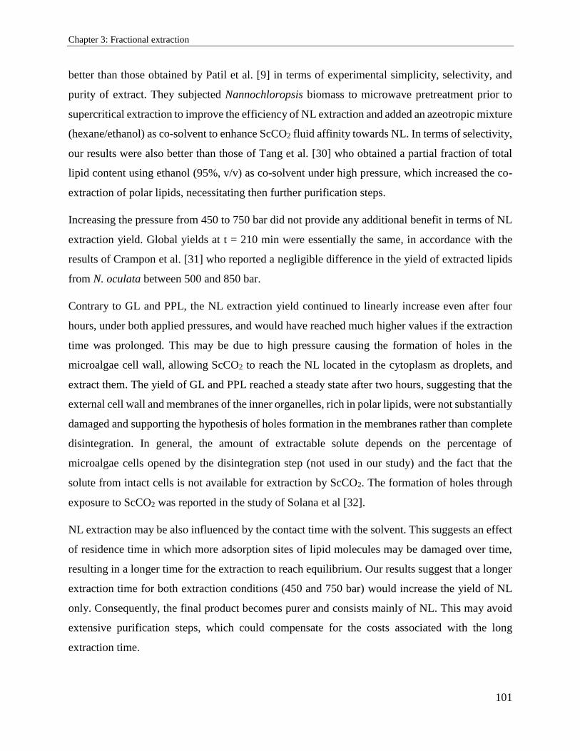
Chapter 3: Fractional extraction
101
better than those obtained by Patil et al. [9] in terms of experimental simplicity, selectivity, and
purity of extract. They subjected Nannochloropsis biomass to microwave pretreatment prior to
supercritical extraction to improve the efficiency of NL extraction and added an azeotropic mixture
(hexane/ethanol) as co-solvent to enhance ScCO2 fluid affinity towards NL. In terms of selectivity,
our results were also better than those of Tang et al. [30] who obtained a partial fraction of total
lipid content using ethanol (95%, v/v) as co-solvent under high pressure, which increased the co-
extraction of polar lipids, necessitating then further purification steps.
Increasing the pressure from 450 to 750 bar did not provide any additional benefit in terms of NL
extraction yield. Global yields at t = 210 min were essentially the same, in accordance with the
results of Crampon et al. [31] who reported a negligible difference in the yield of extracted lipids
from N. oculata between 500 and 850 bar.
Contrary to GL and PPL, the NL extraction yield continued to linearly increase even after four
hours, under both applied pressures, and would have reached much higher values if the extraction
time was prolonged. This may be due to high pressure causing the formation of holes in the
microalgae cell wall, allowing ScCO2 to reach the NL located in the cytoplasm as droplets, and
extract them. The yield of GL and PPL reached a steady state after two hours, suggesting that the
external cell wall and membranes of the inner organelles, rich in polar lipids, were not substantially
damaged and supporting the hypothesis of holes formation in the membranes rather than complete
disintegration. In general, the amount of extractable solute depends on the percentage of
microalgae cells opened by the disintegration step (not used in our study) and the fact that the
solute from intact cells is not available for extraction by ScCO2. The formation of holes through
exposure to ScCO2 was reported in the study of Solana et al [32].
NL extraction may be also influenced by the contact time with the solvent. This suggests an effect
of residence time in which more adsorption sites of lipid molecules may be damaged over time,
resulting in a longer time for the extraction to reach equilibrium. Our results suggest that a longer
extraction time for both extraction conditions (450 and 750 bar) would increase the yield of NL
only. Consequently, the final product becomes purer and consists mainly of NL. This may avoid
extensive purification steps, which could compensate for the costs associated with the long
extraction time.
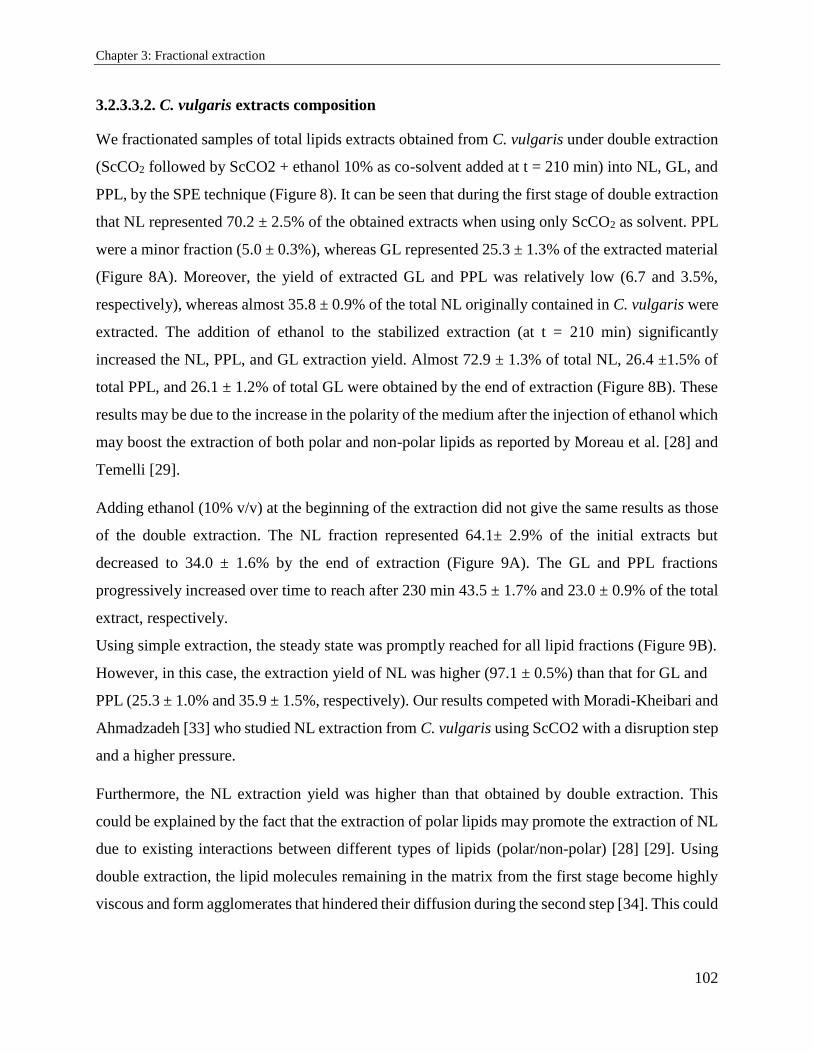
Chapter 3: Fractional extraction
102
3.2.3.3.2. C. vulgaris extracts composition
We fractionated samples of total lipids extracts obtained from C. vulgaris under double extraction
(ScCO2 followed by ScCO2 + ethanol 10% as co-solvent added at t = 210 min) into NL, GL, and
PPL, by the SPE technique (Figure 8). It can be seen that during the first stage of double extraction
that NL represented 70.2 ± 2.5% of the obtained extracts when using only ScCO2 as solvent. PPL
were a minor fraction (5.0 ± 0.3%), whereas GL represented 25.3 ± 1.3% of the extracted material
(Figure 8A). Moreover, the yield of extracted GL and PPL was relatively low (6.7 and 3.5%,
respectively), whereas almost 35.8 ± 0.9% of the total NL originally contained in C. vulgaris were
extracted. The addition of ethanol to the stabilized extraction (at t = 210 min) significantly
increased the NL, PPL, and GL extraction yield. Almost 72.9 ± 1.3% of total NL, 26.4 ±1.5% of
total PPL, and 26.1 ± 1.2% of total GL were obtained by the end of extraction (Figure 8B). These
results may be due to the increase in the polarity of the medium after the injection of ethanol which
may boost the extraction of both polar and non-polar lipids as reported by Moreau et al. [28] and
Temelli [29].
Adding ethanol (10% v/v) at the beginning of the extraction did not give the same results as those
of the double extraction. The NL fraction represented 64.1± 2.9% of the initial extracts but
decreased to 34.0 ± 1.6% by the end of extraction (Figure 9A). The GL and PPL fractions
progressively increased over time to reach after 230 min 43.5 ± 1.7% and 23.0 ± 0.9% of the total
extract, respectively.
Using simple extraction, the steady state was promptly reached for all lipid fractions (Figure 9B).
However, in this case, the extraction yield of NL was higher (97.1 ± 0.5%) than that for GL and
PPL (25.3 ± 1.0% and 35.9 ± 1.5%, respectively). Our results competed with Moradi-Kheibari and
Ahmadzadeh [33] who studied NL extraction from C. vulgaris using ScCO2 with a disruption step
and a higher pressure.
Furthermore, the NL extraction yield was higher than that obtained by double extraction. This
could be explained by the fact that the extraction of polar lipids may promote the extraction of NL
due to existing interactions between different types of lipids (polar/non-polar) [28] [29]. Using
double extraction, the lipid molecules remaining in the matrix from the first stage become highly
viscous and form agglomerates that hindered their diffusion during the second step [34]. This could
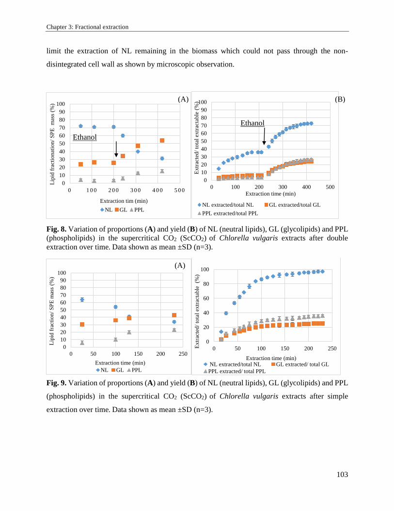
Chapter 3: Fractional extraction
103
limit the extraction of NL remaining in the biomass which could not pass through the non-
disintegrated cell wall as shown by microscopic observation.
Fig. 8. Variation of proportions (A) and yield (B) of NL (neutral lipids), GL (glycolipids) and PPL
(phospholipids) in the supercritical CO2 (ScCO2) of Chlorella vulgaris extracts after double
extraction over time. Data shown as mean ±SD (n=3).
Fig. 9. Variation of proportions (A) and yield (B) of NL (neutral lipids), GL (glycolipids) and PPL
(phospholipids) in the supercritical CO2 (ScCO2) of Chlorella vulgaris extracts after simple
extraction over time. Data shown as mean ±SD (n=3).
0
10
20
30
40
50
60
70
80
90
100
0 1 0 0 2 0 0 3 0 0 4 0 0 5 0 0
Lip
id f
ract
ion
atio
n/
SP
E m
ass
(%)
Extraction tim (min)
NL GL PPL
0
10
20
30
40
50
60
70
80
90
100
0 100 200 300 400 500
Extr
acte
d/
tota
l ex
trac
tab
le (
%)
Extraction time (min)
NL extracted/total NL GL extracted/total GL
PPL extracted/total PPL
0
10
20
30
40
50
60
70
80
90
100
0 50 100 150 200 250
Lip
id f
ract
ion
/ S
PE
mas
s (%
)
Extraction time (min)
NL GL PPL
0
20
40
60
80
100
0 50 100 150 200 250Extr
acte
d/
tota
l ex
trac
tab
le
(%)
Extraction time (min) NL extracted/total NL GL extracted/ total GL
PPL extracted/ total PPL
(A) (B)
Ethanol
addition
Ethanol
addition
(A)
(B)
Chapter 3: Fractional extraction
104
3.2.4. Conclusion
During this study, ScCO2 extraction was carried out for selective recovery of neutral lipids from
C. vulgaris and N. oculata microalgae without preliminary cell disruption step. Extraction yield of
total lipids from N. oculata at 450 and 750 bar (20 %) was higher than that at 250 bar (15%).
Neutral lipids extracts from N. oculata represented 83% of total lipids extracts, whereas the
proportion of glycolipids and phospholipids did not exceed 12.1% and 5.3%, respectively. Adding
ethanol (10% v/v) as co-solvent led to the extraction of 97% of neutral lipids from C. vulgaris.
Residues from ScCO2 extraction, composed mainly of hydrophilic fraction (proteins,
polysaccharides), will undergo in future study an optimized aqueous extraction, in aim to recover
total proteins fraction without any cell disruption pretreatment. This deals with the fundamental
algorefinery concept, by taking advantages from the whole biomass components, making the large
scale application of microalgae feasible and economically viable.
3.2.5. References
[1] S. Bellou, M. N. Baeshen, A. M. Elazzazy, D. Aggeli, F. Sayegh, and G. Aggelis, “Microalgal
lipids biochemistry and biotechnological perspectives,” Biotechnol. Adv., vol. 32, no. 8, pp. 1476–
1493, Dec. 2014.
[2] T. Bradley, D. Maga, and S. Antón, “Unified approach to Life Cycle Assessment between
three unique algae biofuel facilities,” Appl. Energy, vol. 154, pp. 1052–1061, Sep. 2015.
[3] L. Brennan and P. Owende, “Biofuels from microalgae—A review of technologies for
production, processing, and extractions of biofuels and co-products,” Renew. Sustain. Energy Rev.,
vol. 14, no. 2, pp. 557–577, Feb. 2010.
[4] L. Lardon, A. Hélias, B. Sialve, J.-P. Steyer, and O. Bernard, “Life-Cycle Assessment of
Biodiesel Production from Microalgae,” Environ. Sci. Technol., vol. 43, no. 17, pp. 6475–6481,
Sep. 2009.
[5] R. Ranjith Kumar, P. Hanumantha Rao, and M. Arumugam, “Lipid Extraction Methods from
Microalgae: A Comprehensive Review,” Front. Energy Res., vol. 2, Jan. 2015.
[6] M. K. Lam and K. T. Lee, “Microalgae biofuels: A critical review of issues, problems and the
way forward,” Biotechnol. Adv., vol. 30, no. 3, pp. 673–690, May 2012.
[7] A. García, A. de Lucas, J. Rincón, A. Alvarez, I. Gracia, and M. A. García, “Supercritical
carbon dioxide extraction of fatty and waxy material from rice bran,” J. Am. Oil Chem. Soc., vol.
73, no. 9, pp. 1127–1131, Sep. 1996.
[8] J. Lorenzen et al., “Extraction of microalgae derived lipids with supercritical carbon dioxide
in an industrial relevant pilot plant,” Bioprocess Biosyst. Eng., vol. 40, no. 6, pp. 911–918, 2017.
Chapter 3: Fractional extraction
105
[9] P. D. Patil, K. P. R. Dandamudi, J. Wang, Q. Deng, and S. Deng, “Extraction of bio-oils from
algae with supercritical carbon dioxide and co-solvents,” J. Supercrit. Fluids, vol. 135, pp. 60–68,
May 2018.
[10] C.-H. Cheng, T.-B. Du, H.-C. Pi, S.-M. Jang, Y.-H. Lin, and H.-T. Lee, “Comparative study
of lipid extraction from microalgae by organic solvent and supercritical CO2,” Bioresour.
Technol., vol. 102, no. 21, pp. 10151–10153, Nov. 2011.
[11] C. Crampon et al., “Oil extraction from enriched Spirulina platensis microalgae using
supercritical carbon dioxide,” J. Supercrit. Fluids, vol. 119, pp. 289–296, Jan. 2017.
[12] C. Safi et al., “Aqueous extraction of proteins from microalgae: Effect of different cell
disruption methods,” Algal Res., vol. 3, pp. 61–65, Jan. 2014.
[13] K. Ramluckan, K. G. Moodley, and F. Bux, “An evaluation of the efficacy of using selected
solvents for the extraction of lipids from algal biomass by the soxhlet extraction method,” Fuel,
vol. 116, pp. 103–108, Jan. 2014.
[14] J. Jeon et al., “New mixture composition of organic solvents for efficient extraction of lipids
from Chlorella vulgaris,” Biomass Bioenergy, vol. 59, pp. 279–284, Dec. 2013.
[15] Y. Du, B. Schuur, S. R. A. Kersten, and D. W. F. Brilman, “Opportunities for switchable
solvents for lipid extraction from wet algal biomass: An energy evaluation,” Algal Res., vol. 11,
pp. 271–283, Sep. 2015.
[16] P. Juaneda and G. Rocquelin, “Rapid and convenient separation of phospholipids and non
phosphorus lipids from rat heart using silica cartridges,” Lipids, vol. 20, no. 1, pp. 40–41.
[17] M. Olofsson et al., “Combined Effects of Nitrogen Concentration and Seasonal Changes on
the Production of Lipids in Nannochloropsis oculata,” Mar. Drugs, vol. 12, no. 4, pp. 1891–1910,
Mar. 2014.
[18] A. Widjaja, C.-C. Chien, and Y.-H. Ju, “Study of increasing lipid production from fresh water
microalgae Chlorella vulgaris,” J. Taiwan Inst. Chem. Eng., vol. 40, no. 1, pp. 13–20, Jan. 2009.
[19] M. Piorreck, K.-H. Baasch, and P. Pohl, “Biomass production, total protein, chlorophylls,
lipids and fatty acids of freshwater green and blue-green algae under different nitrogen regimes,”
Phytochemistry, vol. 23, no. 2, pp. 207–216, Jan. 1984.
[20] S. Ötleş and R. Pire, “Fatty Acid Composition of Chlorella and Spirulina Microalgae
Species,” Nov-2001.
[21] G. Van Vooren, F. Le Grand, J. Legrand, S. Cuiné, G. Peltier, and J. Pruvost, “Investigation
of fatty acids accumulation in Nannochloropsis oculata for biodiesel application,” Bioresour.
Technol., vol. 124, pp. 421–432, Nov. 2012.
[22] A. L. Stephenson, J. S. Dennis, C. J. Howe, S. A. Scott, and A. G. Smith, “Influence of
nitrogen-limitation regime on the production by Chlorella vulgaris of lipids for biodiesel
feedstocks,” Biofuels, vol. 1, no. 1, pp. 47–58, Jan. 2010.
Chapter 3: Fractional extraction
106
[23] J. Shi, C. Yi, S. J. Xue, Y. Jiang, Y. Ma, and D. Li, “Effects of modifiers on the profile of
lycopene extracted from tomato skins by supercritical CO2,” J. Food Eng., vol. 93, no. 4, pp. 431–
436, Aug. 2009.
[24] A. Cháfer, T. Fornari, A. Berna, E. Ibañez, and G. Reglero, “Solubility of solid carnosic acid
in supercritical CO2 with ethanol as a co-solvent,” J. Supercrit. Fluids, vol. 34, no. 3, pp. 323–
329, Jul. 2005.
[25] G. Andrich, U. Nesti, F. Venturi, A. Zinnai, and R. Fiorentini, “Supercritical fluid extraction
of bioactive lipids from the microalga Nannochloropsis sp.,” Eur. J. Lipid Sci. Technol., vol. 107,
no. 6, pp. 381–386.
[26] H. Xing, B. Su, Q. Ren, and Y. Yang, “Adsorption equilibria of artemisinin from supercritical
carbon dioxide on silica gel,” J. Supercrit. Fluids, vol. 49, no. 2, pp. 189–195, Jun. 2009.
[27] M. Perrut, J. Y. Clavier, M. Poletto, and E. Reverchon, “Mathematical Modeling of Sunflower
Seed Extraction by Supercritical CO2,” Ind. Eng. Chem. Res., vol. 36, no. 2, pp. 430–435, Feb.
1997.
[28] R. A. Moreau, M. J. Powell, and V. Singh, “Pressurized liquid extraction of polar and
nonpolar lipids in corn and oats with hexane, methylene chloride, isopropanol, and ethanol,” J.
Am. Oil Chem. Soc., vol. 80, no. 11, pp. 1063–1067, Nov. 2003.
[29] F. Temelli, “Extraction of Triglycerides and Phospholipids from Canola with Supercritical
Carbon Dioxide and Ethanol,” J. Food Sci., vol. 57, no. 2, pp. 440–443, 1992.
[30] S. Tang, C. Qin, H. Wang, S. Li, and S. Tian, “Study on supercritical extraction of lipids and
enrichment of DHA from oil-rich microalgae,” J. Supercrit. Fluids, vol. 57, no. 1, pp. 44–49, May
2011.
[31] C. Crampon, A. Mouahid, S.-A. A. Toudji, O. Lépine, and E. Badens, “Influence of
pretreatment on supercritical CO2 extraction from Nannochloropsis oculata,” J. Supercrit. Fluids,
vol. 79, pp. 337–344, Jul. 2013.
[32] M. Solana, C. S. Rizza, and A. Bertucco, “Exploiting microalgae as a source of essential fatty
acids by supercritical fluid extraction of lipids: Comparison between Scenedesmus obliquus,
Chlorella protothecoides and Nannochloropsis salina,” J. Supercrit. Fluids, vol. 92, pp. 311–318,
Aug. 2014.
[33] N. Moradi-kheibari and H. Ahmadzadeh, “Supercritical carbon dioxide extraction and
analysis of lipids from Chlorella vulgaris using gas chromatography,” J. Iran. Chem. Soc., vol.
14, no. 11, pp. 2427–2436, Nov. 2017.
[34] G. Yoo et al., “An effective, cost-efficient extraction method of biomass from wet microalgae
with a functional polymeric membrane,” Green Chem., vol. 16, no. 1, pp. 312–319, 2014.
Chapter 3: Fractional extraction
107
3.3. Supercritical fluid extraction and solid phase fractionation of pigments
from Chlorella vulgaris and Nannochloropsis oculata
(Article submitted to Algal research)
Sara Obeida, b, c, 1 Nicholas Beaufils a, Séverine Camy d, Hosni Takache b, c, Ali Ismail b, c,
Pierre-Yves Pontalier a.
a Laboratoire de Chimie Agro-industrielle (LCA), Université de Toulouse, INRA, INPT, Toulouse, France
4 allée Emile Monso, 31030 Toulouse cedex 4, France b Département des Sciences et Technologies Alimentaires, Faculté des Sciences Agronomiques, Université
Libanaise, Allée Georges Matta, 1500, Hadath, Liban. c Université Libanaise, Ecole Doctorale de Sciences et Technologies EDST-PRASE (Plateforme de
Recherches et Analyses en Sciences de l’Environnement), Campus Rafic Hariri, 1500, Hadath, Liban. d Université de Toulouse, INP-ENSIACET, LGC (Laboratoire de Génie Chimique), 4 allée Emile Monso,
31030 Toulouse cedex 4, France
Abstract:
The aim of this study was to determine the influence of pressure and co-solvent on the supercritical
fluid extraction of chlorophyll a, chlorophyll b, lutein and β-carotene from freeze-dried
Nannochloropsis oculata and Chlorella vulgaris. Bypassing the mechanical cells disintegration
showed to be good operational conditions for the selective extraction of chlorophyll a. In
comparison with the reference extraction process used (Soxhlet), 43% of total chlorophyll a were
selectively extracted under 750 bar from freeze-dried Nannochloropsis oculata with traceable
amount of chlorophyll b, β-carotene and lutein. The effect of adding ethanol (10% v/v) on pigments
extraction selectivity was also studied on Chlorella vulgaris. When supercritical CO2 doped with
ethanol was used from the beginning of the extraction, it was possible to selectively extract 30%
of chlorophyll a comparing to 5% only by adding ethanol at 210 min from the beginning of the
SFE: supercritical fluid extraction
NL: neutral Lipids
GL: glycolipids
PPL: phospholipids
ScCO2: supercritical carbon dioxide
SPE: solid phase extraction
Chapter 3: Fractional extraction
108
extraction. The higher selectivity of chlorophyll a extraction should facilitate the recovery of
pigments.
Keywords: Supercritical fluid extraction, solid phase extraction, microalgae, pigments, cell wall
integrity.
3.3.1. Introduction
Over the last decade, microalgae showed their potential of satisfying the industrial growing
demand in natural products beneficial for human health and environment. The algae biomass is
widely studied as a source of highly valuable components such as lipids considered as renewable
biofuel [1]. Besides to the energy potential, these microorganisms have the capacity to synthesize
bioactive molecules such as pigments where particular attention has been given to carotenoids and
chlorophylls. Carotenoids are regarded as cancer and health disorders preventives [2] [3], while
chlorophylls are widely used in food, feed, cosmetics, biomaterials, nanostructures and
pharmaceutical industry [1] [4]. Moreover, microalgal pigments becomes an attractive option due
to problems associated with the harmful effects of synthetic colorants and their limited applications
due to their negative effect on human health [5].
Extraction of pigments from microalgal biomass is usually achieved using chemical solvents.
However, these methods are nonselective and generally accompanied by cell-wall destruction,
leading to complex extracts of hydrophilic and hydrophobic components. Such treatments require
additional costly stages in downstream processing for phase separation and compounds
purification. In addition, a large amount of toxic solvents is needed. For that, this technique is
currently being phased out for their environmental, health and safety impacts. Recently,
supercritical carbon dioxide (ScCO2) extraction has become an important alternative to the
traditional separation methods. CO2 is among the most used supercritical solvent because it
engenders solvent free product compatible with the use of the label ‘‘natural’’ [6]. In addition, the
use of CO2 gives an advantage in terms of quality, as extracts are not submitted to excessive
heating, which would destroy heat sensitive compounds. Many studies have tested ScCO2 due to
its remarkable characteristics like low viscosity, high diffusivity and low surface tension [7] which
could improve pigments extraction deep inside the microalgal cell matrix. Furthermore, ScCO2

Chapter 3: Fractional extraction
109
enables the recovery of molecules of interest by a simple pressure drop, avoiding additional energy
consumption step [8].
Since ScCO2 is a non-polar solvent, a polar co-solvent is often used in order to increase its
extraction capacity. The most often used is ethanol, a green solvent, generally recognized as safe
for human and environment. Despite these studies, understanding the extraction and its selectivity
under certain supercritical parameters still not well described and only limited information exists
[8]. Besides, in supercritical conditions, the degree of cell wall disintegration strongly influenced
the pigment (chlorophyll and carotenoids) recoveries [9] [10] [11]. In addition, the before-
mentioned studies did neither involve the quantification of several specific pigments nor their
fractionation during the extraction process.
The aim of this work is to study the extraction of four pigments (chlorophyll a, chlorophyll b,
lutein and β-carotene) from non-disintegrated C. vulgaris and N. oculata by ScCO2 followed by
solid phase extraction in aim to understand the evolution of their release with manipulation time.
Different ScCO2 extraction parameters were studied on non-disintegrated biomass of both
microalgae.
3.3.2. Materials and methods
3.3.2.1. Microalgae
Microalgae Chlorella vulgaris and Nannochloropsis oculata were supplied as frozen paste from
Alpha Biotech (Asserac, France). Both species were cultivated in 10 L tubular air-lift
photobioreactor. The Sueoka medium with high N nutrient concentration, and the Conway medium
with N nutrient deficiency to favor the accumulation of lipids were used for C. vulgaris and N.
oculata, respectively. The two cultures were growing at a constant temperature of 25 ± 1 °C and
constant pH of 7.5. The harvesting of the biomass was carried out at exponential growth phase.
The algal suspension was centrifuged at 10,000 g for 15 min. The harvested biomass contained 27
% dry matter (DM). Frozen paste was freeze-dried using Fisher Bioblock Scientific Alpha 2–4 LD
Plus device (Illkirch, France), where the pressure was reduced to 0.01 bar and the temperature
decreased to -80°C for 48 h to give a completely dry biomass. After freeze drying process, the
mean diameter of particles was measured showing a value around 250 μm using Mastersizer 2000
Chapter 3: Fractional extraction
110
granulometer (Malvern Instruments Ltd.). Aggregates were crushed gently with a laboratory knife
grinder and the final size was around 200 μm.
3.3.2.2. Lipids content analysis
Total lipids content in the two microalgae species were measured after extraction in Soxhlet
apparatus using a slightly modified method based on both methods developed by Blight and Dyer
(1959) and Folch (1957) using a mixture of chloroform/methanol (0.35 / 0.65, V/V) [12] [13] [14].
The microalgal biomass (3 g) was placed in a cartridge filter paper using 200 mL of solvent. The
Soxhlet extractor was operated for a number of cycles for 18 hours in order to insure the extraction
of all extractable lipids. The amount of total lipids was determined gravimetrically.
3.3.2.3. Supercritical extraction
The experiments were carried out in pilot plant SF100 (Separex Chimie Fine, France) capable of
operating at pressures of up to 1000 bar and at a maximal temperature of 200°C. Desired operating
pressure in the extractor was adjusted by a backpressure regulator. During this experiment, the
extractor of a 25 mL (internal diameter 2 cm, height 8 cm) was filled with powdered freeze-dried
microalgae and CO2 introduced at the bottom. Ethanol was used as a co-solvent when mixed with
CO2 as shown in table 1.
After extraction, the extracts were collected in the separator and stored in the dark at 4°C to prevent
degradation of samples for further analysis. Cleaning of the extractor and the separator was done
after each experiment.
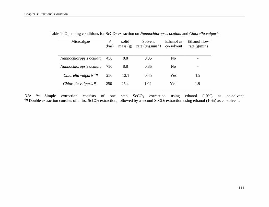
Chapter 3: Fractional extraction
111
Table 1- Operating conditions for ScCO2 extraction on Nannochloropsis oculata and Chlorella vulgaris
NB: (a) Simple extraction consists of one step ScCO2 extraction using ethanol (10%) as co-solvent. (b) Double extraction consists of a first ScCO2 extraction, followed by a second ScCO2 extraction using ethanol (10%) as co-solvent.
Microalgae P
(bar)
solid
mass (g)
Solvent
rate (g/g.min-1)
Ethanol as
co-solvent
Ethanol flow
rate (g/min)
Nannochloropsis oculata 450 8.8 0.35 No -
Nannochloropsis oculata 750 8.8 0.35 No -
Chlorella vulgaris (a) 250 12.1 0.45 Yes 1.9
Chlorella vulgaris (b) 250 25.4 1.02 Yes 1.9
Chapter 3: Fractional extraction
112
3.3.2.4. Lipids classes’ fractionation
Partitioning of lipids to three classes: neutral lipids (NL), glycolipids (GL) and
phospholipids (PPL) was possible using solid phase extraction (SPE) method described
by Juaneda and Rocqueline [15]. This technique consists on putting the mixture of lipids
on the top of a conditioned cartridge (SPE silica cartridges Supelco 500 mg, Sigma-
Aldrich). The separation of lipids fractions was accomplished by eluting NL, GL and PPL
with 10 ml of chloroform, acetone and methanol, respectively. Once eluted and freeze-
dried under liquid nitrogen, fractions were measured gravimetrically and results were
expressed in mg/g dry weight.
3.3.2.5. Pigments analysis
Total biomass content in chlorophyll a, chlorophyll b, lutein and β-carotene were
extracted in cold condition to avoid any denaturation induced by high temperature. For
this, 20 mg of freeze-dried microalgae were mixed with 15 ml of specific solvent (table
2) during 24 min at 10 ºC with continuous gentle stirring. Glass tubes were covered with
aluminum paper to prevent any photodegradation during extraction. Afterward, samples
were centrifuged at 20,000g for 15 min at 5ºC and supernatants were collected for
analysis.
Total pigments (chlorophyll a, chlorophyll b, β-carotenes and lutein) content was
measured by HPLC and UV-Vis spectroscopy according to the general formula
developed by Bidigare [16]: C(mg/l) =OD∗1000
α∗L
Where OD is the optical density read at the specific wavelength in a given solvent, α is
the specific extinction coefficient (1 g-1cm-1) and L is the optical path length (cuvettes) in
cm which is 1 cm in our study.
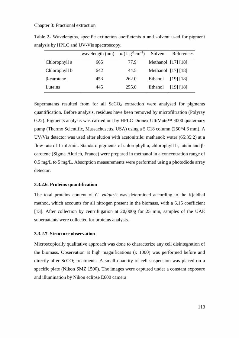
Chapter 3: Fractional extraction
113
Table 2- Wavelengths, specific extinction coefficients α and solvent used for pigment
analysis by HPLC and UV-Vis spectroscopy.
wavelength (nm) α (L g-1cm-1) Solvent References
Chlorophyll a 665 77.9 Methanol [17] [18]
Chlorophyll b 642 44.5 Methanol [17] [18]
β-carotene 453 262.0 Ethanol [19] [18]
Luteins 445 255.0 Ethanol [19] [18]
Supernatants resulted from for all ScCO2 extraction were analysed for pigments
quantification. Before analysis, residues have been removed by microfiltration (Polyray
0.22). Pigments analysis was carried out by HPLC Dionex UltiMate™ 3000 quaternary
pump (Thermo Scientific, Massachusetts, USA) using a 5 C18 column (250*4.6 mm). A
UV/Vis detector was used after elution with acetonitrile: methanol: water (65:35:2) at a
flow rate of 1 mL/min. Standard pigments of chlorophyll a, chlorophyll b, lutein and β-
carotene (Sigma-Aldrich, France) were prepared in methanol in a concentration range of
0.5 mg/L to 5 mg/L. Absorption measurements were performed using a photodiode array
detector.
3.3.2.6. Proteins quantification
The total proteins content of C. vulgaris was determined according to the Kjeldhal
method, which accounts for all nitrogen present in the biomass, with a 6.15 coefficient
[13]. After collection by centrifugation at 20,000g for 25 min, samples of the UAE
supernatants were collected for proteins analysis.
3.3.2.7. Structure observation
Microscopically qualitative approach was done to characterize any cell disintegration of
the biomass. Observation at high magnifications (x 1000) was performed before and
directly after ScCO2 treatments. A small quantity of cell suspension was placed on a
specific plate (Nikon SMZ 1500). The images were captured under a constant exposure
and illumination by Nikon eclipse E600 camera
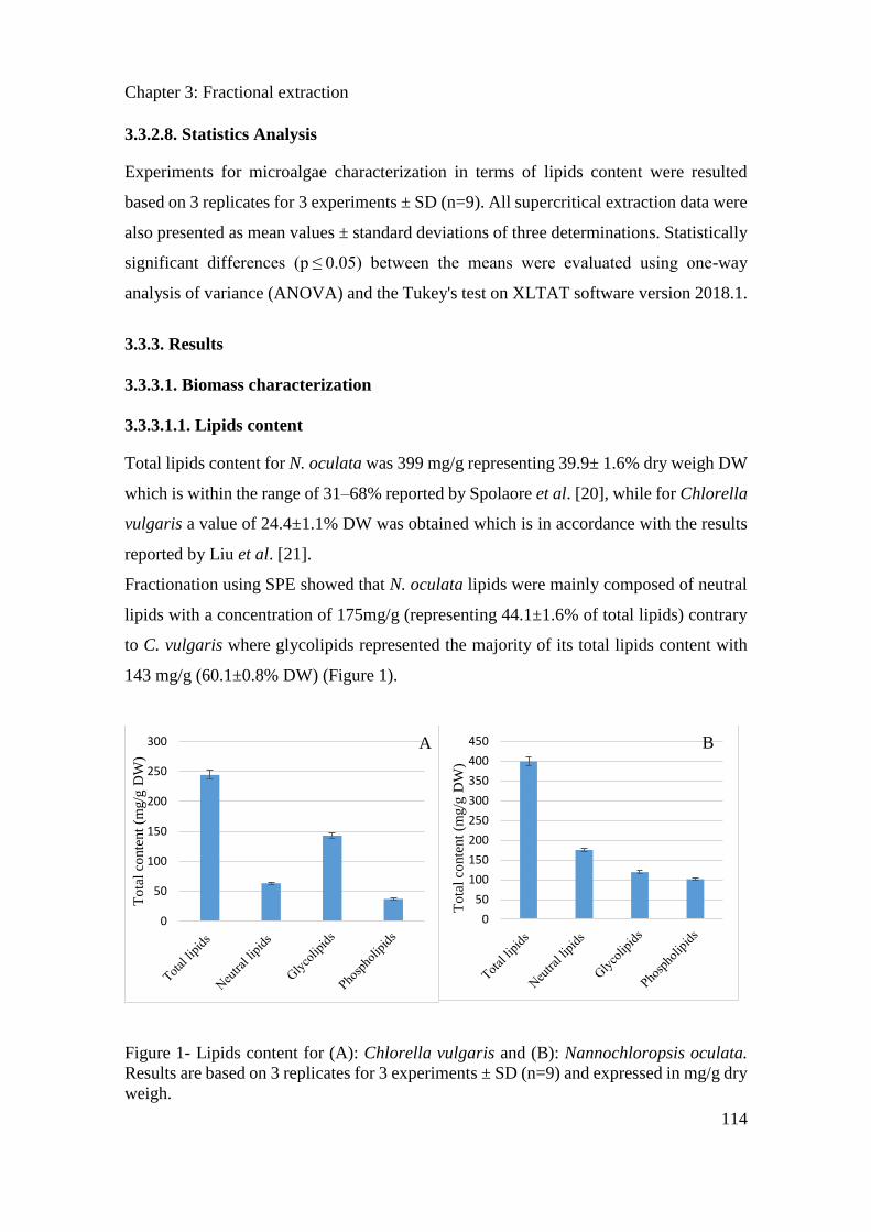
Chapter 3: Fractional extraction
114
3.3.2.8. Statistics Analysis
Experiments for microalgae characterization in terms of lipids content were resulted
based on 3 replicates for 3 experiments ± SD (n=9). All supercritical extraction data were
also presented as mean values ± standard deviations of three determinations. Statistically
significant differences (p ≤ 0.05) between the means were evaluated using one-way
analysis of variance (ANOVA) and the Tukey's test on XLTAT software version 2018.1.
3.3.3. Results
3.3.3.1. Biomass characterization
3.3.3.1.1. Lipids content
Total lipids content for N. oculata was 399 mg/g representing 39.9± 1.6% dry weigh DW
which is within the range of 31–68% reported by Spolaore et al. [20], while for Chlorella
vulgaris a value of 24.4±1.1% DW was obtained which is in accordance with the results
reported by Liu et al. [21].
Fractionation using SPE showed that N. oculata lipids were mainly composed of neutral
lipids with a concentration of 175mg/g (representing 44.1±1.6% of total lipids) contrary
to C. vulgaris where glycolipids represented the majority of its total lipids content with
143 mg/g (60.1±0.8% DW) (Figure 1).
Figure 1- Lipids content for (A): Chlorella vulgaris and (B): Nannochloropsis oculata.
Results are based on 3 replicates for 3 experiments ± SD (n=9) and expressed in mg/g dry
weigh.
0
50
100
150
200
250
300
To
tal
conte
nt
(mg/g
DW
)
0
50
100
150
200
250
300
350
400
450
To
tal
conte
nt
(mg/g
DW
)
A B
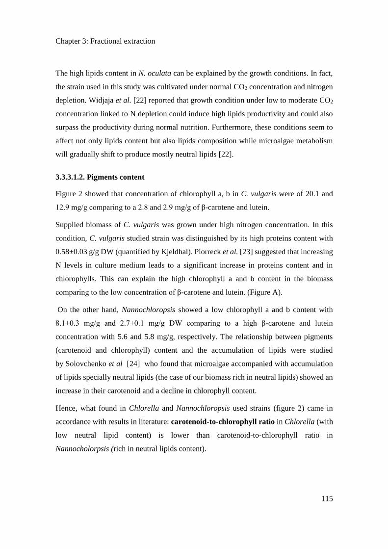
Chapter 3: Fractional extraction
115
The high lipids content in N. oculata can be explained by the growth conditions. In fact,
the strain used in this study was cultivated under normal CO2 concentration and nitrogen
depletion. Widjaja et al. [22] reported that growth condition under low to moderate CO2
concentration linked to N depletion could induce high lipids productivity and could also
surpass the productivity during normal nutrition. Furthermore, these conditions seem to
affect not only lipids content but also lipids composition while microalgae metabolism
will gradually shift to produce mostly neutral lipids [22].
3.3.3.1.2. Pigments content
Figure 2 showed that concentration of chlorophyll a, b in C. vulgaris were of 20.1 and
12.9 mg/g comparing to a 2.8 and 2.9 mg/g of β-carotene and lutein.
Supplied biomass of C. vulgaris was grown under high nitrogen concentration. In this
condition, C. vulgaris studied strain was distinguished by its high proteins content with
0.58±0.03 g/g DW (quantified by Kjeldhal). Piorreck et al. [23] suggested that increasing
N levels in culture medium leads to a significant increase in proteins content and in
chlorophylls. This can explain the high chlorophyll a and b content in the biomass
comparing to the low concentration of β-carotene and lutein. (Figure A).
On the other hand, Nannochloropsis showed a low chlorophyll a and b content with
8.1±0.3 mg/g and 2.7±0.1 mg/g DW comparing to a high β-carotene and lutein
concentration with 5.6 and 5.8 mg/g, respectively. The relationship between pigments
(carotenoid and chlorophyll) content and the accumulation of lipids were studied
by Solovchenko et al [24] who found that microalgae accompanied with accumulation
of lipids specially neutral lipids (the case of our biomass rich in neutral lipids) showed an
increase in their carotenoid and a decline in chlorophyll content.
Hence, what found in Chlorella and Nannochloropsis used strains (figure 2) came in
accordance with results in literature: carotenoid-to-chlorophyll ratio in Chlorella (with
low neutral lipid content) is lower than carotenoid-to-chlorophyll ratio in
Nannocholorpsis (rich in neutral lipids content).
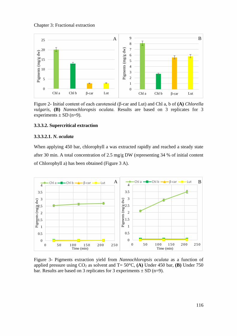
Chapter 3: Fractional extraction
116
Figure 2- Initial content of each carotenoid (β-car and Lut) and Chl a, b of (A) Chlorella
vulgaris, (B) Nannochloropsis oculata. Results are based on 3 replicates for 3
experiments ± SD (n=9).
3.3.3.2. Supercritical extraction
3.3.3.2.1. N. oculata
When applying 450 bar, chlorophyll a was extracted rapidly and reached a steady state
after 30 min. A total concentration of 2.5 mg/g DW (representing 34 % of initial content
of Chlorophyll a) has been obtained (Figure 3 A).
Figure 3- Pigments extraction yield from Nannochloropsis oculata as a function of
applied pressure using CO2 as solvent and T= 50°C, (A) Under 450 bar, (B) Under 750
bar. Results are based on 3 replicates for 3 experiments ± SD (n=9).
0
5
10
15
20
25
Chl a Chl b β-car Lut
Pig
men
ts (
mg/g
dw
)
0
1
2
3
4
5
6
7
8
9
Chl a Chl b β-car Lut
Pig
men
ts (
mg/g
dw
)
0
0.5
1
1.5
2
2.5
3
3.5
4
0 5 0 1 0 0 1 5 0 2 0 0 2 5 0
Pig
men
ts (
mg/g
dw
)
Time (min)
Chl a Chl b β-car Lut
0
0.5
1
1.5
2
2.5
3
3.5
4
0 5 0 1 0 0 1 5 0 2 0 0 2 5 0
Pig
men
ts (
mg/g
dw
)
Time (min)
Chl a Chl b β-car Lut
B A
A B
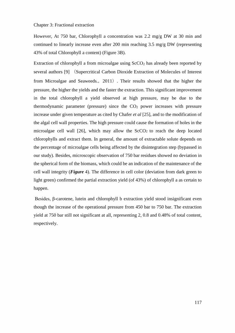
Chapter 3: Fractional extraction
117
However, At 750 bar, Chlorophyll a concentration was 2.2 mg/g DW at 30 min and
continued to linearly increase even after 200 min reaching 3.5 mg/g DW (representing
43% of total Chlorophyll a content) (Figure 3B).
Extraction of chlorophyll a from microalgae using ScCO2 has already been reported by
several authors [9] (Supercritical Carbon Dioxide Extraction of Molecules of Interest
from Microalgae and Seaweeds.,2011). Their results showed that the higher the
pressure, the higher the yields and the faster the extraction. This significant improvement
in the total chlorophyll a yield observed at high pressure, may be due to the
thermodynamic parameter (pressure) since the CO2 power increases with pressure
increase under given temperature as cited by Chafer et al [25], and to the modification of
the algal cell wall properties. The high pressure could cause the formation of holes in the
microalgae cell wall [26], which may allow the ScCO2 to reach the deep located
chlorophylls and extract them. In general, the amount of extractable solute depends on
the percentage of microalgae cells being affected by the disintegration step (bypassed in
our study). Besides, microscopic observation of 750 bar residues showed no deviation in
the spherical form of the biomass, which could be an indication of the maintenance of the
cell wall integrity (Figure 4). The difference in cell color (deviation from dark green to
light green) confirmed the partial extraction yield (of 43%) of chlorophyll a as certain to
happen.
Besides, β-carotene, lutein and chlorophyll b extraction yield stood insignificant even
though the increase of the operational pressure from 450 bar to 750 bar. The extraction
yield at 750 bar still not significant at all, representing 2, 0.8 and 0.48% of total content,
respectively.
Chapter 3: Fractional extraction
118
Figure 4 - Microscopic observation at high magnifications (x 1000) of Nannochloropsis
oculata cells after supercritical CO2 treatment. (A) Under 450 bar, (B) Under 750 bar.
3.3.3.2.2. Chlorella vulgaris
Two experiments were performed to study the influence of the solvent polarity on
pigments extraction yield: double and simple extractions. Double extraction (A) was
managed by injecting only CO2 at the beginning and then, after 210 minutes, a mixture of
CO2 and ethanol (10% v/v) are added. Simple extraction (B) was carried out by injecting
the mixture of ethanol and CO2 at the beginning of the experiment.
Under double extraction, during the initial phase, pigments concentration was so low.
Analysis showed that pigments were mainly constituted of lutein and -carotene. When
injecting ethanol (10%) with CO2 at t=210 min (Figure 5 A), the concentration
significantly increased (p<0.01) especially for chlorophyll a, with 1.1 mg/g DW obtained,
representing 6% of its initial content. Besides, lutein concentration increased after ethanol
injection from 170 μg /g dry to 385 μg /g DW (representing 8.1% of its total content).
Chlorophyll b extraction yield slightly increased to 0.02 mg/g (representing 0.17 % of its
total content). Whereas, -carotene extraction yield does not change after ethanol
injection.
B A
10 µm 10 µm
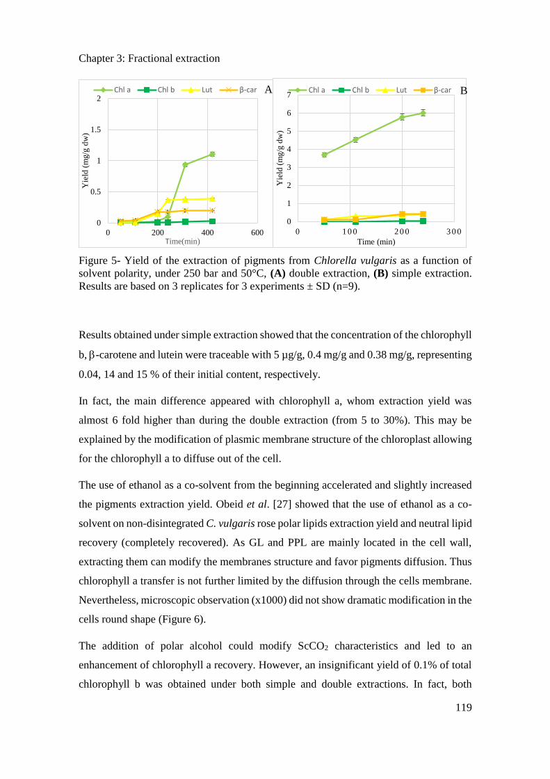
Chapter 3: Fractional extraction
119
Figure 5- Yield of the extraction of pigments from Chlorella vulgaris as a function of
solvent polarity, under 250 bar and 50°C, (A) double extraction, (B) simple extraction.
Results are based on 3 replicates for 3 experiments ± SD (n=9).
Results obtained under simple extraction showed that the concentration of the chlorophyll
b, -carotene and lutein were traceable with 5 µg/g, 0.4 mg/g and 0.38 mg/g, representing
0.04, 14 and 15 % of their initial content, respectively.
In fact, the main difference appeared with chlorophyll a, whom extraction yield was
almost 6 fold higher than during the double extraction (from 5 to 30%). This may be
explained by the modification of plasmic membrane structure of the chloroplast allowing
for the chlorophyll a to diffuse out of the cell.
The use of ethanol as a co-solvent from the beginning accelerated and slightly increased
the pigments extraction yield. Obeid et al. [27] showed that the use of ethanol as a co-
solvent on non-disintegrated C. vulgaris rose polar lipids extraction yield and neutral lipid
recovery (completely recovered). As GL and PPL are mainly located in the cell wall,
extracting them can modify the membranes structure and favor pigments diffusion. Thus
chlorophyll a transfer is not further limited by the diffusion through the cells membrane.
Nevertheless, microscopic observation (x1000) did not show dramatic modification in the
cells round shape (Figure 6).
The addition of polar alcohol could modify ScCO2 characteristics and led to an
enhancement of chlorophyll a recovery. However, an insignificant yield of 0.1% of total
chlorophyll b was obtained under both simple and double extractions. In fact, both
0
0.5
1
1.5
2
0 200 400 600
Yie
ld (
mg/g
dw
)
Time(min)
Chl a Chl b Lut β-car
0
1
2
3
4
5
6
7
0 1 0 0 2 0 0 3 0 0
Yie
ld (
mg/g
dw
)
Time (min)
Chl a Chl b Lut β-carA B
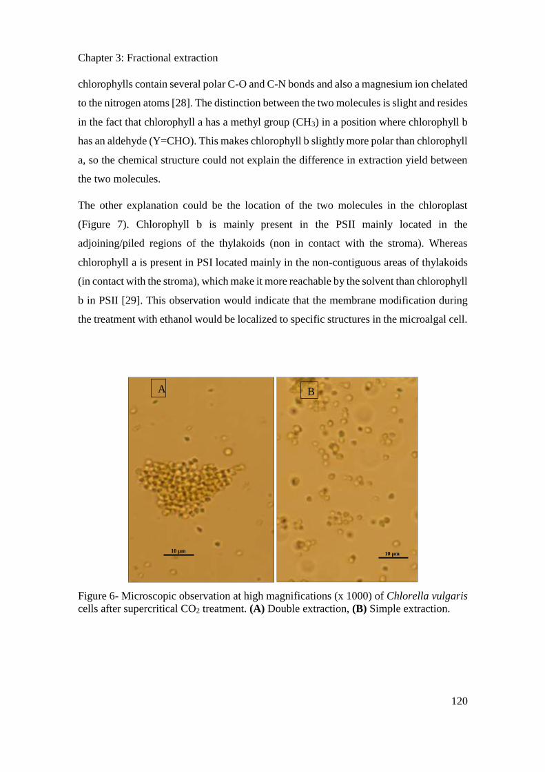
Chapter 3: Fractional extraction
120
chlorophylls contain several polar C-O and C-N bonds and also a magnesium ion chelated
to the nitrogen atoms [28]. The distinction between the two molecules is slight and resides
in the fact that chlorophyll a has a methyl group (CH3) in a position where chlorophyll b
has an aldehyde (Y=CHO). This makes chlorophyll b slightly more polar than chlorophyll
a, so the chemical structure could not explain the difference in extraction yield between
the two molecules.
The other explanation could be the location of the two molecules in the chloroplast
(Figure 7). Chlorophyll b is mainly present in the PSII mainly located in the
adjoining/piled regions of the thylakoids (non in contact with the stroma). Whereas
chlorophyll a is present in PSI located mainly in the non-contiguous areas of thylakoids
(in contact with the stroma), which make it more reachable by the solvent than chlorophyll
b in PSII [29]. This observation would indicate that the membrane modification during
the treatment with ethanol would be localized to specific structures in the microalgal cell.
Figure 6- Microscopic observation at high magnifications (x 1000) of Chlorella vulgaris
cells after supercritical CO2 treatment. (A) Double extraction, (B) Simple extraction.
A B
10 µm 10 µm
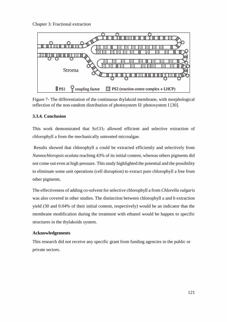
Chapter 3: Fractional extraction
121
Figure 7- The differentiation of the continuous thylakoid membrane, with morphological
reflection of the non-random distribution of photosystem II/ photosystem I [30].
3.3.4. Conclusion
This work demonstrated that ScCO2 allowed efficient and selective extraction of
chlorophyll a from the mechanically untreated microalgae.
Results showed that chlorophyll a could be extracted efficiently and selectively from
Nannochloropsis oculata reaching 43% of its initial content, whereas others pigments did
not come out even at high pressure. This study highlighted the potential and the possibility
to eliminate some unit operations (cell disruption) to extract pure chlorophyll a free from
other pigments.
The effectiveness of adding co-solvent for selective chlorophyll a from Chlorella vulgaris
was also covered in other studies. The distinction between chlorophyll a and b extraction
yield (30 and 0.04% of their initial content, respectively) would be an indicator that the
membrane modification during the treatment with ethanol would be happen to specific
structures in the thylakoids system.
Acknowledgements
This research did not receive any specific grant from funding agencies in the public or
private sectors.
Stroma
Chapter 3: Fractional extraction
122
3.3.5. References
[1] Nobre et al., “A biorefinery from Nannochloropsis sp. microalga – Extraction of oils
and pigments. Production of biohydrogen from the leftover biomass,” Bioresour.
Technol., vol. 135, pp. 128–136, May 2013.
[2] Wilhelm Stahl and Helmut Sies, “Bioactivity and protective effects of natural
carotenoids,” Biochim. Biophys. Acta BBA - Mol. Basis Dis., vol. 1740, no. 2, pp.
101–107, May 2005.
[3] J. A. Olson and N. I. Krinsky, “Introduction: the colorful, fascinating world of the
carotenoids: important physiologic modulators.,” FASEB J., vol. 9, no. 15, pp.
1547–1550, Dec. 1995.
[4] R. Harun, M. Singh, G. M. Forde, and M. K. Danquah, “Bioprocess engineering of
microalgae to produce a variety of consumer products,” Renew. Sustain. Energy
Rev., vol. 14, no. 3, pp. 1037–1047, Apr. 2010.
[5] Petra Amchova, Hana Kotolova, Jana Ruda-Kucerova, “Health safety issues of
synthetic food colorants,” Regul. Toxicol. Pharmacol., vol. 73, no. 3, pp. 914–922,
Dec. 2015.
[6] S. B. Hawthorne, “Analytical-scale supercritical fluid extraction,” 06-Jun-2012.
[Online]. Available: https://pubs.acs.org/doi/abs/10.1021/ac00210a722. [Accessed:
22-Aug-2018].
[7] J. P. Friedrich and E. H. Pryde, “Supercritical CO2 extraction of lipid-bearing
materials and characterization of the products,” J. Am. Oil Chem. Soc., vol. 61, no.
2, pp. 223–228, Feb. 1984.
[8] Victor Abrahamsson, Firas Jumaah, Charlotta Turner, “Continuous multicomponent
quantification during supercritical fluid extraction applied to microalgae using in-
line UV/Vis absorption spectroscopy and on-line evaporative light scattering
detection,” J. Supercrit. Fluids, vol. 131, pp. 157–165, Jan. 2018.
[9] M.D. Macı́as-Sánchez et al, “Supercritical fluid extraction of carotenoids and
chlorophyll a from Nannochloropsis gaditana,” J. Food Eng., vol. 66, no. 2, pp. 245–
251, Jan. 2005.
[10] L. Gouveia and A. C. Oliveira, “Microalgae as a raw material for biofuels
production,” J. Ind. Microbiol. Biotechnol., vol. 36, no. 2, pp. 269–274, Feb. 2009.
[11] Reyes et al., “Astaxanthin extraction from Haematococcus pluvialis using CO2-
expanded ethanol,” J. Supercrit. Fluids, vol. 92, pp. 75–83, Aug. 2014.
[12] K. Ramluckan, K. G. Moodley, and F. Bux, “An evaluation of the efficacy of using
selected solvents for the extraction of lipids from algal biomass by the soxhlet
extraction method,” Fuel, vol. 116, pp. 103–108, Jan. 2014.
[13] J. Jeon et al., “New mixture composition of organic solvents for efficient extraction
of lipids from Chlorella vulgaris,” Biomass Bioenergy, vol. 59, pp. 279–284, Dec.
2013.
[14] Y. Du, B. Schuur, S. R. A. Kersten, and D. W. F. Brilman, “Opportunities for
switchable solvents for lipid extraction from wet algal biomass: An energy
evaluation,” Algal Res., vol. 11, pp. 271–283, Sep. 2015.
[15] P. Juaneda and G. Rocquelin, “Rapid and convenient separation of phospholipids
and non phosphorus lipids from rat heart using silica cartridges,” Lipids, vol. 20, no.
1, pp. 40–41.
Chapter 3: Fractional extraction
123
[16] R. R. Bidigare, “Analysis of Algal Chlorophylls and Carotenoids,” in Marine
Particles: Analysis and Characterization, American Geophysical Union (AGU),
2013, pp. 119–123.
[17] O. Holm-Hansen and B. Riemann, “Chlorophyll a Determination: Improvements in
Methodology,” Oikos, vol. 30, no. 3, pp. 438–447, 1978.
[18] V. Brotas and M.-R. Plante-Cuny, “Identification and quantification of chlorophyll
and carotenoid pigments in marine sediments. A protocol for HPLC analysis.,”
Oceanol. Acta Paris, vol. 19, no. 6, pp. 623–634, 1996.
[19] T. W. Goodwin, “Biosynthesis of Carotenoids,” in The Biochemistry of the
Carotenoids, Springer, Dordrecht, 1980, pp. 33–76.
[20] P. Spolaore, C. Joannis-Cassan, E. Duran, and A. Isambert, “Commercial
applications of microalgae,” J. Biosci. Bioeng., vol. 101, no. 2, pp. 87–96, Feb. 2006.
[21] Z.-Y. Liu, G.-C. Wang, and B.-C. Zhou, “Effect of iron on growth and lipid
accumulation in Chlorella vulgaris,” Bioresour. Technol., vol. 99, no. 11, pp. 4717–
4722, Jul. 2008.
[22] A. Widjaja, C.-C. Chien, and Y.-H. Ju, “Study of increasing lipid production from
fresh water microalgae Chlorella vulgaris,” J. Taiwan Inst. Chem. Eng., vol. 40, no.
1, pp. 13–20, Jan. 2009.
[23] M. Piorreck, K.-H. Baasch, and P. Pohl, “Biomass production, total protein,
chlorophylls, lipids and fatty acids of freshwater green and blue-green algae under
different nitrogen regimes,” Phytochemistry, vol. 23, no. 2, pp. 207–216, Jan. 1984.
[24] A. E. Solovchenko, I. Khozin-Goldberg, Z. Cohen, and M. N. Merzlyak,
“Carotenoid-to-chlorophyll ratio as a proxy for assay of total fatty acids and
arachidonic acid content in the green microalga Parietochloris incisa,” J. Appl.
Phycol., vol. 21, no. 3, pp. 361–366, Jun. 2009.
[25] A. Cháfer et al, “High-pressure solubility data of system ethanol (1)+epicatechin
(2)+CO2 (3),” J. Supercrit. Fluids, vol. 24, no. 2, pp. 103–109, Nov. 2002.
[26] M. Solana, C. S. Rizza, and A. Bertucco, “Exploiting microalgae as a source of
essential fatty acids by supercritical fluid extraction of lipids: Comparison between
Scenedesmus obliquus, Chlorella protothecoides and Nannochloropsis salina,” J.
Supercrit. Fluids, vol. 92, pp. 311–318, Aug. 2014.
[27] Obeid et al., “Supercritical carbon dioxide extraction and fractionation of lipids from
freeze-dried microalgae Nannochloropsis oculata and Chlorella vulgaris,” Algal
Res., vol. 34, pp. 49–56, Sep. 2018.
[28] “Spinach_Pigments handout_Spring 2016 (1) - Isolation of Chlorophyll and
Carotenoid Pigments from Spinach Adapted from Pavia D.L Lampman G.M Kriz
G.S and.” [Online]. Available:
https://www.coursehero.com/file/17807812/Spinach-Pigments-handout-Spring-
2016-1/. [Accessed: 29-Aug-2018].
[29] R. Bassi, F. Rigoni, and G. M. Giacometti, “chlorophyll binding proteins with
antenna function in higher plants and green algae,” photochem. photobiol., vol. 52,
no. 6, pp. 1187–1206, dec. 1990.
[30] J. M. Anderson, “Lateral heterogeneity of plant thylakoid protein complexes: early
reminiscences,” Philos. Trans. R. Soc. Lond. B Biol. Sci., vol. 367, no. 1608, pp.
3384–3388, Dec. 2012.
Chapter 3: Fractional extraction
124
3.4. Extraction and purification of soluble proteins from
Nannochlororopsis oculata by pH-shifting and ultrafiltration
(Article submitted to Biotechnology and Bioengineering Journal)
Sara Obeida, b, c, 1, Hosni Takache b, c, Nicholas Beaufils a, Séverine Camy d, Pierre-
Yves Pontalier a, Ali Ismail b, c
a Laboratoire de Chimie Agro-industrielle (LCA), Université de Toulouse, INRA, INPT,
Toulouse, France 4 allée Emile Monso, 31030 Toulouse cedex 4, France b Département des Sciences et Technologies Alimentaires, Faculté des Sciences Agronomiques,
Université Libanaise, Allée Georges Matta, 1500, Hadath, Liban. c Université Libanaise, Ecole Doctorale de Sciences et Technologies EDST-PRASE (Plateforme
de Recherches et Analyses en Sciences de l’Environnement), Campus Rafic Hariri, 1500,
Hadath, Liban. d Université de Toulouse, INP-ENSIACET, LGC (Laboratoire de Génie Chimique), 4 allée Emile
Monso, 31030 Toulouse cedex 4, France
Abstract
A mild biorefinery process was investigated to obtain purified enriched fraction of water
soluble proteins from Nannochloropsis oculata without prior cell disruption step.
Solubilization of proteins has been achieved under double stage extraction from defatted,
semi defatted and non-defatted biomasses, in aim to understand the effect of defatting
degree and pH shifting on proteins extraction yield. 73.7% of total proteins were released
using a combination of aqueous/alkaline condition from defatted biomass. A dead-end
ultrafiltration was then performed on the supernatants obtained by testing different
membrane cut-off (100, 30, 10 kDa). Enriched fraction of water soluble proteins (83%)
free from lipids and pigments was obtained using 100 kDa membrane.
Keywords:
Microalgae; N. oculata; extraction; pH shifting; purification; ultrafiltration.
Chapter 3: Fractional extraction
125
3.4.1. Introduction
Microalgae biomass have been excessively exploited during the last decades regarding
their high lipids content for biodiesel production as a substitute for fossil oil [1]. Hence,
many studies were focused on lipids, neglecting the potential of other high-value
components like proteins. Aside from biofuels and energy sectors, through time, interest
in this biomass has been taking a new course especially for its nutritional values. To
satisfy the increase on proteins demand as a consequence of rapid world population
growth, microalgae became an important proteins source seeing its high proteins content
reaching up to 71% dry weight (DW) [2] [3]. Microalgal proteins can be used to enhance
the nutritional value of animal feed owing to their chemical composition [4]. They also
play a crucial role in aquaculture and they can be incorporated into cosmetics [5].
Among numerous microalgae species, Nannochloropsis oculata is one of the few species
permitted to be used as a source of proteins for food or feed formulations [6]. In order to
improve the nutritional value of proteins fraction, it should be isolated from the rest of
biomass components. However, proteins extraction from N. oculata is often prevented by
the intrinsic rigidity of its cell wall mainly composed of cellulose and hemicellulose [7].
To overcome this barrier, cell disruption should be used to facilitate proteins extraction
[8]. Numerous disruption methods have been investigated involving ultra-sonication,
high pressure homogenization, bead-beating, blender, manual grinding, etc. [9] [8]. Gerde
et al. [6] obtained 30% of total proteins content from Nannochloropsis spp. using
ultrasound. Safi et al. [10], [11] obtained 49-52.8% of total proteins content from N.
oculata using high-pressure homogenization. Furthermore, Cavonuis et al. [12] studied
proteins extraction by pH-shift process on N. oculata where cells disruption was carried
out with glass beads. As a result, up to 85% of proteins and total fatty acids were
recovered in the final product.
The main drawbacks in all works cited above could be resumed by: (i) the high cost of
cell disintegration process (Growth aspects of the marine microalga Nannochloropsis
gaditana. ,2003), (ii) the non-selectivity of the extraction and co-release of undesirable
components, (iii) the difficulty in the downstream separation processes due to the
micronization of cell debris [13] [14], (iv) the denaturation of heat sensitive proteins
[15].
Chapter 3: Fractional extraction
126
Once extraction is done, it is necessary to establish a process for proteins
fractionation/concentration. Many studies have tested the separation of microalgae
proteins by pH shifting [12], three phase partitioning [16] and aqueous two-phase
extraction [17]. Nevertheless, these methods required large amount of solvents and other
chemicals especially at industrial scale. Contrary to other separation techniques,
membrane technology process is isothermal, involves no phase change or chemical agents
[16], and it is considered as relatively cheap compared to other separation techniques [18].
However, membrane separation technology is not well explored and remains not mature
for microalgae applications [11]. Few examples have been reported about the use of
membrane process for the purification or concentration of microalgal components:
polysaccharides from Porphyridium cruentum [19][20], Spirulina platensis and Chlorella
pyrenoidosa [21], proteins from Chlorella vulgaris and Haematococcus pluvialis [22]
[23].
The application of membrane process such as ultrafiltration is largely used at industrial
level [24] mainly in the dairy industry. However, this technology has not been
investigated for the separation/purification of microalgal proteins, despite this technique
could offer some sources of specific industrial interest (efficiency and facility of
exploitation)
The goal of the present work is to study the extraction/purification of proteins from N.
oculata while maintaining the integrity of the cell wall and minimizing as possible the
fractionation steps. Extraction has been achieved by double stage and pH shifting on
defatted, non-defatted and partially defatted biomass to understand the effect of lipids
content on proteins release. Besides, proteins rich fraction have been subjected to further
purification by semi-frontal ultrafiltration. Size-exclusion chromatography (SEC)
analysis has been used for proteins extracts characterization.
3.4.2. Materials and methods
3.4.2.1. Microalga
Nannochloropsis oculata were provided freeze-dried by Alpha Biotech (Assérac,
France). After being harvested during the exponential growth phase, the biomass was
concentrated by centrifugation to obtain a paste of 20-24% DW. The frozen paste was
Chapter 3: Fractional extraction
127
then freeze-dried in a Fisher Bioblock Scientific Alpha 2–4 LD Plus device (Illkirch,
France). During freeze drying process, the pressure was reduced to 0.010 bar, the
temperature further decreased to -80°C, for 48 h to give a completely dry biomass. Freeze-
dried aggregates were then slightly spread with a laboratory spatula. The mean diameter
of particles was measured using Mastersizer 2000 granulometer (Malvern Instruments
Ltd.) showing a value around 250 μm.
3.4.2.2. Chemicals
The Lowry kit (Lowry reagent plus bovine standard albumin (BSA) standards and 2 N
Folin–Ciocalteu reagents) was purchased from Thermo Fisher Scientific.
3.4.2.3. Proteins content calculation
Quantitation of total proteins content of the biomass was performed according to
Kjeldahl analysis [25] which take into account the totality of the nitrogen present in the
biomass, from which 59–98 % of total nitrogen (TN) can belong to proteins [26] [27].
After evaluating the nitrogen content, the proteins content is estimated by multiplying
Kjeldahl TN by NTP (nitrogen-to-protein) factor. In case of N. oculata, NTP conversion
factor is estimated to be 6.28 [28]. Biomass was analyzed in triplicate, using each time
200 mg samples of dry biomass.
Water-soluble proteins were quantified by Lowry method [29] using a standard curve
of bovine serum albumin (BSA). This method is based on a reaction catalyzed by the
copper content in the Folin and oxidation of the peptide bonds with a Folin-Ciocalteu
reagent [30]. Absorbance was measured at 750 nm using a Shimadzu UV-1700
spectrophotometer.
3.4.2.4. Biomass defatting
Proteins extraction has been performed on the freeze-dried biomass, partially defatted
biomass and defatted biomass.
Two methods of partial defatting have been used: A) supercritical carbon dioxide (ScCO2)
extraction, B) Soxhlet extraction,
Chapter 3: Fractional extraction
128
A) ScCO2 were carried out on an SFE100 apparatus supplied from Separex (Nancy,
France). The pilot was composed of a 25 cm3 autoclave. This unit is able to work at
pressures up to 1000 bar and temperatures up to 250°C. At the beginning of the
experiment, the extractor was filled with powdered freeze-dried microalgae. The module
was supplied with liquid CO2 through a pump maintained at a temperature below 7°C to
keep the CO2 in the liquid state. The operating conditions are presented in table 1.
B) Soxhlet apparatus using a slightly modified method of Blight & Dyer (1959) (A rapid
method of total lipid extractionand purification.,1959) using mixture of polar and non-
polar solvent [31] [32] chloroform / methanol (0.35 / 0.65, v/v). The microalgal biomass
(3 g) was placed in a cartridge filter paper using 200 mL of solvent. The Soxhlet extractor
was operated for a number of cycles for 2 and 6 hours for partial lipid extraction.
Table 1- Various ScCO2 operting conditions applied on freeze-dried biomass of
N.oculata. All experiments were performed under a constant temperature of 50ºC and a
constant CO2 flow rate of 25 g/min.
Defatted biomass was obtained using the Soxhlet method described above but operated
for 18 hours in order to insure the extraction of total lipids [33].
3.4.2.5. Proteins extraction by pH shifting
Double stage proteins extraction have been performed using aqueous medium at two pH:
neutral (pH 7) and alkaline (pH 11). Stock solutions were prepared with ultrapure water.
The pH was adjusted using 1N NaOH solution. All the conditions are summarized in
Table 2. In order to limit proteins damage during extraction, temperature was maintained
at 25°C. After extraction, the microalgae suspensions were centrifuged at 20,000 g for 15
min at 5°C. Protein-rich supernatants were taken for analysis. It should be emphasized
that no cell lysis has been conducted before proteins extractions.
Pressure
(bar)
Biomass
(g)
Solvent rate
(min-1)
250 25.4 1.02
450 8.8 0.35
750 8.8 0.35
Chapter 3: Fractional extraction
129
Table 2- Operating conditions for double extraction on non-defatted, partially defatted
and defatted N. oculata.
In order to define proteins solubility and efficiency of the two stage of double extraction
process, partition-coefficient (P) or distribution-coefficient is calculated. The proteins
concentration in supernatant 1 and supernatant 2 was divided by the proteins
concentration presented in the residue from the first extraction step (R1) and the residue
form the second extraction step (R2) respectively, expressed in percent [12]:
𝑃1= [𝑃𝑟𝑜𝑡𝑒𝑖𝑛 𝑆1]
[𝑃𝑟𝑜𝑡𝑒𝑖𝑛 𝑅1]∗ 100 𝑃2 =
[𝑃𝑟𝑜𝑡𝑒𝑖𝑛 𝑆2]
[𝑃𝑟𝑜𝑡𝑒𝑖𝑛 𝑅2]∗ 100
3.4.2.6. Proteins molecular size determination
In order to determine the molecular weight of extracted proteins, gel electrophoresis and
size exclusion chromatography were used. Molecular weight distribution of soluble
proteins was estimated through size exclusion chromatography using a capillary HPLC
system Dionex UltiMate™ (Thermo Scientific, Massachusetts, USA), equipped with a
UV / VIS detector.
Experiment
number
Defatting
mode
Extraction
stage pH
Ratio
Liquid/Solid
Duration
(hours)
1 ScCO2 1 7 20 20
2 7 10 1
2 ScCO2 1 7 20 20
2 11 10 1
3 ScCO2 1 7 20 5
2 7 10 5
4 ScCO2 1 11 20 5
2 7 10 5
5 Soxhlet 2 h 1 7 20 20
2 7 10 1
6 Soxhlet 6 h 1 7 20 20
2 7 10 1
7 Soxhlet 18 h 1 7 20 20
2 7 10 1
8 -
-
1 7 20 20
2 7 10 1
Chapter 3: Fractional extraction
130
Proteins samples filtered to 0.2 microns were loaded on Agilent column (SEC-3, 100Å,
7.8 x 300 mm) with a flow rate of 1.2 ml / min of a solution (0.1 M phosphate and 0.15M
NaCl). The column was calibrated with a standard mixture of high molecular weight
proteins (Bio-Rad Laboratories) containing thyroglobulin (670 kDa), c-globulin (158
kDa), ovalbumin (44 kDa), myoglobin (17 kDa) and vitamin B12 (1.35 kDa). The eluted
proteins were detected at 220 and 280 nm.
3.4.2.7. Proteins purification
To fulfil the purification operation, protein-rich supernatants from both stages were
collected and mixed together, then underwent a semi-frontal ultrafiltration using
Amicon® Stirred Cells size 200 mL. Membranes with a molecular weight of 10, 30 and
100 kDa cutoff and 30.17 cm2 filtration area were used, under a transmembrane pressure
(TMP) from 0.5 to 4 bar. A stirring of 100 rpm was applied over the membrane to
minimize concentration polarization effect due to the densification of macromolecules at
the membrane surface as reported by Burba et al. [34] Before ultrafiltration experiments,
semi-frontal ultrafiltration membranes were floated with distilled water for at least 1 hour.
After being used, all membranes were immersed in 0.1 M NaOH for 1 hour, then washed
and stood in Milli-Q water for 24 h before their storage at 5ºC in ethanol/water (1:9)
mixtures.
3.4.2.8. Statistical analysis
All treatments and analyses were run in triplicate. Statistical analyses were carried out
using Minitab 17 software. ANOVA and Tukey test were carried out and measurements
of triplicate for each sample were reproducible for ±5% of the respective mean values.
3.4.3. Results and discussion
Total proteins content of the initial biomass were evaluated at 22% DW. This result is in
accordance with what is usually described for N. oculata proteins content [12].
3.4.3.1. Effect of extraction time
Proteins extraction test was realized by placing the partially defatted N. oculata biomass
(by ScCO2) in contact with water for 20 h, liquid/solid ratio of 20 and T=20°C. Results
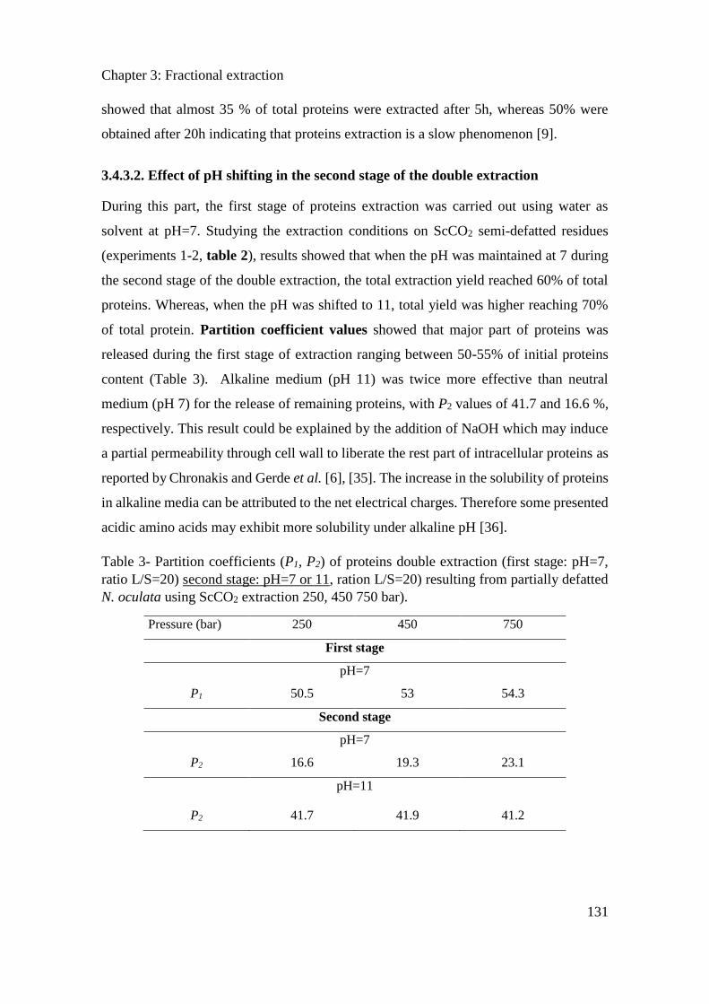
Chapter 3: Fractional extraction
131
showed that almost 35 % of total proteins were extracted after 5h, whereas 50% were
obtained after 20h indicating that proteins extraction is a slow phenomenon [9].
3.4.3.2. Effect of pH shifting in the second stage of the double extraction
During this part, the first stage of proteins extraction was carried out using water as
solvent at pH=7. Studying the extraction conditions on ScCO2 semi-defatted residues
(experiments 1-2, table 2), results showed that when the pH was maintained at 7 during
the second stage of the double extraction, the total extraction yield reached 60% of total
proteins. Whereas, when the pH was shifted to 11, total yield was higher reaching 70%
of total protein. Partition coefficient values showed that major part of proteins was
released during the first stage of extraction ranging between 50-55% of initial proteins
content (Table 3). Alkaline medium (pH 11) was twice more effective than neutral
medium (pH 7) for the release of remaining proteins, with P2 values of 41.7 and 16.6 %,
respectively. This result could be explained by the addition of NaOH which may induce
a partial permeability through cell wall to liberate the rest part of intracellular proteins as
reported by Chronakis and Gerde et al. [6], [35]. The increase in the solubility of proteins
in alkaline media can be attributed to the net electrical charges. Therefore some presented
acidic amino acids may exhibit more solubility under alkaline pH [36].
Table 3- Partition coefficients (P1, P2) of proteins double extraction (first stage: pH=7,
ratio L/S=20) second stage: pH=7 or 11, ration L/S=20) resulting from partially defatted
N. oculata using ScCO2 extraction 250, 450 750 bar).
Pressure (bar) 250 450 750
First stage
pH=7
P1 50.5 53 54.3
Second stage
pH=7
P2 16.6 19.3 23.1
pH=11
P2 41.7 41.9 41.2
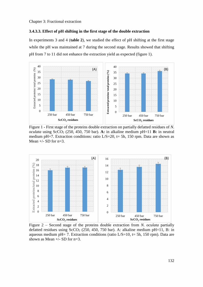
Chapter 3: Fractional extraction
132
3.4.3.3. Effect of pH shifting in the first stage of the double extraction
In experiments 3 and 4 (table 2), we studied the effect of pH shifting at the first stage
while the pH was maintained at 7 during the second stage. Results showed that shifting
pH from 7 to 11 did not enhance the extraction yield as expected (figure 1).
Figure 1 - First stage of the proteins double extraction on partially defatted residues of N.
oculata using ScCO2 (250, 450, 750 bar). A: in alkaline medium pH=11 B: in neutral
medium pH=7. Extraction conditions: ratio L/S=20, t= 5h, 150 rpm. Data are shown as
Mean +/- SD for n=3.
Figure 2 – Second stage of the proteins double extraction from N. oculata partially
defatted residues using ScCO2 (250, 450, 750 bar). A: alkaline medium pH=11, B: in
aqueous medium pH= 7. Extraction conditions (ratio L/S=10, t= 5h, 150 rpm). Data are
shown as Mean +/- SD for n=3.
0
5
10
15
20
25
30
35
40
250 bar 450 bar 750 bar
Extr
acte
d p
rote
ins/
tota
l p
rote
ins
(%
)
ScCO2 residues
(A)
0
5
10
15
20
25
30
35
40
250 bar 450 bar 750 bar
Extr
acte
d p
ro
tein
s/ t
ota
l p
ro
tein
s (%
)
ScCO2 residues
(B)
0
2
4
6
8
10
12
14
16
18
20
250 bar 450 bar 750 bar
Extr
acte
d p
rote
ins/
tota
l p
rote
ins
(%)
ScCO2 residues
(A)
0
2
4
6
8
10
12
14
16
250 bar 450 bar 750 bar
ScCO2 residues
(B)
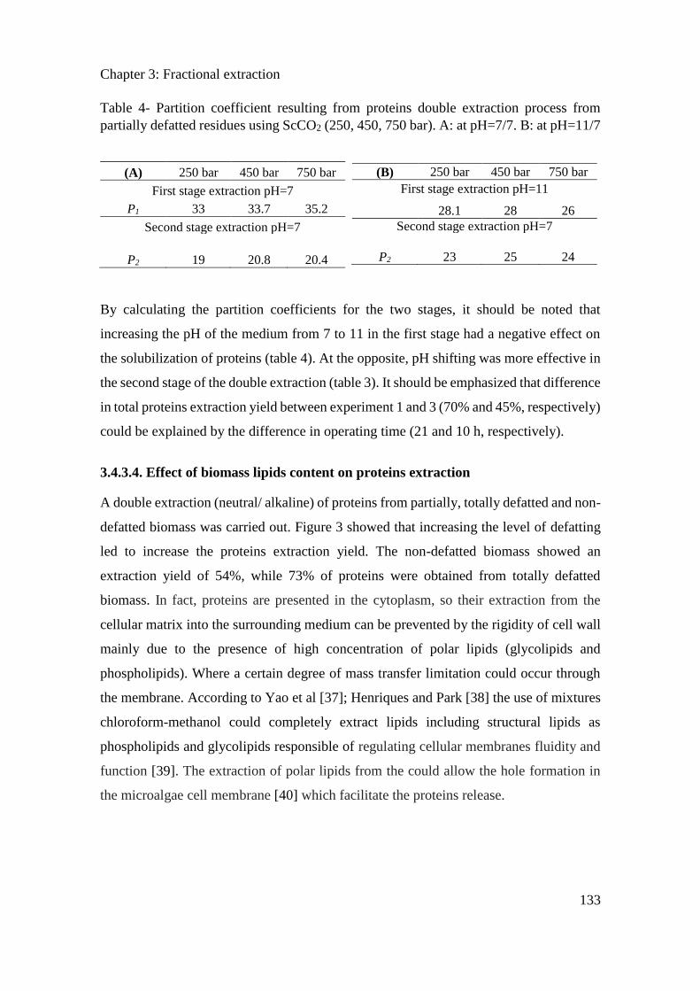
Chapter 3: Fractional extraction
133
Table 4- Partition coefficient resulting from proteins double extraction process from
partially defatted residues using ScCO2 (250, 450, 750 bar). A: at pH=7/7. B: at pH=11/7
By calculating the partition coefficients for the two stages, it should be noted that
increasing the pH of the medium from 7 to 11 in the first stage had a negative effect on
the solubilization of proteins (table 4). At the opposite, pH shifting was more effective in
the second stage of the double extraction (table 3). It should be emphasized that difference
in total proteins extraction yield between experiment 1 and 3 (70% and 45%, respectively)
could be explained by the difference in operating time (21 and 10 h, respectively).
3.4.3.4. Effect of biomass lipids content on proteins extraction
A double extraction (neutral/ alkaline) of proteins from partially, totally defatted and non-
defatted biomass was carried out. Figure 3 showed that increasing the level of defatting
led to increase the proteins extraction yield. The non-defatted biomass showed an
extraction yield of 54%, while 73% of proteins were obtained from totally defatted
biomass. In fact, proteins are presented in the cytoplasm, so their extraction from the
cellular matrix into the surrounding medium can be prevented by the rigidity of cell wall
mainly due to the presence of high concentration of polar lipids (glycolipids and
phospholipids). Where a certain degree of mass transfer limitation could occur through
the membrane. According to Yao et al [37]; Henriques and Park [38] the use of mixtures
chloroform-methanol could completely extract lipids including structural lipids as
phospholipids and glycolipids responsible of regulating cellular membranes fluidity and
function [39]. The extraction of polar lipids from the could allow the hole formation in
the microalgae cell membrane [40] which facilitate the proteins release.
(A) 250 bar 450 bar 750 bar
First stage extraction pH=7
P1 33 33.7 35.2
Second stage extraction pH=7
P2 19 20.8 20.4
(B) 250 bar 450 bar 750 bar
First stage extraction pH=11
28.1 28 26
Second stage extraction pH=7
P2 23 25 24
(A)
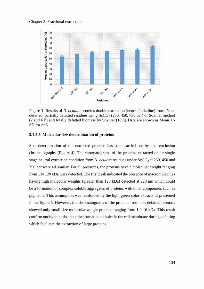
Chapter 3: Fractional extraction
134
Figure 3- Results of N. oculata proteins double extraction (neutral/ alkaline) from: Non-
defatted; partially defatted residues using ScCO2 (250, 450, 750 bar) or Soxhlet method
(2 and 6 h) and totally defatted biomass by Soxhlet (18 h). Data are shown as Mean +/-
SD for n=3.
3.4.3.5. Molecular size determination of proteins
Size determination of the extracted proteins has been carried out by size exclusion
chromatography (Figure 4). The chromatograms of the proteins extracted under single
stage neutral extraction condition from N. oculata residues under ScCO2 at 250, 450 and
750 bar were all similar. For all pressures, the proteins have a molecular weight ranging
from 1 to 120 kDa were detected. The first peak indicated the presence of macromolecules
having high molecular weights (greater than 120 kDa) detected at 220 nm which could
be a formation of complex soluble aggregates of proteins with other compounds such as
pigments. This assumption was reinforced by the light green color extracts as presented
in the figure 5. However, the chromatograms of the proteins from non-defatted biomass
showed only small size molecular weight proteins ranging from 1.0-16 kDa. This result
confirm our hypothesis about the formation of holes in the cell membrane during defatting
which facilitate the extraction of large proteins.
0
10
20
30
40
50
60
70
80
90
100
Pro
tein
s e
xtra
cte
d/
Tota
l pro
tein
s (%
)
Residues
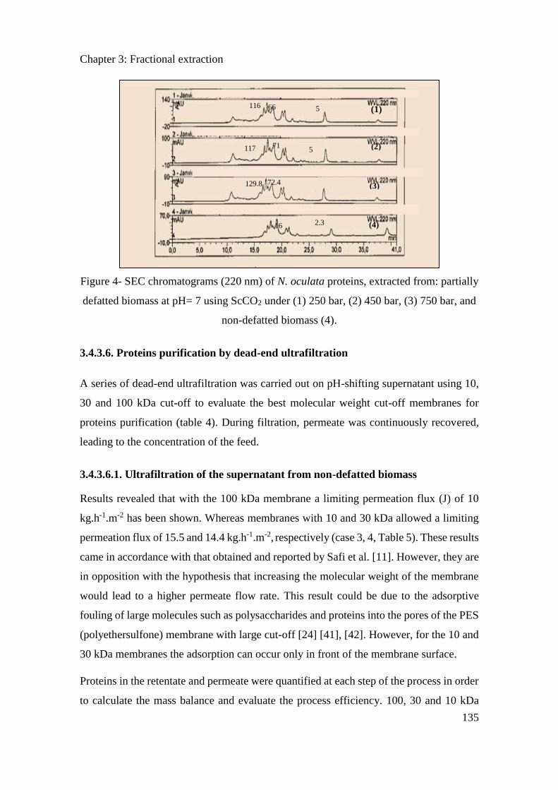
Chapter 3: Fractional extraction
135
Figure 4- SEC chromatograms (220 nm) of N. oculata proteins, extracted from: partially
defatted biomass at pH= 7 using ScCO2 under (1) 250 bar, (2) 450 bar, (3) 750 bar, and
non-defatted biomass (4).
3.4.3.6. Proteins purification by dead-end ultrafiltration
A series of dead-end ultrafiltration was carried out on pH-shifting supernatant using 10,
30 and 100 kDa cut-off to evaluate the best molecular weight cut-off membranes for
proteins purification (table 4). During filtration, permeate was continuously recovered,
leading to the concentration of the feed.
3.4.3.6.1. Ultrafiltration of the supernatant from non-defatted biomass
Results revealed that with the 100 kDa membrane a limiting permeation flux (J) of 10
kg.h-1.m-2 has been shown. Whereas membranes with 10 and 30 kDa allowed a limiting
permeation flux of 15.5 and 14.4 kg.h-1.m-2, respectively (case 3, 4, Table 5). These results
came in accordance with that obtained and reported by Safi et al. [11]. However, they are
in opposition with the hypothesis that increasing the molecular weight of the membrane
would lead to a higher permeate flow rate. This result could be due to the adsorptive
fouling of large molecules such as polysaccharides and proteins into the pores of the PES
(polyethersulfone) membrane with large cut-off [24] [41], [42]. However, for the 10 and
30 kDa membranes the adsorption can occur only in front of the membrane surface.
Proteins in the retentate and permeate were quantified at each step of the process in order
to calculate the mass balance and evaluate the process efficiency. 100, 30 and 10 kDa
5
72.4
16
117 71
129.8
116 (1) 66
5
2.3
(2)
(3)
(4)
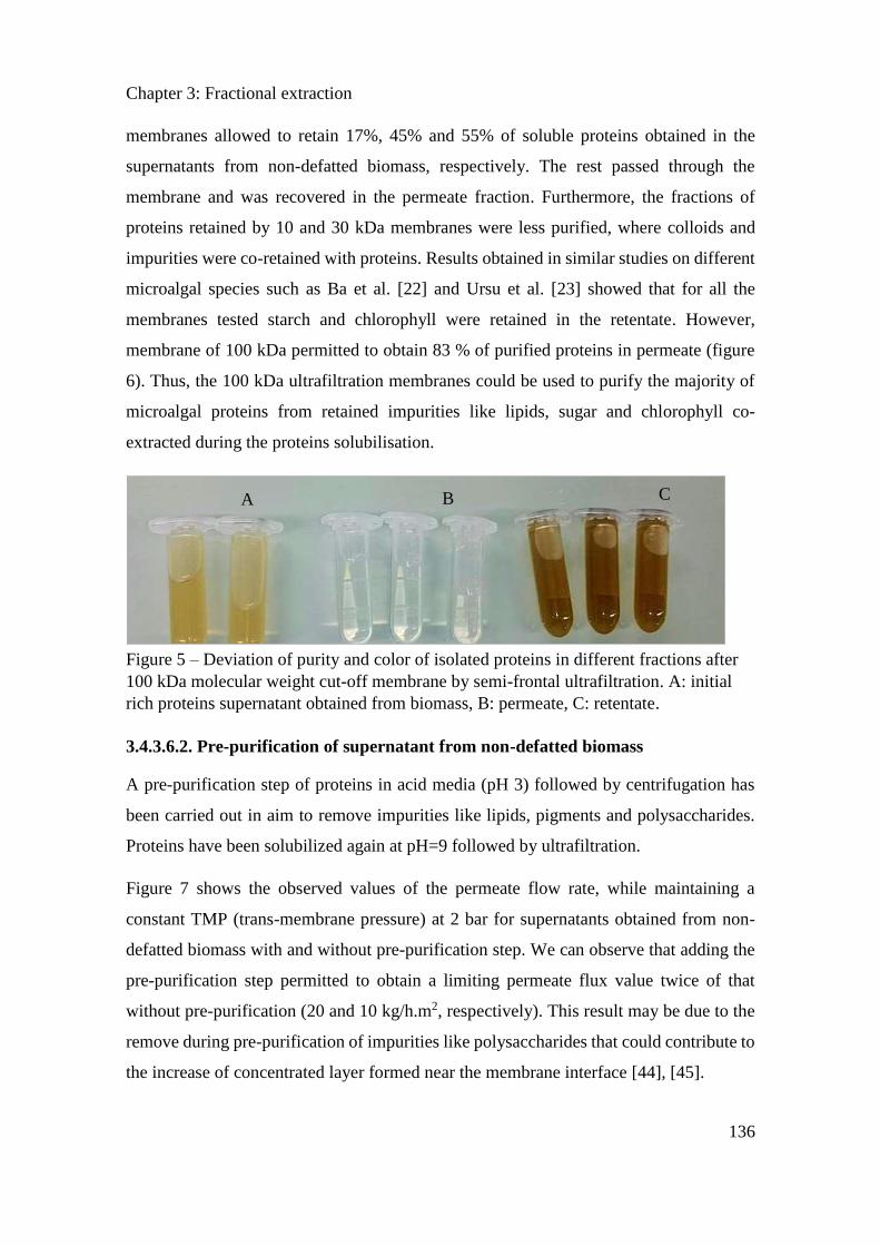
Chapter 3: Fractional extraction
136
membranes allowed to retain 17%, 45% and 55% of soluble proteins obtained in the
supernatants from non-defatted biomass, respectively. The rest passed through the
membrane and was recovered in the permeate fraction. Furthermore, the fractions of
proteins retained by 10 and 30 kDa membranes were less purified, where colloids and
impurities were co-retained with proteins. Results obtained in similar studies on different
microalgal species such as Ba et al. [22] and Ursu et al. [23] showed that for all the
membranes tested starch and chlorophyll were retained in the retentate. However,
membrane of 100 kDa permitted to obtain 83 % of purified proteins in permeate (figure
6). Thus, the 100 kDa ultrafiltration membranes could be used to purify the majority of
microalgal proteins from retained impurities like lipids, sugar and chlorophyll co-
extracted during the proteins solubilisation.
Figure 5 – Deviation of purity and color of isolated proteins in different fractions after
100 kDa molecular weight cut-off membrane by semi-frontal ultrafiltration. A: initial
rich proteins supernatant obtained from biomass, B: permeate, C: retentate.
3.4.3.6.2. Pre-purification of supernatant from non-defatted biomass
A pre-purification step of proteins in acid media (pH 3) followed by centrifugation has
been carried out in aim to remove impurities like lipids, pigments and polysaccharides.
Proteins have been solubilized again at pH=9 followed by ultrafiltration.
Figure 7 shows the observed values of the permeate flow rate, while maintaining a
constant TMP (trans-membrane pressure) at 2 bar for supernatants obtained from non-
defatted biomass with and without pre-purification step. We can observe that adding the
pre-purification step permitted to obtain a limiting permeate flux value twice of that
without pre-purification (20 and 10 kg/h.m2, respectively). This result may be due to the
remove during pre-purification of impurities like polysaccharides that could contribute to
the increase of concentrated layer formed near the membrane interface [44], [45].
A B C
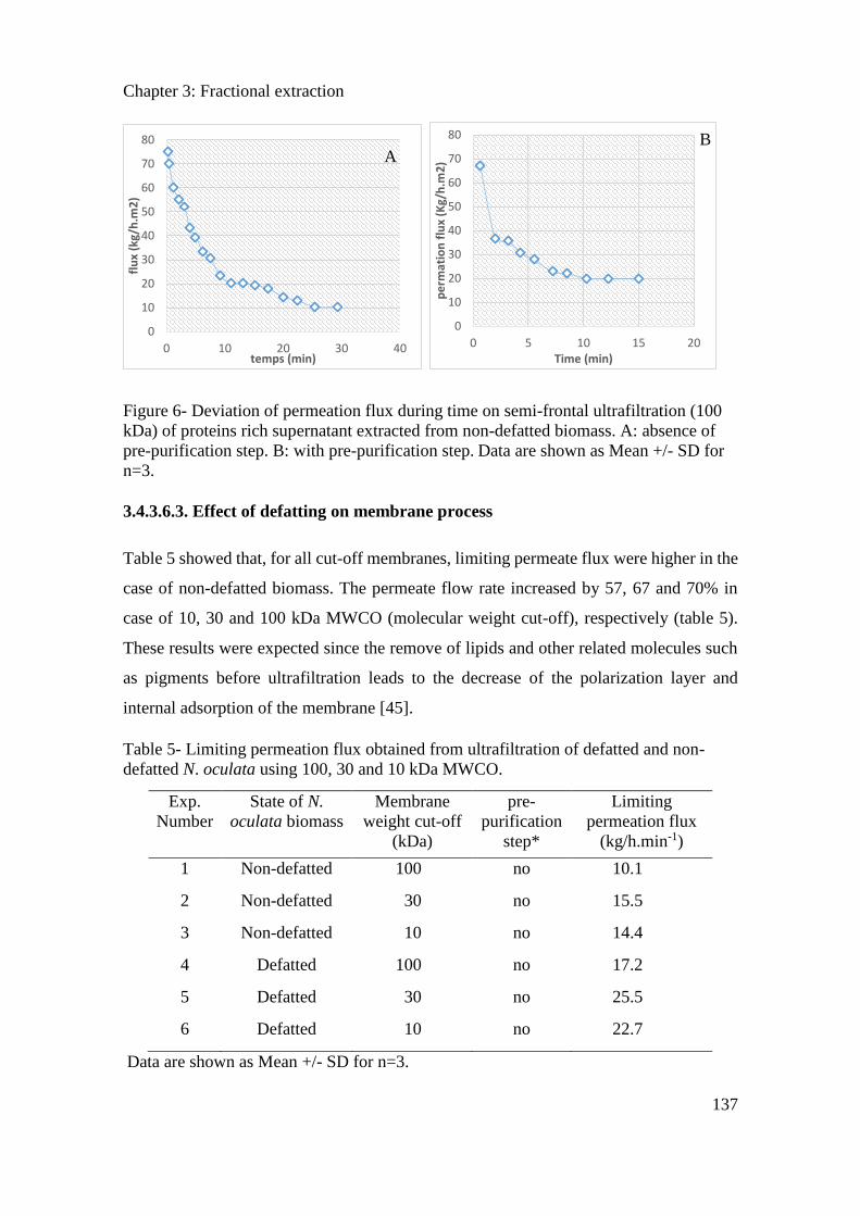
Chapter 3: Fractional extraction
137
Figure 6- Deviation of permeation flux during time on semi-frontal ultrafiltration (100
kDa) of proteins rich supernatant extracted from non-defatted biomass. A: absence of
pre-purification step. B: with pre-purification step. Data are shown as Mean +/- SD for
n=3.
3.4.3.6.3. Effect of defatting on membrane process
Table 5 showed that, for all cut-off membranes, limiting permeate flux were higher in the
case of non-defatted biomass. The permeate flow rate increased by 57, 67 and 70% in
case of 10, 30 and 100 kDa MWCO (molecular weight cut-off), respectively (table 5).
These results were expected since the remove of lipids and other related molecules such
as pigments before ultrafiltration leads to the decrease of the polarization layer and
internal adsorption of the membrane [45].
Table 5- Limiting permeation flux obtained from ultrafiltration of defatted and non-
defatted N. oculata using 100, 30 and 10 kDa MWCO.
Exp.
Number
State of N.
oculata biomass
Membrane
weight cut-off
(kDa)
pre-
purification
step*
Limiting
permeation flux
(kg/h.min-1)
1 Non-defatted 100 no 10.1
2 Non-defatted 30 no 15.5
3 Non-defatted 10 no 14.4
4 Defatted 100 no 17.2
5 Defatted 30 no 25.5
6 Defatted 10 no 22.7
Data are shown as Mean +/- SD for n=3.
0
10
20
30
40
50
60
70
80
0 10 20 30 40
flu
x (k
g/h
.m2
)
temps (min)
0
10
20
30
40
50
60
70
80
0 5 10 15 20
pe
rmat
ion
flu
x (K
g/h
.m2
)
Time (min)
A B
Chapter 3: Fractional extraction
138
3.4.4. Conclusion
In this study we showed that pH shifting process, under neutral condition (pH 7) and using
double stage extraction, was more effective than under alkaline conditions. Furthermore,
the use of defatted N. oculata biomass increased the release of proteins extraction yield.
The remove of lipids and other materials by precipitation or defatting improved the
performance of membrane process for proteins purification. Increasing the MWCO
membranes led to the increase of proteins purity in the permeate. These results suggest
that defatting step is a mandatory stage for high performance extraction/purification of
microalgal proteins.
Acknowledgements
Authors gratefully acknowledge GE Healthcare Life Sciences, Laurent VIALAT is
gratefully acknowledged for his help during this study.
3.4.5. References
[1] E. Suali and R. Sarbatly, “Conversion of microalgae to biofuel,” Renewable and Sustainable
Energy Reviews, vol. 16, no. 6, pp. 4316–4342, Aug. 2012.
[2] E. W. Becker, “Micro-algae as a source of protein,” Biotechnology Advances, vol. 25, no.
2, pp. 207–210, Mar. 2007.
[3] J. de la Noue and N. de Pauw, “The potential of microalgal biotechnology: A review of
production and uses of microalgae,” Biotechnology Advances, vol. 6, no. 4, pp. 725–770,
Jan. 1988.
[4] J. L. Guil-Guerrero, R. Navarro-Juárez, J. C. López-Martı́nez, P. Campra-Madrid, and M.
Rebolloso-Fuentes, “Functional properties of the biomass of three microalgal species,”
Journal of Food Engineering, vol. 65, no. 4, pp. 511–517, Dec. 2004.
[5] P. Spolaore, C. Joannis-Cassan, E. Duran, and A. Isambert, “Commercial applications of
microalgae,” Journal of Bioscience and Bioengineering, vol. 101, no. 2, pp. 87–96, Feb.
2006.
[6] J. A. Gerde, T. Wang, L. Yao, S. Jung, L. A. Johnson, and B. Lamsal, “Optimizing protein
isolation from defatted and non-defatted Nannochloropsis microalgae biomass,” Algal
Research, vol. 2, no. 2, pp. 145–153, Mar. 2013.
[7] M. F. Payne and R. J. Rippingale, “Evaluation of diets for culture of the calanoid copepod
Gladioferens imparipes,” Aquaculture, vol. 187, no. 1, pp. 85–96, Jul. 2000.
[8] J. R. McMillan, I. A. Watson, M. Ali, and W. Jaafar, “Evaluation and comparison of algal
cell disruption methods: Microwave, waterbath, blender, ultrasonic and laser treatment,”
Applied Energy, vol. 103, pp. 128–134, Mar. 2013.
[9] C. Safi et al., “Understanding the effect of cell disruption methods on the diffusion of
Chlorella vulgaris proteins and pigments in the aqueous phase,” Algal Research, vol. 8, pp.
61–68, Mar. 2015.
[10] C. Safi et al., “Aqueous extraction of proteins from microalgae: Effect of different cell
disruption methods,” Algal Research, vol. 3, pp. 61–65, Jan. 2014.
[11] C. Safi et al., “Biorefinery of microalgal soluble proteins by sequential processing and
membrane filtration,” Bioresource Technology, vol. 225, pp. 151–158, Feb. 2017.
Chapter 3: Fractional extraction
139
[12] L. R. Cavonius, E. Albers, and I. Undeland, “pH-shift processing of Nannochloropsis
oculata microalgal biomass to obtain a protein-enriched food or feed ingredient,” Algal
Research, vol. 11, pp. 95–102, Sep. 2015.
[13] G. H. Harrison, E. K. Balcer-Kubiczek, and H. A. Eddy, “Potentiation of Chemotherapy by
Low-level Ultrasound,” International Journal of Radiation Biology, vol. 59, no. 6, pp.
1453–1466, Jan. 1991.
[14] B. Balasundaram, S. Harrison, and D. G. Bracewell, “Advances in product release strategies
and impact on bioprocess design,” Trends in Biotechnology, vol. 27, no. 8, pp. 477–485,
Aug. 2009.
[15] A. K. Lee, D. M. Lewis, and P. J. Ashman, “Force and energy requirement for microalgal
cell disruption: An atomic force microscope evaluation,” Bioresource Technology, vol. 128,
pp. 199–206, Jan. 2013.
[16] A. G. Waghmare, M. K. Salve, J. G. LeBlanc, and S. S. Arya, “Concentration and
characterization of microalgae proteins from Chlorella pyrenoidosa,” Bioresour.
Bioprocess., vol. 3, no. 1, p. 16, Mar. 2016.
[17] R. K. Desai, M. Streefland, R. H. Wijffels, and M. H. M. Eppink, “Extraction and stability
of selected proteins in ionic liquid based aqueous two phase systems,” Green Chem., vol.
16, no. 5, pp. 2670–2679, Apr. 2014.
[18] D. MacKay and T. Salusbury, “Choosing between centrifugation and crossflow
microfiltration,” Chemical engineer, no. 447, pp. 45–50, 1988.
[19] Marcati et al., “Extraction and fractionation of polysaccharides and B-phycoerythrin from
the microalga Porphyridium cruentum by membrane technology,” Algal Research, vol. 5,
pp. 258–263, Jul. 2014.
[20] Patel et al., “Separation and fractionation of exopolysaccharides from Porphyridium
cruentum,” Bioresource Technology, vol. 145, pp. 345–350, Oct. 2013.
[21] Pugh et al., “(9) Isolation of Three High Molecular Weight Polysaccharide Preparations
with Potent Immunostimulatory Activity from Spirulina platensis , Aphanizomenon flos-
aquae and Chlorella pyrenoidosa | Request PDF,” ResearchGate. [Online]. Available:
https://www.researchgate.net/publication/11624110_Isolation_of_Three_High_Molecular
_Weight_Polysaccharide_Preparations_with_Potent_Immunostimulatory_Activity_from_
Spirulina_platensis_Aphanizomenon_flos-aquae_and_Chlorella_pyrenoidosa. [Accessed:
22-Aug-2018].
[22] Ba, Fatou, Ursu, Alina Violetta, Laroche, Celine, and Djelveh, Gholamreza,
“Haematococcus pluvialis soluble proteins: Extraction, characterization,
concentration/fractionation and emulsifying properties,” Bioresource Technology, vol. 200,
pp. 147–152, Jan. 2016.
[23] A.-V. Ursu, A. Marcati, T. Sayd, V. Sante-Lhoutellier, G. Djelveh, and P. Michaud,
“Extraction, fractionation and functional properties of proteins from the microalgae
Chlorella vulgaris,” Bioresource Technology, vol. 157, pp. 134–139, Apr. 2014.
[24] H. Susanto, H. Arafat, E. M. L. Janssen, and M. Ulbricht, “Ultrafiltration of polysaccharide–
protein mixtures: Elucidation of fouling mechanisms and fouling control by membrane
surface modification,” Separation and Purification Technology, vol. 63, no. 3, pp. 558–565,
Nov. 2008.
[25] R. Owusu-Apenten, Food Protein Analysis: Quantitative Effects On Processing. CRC
Press, 2002.
[26] S. Fujihara, A. Kasuga, and Y. Aoyagi, “Nitrogen-to-Protein Conversion Factors for
Common Vegetables in Japan,” Journal of Food Science, vol. 66, no. 3, pp. 412–415, Apr.
2001.
[27] S. O. Lourenço, E. Barbarino, U. M. L. Marquez, and E. Aidar, “Distribution of Intracellular
Nitrogen in Marine Microalgae: Basis for the Calculation of Specific Nitrogen-to-Protein
Conversion Factors,” Journal of Phycology, vol. 34, no. 5, pp. 798–811, Oct. 1998.
[28] C. Safi, M. Charton, O. Pignolet, F. Silvestre, C. Vaca-Garcia, and P.-Y. Pontalier,
“Influence of microalgae cell wall characteristics on protein extractability and determination
Chapter 3: Fractional extraction
140
of nitrogen-to-protein conversion factors,” J Appl Phycol, vol. 25, no. 2, pp. 523–529, Apr.
2013.
[29] O. H. Lowry, A. L. Rosbrough, and R. J. Randall, “Lowry assay protocol,” J. Biol. Chem.,
Lond, vol. 193, p. 265, 1951.
[30] E. Barbarino and S. O. Lourenço, “An evaluation of methods for extraction and
quantification of protein from marine macro- and microalgae,” J Appl Phycol, vol. 17, no.
5, pp. 447–460, Oct. 2005.
[31] E. Ryckebosch, K. Muylaert, and I. Foubert, “Optimization of an Analytical Procedure for
Extraction of Lipids from Microalgae,” Journal of the American Oil Chemists’ Society, vol.
89, no. 2, pp. 189–198, Feb. 2012.
[32] Y. Du, B. Schuur, S. R. A. Kersten, and D. W. F. Brilman, “Opportunities for switchable
solvents for lipid extraction from wet algal biomass: An energy evaluation,” Algal Research,
vol. 11, pp. 271–283, Sep. 2015.
[33] C. Enterman, “General procedures f? r separating lipid components of tissues. 299-317 IN
S. Colow1ck∼ nd N. Kaplan,” Methods in enzymology, vol. 3, 1957.
[34] Burba et al., “Membrane filtration studies of aquatic humic substances and their metal
species: a concise overview: Part 1. Analytical fractionation by means of sequential-stage
ultrafiltration,” Talanta, vol. 45, no. 5, pp. 977–988, Mar. 1998.
[35] I. S. Chronakis, “Gelation of Edible Blue-Green Algae Protein Isolate (Spirulina platensis
Strain Pacifica): Thermal Transitions, Rheological Properties, and Molecular Forces
Involved,” J. Agric. Food Chem., vol. 49, no. 2, pp. 888–898, Feb. 2001.
[36] K. J. Flynn, “Composition of intracellular and extracellular pools of amino acids, and amino
acid utilization of microalgae of different sizes,” Journal of Experimental Marine Biology
and Ecology, vol. 139, no. 3, pp. 151–166, Jul. 1990.
[37] L. Yao, J. A. Gerde, and T. Wang, “Oil extraction from microalga Nannochloropsis sp. with
isopropyl alcohol,” Journal of the American Oil Chemists’ Society, vol. 89, no. 12, pp.
2279–2287, Dec. 2012.
[38] F. Henriques and R. B. Park, “Compositional characteristics of a chloroform/methanol
soluble protein fraction from spinach chloroplast membranes,” Biochimica et Biophysica
Acta (BBA) - Bioenergetics, vol. 430, no. 2, pp. 312–320, May 1976.
[39] S. Bellou, M. N. Baeshen, A. M. Elazzazy, D. Aggeli, F. Sayegh, and G. Aggelis,
“Microalgal lipids biochemistry and biotechnological perspectives,” Biotechnology
Advances, vol. 32, no. 8, pp. 1476–1493, Dec. 2014.
[40] Obeid et al., “Supercritical carbon dioxide extraction and fractionation of lipids from freeze-
dried microalgae Nannochloropsis oculata and Chlorella vulgaris,” Algal Research, vol.
34, pp. 49–56, Sep. 2018.
[41] T. D. la Torre, V. Iversen, A. Moreau, and J. Stüber, “Filtration charaterization methods in
MBR systems: A practical comparison,” Desalination and Water Treatment, vol. 9, no. 1–
3, pp. 15–21, Sep. 2009.
[42] H. Susanto, S. Franzka, and M. Ulbricht, “Dextran fouling of polyethersulfone ultrafiltration
membranes—Causes, extent and consequences,” Journal of Membrane Science, vol. 296,
no. 1, pp. 147–155, Jun. 2007.
[43] O. Morineau-Thomas, P. Jaouen, and P. Legentilhomme, “The role of exopolysaccharides
in fouling phenomenon during ultrafiltration of microalgae (Chlorella sp. and Porphyridium
purpureum): Advantage of a swirling decaying flow,” Bioprocess Biosyst Eng, vol. 25, no.
1, pp. 35–42, Apr. 2002.
[44] W. Richard Bowen and P. M. Williams, “Obtaining the Osmotic Pressure of
Electrostatically Stabilized Colloidal Dispersions from Frontal Ultrafiltration
Experiments,” Journal of Colloid and Interface Science, vol. 233, no. 1, pp. 159–163, Jan.
2001.
[45] G. B. van den Berg and C. A. Smolders, “Flux decline in ultrafiltration processes,”
Desalination, vol. 77, pp. 101–133, Mar. 1990.
Chapter 3: Fractional extraction
141
3.5. Chapter conclusion
During this study, ScCO2 extraction showed a selective recovery of neutral lipids from
C. vulgaris and N. oculata microalgae without preliminary cell disruption step. Neutral
lipids extracts from N. oculata represented 83% of total lipids extracts, whereas the
proportion of glycolipids and phospholipids did not exceed 12.1% and 5.3%, respectively.
Adding ethanol (10% v/v) as co-solvent led to the extraction of 97% of neutral lipids from
C. vulgaris.
Besides, ScCO2 allowed efficient and selective extraction of chlorophyll a from the
mechanically untreated N. oculata. Chlorophyll a could be extracted efficiently and
selectively, whereas other pigments did not come out even at high pressure. This study
highlighted the potential and the possibility to eliminate some unit operations (cell
disruption) to selectively extract chlorophyll a free from other pigments. The
effectiveness of adding co-solvent for selective chlorophyll a (free from chlorophyll b)
from C. vulgaris was also covered. The distinction between the extraction yields of the
two chlorophylls a and b would be an indicator of the cell membrane modification during
the treatment with ethanol which could localized at specific structures.
A plausible line of unit operations combined pH-shifting and defatting showed to increase
the amount of proteins released in the supernatant of ScCO2 residues of N. oculata. Hence,
by following the proposed sequence of unit operations, 83% of extracted proteins were
obtained in the permeate. Biomass defatting had the advantage to facilitate the proteins
extraction. Over this study, semi-frontal ultrafiltration have proved their efficiency to
separate proteins from the various coloring fractions and co-extracted components.
Chapter 4: Extracts purification
142
Chapter 4: Extracts purification
4.1. Introduction
One of the special characteristics of microalgae biomass is its cell wall compositions and
rigidity, which differs from one species to another according to their growth conditions.
Some species are known for their rigid cell wall, contrary to others with fragile one. Cell
wall plays an important role as barrier to hinder the passage of valuable inner compounds.
This matter was well considered in this chapter due to the important role of the cell wall
on extraction selectivity.
Therefore, the main objective of this chapter is to understand the effect of cell wall
structures of Chlorella vulgaris and Spirulina sp. on the extraction of proteins in the
aqueous medium.
Besides, while all UAE studies conducted for proteins release have been performed with
frequencies comprised between 20 kHz and 100 kHz [24]. UAE treatments for microalgal
proteins extraction at audible frequencies (20 Hz-20 kHz) have never been reported in the
literature. A soft ultrasound assisted extraction at acoustic frequency (12 kHz) and low
intensities treatment was applied in this part of our study.
The results in this chapter are exposed in the form of three publications. The aim of first
publication was to investigate the effect of working at nearly audible frequency (12 kHz)
and under hydrostatic pressure on total extraction of Spirulina proteins including C-
phycocyanin. Besides, extraction efficiency has been evaluated by measuring the
influence of extraction operating parameters (ultrasound power, hydrostatic pressure,
stirring) and conditioning methods (fresh, frozen or freeze-dried) on the extracts
compositions.
While all conducted studies have focused on the recovery of pigmented proteins (mainly
C-phycocyanin) from Spirulina sp., the non-pigmented proteins have been neglected
without any valorization. For that, the second publication focused on the highly
clarification of the non-pigmented proteins using membrane process.
Contrary to Spirulina sp. fragile cell wall, the apparent rigidity of C. vulgaris was
exploited for selective extraction of the intracellular proteins, alleviating then the key
Chapter 4: Extracts purification
143
challenges in the final-stream steps of purification. Hence, in this context, the third
publication proposed to evaluate the potential of UAE for proteins selective extraction
and high recovery yield. In addition, the pigments and UAE residues were quantified to
be used as a marker for inner organelle maintenance, cell wall integrity, and absence of
lipids liberation.
Chapter 4: Extracts purification
144
4.2. Proteins and pigments extraction from microalgae Spirulina sp.
assisted by ultrasound irradiation: effect of variable low power and
pre-treatment conditioning
(Article published in Récents Progrès en Génie des Procédés, Numéro 110 - 2017
ISSN: 1775-335X; ISBN: 978-2-910239-85-5, Ed. SFGP, Paris, France)
OBEID Saraa,b,c*, BENHAMED Imaned, CHARTON Michaela, PEYDECASTAING
Jérômea, TAKACHE Hosnib,c, ISMAIL Alib,c, PONTALIER Pierre-Yvesa, BARTHE
Lauried a Laboratoire de Chimie Agro-industrielle (LCA), Université de Toulouse, INRA, INPT,
Toulouse, France 4 allée Emile Monso, 31 030 Toulouse cedex 4, France bUniversité Libanaise, Faculté de Génie Agronomique et de Médecine Vétérinaire,
Département des Sciences et Technologie Alimentaires, Hadath, Liban. Allée Georges
Matta, 1500, Hadath, Liban. cUniversité Libanaise, Ecole Doctorale de Sciences et Technologies EDST-PRASE
(Platform de Recherches et d'Analyses en Sciences de l'Environnement), Hadath, Liban.
Campus Rafic Hariri, 1500, Hadath, Liban dLaboratoire de Génie Chimique, Université de Toulouse, CNRS, INPT, UPS, Toulouse,
France. 4 allée Emile Monso, CS 84234, 31 432 Toulouse cedex 4, France
Abstract
Spirulina sp. is a marine biomass with high valuable molecules content, such as proteins
and pigments. Low frequency ultrasound has been investigated as selective extraction
method in order to reduce the number of downstream steps. In the case of Spirulina, the
results indicated that proteins extraction was high, at a value of almost 95% of the initial
proteins content. According to the cell disruption degree, pigments extraction yield can
be favoured under sonication, mainly chlorophylls rather than carotenoids. Storage
conditions had a large effect on extraction efficiency, particularly freezing could partially
lyse the cell wall and favoured soluble molecules extraction. Application of ultrasound
permitted to obtain a high extraction yield of the pigments like chlorophyll a with a
concentration in the supernatant 100 times higher. A microscopic structural study for
biomass after each ultrasound extraction shows that pigments extraction required a higher
disintegration of the cell wall, while proteins are extracted with a low cell lysis.
Keywords: Spirulina sp., ultrasound, proteins, pigments, cell membrane
disintegration
* Auteur/s à qui la correspondance devrait être adressée : [email protected]
Chapter 4: Extracts purification
145
4.2.1. Introduction
Cyanobacteria and algae are a source of highly precious molecules like polyunsaturated
fatty acid oils as nutritional supplements and pigments including carotenoids,
chlorophylls and phycobiliproteins (Henrikson, 1989; Spolaore et al., 2006). Spirulina is
a cyanobacteria that has been widely studied because of its nutritional and medicinal
properties (Piñero Estrada et al., 2001), but also due to its composition in valuable
constituents particularly proteins, vitamins and pigments such as β-carotene and
phycocyanin. The latter is one of the most promising biochemicals in Spirulina because
it could be used as natural colorant in food and for its fluorescence properties that could
improve the development of phycofluor probes for immunodiagnostics (Kronik and
Grossman, 1983). However, the recovery of Spirulina molecules usually requires
numerous extraction/fractionation methods that have hindered its industrial production.
Different methods have been used for Spirulina components extraction, like γ-linolenic
acid extraction or fatty acids and carotenoids, by supercritical CO2 (Canela et al., 2002;
Sajilata et al., 2008). However, in addition to process cost, supercritical extraction without
co-solvent gives a low yield compared to extraction using a mixture of solvents (Mendes
et al., 2006). Spirulina cells could also be destroyed using different methods as
homogenization in Virtimixer (Sarada et al., 1999), releasing all the cell components in
the solvent, leading to a complex of molecules difficult to purify. Hence there is a need
for technology development allowing for molecules selective extraction that would
simplify the downstream processing like ultrasound assisted extraction (UAE)
technology.
Ultrasounds (US) are giving rise to the cavitation phenomenon where bubbles collapses
and liquid micro-jets generate interesting mechanical effects, localized on
particles/entities of micron size, which can be different according to the ultrasonic
irradiation (frequency, power, intensity, etc.) (Mason, 2013). Previous studies have
already shown that UAE is efficient in Spirulina extraction of carotenes (Dey et al., 2013)
and phycocyanin (Furuki et al., 2003; Moraes et al., 2011) but always at frequency higher
than 20 kHz and under atmospheric pressure.
The aim of this study is to investigate how UAE could be efficient to selectively extract
the main components of Spirulina, such as proteins and pigments notably phycocyanin.
Chapter 4: Extracts purification
146
In order to control the extraction, an innovative process, for cavitation intensification, has
been studied by working at nearly audible frequency (12 kHz) and under hydrostatic
pressure. Extraction efficiency has been evaluated by measuring the influence of
extraction operating parameters (ultrasound power, hydrostatic pressure, stirring) and
conditioning methods (fresh, frozen or freeze-dried) on the extracts composition.
4.2.2. Material and methods
4.2.2.1 Microalgae
Fresh Spirulina Sp. was provided by Alg&You company (Toulouse, France) in a culture
solution at a concentration of 5% (dry matter). Fractions of the culture has been frozen or
freeze-dried.
4.2.2.2 UAE
In this study, UAE extraction was carried out with an ultrasonic reactor of 1L capacity
that was connected to a pressurized N2 bottle. The emitting surface of the ultrasound was
located at the bottom. The solution temperature was controlled at 30 ± 2°C by a
circulation of cooling water stream in an internal coil. A constant volume of biomass
solution (600 mL) was stirred at 500 rpm and sonicated at 12 kHz frequency during 1
hour. Two ultrasonic parameters were investigated to determine their effect on the
proteins extraction yield: ultrasound power (100, 200 and 300 W) and relative hydrostatic
pressure (0, 1.5 and 3 bar). Moreover, pre-treatment conditions of microalgae were also
studied in aim to see the effect of biomass state on molecules extraction yield (table 1).
4.2.2.3 Analysis
a. Total proteins
Total proteins content of Spirulina sp. was quantified according to Kjeldhal method that
take into account the totality of the nitrogen present in the biomass with a 6.15 coefficient
(Fujihara et al., 2001) because only 59–98 % of total nitrogen can belong to proteins (Jeon
et al., 2013; Fujihara et al., 2001; Lourenço et al., 1998). Dry Spirulina sp. was washed
with water and separated from supernatant (culture medium) rich in nitrogen by

Chapter 4: Extracts purification
147
centrifugation (20,000 g, 15 min, 5C). Biomass was analyzed in triplicate using each time
50 mg samples.
b. Soluble protein
Water-soluble proteins were quantified by Kjeldhal analysis (with a 6.15 coefficient) and
Lowry method (Lowry et al., 1951) using a standard curve of bovine serum albumin
(BSA) and a Lowry kit (Lowry reagent plus bovine standard albumin (BSA) standards
and 2 N Folin–Ciocalteu reagents) purchased from Thermo Fisher Scientific. This method
is based on a reaction catalyzed by the copper content in the Folin and oxidation of the
peptide bonds with a Folin-Ciocalteu reagent (Barbarino and Laurenco, 2005). The
hydro-soluble proteins fraction was evaluated after residues elimination by centrifugation
at 15,000g for 15 min. Samples of the supernatant rich in hydro-soluble proteins were
taken for analysis by Lowry method. Absorbance was measured at 750 nm using a
Shimadzu UV-1700 spectrophotometer
c. Pigments
Samples were analyzed in HPLC Dionex apparatus after filtration using micro-filtration
cartridge Polyray 0.22 micron was done to eliminate cell residues. Filtered sample were
injected on a 5C18, 250*4.6 mm column and quantified by UV/VIS detector after elution
with acetonitrile: methanol: water (65:35:2) at a flow rate of 1 ml/min. The standard
pigments, lutein, beta-carotene, chlorophyll a and chlorophyll b (supplied by Sigma-
Aldrich) were prepared by dilution of a stock solution (0.1 g/L in acetone) of each of the
standard pigments in methanol to a concentration range of 0.5 mg/L to 5 mg/L.
d. Structural characterization
Microscopic observations represent a qualitative approach to characterize the cell
disruption. Microalgae observation at high magnifications (* 1000) was performed before
and after treatment on a small quantity of cell suspension placed on a specific plate (Nikon
SMZ 1500). The images were captured under a constant exposure and illumination by
(Nikon eclipse E600) camera.
Chapter 4: Extracts purification
148
4.2.3. Results and Discussion
4.2.3.1 Proteins extraction
Characterization of Spirulina sp. by Kjeldhal method indicated that this microalgae has a
high proteins content at about 70 % dry weight (dw). This result is usually described on
Spirulina, with low lipids content. The fraction of hydro-soluble proteins released in
aqueous medium after each UAE condition has been evaluated by the Lowry and Kjeldhal
methods. No significant difference has been detected between values using those two
methods.
The ease or difficulty of extracting proteins is due to their location in the cell. Proteins of
Spirulina with pluri-stratified cell wall, thylakoid lamella system and numerous
inclusions, are localized in various compartments. Proteins like ribosome are found in the
cytoplasm or bound to the plasma membrane. Phycobiliproteins are located in the
thylakoid system or photosynthetic lamellas. The inter-thylakoid space is limited by
proteinaceous gas vesicles that give Spirulina its floating capacity. Fibrils of DNA and
related proteins (Chaperones, and proteins secreted under stress) are generally of central
localization (Van Eykelenburg, 1977). The proteins located in the plasma are easily
recovered while for the other it requires a higher disruption degree.
Total proteins released content was high in all cases, ranging from 57.9 to 69.8 % of the
introduced dry weight (Table 1). When the cells are placed in distilled water in the
ultrasound reactor the extracted proteins represent 58% of the introduced matter.
Spirulina is a filamentous microalgae characterized by its shear-sensitive and fragile
membrane. It does not have cellulose walls, but a relatively fragile envelope of murein
(Madkourb et al., 2012). This biomass is generally recommended for their high proteins
content that can reach up to 70 % of dry weight (Hoseini et al., 2013) and the absence of
membrane rigidity (Moreas et al., 2011).
When the ultrasound system is activated, the amount of released proteins increases
slightly with increasing the US power and the pressure. Among the tested UAE conditions
on Spirulina sp., C31 (frozen-unfrozen biomass-highest pressure applied of 3bar- 100 W)
and C1.53 (frozen-unfrozen biomass -1.5 bar- 300 W) were the best conditions for the
cyanobacteria proteins release with a recovery yield of 69.8 %w equal to almost the total
Chapter 4: Extracts purification
149
proteins content. Under hydrostatic relative pressure of 1.5 or 3 bar the cavitation bubbles
expand to its maximum volume and collapse violently accompanied by strong shock wave
emission (Brujana, et al., 2008) which make the release of proteins more significant, that
explain the yield difference between experiments under same power but different pressure
(C01 and C31; C03 and C1.53).
The proteins extraction is more efficient from fresh biomass (67.5 %w) than from frozen-
unfrozen biomass (64.4 %w) at 300 W, 500 rpm. Ultrasounds are characterized by the
mechanical/physical effects: implosion of cavitation bubble which leads to powerful
shock waves and fragmentation of interfaces (solid-liquid) and the effects produced on
hydrodynamics improve the mass transfer in the solvent (water) (Suslick et al., 1994). A
plausible explanation could be that the brutal and rapid cell degradation on the frozen-
unfrozen fragile biomass hindered the proteins diffusion to the liquid phase and left a part
of proteins attached to the membrane or non-purified detectible molecules.
4.2.3.2 Pigments extraction
The marine microalga Spirulina used for this study is a marine biomass richer in
chlorophyll (b and a) rather than carotenoids (lutein and -carotene). Extraction of these
pigments requires disintegration of internal compartments because they are mainly
located in thylakoid membranes. UAE efficiency has been evaluated for pigments
extraction (Table 1).
Among several factors tested, pigments extraction from frozen biomass at a power
intensity of 300 watt (C03- 500 rpm) or 100 watt with 3 bar as relative pressure (C31-
500 rpm) were shown to be the most suitable conditions to extract chlorophyll A and -
carotenes from this strain. The results show large influence of the operating conditions
on pigments extraction yields.
Contrary to proteins extraction, freeze-drying degradation only allows a small extraction
of the pigments, mainly chlorophyll a with 2.15 mg/l. Freezing seems to lead to higher
degradation level than freeze-drying with 1137.04 mg/l and 590.74 mg/l of Chlorophyll
a under C03 and F03 conditions respectively.
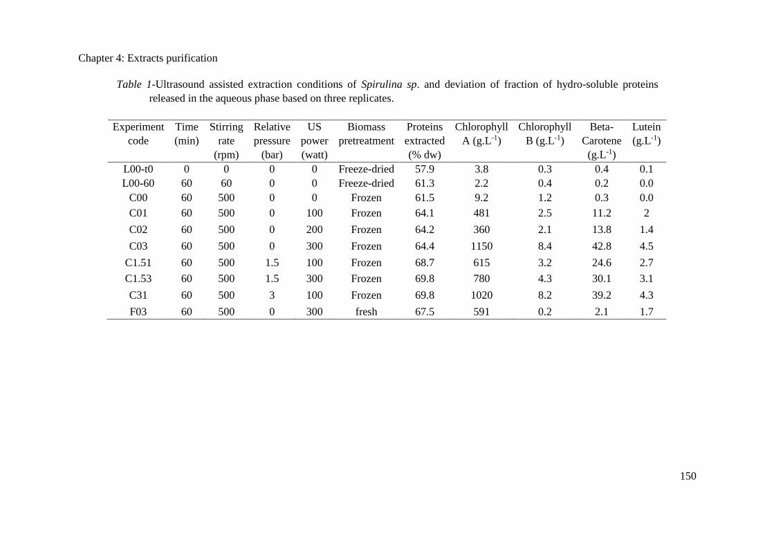
Chapter 4: Extracts purification
150
Table 1-Ultrasound assisted extraction conditions of Spirulina sp. and deviation of fraction of hydro-soluble proteins
released in the aqueous phase based on three replicates.
Experiment
code
Time
(min)
Stirring
rate
(rpm)
Relative
pressure
(bar)
US
power
(watt)
Biomass
pretreatment
Proteins
extracted
(% dw)
Chlorophyll
A (g.L-1)
Chlorophyll
B (g.L-1)
Beta-
Carotene
(g.L-1)
Lutein
(g.L-1)
L00-t0 0 0 0 0 Freeze-dried 57.9 3.8 0.3 0.4 0.1
L00-60 60 60 0 0 Freeze-dried 61.3 2.2 0.4 0.2 0.0
C00 60 500 0 0 Frozen 61.5 9.2 1.2 0.3 0.0
C01 60 500 0 100 Frozen 64.1 481 2.5 11.2 2
C02 60 500 0 200 Frozen 64.2 360 2.1 13.8 1.4
C03 60 500 0 300 Frozen 64.4 1150 8.4 42.8 4.5
C1.51 60 500 1.5 100 Frozen 68.7 615 3.2 24.6 2.7
C1.53 60 500 1.5 300 Frozen 69.8 780 4.3 30.1 3.1
C31 60 500 3 100 Frozen 69.8 1020 8.2 39.2 4.3
F03 60 500 0 300 fresh 67.5 591 0.2 2.1 1.7
Chapter 4: Extracts purification
151
Extraction efficiency of all pigments strongly increased when ultrasounds are applied at a power
of 100 W, since the concentrations shifted from 9 g.L-1 and 0.3 g.L-1 to 480 g.L-1 and 11 g.L-1 for
chlorophyll A and -carotene respectively. Increasing the power has no effect until 300 W (like a
threshold), that allows for the release of 4 times more pigments, like some new specific membranes
has been broken above a limit energy received. A similar effect is observed when increasing the
pressure to 3 bars with a power of 100 W but with a much more progressive influence. It is
therefore possible to obtain the same extraction yield at 300W under atmospheric pressure or 100
W and 3 bars. The second condition will be preferred as it is more economic.
In all cases, the extraction is much more efficient after freezing than after freeze-drying. When the
extraction is operated with fresh Spirulina, pigments extraction yield is much smaller, particularly
for chlorophyll B and -carotene that are more difficult to extract. So ultrasound is an efficient
method to extract pigment and freezing pre-treatment can enhance its performances.
4.2.3.3 Microscopic observation
During this work, after each UAE treatment, microalgae were then observed under a microscope
to determine cell membrane damaging mark (Figure 1). The first photography indicates that
Spirulina after freezing has still a normal shape, and it can be concluded that the degradation must
be only like small holes in the membranes. These holes would allow for the diffusion out of the
cell of the soluble molecules, like the proteins, but not for the molecules entrapped in the
membranes systems of the cells like the pigments.
Spirulina sp. showed a sensitive cell wall, at frequency of 12 kHz, cell integrity degree decreased
with increasing power dose (100 to 300 W). After treatment, the structure of Spirulina completely
disappeared, which can explain that the pigments are extracted.
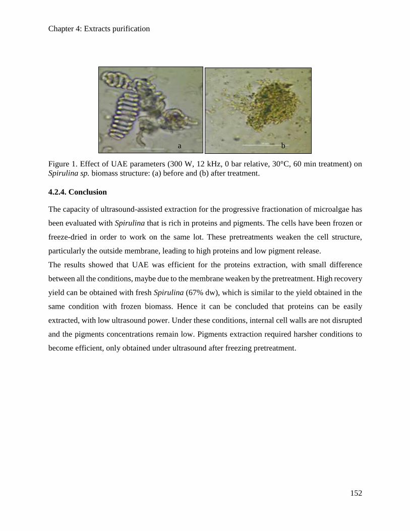
Chapter 4: Extracts purification
152
Figure 1. Effect of UAE parameters (300 W, 12 kHz, 0 bar relative, 30°C, 60 min treatment) on
Spirulina sp. biomass structure: (a) before and (b) after treatment.
4.2.4. Conclusion
The capacity of ultrasound-assisted extraction for the progressive fractionation of microalgae has
been evaluated with Spirulina that is rich in proteins and pigments. The cells have been frozen or
freeze-dried in order to work on the same lot. These pretreatments weaken the cell structure,
particularly the outside membrane, leading to high proteins and low pigment release.
The results showed that UAE was efficient for the proteins extraction, with small difference
between all the conditions, maybe due to the membrane weaken by the pretreatment. High recovery
yield can be obtained with fresh Spirulina (67% dw), which is similar to the yield obtained in the
same condition with frozen biomass. Hence it can be concluded that proteins can be easily
extracted, with low ultrasound power. Under these conditions, internal cell walls are not disrupted
and the pigments concentrations remain low. Pigments extraction required harsher conditions to
become efficient, only obtained under ultrasound after freezing pretreatment.
b a
Chapter 4: Extracts purification
153
4.2.5. References
Barbarino, E. and S.O. Lourenço, 2005, An evaluation of methods for extraction and quantification
of protein from marine macro- and microalgae, Journal of Applied Phycology, 17, no. 5, 447-460.
Brujana, E. A., T. Ikedab and Y. Matsumoto, 2008, On the pressure of cavitation bubbles,"
Experimental Thermal and Fluid Science, 32, 1188-1191.
Canela, A.P.R.F., P.T.V. Rosa, M.O.M. Marques and M.A.A. Meireles, 2002, Supercritical Fluid
Extraction of Fatty Acids and Carotenoids from the Microalgae Spirulina maxima Ind, Eng. Chem.
Res. 41, 3012.
Dey, S. and V.K. Rathod, 2013, Ultrasound assisted extraction of -carotene from Spirulina
platensis. Ultrasonics Sonochemistry 20, 271
Fujihara, S., A. Kasuga. and Y. Aoyagi, 2001, Nitrogen-to-Protein Conversion Factors for
Common Vegetables in Japan, J Food Sci., 66, 412-415.
Furuki T., S. Maeda, S. Imajo, T. Hiroi, T. Amaya, T. Hirokawa, K. Ito and H. Nozawa, 2003,
Rapid and selective extraction of phycocyanin from Spirulina platensis with ultrasonic cell
disruption, J. Appl. Phycol. 15, 319.
Henrikson R., 1989, Earth food Spirulina. Ronore Enterprises Inc, Laguna Beach, CA.
Hoseini, S.M., K. Khosravi-Darani, M.R. Mozafari, 2013, Nutritional and Medical Applications
of Spirulina Microalgae, Mini Reviews in Medicinal Chemistry, 13, 1231-1237(7).
Jeon, J.-M., H.-W. Choi, G.-C. Yoo, Y.-K. Choi, K.-Y. Choi, H.-Y. Park, S.-H. Park, Y.-G. Kim,
H.J. Kim and S.H. Lee, 2013, New mixture composition of organic solvents for efficient extraction
of lipids from Chlorella vulgaris, Biomass Bioenergy, 29, 279-284.
Kronik, M. and P. Grossman, 1983, Immunoassay techniques with fluorescent phycobiliprotein
conjugates, Clin Chem 29, 582.
Lourenço, SO., E. Barbarino., P.L. Lavín, U.M. Lanfer Marquez and E. Aidar, 1998, Distribution
of intracellular nitrogen in marine microalgae: basis for the calculation of specific nitrogen-to-
protein conversion factors, J Phycol, 37, 798-811.
Lowry, O.H., N.J. Rosebrough, A.L. Far and R.J. Randall, 1951, Protein measurement with the
Folin phenol reagent," J Biol Chem, 193, 265-275.
Mason, T.J., 2003, Sonochemistry and sonoprocessing: the link, the trends and (probably) the
future, Ultrason. Sonochem. 10, 175.
Madkourb, F., A. Kamila, and H.S. Nasr, 2012, Production and nutritive value of Spirulina
platensis in reduced cost media, The Egyptian Journal of Aquatic Research, 38, no. 1, 51-57.
Mendes R.L., A.D. Reis and A. F. Palavra, 2006, Supercritical CO2 extraction of γ-linolenic acid
and other lipids from Arthrospira (Spirulina) maxima: Comparison with organic solvent
extraction, Food Chemistry, 99, 57
Moraes, C.C., L. Sala, G.P. Cerveira and S.J. Kalil, 2011, C-phycocyanin extraction from Spirulina
platensis wet biomass, Braz. J. Chem. Eng. 28, 45.
Piñero Estrada, J.E., P. Bermejo Bescós and A.M. Villar Del Fresno, 2001, Antioxidant activity of
different fractions of Spirulina platensis protean extract, Il Farmaco 56, 497.
Chapter 4: Extracts purification
154
Sajilata M.G., R.S. Singhal and M.Y. Kamat, 2008, Supercritical CO2 extraction of γ-linolenic
acid (GLA) from Spirulina platensis ARM 740 using response surface methodology, J. Food Eng.
84, 321.
Sarada, R., M. G. Pillai and G.A. Ravishankar, 1999, Phycocyanin from Spirulina sp: influence of
processing of biomass on phycocyanin yield, analysis of efficacy of extraction methods and
stability studies on phycocyanin, Process Biochemistry 34 795.
Spolaore, P., C. Joannis-Cassan, E. Duran and A. Isambert, 2006, Commercial applications of
microalgae, J. Biosci. Bioeng. 101, 87.
Suslick, K.S., 1994, The Chemistry of Ultrasound, Encyclopaedia Britannica Yearbook, Chicago,
138-155.
Van Eykelenburg, C., 1977, On the morphology and ultrastructure of the cell wall of Spirulina
platensis, Antonie van Leeuwenhoek, 43, 89.
Acknowledgements
The authors would like to gratefully thank Alg&You society (Toulouse, France) for providing
Spirulina sp. biomass.
Chapter 4: Extracts purification
155
4.3. Clarification of non-pigmented proteins from Spirulina sp. using
ultrasound assisted extraction coupled with ultrafiltration
(Article will be submitted to Journal of Bioresource Technology)
S. Obeid a,b,c*, I. Benhamedd , H. Takacheb,c, J. Peydecastinga , M. Chartona, L. Barthed , A. Ismailb,c
and P.Y. Pontaliera, 1
a Laboratoire de Chimie Agro-industrielle (LCA), Université de Toulouse, INRA, INPT,
Toulouse, France 4 allée Emile Monso, 31030 Toulouse cedex 4, France. b Département des Sciences et Technologies Alimentaires, Faculté des Sciences Agronomiques,
Université Libanaise, Dekwaneh, Liban. c Plateforme de Recherches et Analyses en Sciences de l’Environnement (PRASE), Ecole
Doctorale des Sciences et Technologies, Université Libanaise, Hadath, Liban. d Laboratoire de Génie Chimique (LGC), INP-ENSIACET, Université de Toulouse, 31030
Toulouse, France.
Clearly label i.e. Table 1 or Figure 1 (no full stop)
Abstract
A fractionation process was carried out on the cyanobacteria Spirulina sp. using ultrasound
assisted extraction in order to obtain a highly clarified and uncolored proteins free from
chlorophylls, carotenoids, and C-phycocyanin. Effect of pressure and ultrasound intensity were
studied for total proteins release. Total proteins content, up to 97%, was obtained in the aqueous
medium at atmospheric pressure, 12 kHz frequency and 200W intensity. Precipitation with
ammonium sulfate (25% w/v) followed by semi-frontal ultrafiltration (100 kDa) permitted to
obtain 67% of proteins in the permeate, while up to 90% of C-phycocyanin was retained in the
retentate. Using 10 kDa cut-off membrane allowed to obtain a highly clarified proteins fraction
where up to 95% of C-phycocyanin were removed.
Keywords: Proteins purification, Spirulina sp., phycocyanine, ultrasounds, ultrafiltration.
4.3.1. Introduction
Spirulina sp. cyanobacteria has been recognized worldwide as a traditional source of proteins used
for human consumption, given their interesting abundance in proteins and essential amino acids
Chapter 4: Extracts purification
156
[1] [2]. These edible blue-green algae are known for their high proteins content representing up to
70% of the dry biomass, which is equivalent to three times to beef proteins content or ten times to
soy-bean proteins content [3]. Some of Spirulina proteins are found in the cytoplasm or bounded
to the plasma membrane [4]. Others are found covalently attached and located in the thylakoid
system and photosynthetic lamellas like brilliantly colored phycobiliproteins [5].
Phycobiliproteins are water-soluble proteins that play a biological role in collecting and conveying
sunlight to a special pair of chlorophyll molecules located in the photosynthetic reaction center
[5]. Many phycobiliproteins are produced by Spirulina like phycoerythrin (PE), allophycocyanin
(APC) and C-phycocyanin (CPC)[6]. The last proteins fraction represents the major
phycobiliproteins, and can be synthesized to reach 20% of total dry biomass [7] [8].
CPC is produced commercially as a colorant for food and cosmetics and as fluorescent marker for
diagnostic probes. However, other proteins fraction (pigments free proteins) started to find new
applications in cosmetics and pharmaceutical industries. However, the use of such proteins in
industry is still not significant due to the presence of undesirable elements like CPC leading to
significant modifications in flavor and color of the product. Thus, to improve their potential use,
Spirulina proteins have to be clarified from CPC and undesirable coloring components. However,
extraction of proteins from microalgae and cyanobacteria are always challenging. Normally,
proteins located in the plasma are easy to recover, while for the other fractions (located in the
thylakoid system and photosynthetic lamellas) an intensive cells disruption is required. Scientists
are investigating many ways for effective proteins recovery, where cells disintegration is always a
neediness to facilitate the intracellular proteins release and maximize their recovery yield.
Ultrasound-assisted extraction (UAE), as cells disruption technique, is used for microalgae and
cyanobacteria fractionation [9] [10] [11]. UAE technique is known as a scalable processing step
that comply with criteria of green chemistry concept and sustainability [9] [10]. Besides, working
with ultrasonic cavitation induced by low frequency UAE could alleviate the key challenges
associated with cell disruption techniques during the recovery of total proteins content from
Spirulina [12]. However, UAE leads to the liberation of all biomass components in the
supernatants, which requires additional costly stages in downstream processing for phase
separation and compounds purification.
Chapter 4: Extracts purification
157
Ultrafiltration technology is considered as efficient purification technique with relatively moderate
capital and operating costs [13]. Contrary to other separation techniques, membrane processes are
isothermal and involve no phase change or chemical reagents [14]. These separation techniques
are particularly suited to the purification of water-soluble biomolecules without neither thermal
denaturation nor chemical solvents use.
Membrane processes, particularly ultrafiltration, have begun to find new applications in the field
of marine bioresources [15] [16] [17]. Actually, limited studies have investigated the membrane
technology to purify components from microalgae (Gerardo et al., 2014). Many authors have
investigated the use of this technique to purify components from microalgae: polysaccharides from
Porphyridium cruentum [18] [19], Spirulina platensis and Chlorella pyrenoidosa [20]. Other
studies reported the use of membrane processes to concentrate proteins from Chlorella vulgaris
and Haematococcus pluvialis [21] [22]. Membrane processes have also been used for the recovery
of CPC from Spirulina sp. [23][24][8]. However, all these studies have focused on the recovery of
pigmented proteins (mainly CPC) while the non-pigmented proteins have been neglected without
any valorization.
The objective of this work is to study the use of low frequency ultrasound (12 kHz) coupled with
ultrafiltration to the recovery and clarification of non-pigmented proteins from Spirulina sp. To
our best of knowledge no similar studies have been reported on obtaining highly clarified non-
pigmented proteins fraction from Spirulina sp. using the mentioned techniques.
4.3.2. Materials and Methods
The Lowery assay kit was purchased from Fisher Scientific. Chemicals including pigments
(Chlorophyll a, Chlorophyll b, lutein and -carotenes) were purchased from Sigma-Aldrich
(France) then used as received.
4.3.2.1. Microalgae
Fresh cyanobacteria Spirulina Sp. was purchased from by Alg&You company (Toulouse, France).
This strain was grown in photobioreactor under normal growth condition. Fresh biomass was
supplied in culture media solution brought at a concentration of 5% dry weight. To ensure that the
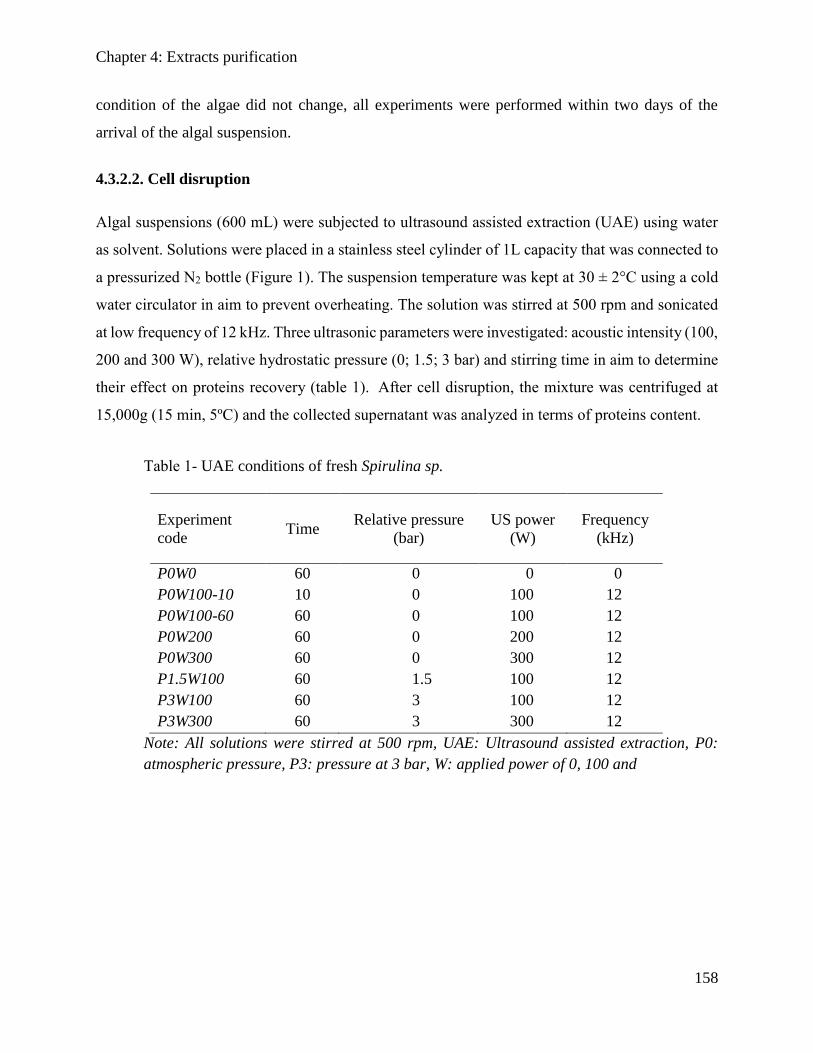
Chapter 4: Extracts purification
158
condition of the algae did not change, all experiments were performed within two days of the
arrival of the algal suspension.
4.3.2.2. Cell disruption
Algal suspensions (600 mL) were subjected to ultrasound assisted extraction (UAE) using water
as solvent. Solutions were placed in a stainless steel cylinder of 1L capacity that was connected to
a pressurized N2 bottle (Figure 1). The suspension temperature was kept at 30 ± 2°C using a cold
water circulator in aim to prevent overheating. The solution was stirred at 500 rpm and sonicated
at low frequency of 12 kHz. Three ultrasonic parameters were investigated: acoustic intensity (100,
200 and 300 W), relative hydrostatic pressure (0; 1.5; 3 bar) and stirring time in aim to determine
their effect on proteins recovery (table 1). After cell disruption, the mixture was centrifuged at
15,000g (15 min, 5ºC) and the collected supernatant was analyzed in terms of proteins content.
Table 1- UAE conditions of fresh Spirulina sp.
Note: All solutions were stirred at 500 rpm, UAE: Ultrasound assisted extraction, P0:
atmospheric pressure, P3: pressure at 3 bar, W: applied power of 0, 100 and
Experiment
code Time
Relative pressure
(bar)
US power
(W)
Frequency
(kHz)
P0W0 60 0 0 0
P0W100-10 10 0 100 12
P0W100-60 60 0 100 12
P0W200 60 0 200 12
P0W300 60 0 300 12
P1.5W100 60 1.5 100 12
P3W100 60 3 100 12
P3W300 60 3 300 12
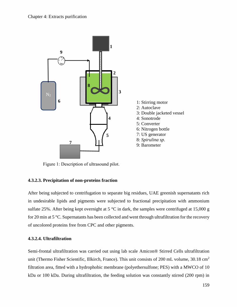
Chapter 4: Extracts purification
159
Figure 1: Description of ultrasound pilot.
4.3.2.3. Precipitation of non-proteins fraction
After being subjected to centrifugation to separate big residues, UAE greenish supernatants rich
in undesirable lipids and pigments were subjected to fractional precipitation with ammonium
sulfate 25%. After being kept overnight at 5 °C in dark, the samples were centrifuged at 15,000 g
for 20 min at 5 °C. Supernatants has been collected and went through ultrafiltration for the recovery
of uncolored proteins free from CPC and other pigments.
4.3.2.4. Ultrafiltration
Semi-frontal ultrafiltration was carried out using lab scale Amicon® Stirred Cells ultrafiltration
unit (Thermo Fisher Scientific, Illkirch, France). This unit consists of 200 mL volume, 30.18 cm2
filtration area, fitted with a hydrophobic membrane (polyethersulfone; PES) with a MWCO of 10
kDa or 100 kDa. During ultrafiltration, the feeding solution was constantly stirred (200 rpm) in
∞ N2
1
2
3
4
5
6 1: Stirring motor
2: Autoclave
3: Double jacketed vessel
4: Sonotrode
5: Converter
6: Nitrogen bottle
7: US generator
8: Spirulina sp.
9: Barometer
7
8
9
Chapter 4: Extracts purification
160
the feeding chamber which allows to minimize the concentration polarization effect at the
membrane surface [25]. Sample of UAE supernatant with the highest proteins extraction yield
went through precipitation of non-proteins fraction before being served as a feed for ultrafiltration
(Figure 2). To underline the efficiency of precipitation step, a sample of the same supernatant went
directly through ultrafiltration step. The transmembrane difference pressure (TMDP) was
maintained constant at 2 bar for all experiments. Free pigments proteins fraction was recovered in
the permeate compartment. Samples from permeate and retentate were taken for analysis of total
chlorophyll, total carotenoids, proteins and CPC content.
4.3.2.5. Analysis
Total proteins content
The total proteins content of Spirulina sp. was determined according to the Kjeldhal method, which
accounts for all nitrogen present in the biomass, with a 6.15 coefficient [26]. Biomass was analyzed
in triplicate using 50 mg samples each time.
Soluble protein
Samples of the supernatants rich in hydro-soluble proteins were taken for analysis and
quantification by Lowry method [27] using a Lowry kit (Lowry reagent plus bovine standard
albumin BSA standards, and 2 N Folin–Ciocalteu reagents) purchased from Thermo Fisher
Scientific (Illkirch, France).
UAE supernatants rich in hydro-soluble proteins were evaluated after residues elimination by
centrifugation at 15,000 g for 15 min. Absorbance was measured at 750 nm using a Shimadzu UV-
1800 spectrophotometer
Phycocyanin quantification
The CPC content in UAE supernatants and following ultrafiltration in the permeate/retentate was
measured using a UV-VIS Shimadzu UV-1800 spectrophotometer at the wavelengths of 620 nm.
The retention rate of phycocyanin was determined using the following equation [7]:
𝑅𝑒𝑡𝑒𝑛𝑡𝑖𝑜𝑛 𝑟𝑎𝑡𝑒(%) = 100 − [1 −𝐴620(𝑝𝑒𝑟𝑚𝑒𝑎𝑡𝑒)
𝐴620(𝑟𝑒𝑡𝑒𝑛𝑡𝑎𝑡𝑒)]
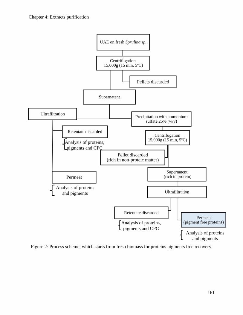
Chapter 4: Extracts purification
161
Figure 2: Process scheme, which starts from fresh biomass for proteins pigments free recovery.
UAE on fresh Sprulina sp.
Centrifugation15,000g (15 min, 5ºC)
Supernatent
Ultrafiltration
Retentate discarded
Permeat
Precipitation with ammonium sulfate 25% (w/v)
Centrifugation 15,000g (15 min, 5ºC)
Supernatent(rich in protein)
Ultrafiltration
Permeat(pigment free proteins)
Retentate discarded
Pellet discarded (rich in non-proteic matter)
Pellets discarded
Analysis of proteins,
pigments and CPC
Analysis of proteins
and pigments
Analysis of proteins,
pigments and CPC Analysis of proteins
and pigments
Chapter 4: Extracts purification
162
Pigments analysis
200 μL of supernatant was mixed with 1300 μL pure methanol, then incubated in dark for 1h at
45°C. Further on, the samples were centrifuged at 10,000 g for 10 min at 20°C. The organic phase
(methanol) containing the pigments was recovered. Chlorophyll and carotenoids were determined
using the equations proposed by Ritchie [28]:
(1) Total chlorophyll μg.L-1 = (9.3443 × 𝐴652) + (4.3481 × 𝐴665)
(2) Total carotenoids μg.L-1 = 4 × 𝐴480
4.3.2.6. Microscopic observation
To determine the efficiency of cell disruption, an observation was performed before and after
treatment on cell suspension placed on a specific plate (Nikon SMZ 1500). Magnification used
was 1000 times. The images were captured under a constant exposure and illumination by Nikon
eclipse E600 camera.
4.3.2.7. Statistical analysis
Experiments were conducted in triplicate (n=3). Statistically significant differences (p ≤ 0.05)
between the means were evaluated using one-way analysis of variance (ANOVA) and the Tukey's
test on XLTAT software version 2018.1.
4.3.3. Results and discussions
4.3.3.1. Influence of ultrasonic parameter on proteins recovery
Crude proteins content of the initial biomass was estimated at 71 %. This value is close to what
reported for Spirulina in several studies [29] [30].
Effect of stirring rate. Figure 3 shows that, under 500 rpm stirring rate and in the absence of any
UAE treatment, the yield of soluble proteins extraction was around 34% of total proteins content
after 60 min of treatment (P0W0).
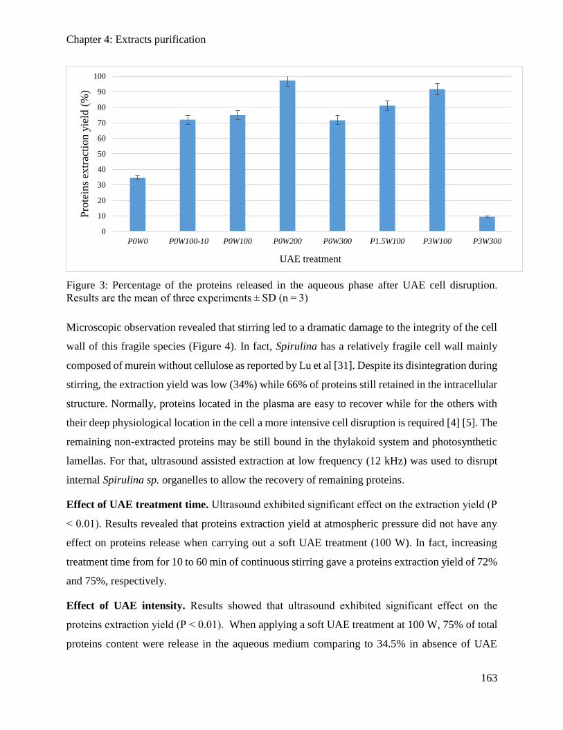
Chapter 4: Extracts purification
163
Figure 3: Percentage of the proteins released in the aqueous phase after UAE cell disruption.
Results are the mean of three experiments ± SD (n = 3)
Microscopic observation revealed that stirring led to a dramatic damage to the integrity of the cell
wall of this fragile species (Figure 4). In fact, Spirulina has a relatively fragile cell wall mainly
composed of murein without cellulose as reported by Lu et al [31]. Despite its disintegration during
stirring, the extraction yield was low (34%) while 66% of proteins still retained in the intracellular
structure. Normally, proteins located in the plasma are easy to recover while for the others with
their deep physiological location in the cell a more intensive cell disruption is required [4] [5]. The
remaining non-extracted proteins may be still bound in the thylakoid system and photosynthetic
lamellas. For that, ultrasound assisted extraction at low frequency (12 kHz) was used to disrupt
internal Spirulina sp. organelles to allow the recovery of remaining proteins.
Effect of UAE treatment time. Ultrasound exhibited significant effect on the extraction yield (P
< 0.01). Results revealed that proteins extraction yield at atmospheric pressure did not have any
effect on proteins release when carrying out a soft UAE treatment (100 W). In fact, increasing
treatment time from for 10 to 60 min of continuous stirring gave a proteins extraction yield of 72%
and 75%, respectively.
Effect of UAE intensity. Results showed that ultrasound exhibited significant effect on the
proteins extraction yield (P < 0.01). When applying a soft UAE treatment at 100 W, 75% of total
proteins content were release in the aqueous medium comparing to 34.5% in absence of UAE
0
10
20
30
40
50
60
70
80
90
100
P0W0 P0W100-10 P0W100 P0W200 P0W300 P1.5W100 P3W100 P3W300
Pro
tein
s ex
trac
tion y
ield
(%
)
UAE treatment
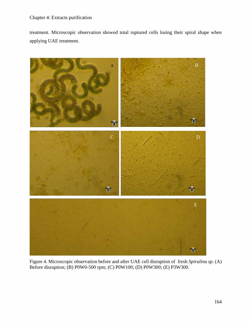
Chapter 4: Extracts purification
164
treatment. Microscopic observation showed total ruptured cells losing their spiral shape when
applying UAE treatment.
Figure 4. Microscopic observation before and after UAE cell disruption of fresh Spirulina sp. (A)
Before disruption; (B) P0W0-500 rpm; (C) P0W100; (D) P0W300; (E) P3W300.
A B
C D
E
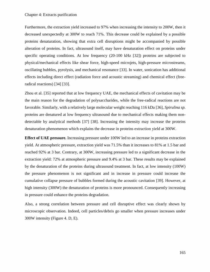
Chapter 4: Extracts purification
165
Furthermore, the extraction yield increased to 97% when increasing the intensity to 200W, then it
decreased unexpectedly at 300W to reach 71%. This decrease could be explained by a possible
proteins denaturation, showing that extra cell disruptions might be accompanied by possible
alteration of proteins. In fact, ultrasound itself, may have denaturation effect on proteins under
specific operating conditions. At low frequency (20-100 kHz [32]) proteins are subjected to
physical/mechanical effects like shear force, high-speed microjets, high-pressure microstreams,
oscillating bubbles, pyrolysis, and mechanical resonance [33]. In water, sonication has additional
effects including direct effect (radiation force and acoustic streaming) and chemical effect (free-
radical reactions) [34] [33].
Zhou et al. [35] reported that at low frequency UAE, the mechanical effects of cavitation may be
the main reason for the degradation of polysaccharides, while the free-radical reactions are not
favorable. Similarly, with a relatively large molecular weight reaching 116 kDa [36], Spirulina sp.
proteins are denatured at low frequency ultrasound due to mechanical effects making them non-
detectable by analytical methods [37] [38]. Increasing the intensity may increase the proteins
denaturation phenomenon which explains the decrease in proteins extraction yield at 300W.
Effect of UAE pressure. Increasing pressure under 100W led to an increase in proteins extraction
yield. At atmospheric pressure, extraction yield was 71.5% than it increases to 81% at 1.5 bar and
reached 92% at 3 bar. Contrary, at 300W, increasing pressure led to a significant decrease in the
extraction yield: 72% at atmospheric pressure and 9.4% at 3 bar. These results may be explained
by the denaturation of the proteins during ultrasound treatment. In fact, at low intensity (100W)
the pressure phenomenon is not significant and in increase in pressure could increase the
cumulative collapse pressure of bubbles formed during the acoustic cavitation [39]. However, at
high intensity (300W) the denaturation of proteins is more pronounced. Consequently increasing
in pressure could enhance the proteins degradation.
Also, a strong correlation between pressure and cell disruptive effect was clearly shown by
microscopic observation. Indeed, cell particles/debris go smaller when pressure increases under
300W intensity (Figure 4. D, E).
Chapter 4: Extracts purification
166
4.3.3.3. Ultrafiltration
After extraction with UAE and centrifugation, supernatant from experiment at 200W and
atmospheric pressure (P0W200) was taken for UF since it has the highest proteins content. Results
showed that proteins extract still contained small cell fragments and coloring pigments. To obtain
a clarified crude proteins extracts, a precipitation with ammonium sulfate 25% (w/v) was carried
out before UF.
As shown in Figure 5, in the absence of precipitation, the permeate flux decreased quickly to reach
2 kg/h.m2 whatever the cut-off membrane. However, when applying a precipitation on crude
extract before UF, the permeate flux decreased but remained high and reached 4 and 6 kg/h.m2
using 10 and 100 kDa cut-off membranes, respectively. This result may be due to the remove of
pigments (Figure 6) and cell debris (Figure 7) which may decrease the cake and gel forming on
the membrane surface.
Precipitation supernatant extract went through membrane filtration of 10 and 100 kDa molecular
weight cut-off. Figure 6 showed that 91% and 95% of CPC were removed in the retentate using
100 and 10 kDa MWCO membranes. This allowed to obtain a CPC free proteins fractions (Figure
8). This results came in accordance with MacColl [40] who reported that CPC can be found as a
complex solution of trimers, hexamers and other oligomers with a molecular weight in the range
of 44–260 kDa.
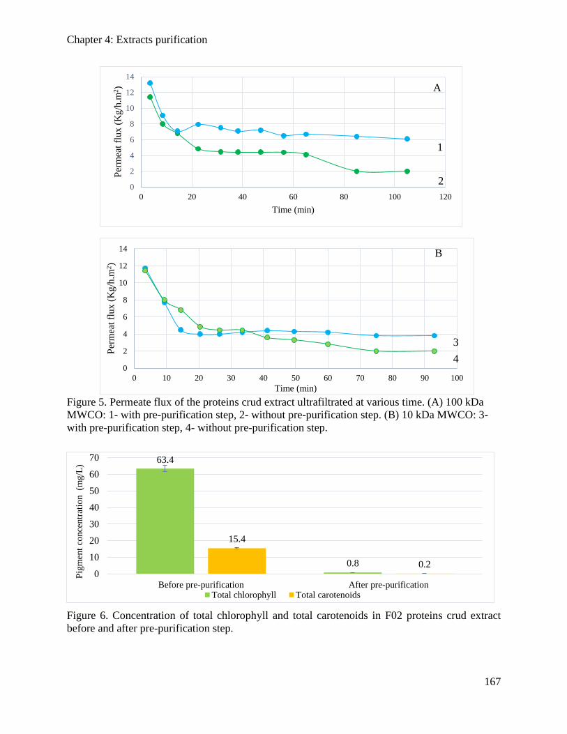
Chapter 4: Extracts purification
167
Figure 5. Permeate flux of the proteins crud extract ultrafiltrated at various time. (A) 100 kDa
MWCO: 1- with pre-purification step, 2- without pre-purification step. (B) 10 kDa MWCO: 3-
with pre-purification step, 4- without pre-purification step.
Figure 6. Concentration of total chlorophyll and total carotenoids in F02 proteins crud extract
before and after pre-purification step.
0
2
4
6
8
10
12
14
0 20 40 60 80 100 120
Per
mea
t fl
ux
(K
g/h
.m2)
Time (min)
0
2
4
6
8
10
12
14
0 10 20 30 40 50 60 70 80 90 100
Per
mea
t fl
ux (
Kg/h
.m2)
Time (min)
63.4
0.8
15.4
0.20
10
20
30
40
50
60
70
Before pre-purification After pre-purification
Pig
men
t co
nce
ntr
atio
n
(mg/L
)
Total chlorophyll Total carotenoids
A
B
1
2
3
4
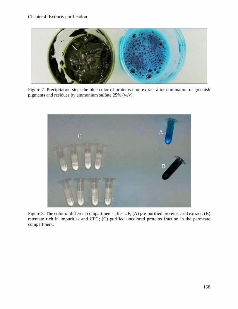
Chapter 4: Extracts purification
168
Figure 7. Precipitation step: the blue color of proteins crud extract after elimination of greenish
pigments and residues by ammonium sulfate 25% (w/v).
Figure 8. The color of different compartments after UF, (A) pre-purified proteins crud extract; (B)
retentate rich in impurities and CPC; (C) purified uncolored proteins fraction in the permeate
compartment.
C A
B
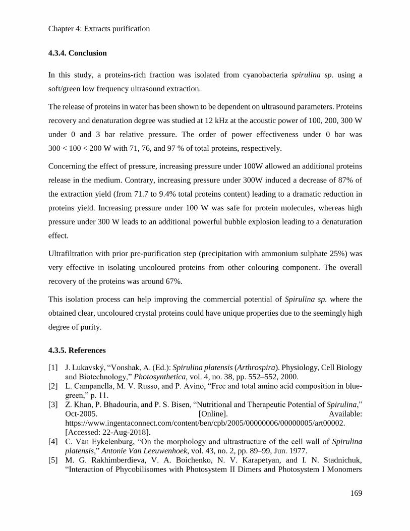
Chapter 4: Extracts purification
169
4.3.4. Conclusion
In this study, a proteins-rich fraction was isolated from cyanobacteria spirulina sp. using a
soft/green low frequency ultrasound extraction.
The release of proteins in water has been shown to be dependent on ultrasound parameters. Proteins
recovery and denaturation degree was studied at 12 kHz at the acoustic power of 100, 200, 300 W
under 0 and 3 bar relative pressure. The order of power effectiveness under 0 bar was
300 < 100 < 200 W with 71, 76, and 97 % of total proteins, respectively.
Concerning the effect of pressure, increasing pressure under 100W allowed an additional proteins
release in the medium. Contrary, increasing pressure under 300W induced a decrease of 87% of
the extraction yield (from 71.7 to 9.4% total proteins content) leading to a dramatic reduction in
proteins yield. Increasing pressure under 100 W was safe for protein molecules, whereas high
pressure under 300 W leads to an additional powerful bubble explosion leading to a denaturation
effect.
Ultrafiltration with prior pre-purification step (precipitation with ammonium sulphate 25%) was
very effective in isolating uncoloured proteins from other colouring component. The overall
recovery of the proteins was around 67%.
This isolation process can help improving the commercial potential of Spirulina sp. where the
obtained clear, uncoloured crystal proteins could have unique properties due to the seemingly high
degree of purity.
4.3.5. References
[1] J. Lukavský, “Vonshak, A. (Ed.): Spirulina platensis (Arthrospira). Physiology, Cell Biology
and Biotechnology,” Photosynthetica, vol. 4, no. 38, pp. 552–552, 2000.
[2] L. Campanella, M. V. Russo, and P. Avino, “Free and total amino acid composition in blue-
green,” p. 11.
[3] Z. Khan, P. Bhadouria, and P. S. Bisen, “Nutritional and Therapeutic Potential of Spirulina,”
Oct-2005. [Online]. Available:
https://www.ingentaconnect.com/content/ben/cpb/2005/00000006/00000005/art00002.
[Accessed: 22-Aug-2018].
[4] C. Van Eykelenburg, “On the morphology and ultrastructure of the cell wall of Spirulina
platensis,” Antonie Van Leeuwenhoek, vol. 43, no. 2, pp. 89–99, Jun. 1977.
[5] M. G. Rakhimberdieva, V. A. Boichenko, N. V. Karapetyan, and I. N. Stadnichuk,
“Interaction of Phycobilisomes with Photosystem II Dimers and Photosystem I Monomers
Chapter 4: Extracts purification
170
and Trimers in the Cyanobacterium Spirulina platensis,” Biochemistry (Mosc.), vol. 40, no.
51, pp. 15780–15788, Dec. 2001.
[6] M. Kissoudi, I. Sarakatsianos, and V. Samanidou, “Isolation and purification of food-grade
C-phycocyanin from Arthrospira platensis and its determination in confectionery by HPLC
with diode array detection,” J. Sep. Sci., vol. 41, no. 4, pp. 975–981, Feb. 2018.
[7] P. Jaouen, B. Lépine, N. Rossignol, R. Royer, and F. Quéméneur, “Clarification and
concentration with membrane technology of a phycocyanin solution extracted from Spirulina
platensis,” Biotechnol. Tech., vol. 13, no. 12, pp. 877–881, Dec. 1999.
[8] Chaiklahan et al., “Separation and purification of phycocyanin from Spirulina sp. using a
membrane process,” Bioresour. Technol., vol. 102, no. 14, pp. 7159–7164, Jul. 2011.
[9] J. A. Gerde, L. Yao, J. Lio, Z. Wen, and T. Wang, “Microalgae flocculation: Impact of
flocculant type, algae species and cell concentration,” Algal Res., vol. 3, pp. 30–35, Jan. 2014.
[10] R. Giordano, U. Leuzzi, and F. Wanderlingh, “Effects of ultrasound on unicellular algae,” J.
Acoust. Soc. Am., vol. 60, no. 1, pp. 275–278, Jul. 1976.
[11] Araujo et al., “Extraction of lipids from microalgae by ultrasound application: Prospection of
the optimal extraction method,” Ultrason. Sonochem., vol. 20, no. 1, pp. 95–98, Jan. 2013.
[12] E. Roselló-Soto et al., “Clean recovery of antioxidant compounds from plant foods, by-
products and algae assisted by ultrasounds processing. Modeling approaches to optimize
processing conditions,” Trends Food Sci. Technol., vol. 42, no. 2, pp. 134–149, Apr. 2015.
[13] Rossignol et al, “Membrane technology for the continuous separation microalgae/culture
medium: compared performances of cross-flow microfiltration and ultrafiltration,” Aquac.
Eng., vol. 20, no. 3, pp. 191–208, Aug. 1999.
[14] A. G. Waghmare, M. K. Salve, J. G. LeBlanc, and S. S. Arya, “Concentration and
characterization of microalgae proteins from Chlorella pyrenoidosa,” Bioresour. Bioprocess,
vol. 3, no. 1, p. 16, Mar. 2016.
[15] bilad et al., “Membrane technology in microalgae cultivation and harvesting: A review,”
Biotechnol. Adv., vol. 32, no. 7, pp. 1283–1300, Nov. 2014.
[16] Gerardo et al., “Integration of membrane technology in microalgae biorefineries,” J. Membr.
Sci., vol. 464, pp. 86–99, Aug. 2014.
[17] Zhang et al., “Harvesting algal biomass for biofuels using ultrafiltration membranes,”
Bioresour. Technol., vol. 101, no. 14, pp. 5297–5304, Jul. 2010.
[18] Marcati et al., “Extraction and fractionation of polysaccharides and B-phycoerythrin from the
microalga Porphyridium cruentum by membrane technology,” Algal Res., vol. 5, pp. 258–
263, Jul. 2014.
[19] Patel et al., “Separation and fractionation of exopolysaccharides from Porphyridium
cruentum,” Bioresour. Technol., vol. 145, pp. 345–350, Oct. 2013.
[20] Pugh et al., “(9) Isolation of Three High Molecular Weight Polysaccharide Preparations with
Potent Immunostimulatory Activity from Spirulina platensis , Aphanizomenon flos-aquae
and Chlorella pyrenoidosa | Request PDF,” ResearchGate. [Online]. Available:
https://www.researchgate.net/publication/11624110_Isolation_of_Three_High_Molecular_
Weight_Polysaccharide_Preparations_with_Potent_Immunostimulatory_Activity_from_Spi
rulina_platensis_Aphanizomenon_flos-aquae_and_Chlorella_pyrenoidosa. [Accessed: 22-
Aug-2018].
[21] Ba, Fatou, Ursu, Alina Violetta, Laroche, Celine, and Djelveh, Gholamreza, “Haematococcus
pluvialis soluble proteins: Extraction, characterization, concentration/fractionation and
emulsifying properties,” Bioresour. Technol., vol. 200, pp. 147–152, Jan. 2016.
Chapter 4: Extracts purification
171
[22] A.-V. Ursu, A. Marcati, T. Sayd, V. Sante-Lhoutellier, G. Djelveh, and P. Michaud,
“Extraction, fractionation and functional properties of proteins from the microalgae Chlorella
vulgaris,” Bioresour. Technol., vol. 157, pp. 134–139, Apr. 2014.
[23] Soni et al., “Extraction, purification and characterization of phycocyanin from Oscillatoria
quadripunctulata—Isolated from the rocky shores of Bet-Dwarka, Gujarat, India,” Process
Biochem., vol. 41, no. 9, pp. 2017–2023, Sep. 2006.
[24] A. Herrera, S. Boussiba, V. Napoleone, and A. Hohlberg, “Recovery of c-phycocyanin from
the cyanobacterium Spirulina maxima,” J. Appl. Phycol., vol. 1, no. 4, pp. 325–331, Dec.
1989.
[25] Burba et al., “Membrane filtration studies of aquatic humic substances and their metal
species: a concise overview: Part 1. Analytical fractionation by means of sequential-stage
ultrafiltration,” Talanta, vol. 45, no. 5, pp. 977–988, Mar. 1998.
[26] J. Jeon et al., “New mixture composition of organic solvents for efficient extraction of lipids
from Chlorella vulgaris,” Biomass Bioenergy, vol. 59, pp. 279–284, Dec. 2013.
[27] O. H. Lowry, A. L. Rosbrough, and R. J. Randall, “Lowry assay protocol,” J Biol Chem Lond,
vol. 193, p. 265, 1951.
[28] R. J. Ritchie, “Consistent Sets of Spectrophotometric Chlorophyll Equations for Acetone,
Methanol and Ethanol Solvents,” Photosynth. Res., vol. 89, no. 1, pp. 27–41, Jul. 2006.
[29] Bao et al., “Mixed fermentation of Spirulina platensis with Lactobacillus plantarum and
Bacillus subtilis by random-centroid optimization,” Food Chem., vol. 264, pp. 64–72, Oct.
2018.
[30] Danesi et al., “An investigation of effect of replacing nitrate by urea in the growth and
production of chlorophyll by Spirulina platensis,” Biomass Bioenergy, vol. 23, no. 4, pp.
261–269, Oct. 2002.
[31] H.-K. Lu, C.-C. Hsieh, J.-J. Hsu, Y.-K. Yang, and H.-N. Chou, “Preventive effects of
Spirulina platensis on skeletal muscle damage under exercise-induced oxidative stress,” Eur.
J. Appl. Physiol., vol. 98, no. 2, p. 220, Aug. 2006.
[32] O. Ngo et al., “Development of low frequency (20-100 kHz) clinically viable ultrasound
applicator for chronic wound treatment,” IEEE Trans. Ultrason. Ferroelectr. Freq. Control,
pp. 1–1, 2018.
[33] Kurokawa et al., “Effect of sonication frequency on the disruption of algae,” Ultrason.
Sonochem., vol. 31, pp. 157–162, Jul. 2016.
[34] Papadopoulos et al., “Sonochemical degradation of ethyl paraben in environmental samples:
Statistically important parameters determining kinetics, by-products and pathways,”
Ultrason. Sonochem., vol. 31, pp. 62–70, Jul. 2016.
[35] Zou and Ma, “Ultrasonic Degradation of Polysaccharide from a Red Algae (Porphyra
yezoensis),” 01-Mar-2006. [Online]. Available:
https://pubs.acs.org/doi/abs/10.1021/jf052763h. [Accessed: 22-Aug-2018].
[36] SChronakis et al., “The behaviour of protein preparations from blue-green algae (Spirulina
platensis strain Pacifica) at the air/water interface,” Colloids Surf. Physicochem. Eng. Asp.
vol. 173, no. 1–3, pp. 181–192, Nov. 2000.
[37] Liu et al., “The effects and mechanism of phycocyanin removal from water by high-
frequency ultrasound treatment,” Ultrason. Sonochem., vol. 41, pp. 303–309, Mar. 2018.
[38] Liu et al., “Performance and mechanism of phycocyanin removal from water by low-
frequency ultrasound treatment,” Ultrason. Sonochem., vol. 34, pp. 214–221, Jan. 2017.
Chapter 4: Extracts purification
172
[39] E. B. Flint and K. S. Suslick, “The Temperature of Cavitation,” Science, vol. 253, no. 5026,
pp. 1397–1399, Sep. 1991.
[40] MacColl, “Cyanobacterial Phycobilisomes,” J. Struct. Biol., vol. 124, no. 2–3, pp. 311–334,
Dec. 1998.
Chapter 4: Extracts purification
173
4.4. Selectivity of low-frequency ultrasound-assisted extraction for proteins
recovery from the microalga Chlorella vulgaris
(Article to be submitted to Journal of Applied Phycology)
Sara Obeida, b, c, Imane Benhamedd, Hosni Takacheb, c, Jérôme Peydecastainga, Ali Ismailb, c, Pierre-
Yves Pontalier1, a, Laurie Barthed.
a Laboratoire de Chimie Agro-industrielle (LCA), Université de Toulouse, INRA, INPT, 4 allée
Emile Monso, 31030 Toulouse cedex 4, France. b Département des Sciences et Technologies Alimentaires, Faculté des Sciences Agronomiques,
Université Libanaise, Dekwaneh, Lebanon. c Plateforme de Recherches et d’Analyses en Sciences de l’Environnement (PRASE), Ecole
Doctorale des Sciences et Technologies, Université Libanaise, Hadath, Lebanon. d Laboratoire de Génie Chimique (LGC), INP-ENSIACET, Université de Toulouse, 31030
Toulouse, France.
Abstract
The objective of this study was to evaluate the effectiveness of low-frequency ultrasound assisted
extraction (UAE) for selective proteins extraction. The microalga Chlorella vulgaris was subjected
to a low acoustic frequency (12 kHz) at low power intensity (0-100-300 W) and relative hydrostatic
pressure (0-3 bar). UAE was effective in the selective extraction of 96% of total proteins content,
shown by limited co-extraction of pigments and lipids. Increasing pressure at low power intensity
was highly effective, indicating that pressure played a critical role in proteins extraction yield. The
wall structure of Chlorella vulgaris plays an important role in the phenomena of selective
extraction. Cell integrity was maintained during total proteins extraction.
Keywords: Chlorella vulgaris, sono-extraction, selectivity, proteins, cell integrity.
4.4.1. Introduction
Over the last years, several research groups have investigated the use of simple technologies to
improve the extraction yield of specific compounds, such as lipids, proteins and intracellular
pigments, enclosed in microalgae structures [1]. In fact, microalgae may become a sustainable
source for many components by simplifying their recovery using reduced processing temperature,
Chapter 4: Extracts purification
174
green extraction solvent, short treatment time, and selective extraction methods with limited
purification steps [2] [3] [4].
Chlorella vulgaris is a microalga produced at industrial scale for its high proteins contents used in
human food [5] [6] [7] [8]. However, the limiting factor for the valorization of this compound is
the difficulty associate to its extraction due to the rigid cell wall of C. vulgaris [9]. For that, most
of the extraction methods are focused on the disruption of the cell wall before the recovery of the
proteins fraction [10]. The most used techniques are high pressure homogenizers, bead mills,
enzymatic or chemical treatments, thermal or osmotic shocks (repeated freezing/thawing) [11] [10]
[12][13]. Whatever the used method, once proteins fraction is recovered, it is necessary to setup
additional purification steps to separate the released proteins from the co-extracted components
[14].
Ultrasonication as an environmental friendly mechanical method, can be universally applied to
any type of microalgae for cell disintegration [15]. Several studies have reported the use of
ultrasound assisted extraction (UAE) on cyanobacteria in aim to extract intensely fluorescent
hydrosoluble proteins known as phycobiliproteins [16][17][18][19]. However, further purification
steps are needed due to dramatic cell disruption and the absence of any extraction selectivity.
Contrary to fragile cell wall, UAE treatment has a little acoustic cavitation effect on rigid cell wall
microalgae like Chlorella vulgaris [20] [21]. In fact, sonication offers a significant degree of cell
disruption on weaker walls that allows cells to break more readily [21].
Recently, Duan et al [22] investigated the effect of hydroxyl‐peroxide‐aided ultrasonic treatment
for proteins release from Chlorella sp. resulting in partial rupture of the cell wall with low proteins
extraction yield and the liberation of many other components. However, the apparent rigidity of
C. vulgaris could be exploited for selective extraction of the intracellular proteins, alleviating then
the key challenges in the final-stream steps of purification [23]. Hence, in this context, this study
proposes to evaluate the potential of UAE for high yield and selective extraction of proteins.
Furthermore, all UAE studies conducted for proteins release have been performed with frequencies
comprised between 20 and 100 kHz as reported lately by Chemat et al. [24]. However, to our best
of knowledge, UAE treatments for microalgal proteins extraction at audible frequencies (20 Hz-
20 kHz) have never been reported in the literature. Hence, we investigated the impact and
effectiveness of nearly audible frequency (12 kHz) on C. vulgaris suspension for selective
Chapter 4: Extracts purification
175
proteins extraction. In addition, to comply with the criteria of the green-chemistry concept, water
was used as the extraction solvent [25]. Besides, the effect operating conditions of UAE (e.g.
intensity, hydrostatic pressure) (have been studied. To avoid any thermal denaturation, the
temperature was kept at 30 ± 2°C during UAE treatments. Proteins extraction yield for all
supernatants was determined in order to evaluate the effectiveness of this technique. In addition,
the pigments and UAE residues were quantified to be used as a marker for inner organelles
alteration, cell wall disintegration, and lipids liberation. Microscopic observations have been
conducted to evaluate any cell disruption or cell morphology changes.
4.4.2. Methods
4.4.2.1. Microalgae Chlorella vulgaris
The selected Chlorophyceae, C. vulgaris (strain SAG 211-19), was supplied as a frozen paste from
Alpha Biotech (Asserac, France). This strain was cultivated in Sueoka media under conditions of
high N nutrient concentration to favor protein accumulation, and grown in batch mode in an indoor
tubular Air-Lift photobioreactor (10 L) at 25°C, harvested during the exponential growth phase,
concentrated by centrifugation, and then frozen. The harvested biomass contained 27% dry matter.
For UAE experiments, the frozen algal paste of C. vulgaris was first thawed at ambient temperature
and then diluted with water to prepare algal suspensions with a final concentration of 5% by
weight.
4.4.2.2. UAE procedure
Algal suspensions (600 mL) were placed in a 1L-capacity stainless steel ultrasonic reactor
connected to a pressurized N2 bottle allowing adjustment of the relative hydrostatic pressure (0-3
bar). Ultrasonic irradiation was emitted at the reactor bottom (cup-horn type) providing an
intensity power of 100, 200, or 300W at a very low frequency of 12 kHz. The constant volume of
biomass solution was stirred at 500 rpm and sonicated for 1 h (table 1). The suspension temperature
was kept at 30 ± 2°C by cool water circulation in an internal coil to limit denaturation. After each
treatment, samples were centrifuged at 20,000 g for 20 min at 5 °C. The mass of the pellets
(residues) was measured after all treatments and the supernatants analyzed for proteins and
pigment content.
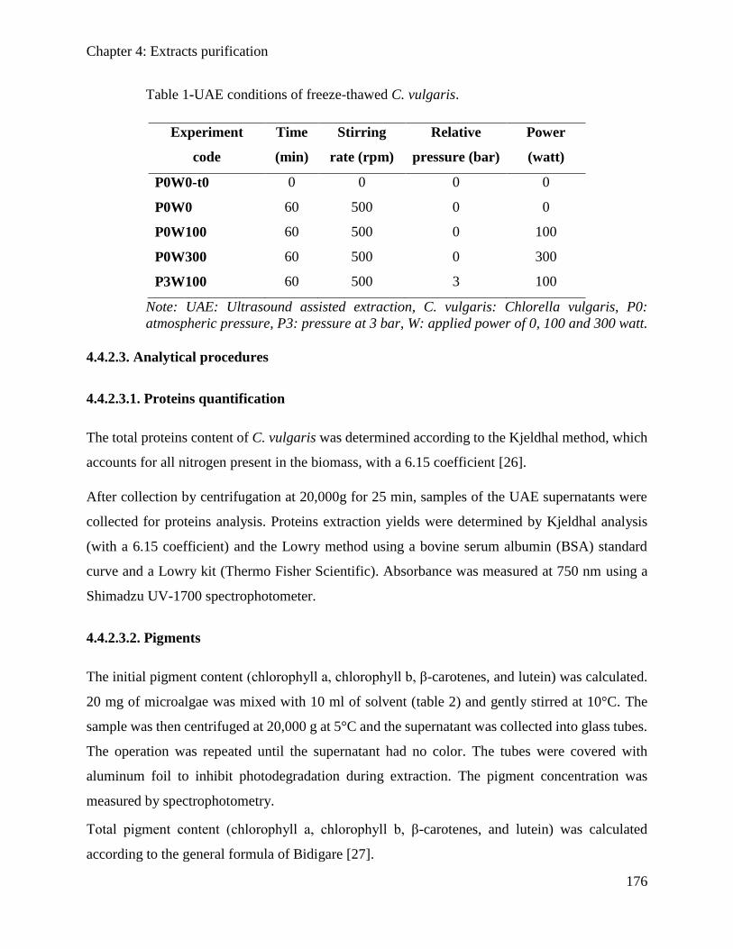
Chapter 4: Extracts purification
176
Table 1-UAE conditions of freeze-thawed C. vulgaris.
Experiment
code
Time
(min)
Stirring
rate (rpm)
Relative
pressure (bar)
Power
(watt)
P0W0-t0 0 0 0 0
P0W0 60 500 0 0
P0W100 60 500 0 100
P0W300 60 500 0 300
P3W100 60 500 3 100
Note: UAE: Ultrasound assisted extraction, C. vulgaris: Chlorella vulgaris, P0:
atmospheric pressure, P3: pressure at 3 bar, W: applied power of 0, 100 and 300 watt.
4.4.2.3. Analytical procedures
4.4.2.3.1. Proteins quantification
The total proteins content of C. vulgaris was determined according to the Kjeldhal method, which
accounts for all nitrogen present in the biomass, with a 6.15 coefficient [26].
After collection by centrifugation at 20,000g for 25 min, samples of the UAE supernatants were
collected for proteins analysis. Proteins extraction yields were determined by Kjeldhal analysis
(with a 6.15 coefficient) and the Lowry method using a bovine serum albumin (BSA) standard
curve and a Lowry kit (Thermo Fisher Scientific). Absorbance was measured at 750 nm using a
Shimadzu UV-1700 spectrophotometer.
4.4.2.3.2. Pigments
The initial pigment content (chlorophyll a, chlorophyll b, β-carotenes, and lutein) was calculated.
20 mg of microalgae was mixed with 10 ml of solvent (table 2) and gently stirred at 10°C. The
sample was then centrifuged at 20,000 g at 5°C and the supernatant was collected into glass tubes.
The operation was repeated until the supernatant had no color. The tubes were covered with
aluminum foil to inhibit photodegradation during extraction. The pigment concentration was
measured by spectrophotometry.
Total pigment content (chlorophyll a, chlorophyll b, β-carotenes, and lutein) was calculated
according to the general formula of Bidigare [27].
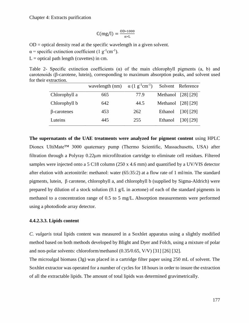
Chapter 4: Extracts purification
177
C(mg/l) =OD∗1000
α∗L
OD = optical density read at the specific wavelength in a given solvent.
α = specific extinction coefficient (1 g-1cm-1).
L = optical path length (cuvettes) in cm.
Table 2- Specific extinction coefficients (α) of the main chlorophyll pigments (a, b) and
carotenoids (β-carotene, lutein), corresponding to maximum absorption peaks, and solvent used
for their extraction.
wavelength (nm) α (1 g-1cm-1) Solvent Reference
Chlorophyll a 665 77.9 Methanol [28] [29]
Chlorophyll b 642 44.5 Methanol [28] [29]
β-carotenes 453 262 Ethanol [30] [29]
Luteins 445 255 Ethanol [30] [29]
The supernatants of the UAE treatments were analyzed for pigment content using HPLC
Dionex UltiMate™ 3000 quaternary pump (Thermo Scientific, Massachusetts, USA) after
filtration through a Polyray 0.22m microfiltration cartridge to eliminate cell residues. Filtered
samples were injected onto a 5 C18 column (250 x 4.6 mm) and quantified by a UV/VIS detector
after elution with acetonitrile: methanol: water (65:35:2) at a flow rate of 1 ml/min. The standard
pigments, lutein, β carotene, chlorophyll a, and chlorophyll b (supplied by Sigma-Aldrich) were
prepared by dilution of a stock solution (0.1 g/L in acetone) of each of the standard pigments in
methanol to a concentration range of 0.5 to 5 mg/L. Absorption measurements were performed
using a photodiode array detector.
4.4.2.3.3. Lipids content
C. vulgaris total lipids content was measured in a Soxhlet apparatus using a slightly modified
method based on both methods developed by Blight and Dyer and Folch, using a mixture of polar
and non-polar solvents: chloroform/methanol (0.35/0.65, V/V) [31] [26] [32].
The microalgal biomass (3g) was placed in a cartridge filter paper using 250 mL of solvent. The
Soxhlet extractor was operated for a number of cycles for 18 hours in order to insure the extraction
of all the extractable lipids. The amount of total lipids was determined gravimetrically.
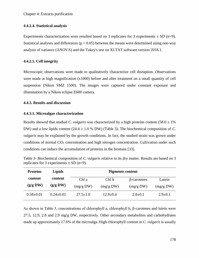
Chapter 4: Extracts purification
178
4.4.2.4. Statistical analysis
Experiments characterization were resulted based on 3 replicates for 3 experiments ± SD (n=9).
Statistical analyses and differences (p < 0.05) between the means were determined using one-way
analysis of variance (ANOVA) and the Tukey's test on XLTAT software version 2018.1.
4.4.2.5. Cell integrity
Microscopic observations were made to qualitatively characterize cell disruption. Observations
were made at high magnification (x1000) before and after treatment on a small quantity of cell
suspension (Nikon SMZ 1500). The images were captured under constant exposure and
illumination by a Nikon eclipse E600 camera.
4.4.3. Results and discussion
4.4.3.1. Microalgae characterization
Results showed that studied C. vulgaris was characterized by a high proteins content (58.0 ± 1%
DW) and a low lipids content (24.4 ± 1.0 % DW) (Table 3). The biochemical composition of C.
vulgaris may be explained by the growth conditions. In fact, the studied strain was grown under
conditions of normal CO2 concentration and high nitrogen concentration. Cultivation under such
conditions can induce the accumulation of proteins in the biomass [33].
Table 3- Biochemical composition of C. vulgaris relative to its dry matter. Results are based on 3
replicates for 3 experiments ± SD (n=9).
Proteins
content
(g/g DW)
Lipids
content
(g/g DW)
Pigments content
Chl a
(mg/g DW)
Chl b
(mg/g DW)
β-carotenes
(mg/g DW)
Lutein
(mg/g DW)
0.58±0.01 0.24±0.01 27.5±1.0 12.9±0.4 2.8±0.1 2.9±0.1
As shown in Table 3, concentrations of chlorophyll a, chlorophyll b, β-carotenes and lutein were
27.5, 12.9, 2.8 and 2.9 mg/g DW, respectively. Other secondary metabolites and carbohydrates
made up approximately 17.6% of the microalga. High chlorophyll content in C. vulgaris is usually
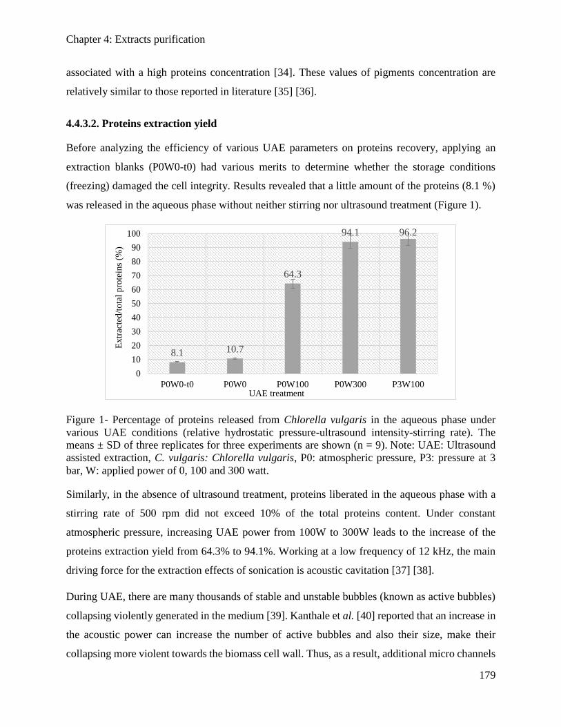
Chapter 4: Extracts purification
179
associated with a high proteins concentration [34]. These values of pigments concentration are
relatively similar to those reported in literature [35] [36].
4.4.3.2. Proteins extraction yield
Before analyzing the efficiency of various UAE parameters on proteins recovery, applying an
extraction blanks (P0W0-t0) had various merits to determine whether the storage conditions
(freezing) damaged the cell integrity. Results revealed that a little amount of the proteins (8.1 %)
was released in the aqueous phase without neither stirring nor ultrasound treatment (Figure 1).
Figure 1- Percentage of proteins released from Chlorella vulgaris in the aqueous phase under
various UAE conditions (relative hydrostatic pressure-ultrasound intensity-stirring rate). The
means ± SD of three replicates for three experiments are shown (n = 9). Note: UAE: Ultrasound
assisted extraction, C. vulgaris: Chlorella vulgaris, P0: atmospheric pressure, P3: pressure at 3
bar, W: applied power of 0, 100 and 300 watt.
Similarly, in the absence of ultrasound treatment, proteins liberated in the aqueous phase with a
stirring rate of 500 rpm did not exceed 10% of the total proteins content. Under constant
atmospheric pressure, increasing UAE power from 100W to 300W leads to the increase of the
proteins extraction yield from 64.3% to 94.1%. Working at a low frequency of 12 kHz, the main
driving force for the extraction effects of sonication is acoustic cavitation [37] [38].
During UAE, there are many thousands of stable and unstable bubbles (known as active bubbles)
collapsing violently generated in the medium [39]. Kanthale et al. [40] reported that an increase in
the acoustic power can increase the number of active bubbles and also their size, make their
collapsing more violent towards the biomass cell wall. Thus, as a result, additional micro channels
8.1 10.7
64.3
94.1 96.2
0
10
20
30
40
50
60
70
80
90
100
P0W0-t0 P0W0 P0W100 P0W300 P3W100
Extr
acte
d/t
ota
l p
rote
ins
(%)
UAE treatment
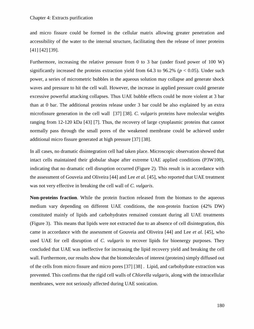
Chapter 4: Extracts purification
180
and micro fissure could be formed in the cellular matrix allowing greater penetration and
accessibility of the water to the internal structure, facilitating then the release of inner proteins
[41] [42] [39].
Furthermore, increasing the relative pressure from 0 to 3 bar (under fixed power of 100 W)
significantly increased the proteins extraction yield from 64.3 to 96.2% (p < 0.05). Under such
power, a series of micrometric bubbles in the aqueous solution may collapse and generate shock
waves and pressure to hit the cell wall. However, the increase in applied pressure could generate
excessive powerful attacking collapses. Thus UAE bubble effects could be more violent at 3 bar
than at 0 bar. The additional proteins release under 3 bar could be also explained by an extra
microfissure generation in the cell wall [37] [38]. C. vulgaris proteins have molecular weights
ranging from 12-120 kDa [43] [7]. Thus, the recovery of large cytoplasmic proteins that cannot
normally pass through the small pores of the weakened membrane could be achieved under
additional micro fissure generated at high pressure [37] [38].
In all cases, no dramatic disintegration cell had taken place. Microscopic observation showed that
intact cells maintained their globular shape after extreme UAE applied conditions (P3W100),
indicating that no dramatic cell disruption occurred (Figure 2). This result is in accordance with
the assessment of Gouveia and Oliveira [44] and Lee et al. [45], who reported that UAE treatment
was not very effective in breaking the cell wall of C. vulgaris.
Non-proteins fraction. While the protein fraction released from the biomass to the aqueous
medium vary depending on different UAE conditions, the non-protein fraction (42% DW)
constituted mainly of lipids and carbohydrates remained constant during all UAE treatments
(Figure 3). This means that lipids were not extracted due to an absence of cell disintegration, this
came in accordance with the assessment of Gouveia and Oliveira [44] and Lee et al. [45], who
used UAE for cell disruption of C. vulgaris to recover lipids for bioenergy purposes. They
concluded that UAE was ineffective for increasing the lipid recovery yield and breaking the cell
wall. Furthermore, our results show that the biomolecules of interest (proteins) simply diffused out
of the cells from micro fissure and micro pores [37] [38] . Lipid, and carbohydrate extraction was
prevented. This confirms that the rigid cell walls of Chlorella vulgaris, along with the intracellular
membranes, were not seriously affected during UAE sonication.
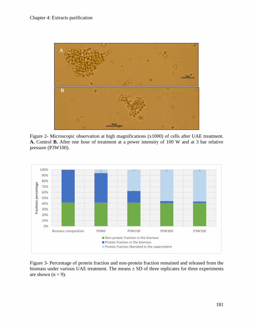
Chapter 4: Extracts purification
181
Figure 2- Microscopic observation at high magnifications (x1000) of cells after UAE treatment.
A. Control B. After one hour of treatment at a power intensity of 100 W and at 3 bar relative
pressure (P3W100).
Figure 3- Percentage of protein fraction and non-protein fraction remained and released from the
biomass under various UAE treatment. The means ± SD of three replicates for three experiments
are shown (n = 9).
0%
10%
20%
30%
40%
50%
60%
70%
80%
90%
100%
Biomass composition P0W0 P0W100 P0W300 P3W100
Frac
tio
ns
pe
rce
nta
ge
Non-proteic fraction in the biomassProteic fraction in the biomassProteic fraction liberated in the supernatent
B
A
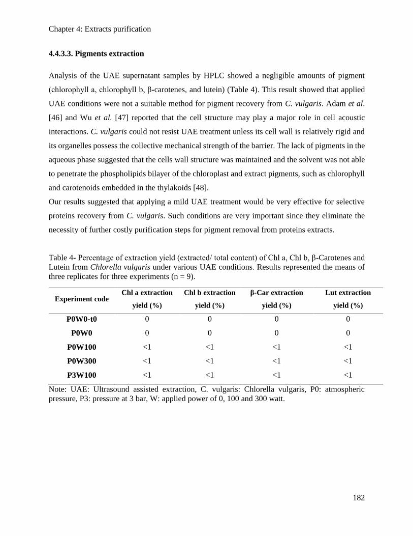
Chapter 4: Extracts purification
182
4.4.3.3. Pigments extraction
Analysis of the UAE supernatant samples by HPLC showed a negligible amounts of pigment
(chlorophyll a, chlorophyll b, β-carotenes, and lutein) (Table 4). This result showed that applied
UAE conditions were not a suitable method for pigment recovery from C. vulgaris. Adam et al.
[46] and Wu et al. [47] reported that the cell structure may play a major role in cell acoustic
interactions. C. vulgaris could not resist UAE treatment unless its cell wall is relatively rigid and
its organelles possess the collective mechanical strength of the barrier. The lack of pigments in the
aqueous phase suggested that the cells wall structure was maintained and the solvent was not able
to penetrate the phospholipids bilayer of the chloroplast and extract pigments, such as chlorophyll
and carotenoids embedded in the thylakoids [48].
Our results suggested that applying a mild UAE treatment would be very effective for selective
proteins recovery from C. vulgaris. Such conditions are very important since they eliminate the
necessity of further costly purification steps for pigment removal from proteins extracts.
Table 4- Percentage of extraction yield (extracted/ total content) of Chl a, Chl b, β-Carotenes and
Lutein from Chlorella vulgaris under various UAE conditions. Results represented the means of
three replicates for three experiments (n = 9).
Experiment code Chl a extraction
yield (%)
Chl b extraction
yield (%)
β-Car extraction
yield (%)
Lut extraction
yield (%)
P0W0-t0 0 0 0 0
P0W0 0 0 0 0
P0W100 <1 <1 <1 <1
P0W300 <1 <1 <1 <1
P3W100 <1 <1 <1 <1
Note: UAE: Ultrasound assisted extraction, C. vulgaris: Chlorella vulgaris, P0: atmospheric
pressure, P3: pressure at 3 bar, W: applied power of 0, 100 and 300 watt.
Chapter 4: Extracts purification
183
4.4.4. Conclusion
This study showed that UAE at a low acoustic frequency of 12 kHz and low intensity power is
very effective for selective extraction of proteins from C. vulgaris. 94.1% and 96.2% of initial
proteins content were released under atmospheric pressure/300W and 3bar/100W, respectively.
Although the two conditions resulted in similar and high proteins yields, the second is preferable,
because it is more efficient economically. In all conditions, negligible amount of pigments and
non-proteins matter were released, showing a selective extraction of protein fraction. The wall
structure of C. vulgaris played an important role in the release of proteins as a specific component.
Algal protein fraction started to find new applications in cosmetics and pharmaceutical industries
[49]. However, current proteins extraction techniques lead to a co-liberation of non-protein
compounds (like pigments and lipids) conferring a significant non-desirable flavors and colors in
the final product. Our results showed that UAE under specific conditions could be applied for
selective proteins recovery with no need of further purification demands. This deals with the
principal of the algorefinery concept, making the large scale of microalgae application more
economically efficient.
4.4.5. References
[1] F. J. Barba, N. Grimi, and E. Vorobiev, “New Approaches for the Use of Non-conventional
Cell Disruption Technologies to Extract Potential Food Additives and Nutraceuticals from
Microalgae,” Food Eng. Rev., vol. 7, no. 1, pp. 45–62, Mar. 2015.
[2] W. N. Phong, P. L. Show, C. F. Le, Y. Tao, J.-S. Chang, and T. C. Ling, “Improving cell
disruption efficiency to facilitate protein release from microalgae using chemical and
mechanical integrated method,” Biochem. Eng. J., vol. 135, pp. 83–90, Jul. 2018.
[3] E. Günerken, E. D’Hondt, M. H. M. Eppink, L. Garcia-Gonzalez, K. Elst, and R. H. Wijffels,
“Cell disruption for microalgae biorefineries,” Biotechnol. Adv., vol. 33, no. 2, pp. 243–260,
Mar. 2015.
[4] M. M. Poojary et al., “Innovative Alternative Technologies to Extract Carotenoids from
Microalgae and Seaweeds,” Mar. Drugs, vol. 14, no. 11, p. 214, Nov. 2016.
[5] E. W. Becker, “Micro-algae as a source of protein,” Biotechnol. Adv., vol. 25, no. 2, pp. 207–
210, Mar. 2007.
[6] J. A. Lubitz, “The Protein Quality, Digestibility, and Composition of Algae, Chlorella
71105a,” J. Food Sci., vol. 28, no. 2, pp. 229–232.
[7] H. J. Morris et al., “Protein hydrolysates from the alga Chlorella vulgaris 87/1 with
potentialities in immunonutrition,” Biotecnol. Apl., vol. 26, no. 2, pp. 162–165, Jun. 2009.
Chapter 4: Extracts purification
184
[8] J. Seyfabadi, Z. Ramezanpour, and Z. A. Khoeyi, “Protein, fatty acid, and pigment content
of <Emphasis Type="Italic">Chlorella vulgaris</Emphasis> under different light regimes,”
J. Appl. Phycol., vol. 23, no. 4, pp. 721–726, Aug. 2011.
[9] H. J. Morris, A. Almarales, O. Carrillo, and R. C. Bermúdez, “Utilisation of
Chlorellavulgaris cell biomass for the production of enzymatic protein hydrolysates,”
Bioresour. Technol., vol. 99, no. 16, pp. 7723–7729, Nov. 2008.
[10] R. Seetharam and S. K. Sharma, Purification and Analysis of Recombinant Proteins. CRC
Press, 1991.
[11] J. Doucha and K. Lívanský, “Influence of processing parameters on disintegration of
<Emphasis Type="Italic">Chlorella</Emphasis> cells in various types of homogenizers,”
Appl. Microbiol. Biotechnol., vol. 81, no. 3, p. 431, Dec. 2008.
[12] A. P. J. Middelberg, “Process-scale disruption of microorganisms,” Biotechnol. Adv., vol.
13, no. 3, pp. 491–551, Jan. 1995.
[13] Y. W. Sari, M. E. Bruins, and J. P. M. Sanders, “Enzyme assisted protein extraction from
rapeseed, soybean, and microalgae meals,” Ind. Crops Prod., vol. 43, pp. 78–83, May 2013.
[14] A.-V. Ursu, A. Marcati, T. Sayd, V. Sante-Lhoutellier, G. Djelveh, and P. Michaud,
“Extraction, fractionation and functional properties of proteins from the microalgae
Chlorella vulgaris,” Bioresour. Technol., vol. 157, pp. 134–139, Apr. 2014.
[15] J. Kim et al., “Methods of downstream processing for the production of biodiesel from
microalgae,” Biotechnol. Adv., vol. 31, no. 6, pp. 862–876, Nov. 2013.
[16] H. A. Tavanandi, R. Mittal, J. Chandrasekhar, and K. S. M. S. Raghavarao, “Simple and
efficient method for extraction of C-Phycocyanin from dry biomass of Arthospira platensis,”
Algal Res., vol. 31, pp. 239–251, Apr. 2018.
[17] R. D. P. Rodrigues, F. C. de Castro, R. S. de Santiago-Aguiar, and M. V. P. Rocha,
“Ultrasound-assisted extraction of phycobiliproteins from Spirulina (Arthrospira) platensis
using protic ionic liquids as solvent,” Algal Res., vol. 31, pp. 454–462, Apr. 2018.
[18] P. Jaouen, B. Lépine, N. Rossignol, R. Royer, and F. Quéméneur, “Clarification and
concentration with membrane technology of a phycocyanin solution extracted from Spirulina
platensis,” Biotechnol. Tech., vol. 13, no. 12, pp. 877–881, Dec. 1999.
[19] R. V. Aftari, K. Rezaei, A. Mortazavi, and A. R. Bandani, “The Optimized Concentration
and Purity of Spirulina platensis C-Phycocyanin: A Comparative Study on Microwave-
Assisted and Ultrasound-Assisted Extraction Methods,” J. Food Process. Preserv., vol. 39,
no. 6, pp. 3080–3091.
[20] P. Rajasekhar, L. Fan, T. Nguyen, and F. A. Roddick, “Impact of sonication at 20 kHz on
Microcystis aeruginosa, Anabaena circinalis and Chlorella sp.,” Water Res., vol. 46, no. 5,
pp. 1473–1481, Apr. 2012.
[21] R. R. dos Santos, D. M. Moreira, C. N. Kunigami, D. A. G. Aranda, and C. M. L. L. Teixeira,
“Comparison between several methods of total lipid extraction from Chlorella vulgaris
biomass,” Ultrason. Sonochem., vol. 22, pp. 95–99, Jan. 2015.
Chapter 4: Extracts purification
185
[22] Z. Duan, X. Tan, K. Dai, H. Gu, and H. Yang, “Evaluation on H2O2-aided ultrasonic
pretreatment for cell disruption of Chlorella pyrenoidosa,” Asia-Pac. J. Chem. Eng., vol. 12,
no. 3, pp. 502–510.
[23] E. Roselló-Soto et al., “Clean recovery of antioxidant compounds from plant foods, by-
products and algae assisted by ultrasounds processing. Modeling approaches to optimize
processing conditions,” Trends Food Sci. Technol., vol. 42, no. 2, pp. 134–149, Apr. 2015.
[24] F. Chemat, N. Rombaut, A.-G. Sicaire, A. Meullemiestre, A.-S. Fabiano-Tixier, and M.
Abert-Vian, “Ultrasound assisted extraction of food and natural products. Mechanisms,
techniques, combinations, protocols and applications. A review,” Ultrason. Sonochem., vol.
34, pp. 540–560, Jan. 2017.
[25] G. Chatel, “How sonochemistry contributes to green chemistry?,” Ultrason. Sonochem., vol.
40, pp. 117–122, Jan. 2018.
[26] J. Jeon et al., “New mixture composition of organic solvents for efficient extraction of lipids
from Chlorella vulgaris,” Biomass Bioenergy, vol. 59, pp. 279–284, Dec. 2013.
[27] R. R. Bidigare, “Analysis of Algal Chlorophylls and Carotenoids,” in Marine Particles:
Analysis and Characterization, American Geophysical Union (AGU), 2013, pp. 119–123.
[28] O. Holm-Hansen and B. Riemann, “Chlorophyll a Determination: Improvements in
Methodology,” Oikos, vol. 30, no. 3, pp. 438–447, 1978.
[29] V. Brotas and M.-R. Plante-Cuny, “Identification and quantification of chlorophyll and
carotenoid pigments in marine sediments. A protocol for HPLC analysis.,” Oceanol. Acta
Paris, vol. 19, no. 6, pp. 623–634, 1996.
[30] T. W. Goodwin, “Biosynthesis of Carotenoids,” in The Biochemistry of the Carotenoids,
Springer, Dordrecht, 1980, pp. 33–76.
[31] K. Ramluckan, K. G. Moodley, and F. Bux, “An evaluation of the efficacy of using selected
solvents for the extraction of lipids from algal biomass by the soxhlet extraction method,”
Fuel, vol. 116, pp. 103–108, Jan. 2014.
[32] Y. Du, B. Schuur, S. R. A. Kersten, and D. W. F. Brilman, “Opportunities for switchable
solvents for lipid extraction from wet algal biomass: An energy evaluation,” Algal Res., vol.
11, pp. 271–283, Sep. 2015.
[33] A. L. Stephenson, J. S. Dennis, C. J. Howe, S. A. Scott, and A. G. Smith, “Influence of
nitrogen-limitation regime on the production by Chlorella vulgaris of lipids for biodiesel
feedstocks,” Biofuels, vol. 1, no. 1, pp. 47–58, Jan. 2010.
[34] N. F. Y. Tam and Y. S. Wong, “Effect of ammonia concentrations on growth of Chlorella
vulgaris and nitrogen removal from media,” Bioresour. Technol., vol. 57, no. 1, pp. 45–50,
Jul. 1996.
[35] W. Kong, H. Yang, Y. Cao, H. Song, and S. Hua, Effects of glycerol and glucose on the
enhancement of biomass, lipid and soluble carbohydrate production by chlorella vulgaris in
mixotrophic culture. Food Technology and Biotechnology. 2013.
Chapter 4: Extracts purification
186
[36] C. Safi, B. Zebib, O. Merah, P.-Y. Pontalier, and C. Vaca-Garcia, “Morphology,
composition, production, processing and applications of Chlorella vulgaris: A review,”
Renew. Sustain. Energy Rev., vol. 35, pp. 265–278, Jul. 2014.
[37] T. J. Mason, A. J. Cobley, J. E. Graves, and D. Morgan, “New evidence for the inverse
dependence of mechanical and chemical effects on the frequency of ultrasound,” Ultrason.
Sonochem., vol. 18, no. 1, pp. 226–230, Jan. 2011.
[38] S. Kentish and M. Ashokkumar, “The Physical and Chemical Effects of Ultrasound,” in
Ultrasound Technologies for Food and Bioprocessing, Springer, New York, NY, 2011, pp.
1–12.
[39] B. K. Tiwari, “Ultrasound: A clean, green extraction technology,” TrAC Trends Anal. Chem.,
vol. 71, pp. 100–109, Sep. 2015.
[40] P. Kanthale, M. Ashokkumar, and F. Grieser, “Sonoluminescence, sonochemistry (H2O2
yield) and bubble dynamics: Frequency and power effects,” Ultrason. Sonochem., vol. 15,
no. 2, pp. 143–150, Feb. 2008.
[41] H. Li, L. Pordesimo, and J. Weiss, “High intensity ultrasound-assisted extraction of oil from
soybeans,” Food Res. Int., vol. 37, no. 7, pp. 731–738, Aug. 2004.
[42] S. Both, F. Chemat, and J. Strube, “Extraction of polyphenols from black tea – Conventional
and ultrasound assisted extraction,” Ultrason. Sonochem., vol. 21, no. 3, pp. 1030–1034,
May 2014.
[43] H. M. Khairy, E. M. Ali, and S. M. Dowidar, “Comparative effects of autotrophic and
heterotrophic growth on some vitamins, 2,2-diphenyl-1-picrylhydrazyl (DPPH) free radical
scavenging activity, amino acids and protein profile of Chlorella vulgaris Beijerinck,” Afr.
J. Biotechnol., vol. 10, no. 62, pp. 13514–13519, Jan. 2011.
[44] L. Gouveia and A. C. Oliveira, “Microalgae as a raw material for biofuels production,” J.
Ind. Microbiol. Biotechnol., vol. 36, no. 2, pp. 269–274, Feb. 2009.
[45] J.-Y. Lee, C. Yoo, S.-Y. Jun, C.-Y. Ahn, and H.-M. Oh, “Comparison of several methods for
effective lipid extraction from microalgae,” Bioresour. Technol., vol. 101, no. 1, Supplement,
pp. S75–S77, Jan. 2010.
[46] F. Adam, M. Abert-Vian, G. Peltier, and F. Chemat, “‘Solvent-free’ ultrasound-assisted
extraction of lipids from fresh microalgae cells: A green, clean and scalable process,”
Bioresour. Technol., vol. 114, pp. 457–465, Jun. 2012.
[47] X. Wu, E. M. Joyce, and T. J. Mason, “Evaluation of the mechanisms of the effect of
ultrasound on Microcystis aeruginosa at different ultrasonic frequencies,” Water Res., vol.
46, no. 9, pp. 2851–2858, Jun. 2012.
[48] C. Safi et al., “Extraction of lipids and pigments of Chlorella vulgaris by supercritical carbon
dioxide: influence of bead milling on extraction performance,” J. Appl. Phycol., vol. 26, no.
4, pp. 1711–1718, Aug. 2014.
[49] P. Mehta et al., “High-Value Coproducts from Algae—An Innovational Way to Deal with
Advance Algal Industry,” in Waste to Wealth, Springer, Singapore, 2018, pp. 343–363.
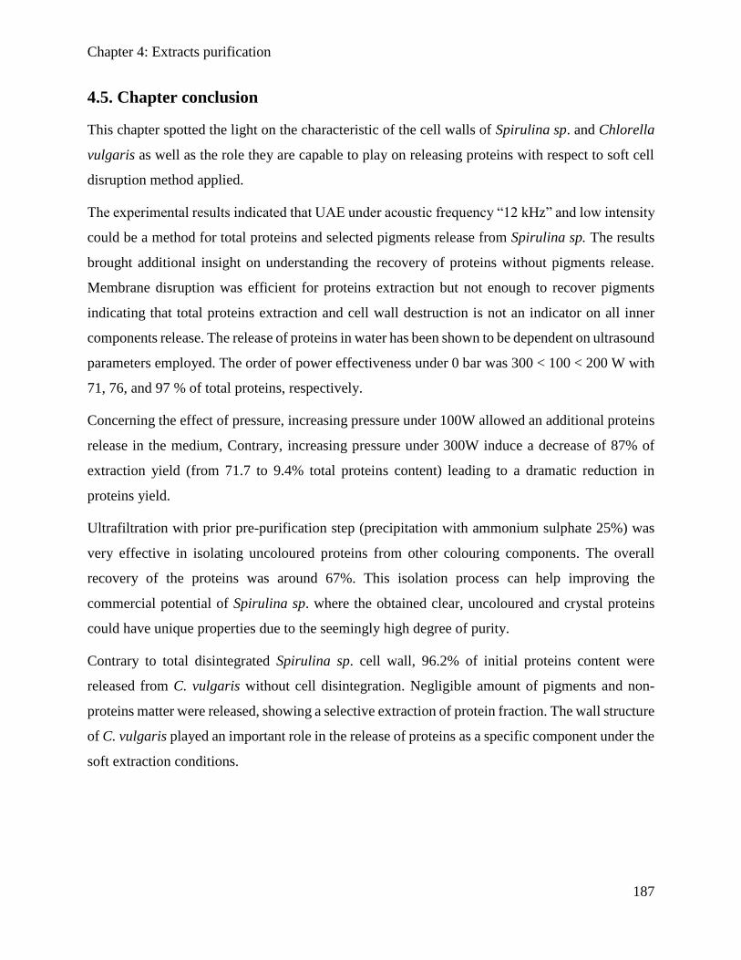
Chapter 4: Extracts purification
187
4.5. Chapter conclusion
This chapter spotted the light on the characteristic of the cell walls of Spirulina sp. and Chlorella
vulgaris as well as the role they are capable to play on releasing proteins with respect to soft cell
disruption method applied.
The experimental results indicated that UAE under acoustic frequency “12 kHz” and low intensity
could be a method for total proteins and selected pigments release from Spirulina sp. The results
brought additional insight on understanding the recovery of proteins without pigments release.
Membrane disruption was efficient for proteins extraction but not enough to recover pigments
indicating that total proteins extraction and cell wall destruction is not an indicator on all inner
components release. The release of proteins in water has been shown to be dependent on ultrasound
parameters employed. The order of power effectiveness under 0 bar was 300 < 100 < 200 W with
71, 76, and 97 % of total proteins, respectively.
Concerning the effect of pressure, increasing pressure under 100W allowed an additional proteins
release in the medium, Contrary, increasing pressure under 300W induce a decrease of 87% of
extraction yield (from 71.7 to 9.4% total proteins content) leading to a dramatic reduction in
proteins yield.
Ultrafiltration with prior pre-purification step (precipitation with ammonium sulphate 25%) was
very effective in isolating uncoloured proteins from other colouring components. The overall
recovery of the proteins was around 67%. This isolation process can help improving the
commercial potential of Spirulina sp. where the obtained clear, uncoloured and crystal proteins
could have unique properties due to the seemingly high degree of purity.
Contrary to total disintegrated Spirulina sp. cell wall, 96.2% of initial proteins content were
released from C. vulgaris without cell disintegration. Negligible amount of pigments and non-
proteins matter were released, showing a selective extraction of protein fraction. The wall structure
of C. vulgaris played an important role in the release of proteins as a specific component under the
soft extraction conditions.
General Conclusion
188
General Conclusion
Bio-refining of microalgae is considered as a potential source of various biomolecules. The
exploitation of this rich biomass does not cease to develop. Numerous research perspectives were
unwrapped in aim to optimize various parameters at the different stages of the algorefinery process
such as cultivation, extraction and fractionation of the metabolites of interest. Microalgae produce
several components whose use is now centered in the markets of biofuels, food and feed, cosmetic,
pharmaceutical and aquaculture
In this context, the objective of the algorefinery is to develop a method to fractionate the major
biomolecules, using a series of unit operations allowing a "selective" extraction of the compounds
of interest without altering the others.
Hence, at the “Laboratoire de Chimie Agro-Industrielle” (LCA) and “Plateforme de Recherche et
d'Analyse en Sciences de l'Environnement” (PRASE), we showed major interest on recovering the
hydrophilic and the lipophilic fractions by taking into consideration the biomass composition, the
cell wall characteristic and the role that can play regarding the extraction technique, the
optimization of extraction parameters, and the purification techniques carried out for the
fractionation of the heterogeneous phase obtained after cell disruption. The additional mission was
to respect the principles of ecoconception and green chemistry.
Since selected species of microalgae and cyanobacteria were investigated in our study, and since
the characteristics of the cell wall vary from one species to another, it was important to understand
the effect of the cell wall rigidity, biomass pretreatment, and the location of the biomolecule of
interest inside cell on extraction efficiency. Furthermore, the internal organelles also showed some
resistance toward cell disruption and extraction yield, and revealed that it would hinder the
liberation of some biomolecules.
Extraction of dry microalgae
Our work consisted of implementing a continuous fractionation process that takes into account
the integrity of the biomolecules in the downstream process. The unit operation of cell disruption
was bypassed. The selectivity of reserve lipid and specific pigments extracts using supercritical
carbon dioxide was possible by setting optimized parameters. Our results showed that selective
General Conclusion
189
recovery from freeze-dried Nannochloropsis oculata and Chlorella vulgaris was possible. This set
of studies gave us a clear idea on the role of the cell wall on the release of the lipids.
Then following the lipophilic extraction, the well maintained cell wall biomass underwent a double
stage aqueous extraction for proteins recovery. It was interesting to look more in depth on their
diffusion behavior regarding the pH of the aqueous media. The effect of biomass defatting degree
(partially or totally) on proteins release was studied. Results showed that total defatting allowed a
higher proteins release in the aqueous phase. Hence, following high proteins extraction, the
aqueous phase of Nannochloropsis oculata containing proteins and impurities like sugars and
pigments was fractionated to obtain proteins separate fractions. The fractionation process is based
on a single ultrafiltration using membrane process.
Therefore, starches and pigments were retained in the retentate while permeate was composed of
cleared proteins fraction.
Extraction of wet microalgae
Moreover, a soft algorefinery process was carried on the cyanobacteria Spirulina sp. and green
algae Chlorella vulgaris. The biomasses were subjected to an innovative green/soft ultrasound
assisted extraction (UAE) for proteins recovery. Total proteins content was released in the aqueous
medium from the dramatically disintegrated cell Spirulina sp. contrary to the well maintained
Chlorella vulgaris allowing a highly selective proteins total release. For clarification/
concentration step, membrane technologies were investigated using cross-flow ultrafiltration
process. Results showed a high yield (reaching 65%) of pigments free proteins of Spirulina sp. in
the permeate compartment. Optimization of the ultrafiltration conditions was made by combining
a pre-purification step (separation of impurities from proteins using ammonium sulfate 25%)
providing higher permeation flux.
The following points could summarize the strong force of our work:
The principles of “green chemistry” were respected in terms of the extracting solvent used.
It has been pointed out that even though the distinguished role of the cell wall on
biomolecules recovery, the ultrastructure of some microalgae could also show a resistance
against some mechanical treatment and allow a selective extraction.
General Conclusion
190
The unit operations of cell disruption could be avoided before supercritical carbon dioxide
extraction for selective reserve lipids recovery.
It was possible to understand the release behavior during extraction of lipids, pigments and
proteins.
A fractionation process with simplified steps has been set and could be optimized for an
industrial scale up.
The selectivity of extraction from robust cell wall was investigated using the low frequency
ultrasound conducting soft green extraction on wet microalgae, which saved the cost of
drying the biomass before the extraction.
Despite all the advantages of our studied process, theoretically every process has its limitations
The supercritical extraction investigated is considered as costly technique and required
high-energy input.
Regardless of the success of the fractionation process, the recovery of components of
interest using membrane process need to be optimized.
These difficulties represent an opportunity to open new perspectives:
Obtain images by electron microscopy that would follow up the modification of the cell
morphology during the extraction.
For rigid cell wall microalgae, it is a serious challenge to understand and optimize other
disruption methods for more selective extraction.
Optimize the membrane process to improve the purity and the flow rate of the components
of interest.
Scientific production
191
Scientific production
Paper accepted in peer-reviewed international scientific journals
1. Supercritical carbon dioxide extraction and fractionation of lipids from freeze dried
microalgae Nannochloropsis oculata and Chlorella vulgaris, Algal Research 34 (2018)
49–56.
S. Obeid, N. Beaufils, S. Camy, H. Takach, A. Ismail, P-Y. Pontalier.
Paper accepted in peer-reviewed international congress
2. Proteins and pigments extraction from microalgae Spirulina sp. assisted by ultrasound
irradiation: effect of variable low power and pre-treatment conditioning,
Récents Progrès en Génie des Procédés, Numéro 110 – 2017
ISSN: 1775-335X; ISBN: 978-2-910239-85-5, Ed. SFGP, Paris, France.
S. Obeid, I. Benhamed, M. Charton, J. Peydecastaing, H. Takache, A. Ismail, P-Y.
Pontalier, L. Barthe
Papers submitted or ready for submission
3. Supercritical fluid extraction and solid phase fractionation of pigments from freeze-dried
Chlorella vulgaris and Nannochloropsis oculata
S. Obeid, H. Takache, N. Beaufils, S. Camy, A. Ismail, P-Y. Pontalier
4. Extraction and purification of soluble proteins from Nannochloropsis oculata by pH-
shifting and ultrafiltration
S. Obeid, H. Takache, N. Beaufills, S. Camy, A. Ismail, P-Y. Pontalier.
5. Selectivity of low-frequency ultrasound-assisted extraction for proteins recovery from the
microalgae Chlorella vulgaris.
S. Obeid, I. Benhamed, M. Charton, J. Peydecastaing, H. Takache, A. Ismail, P-Y.
Pontalier, L. Barthe.
6. Clarification of non-pigmented proteins from Spirulina sp. using ultrasound assisted
extraction coupled with ultrafiltration
S. Obeid, I. Benhamed, M. Charton, J. Peydecastaing, H. Takache, A. Ismail, P-Y.
Pontalier, L. Barthe.
Poster presentations
1. Etude de procédé de quantification et de purification des lipides et des protéines de
microalgues extraites à l’eau et au CO2 supercritique - LAAS congress 2016 (USEK-
Lebanon)
S. Obeid, H. Takache, N. Beaufils, S. Camy, A. Ismail, P-Y. Pontalier
Scientific production
192
2. Ultrasound-assisted extraction for microalgae proteins recovery - SFGP congress 2017
(Nancy- France)
S. Obeid, I. Benhamed, H. Takache, A. Ismail, P-Y. Pontalier, L. Barthe.
3. A primary algorefinery study on Nannochloropsis oculata and Chlorella vulgaris - ISAP
Congress 2017 (Nantes- France)
S. Obeid, H. Takache, N. Beaufils, S. Camy, A. Ismail, P-Y. Pontalier
4. Extraction and fractionation of proteins from Chlorella vulgaris and Spirulina sp. by
membrane technology - FPS congress 2018 (Lyon- France)
S. Obeid, I. Benhamed , H. Takache, J. Peydecasting , M. Charton, L. Barthe, A. Ismail ,
and P.Y. Pontalier.
5. A primary algorefinery study on green microalgae and cyanobacteria using supercritical-
EDST Scientific Forum 2017
S. Obeid, I. Benhamed , H. Takache, J. Peydecasting , M. Charton, L. Barthe, A. Ismail ,
and P.Y. Pontalier
Oral presentations
1. Supercritical carbon dioxide extraction and fractionation of lipids for biofuel production
from untreated microalgae Nannochloropsis oculata and Chlorella vulgaris - 9th Annual
Congress and Expo on Biofuels and Bioenergy 2018 (Dubai, UAE)
S. Obeid, H. Takache, A. Ismail, P-Y. Pontalier.
2. Proteins extraction from microalgae Spirulina sp. assisted by ultrasound irradiation: effect
of variable low power and pre-treatment conditioning - EDST- BAU Scientific Colloquium
2017 (Beirut- Lebanon)
S. Obeid, H. Takache, A. Ismail, P.Y. Pontalier.
3. Quantification et Purification des Lipides et des Protéines Extraits de Microalgues - 2017
EDST (Beirut-Lebanon)
S. Obeid, H. Takache, A. Ismail, P-Y. Pontalier.
Résumé générale en français
193
Résumé générale en français
Chapitre 1: Etat de l'art
Les microalgues sont des organismes aquatiques unicellulaires. Jusqu'à ce jour, environ 30000
espèces sont connues et analysées. Ils sont capables de coloniser tous les types d'environnements,
y compris les eaux salées, douces, saumâtres et même les eaux usées grâce à leurs compétences de
survie. Aujourd’hui, une gamme d'espèces de microalgues peut-être produite dans un
environnement artificiel appelé photobioréacteur (PBR) où elles sont cultivées sous une exposition
lumineuse désirée, une concentration spécifiée d’éléments nutritifs et des facteurs vitaux
convenables. Les microalgues diffèrent principalement des autres plantes par leur vitesse de
croissance très rapide, leur richesse en lipides, protéines, polysaccharides, vitamines, pigments et
antioxydants. Ils sont principalement exploités pour leurs composants primaires : lipides,
protéines, polysaccharides et pigments.
Les microalgues sont divisées en deux groupes : celles appartenant au groupe des eucaryotes, c'est-
à-dire ayant des organites cellulaires (chloroplastes, mitochondries et son noyau entouré de son
enveloppe), et ceux appartenant au groupe des procaryotes, où les organites cellulaires sont
absentes. Les microalgues sont classées sous 11 divisions et 29 classes. Les trois classes les plus
connues sont les « Chlorophyceae » et les « Eustigmatophyceae » qui sont des algues vertes, et
les « Cyanophyceae » ou cyanobactéries. Ces trois classes sont les plus étudiées en raison de leurs
propriétés énergétiques, vue leur teneur élevée en lipides, protéines, pigments, sucre, etc. A côté
du secteur énergétique, Ils offrent des possibilités pour des applications agricoles, alimentaires,
pharmaceutiques et médicales.
La biomasse algale représente un potentiel considérable dans le monde et elle a déjà été identifiée
pour sa capacité à produire des produits de valeur. Les scientifiques cherchent à ouvrir et
développer de procédés innovants pour permettre la production d'une très large gamme de produits,
à l’aide du concept « d’algoraffinerie », mais ils sont loin de garantir une production de masse avec
une rentabilité économique/écologique acceptable. L'algoraffinerie est maintenant au stade de la
recherche et du développement dans le but d'atteindre une rentabilité écologique/économique
suffisante.
Résumé générale en français
194
La transformation des microalgues présente beaucoup d’avantages par rapport à d’autre biomasse
terrestres :
• Au stade du laboratoire, les rendements en huile à partir des microalgues (par hectare) sont
20 à 30 fois plus élevés que ceux d’autres oléagineux terrestres.
• Les zones cultivées en microalgues n'ont pas besoin d'être des terres arables et ne sont pas
donc en concurrence avec les terres agricoles.
• La croissance des microalgues lipidiques nécessite de grandes quantités de CO2, ce qui
ouvre des perspectives pour le recyclage du CO2 émis par les usines ou les centrales
thermiques.
Cependant, plusieurs obstacles se posent encore pour la production de cette biomasse :
• La culture nécessite des conditions spéciales d'ensoleillement et de disponibilité de l'eau,
ce qui limite les emplacements possibles.
• Elle a également besoin en nutriments (nitrates et phosphates) et en dioxyde de carbone.
• L’algoraffinerie (prétraitement - extraction - purification) consomme beaucoup d'énergie.
Différents schémas de couplage pour les techniques d'extraction, prétraitement et
séparation sont rapportés dans la littérature afin d'obtenir des extraits riches en composants
primaires (lipides, protéines et pigments).
Le processus nécessite une séquence d'opérations unitaires à partir du prétraitement de la biomasse
par une désintégration chimique / mécanique de la paroi cellulaire des microalgues. Ceci permet
un accès plus facile du solvant d'extraction face aux composants intracellulaires, et contribue à
améliorer le rendement de récupération des biomolécules dédiées. Les méthodes de désintégration
sont préférées en raison de l'accélération du mécanisme, mais ces méthodes sont gourmandes en
énergie et surchauffent les molécules, ce qui nécessite un refroidissement le long du processus
pour éviter la dénaturation ou la dégradation thermique. Après l'étape de prétraitement et
d'extraction vient le stade de la purification, ce qui est une étape difficile pour de nombreux
scientifiques à l'échelle du laboratoire et à l'échelle industrielle.
Résumé générale en français
195
Plan d'action de la thèse
L'état de l'art apporté dans ce manuscrit a montré que les microalgues représentent une source
renouvelable importante. Certaines microalgues sont riches en protéines, d'autres en pigments et
en lipides.
Cependant, de nombreux verrous doivent être levés du processus d’algoraffinerie pour assurer
autant que possible une viabilité environnementale / économique. Les conditions de prétraitement
et d'extraction des composants d'intérêt restent un défi important dans les processus en aval. De
nos jours, les déficiences dans le domaine de l'extraction des microalgues sèches sont toujours
confrontées.
Actuellement, le prétraitement exécuté avant les procédés d'extraction nécessite une
consommation d'énergie significative, l'addition d'étapes ultérieures pour la séparation et la
purification des composants désirés d'autres composants co-extraits, et augmente la proportion de
produits finaux altérés dus à un effet mécanique ou thermique. Dans cette perspective, il apparaît
nécessaire d'améliorer les processus actuels et de progresser vers une exécution améliorée.
Démarche 1 :
Ainsi, nous avons pris ce problème d'extraction à partir de microalgues sèches en utilisant la
technique du fluide supercritique selon l'approche suivante (Schéma I) :
• L’étape de désintégration des parois cellulaires a été évitée pendant tout le processus
d’extraction : après réduction de la teneur en eau inférieure à 2%, une extraction avec du
CO2 supercritique sans désintégration préalable de la paroi cellulaire a été effectuée pour
récupérer uniquement les lipides neutres et pour éviter la formation d'émulsions, à partir
des microalgues Chlorella vulgaris et Nannochloropsis oculata.
• Le procédé de purification des lipides neutres sera donc plus facile à mettre en œuvre.
• Contrairement aux études reportées dans la littérature, les résidus d'extraction
supercritiques composés de cellules delipidées riche en protéines ne subiront plus une
désintégration, et les protéines seront extraits à l’aide d’une double extraction aqueuse /
alcaline.
• La purification des protéines d’autres composants hydrophiles co-extraits sera exécutée en
utilisant un procédé à membrane.
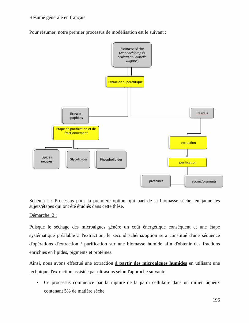
Résumé générale en français
196
Pour résumer, notre premier processus de modélisation est le suivant :
Schéma I : Processus pour la première option, qui part de la biomasse sèche, en jaune les
sujets/étapes qui ont été étudiés dans cette thèse.
Démarche 2 :
Puisque le séchage des microalgues génère un coût énergétique conséquent et une étape
systématique préalable à l'extraction, le second schéma/option sera constitué d'une séquence
d'opérations d'extraction / purification sur une biomasse humide afin d'obtenir des fractions
enrichies en lipides, pigments et protéines.
Ainsi, nous avons effectué une extraction à partir des microalgues humides en utilisant une
technique d'extraction assistée par ultrasons selon l'approche suivante:
• Ce processus commence par la rupture de la paroi cellulaire dans un milieu aqueux
contenant 5% de matière sèche
Biomasse sèche(Nannochloropsis
oculata et Chlorella vulgaris)
Extraitslipophiles
Etape de purification et de fractionnement
Lipides neutres
Glycolipides Phospholipides
Residus
extraction
proteines
purification
sucres/pigments
Extracion supercritique
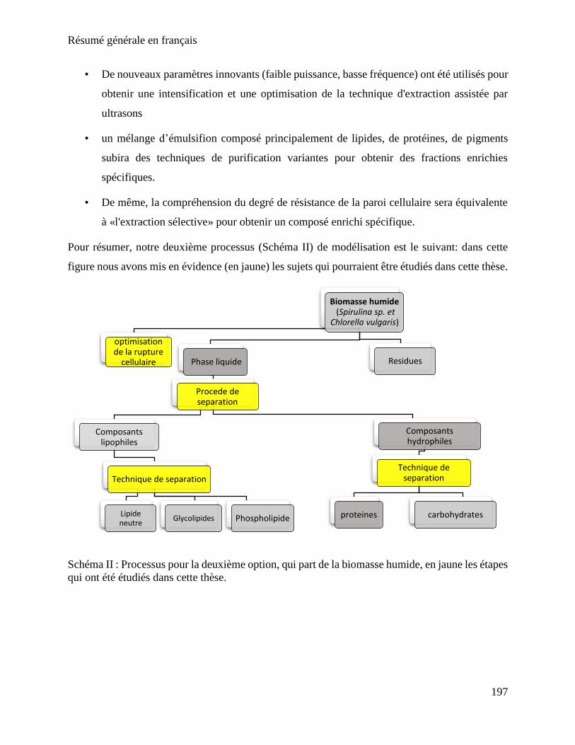
Résumé générale en français
197
• De nouveaux paramètres innovants (faible puissance, basse fréquence) ont été utilisés pour
obtenir une intensification et une optimisation de la technique d'extraction assistée par
ultrasons
• un mélange d’émulsifion composé principalement de lipides, de protéines, de pigments
subira des techniques de purification variantes pour obtenir des fractions enrichies
spécifiques.
• De même, la compréhension du degré de résistance de la paroi cellulaire sera équivalente
à «l'extraction sélective» pour obtenir un composé enrichi spécifique.
Pour résumer, notre deuxième processus (Schéma II) de modélisation est le suivant: dans cette
figure nous avons mis en évidence (en jaune) les sujets qui pourraient être étudiés dans cette thèse.
Schéma II : Processus pour la deuxième option, qui part de la biomasse humide, en jaune les étapes
qui ont été étudiés dans cette thèse.
Biomasse humide(Spirulina sp. et
Chlorella vulgaris)
Phase liquide
Procede de separation
Composants lipophiles
Technique de separation
Lipide neutre
Glycolipides Phospholipide
Composants hydrophiles
Technique de separation
proteines carbohydrates
Residues
optimisationde la rupture
cellulaire
Résumé générale en français
198
Chapitre 2 : Méthodes et matériels
Biomasse microalgale
Des expériences ont été réalisées à partir de différentes espèces de microalgues; chacune d’elle a
des caractéristiques morphologiques (membranaires) et une composition biochimique différentes.
Les trois espèces impliquées dans ce travail sont Chlorella vulgaris, Nannochloropsis oculata et
Spirulina sp.
Les souches de C. vulgaris et N. oculata (Alpha Biotech, Assérac, France) ont été cultivées dans
un milieu riche en nutriments azotés pour favoriser l'accumulation de protéines chez C. vulgaris.
Les souches de N. oculata ont été cultivées dans des conditions de carence en nutriments azotés
pour favoriser l'accumulation de lipides de réserve. La souche de Spirulina sp. (Alg&You,
Toulouse, France), à été fournie dans des milieux de culture à 5% MS .Des fractions ont été laissées
fraiches, d’autre congelées et d’autre lyophilisées.
Méthodes d'extraction
La teneur totale en lipides a été déterminée à l’aide de l’extraction conventionnelle selon Bligh et
Dyer (1959).
Extraction par CO2 supercritique
Les différentes extractions au CO2 supercritique ont été réalisées sur des biomasses lyophilisées
au Laboratoire d'Ingénierie Chimique (LGC) sur un pilote développé par la société Separex
(SFE100, Separex Chimie fine, France), capable de travailler à des pressions jusqu'à 1000 Bar et
températures jusqu'à 250 °C. Plusieurs extractions ont été effectuées en faisant varier la pression
de travail et la présence ou l'absence d’un co-solvant.
Ultrason
Les tests d'extraction ont été réalisés à l'aide d'une installation SinapTec située au Laboratoire de
Génie Chimique (LGC). Un plan expérimental a été réalisé afin de mettre en évidence et
d’optimiser les facteurs influençant l’extraction de protéines à partir de Spirulina sp et de Chlorella
vulgaris. Quatre facteurs ont été étudiés: l’agitation, la fréquence, la pression et la puissance.
Résumé générale en français
199
Extraction totale de pigments
Il existe différents types de pigments dans C. vulgaris, N. oculata et Spirulina sp. Nous nous
sommes concentrés sur certains pigments en particulier: les chlorophylles a et b, les lutéines et les
β-carotène. La teneur totale en ces pigments a été extraite à froid pour les protéger de la
dénaturation thermique.
Technique de purification
Ultrafiltration
Les membranes utilisées sont des planes en polysulfone (dont le seuil de coupure est donné à 10-
30- 100 kDa) à l'aide d'une cellule de filtration de 200 mL (Unités d'Ultrafiltration Agitées -
Amicon® Stirred Cells) correspondant à une surface de filtration de 30,2 cm2. Cette unité offre
une surface de filtration réduite qui permet d'étudier la filtration de quantités relativement faibles
(à partir de 50 mL). De plus, une agitation sur la membrane a été également appliquée pour
minimiser l'effet de la couche de polarisation de concentration due à l’accumulation des
macromolécules à la surface de la membrane
Techniques analytiques
Analyse des protéines : La teneur en protéines totales des biomasses étudiées a été quantifiée selon
la méthode de Kjeldahl. La teneur en protéines brutes des microalgues étudiées est obtenue en
multipliant la valeur obtenue lors de la détermination de la teneur en azote par le facteur 6.2.
Méthode de Lowry (Lowry et al, 1951) : pour la quantification des protéines solubles dans l'eau.
Pour les échantillons solides, l'extraction à l'hydroxyde de sodium est nécessaire pour solubiliser
les protéines hydrosolubles. Au laboratoire, cette extraction est généralement réalisée avec une
solution d'hydroxyde de sodium à pH 11 pendant une heure à 40 °C. Une fois les protéines
extraites, la méthode de Lowry a été utilisée pour quantifier les protéines solubles libérées dans
le milieu.
Analyse SDS-PAGE : Afin d'identifier les protéines extraites par leur poids, une électrophorèse
sur gel de polyacrylamide en présence de dodécylsulfate de sodium (SDS-PAGE) a été réalisée.
Résumé générale en français
200
Chromatographie d'exclusion de taille SEC : La méthode SEC a été étudiée pour séparer les
petites protéines extraites de celles de plus grande taille dans le but de comprendre les
caractéristiques des protéines libérées.
Analyse pigmentaire
La teneur en pigment total (chlorophylles a et b, β-carotènes et lutéine) a été mesurée par HPLC
selon la méthode développée par Bidigare (1991).
Fractionnement des lipides par extraction en phase solide (SPE) : Le fractionnement des extraits
lipidiques est une étape particulièrement importante dans l'identification de la teneur en
biomasse des lipides neutres, des glycolipides et des phospholipides. Il permet aussi d’évaluer
l'efficacité et la sélectivité de l'extraction supercritique. C’est une technique utilisée en raison
de sa vitesse et de son efficacité.
Le fractionnement des lipides en trois classes de lipides neutres, de glycolipides et de
phospholipides était possible. Une fois élues et lyophilisées sous azote liquide, les fractions ont
été mesurées par gravimétrie et exprimées en mg / g de poids sec.
Chapitre 3 : Extraction fractionnée
Ce chapitre est composé de trois publications.
Publication # 1
Cette étude porte sur l'extraction sélective de lipides neutres à partir de microalgues en utilisant
le ScCO2 sans rupture de cellule. Les biomasses étudiées étaient cultivées dans des conditions
de de stress nutritives. Nannochloropsis oculata a été cultivé dans un milieu de Conway modifié
dans des conditions de carence en azote pour favoriser l'accumulation de lipides, alors que la
microalgue Chlorella vulgaris a été cultivée dans des milieux de Sueoka dans des conditions de
teneur élevée en azotés pour favoriser l'accumulation de protéines. La culture dans de telles
conditions induisait l'accumulation de lipides chez Nannochloropsis oculata spécifiquement les
lipides neutres (triglycérides ou lipides de réserve). Contrairement à N. oculata, C. vulgaris
possède des lipides polaires ou structuraux comme lipides prédominants, situés dans les
chloroplastes et les membranes cellulaires
Résumé générale en français
201
L'extraction des lipides de réserve à l’aide d’une méthode qui respecte l'environnement et sans
solvant est un grand défi pour l'industrie d'aujourd'hui. L’extraction à l’aide du CO2
supercritique comme solvant, présente de nombreux avantages, dont le plus important est
l’obtention du produit final sans traces de solvant toxique.
Dans cette partie, nous étudions l'effet des paramètres clés de l’extraction ScCO2 sur le
rendement: la température, la pression et l’ajout de modificateur polaire (éthanol 10%). Une
telle addition de modificateur polaire permet de comprendre l'effet de la co-extraction de lipides
polaires sur le rendement d'extraction de lipides neutres. D'autre part, la sélectivité de
l'extraction de lipides neutres a été évaluée en analysant la composition des extraits en lipides
polaires (glycolipides et phospholipides) tout au long du processus, en utilisant la technique
d'extraction en phase solide (SPE).
Dans des conditions d'exploitation, 97% lipides neutres de C. vulgaris ont été extraits en
utilisant l'éthanol à 10% comme co-solvant. Les lipides neutres de N. oculata représentaient la
plupart des extraits (83%), alors que la proportion de glycolipides et de phospholipides
n'excédait pas 12,1% et 5,3% respectivement. L'observation microscopique a montré que
l'intégrité de la paroi cellulaire était maintenue pendant le processus d'extraction.
Publication # 2
Le but de ce travail est d'étudier la cinétique d’extraction supercritique de plusieurs composants
(chlorophylles a et b, la lutéine, β-carotènes, lipides neutres, glycolipides et phospholipides)
dans. L'extraction des composants à l'extérieur de la paroi cellulaire rigide de N. oculata et C.
vulgaris nécessite un traitement spécifique afin de faciliter leur transfert et d'augmenter le
rendement d’extraction. Ainsi, nous étudions l’évolution d’extraction sans destruction cellulaire
au préalable. Par conséquent, différents paramètres d'extraction de ScCO2 ont été étudiés sur C.
vulgaris et N. oculata.
La souche de C. vulgaris utilisée dans cette étude est distinguée par sa teneur élevée en
protéines, chlorophylles a et b et les lipides structuraux. D'autre part, N. oculata présentait une
teneur en lipides plus élevée notamment en lipides de réserves, et une faible teneur en
chlorophylles a et b. La relation entre la teneur en pigments (caroténoïdes et chlorophylles) et
l'accumulation de lipides a été étudiée dans les souches de Chlorella et Nannochloropsis : le
Résumé générale en français
202
rapport caroténoïde-chlorophylle de C. vulgaris (faible teneur en lipides neutres) est inférieur
au rapport caroténoïde-chlorophylle de N. oculata (riche en lipides neutres).
L'huile et les pigments dérivés de C. vulgaris lyophilisés ont été extraits par du CO2
supercritique. Les résultats obtenus ont montré que le rendement d'extraction des pigments était
si bas et qu'une extraction simple avec ScCO2 + éthanol (10%) comme co-solvant n'a pas
modifié le rendement d'extraction de la chlorophylle b et de la lutéine. Le La principale
différence est apparue avec la chlorophylle a, dont le rendement d'extraction était près de 6 fois
plus élevé que lors de la double extraction (consiste à faire une première extraction supercritique
en utilisant du ScCO2 comme solvant, suivie d'une seconde extraction en utilisant du ScCO2 +
éthanol comme co-solvant 10%). Cette procédure a modifié la structure de la membrane
plasmique du chloroplaste permettant la diffusion hors de la cellule de la chlorophylle a. Les
effets des co-solvants ont été trouvés prononcés pour l'extraction des lipides neutres, sous
simple extraction, atteignant 97% des lipides neutres totaux.
L'extraction au ScCO2 de N. oculata a révélé que 450 bars étaient la pression optimale pour
l'extraction des lipides neutres avec 76 mg / g de poids sec (correspondant à 45% de la teneur
totale en lipides neutres), en plus de 34% de chlorophylle a sélectivement extraites parmi les
autres pigments.
Publication # 3
Le but de cet article est d'étudier la possibilité d'un rendement élevé d'extraction de microalgues
de N. oculata sans endommager les propriétés et l'intégrité de la paroi cellulaire afin de respecter
le concept de bioraffinerie, et de minimiser les étapes possibles de fractionnement et de
purification. L'extraction a été réalisée en double étapes en faisant varier le pH du milieu, sur
une biomasse dégraissée, non dégraissée et semi-dégraissée (par extraction au CO2
supercritique) dans le but de comprendre l'effet de la délipidation sur l’extraction de protéines.
Une fois l'extraction terminée, il est nécessaire d'établir un processus de fractionnement /
concentration des protéines. Quelques travaux ont étudié le procédé membranaire pour purifier
des composants de microalgues. L'application de l'ultrafiltration pour la séparation des protéines
microalgales d'autres composants semble offrir certaines sources d'intérêt industriel spécifique
(efficacité et facilité d'exploitation). Par conséquent, l'ultrafiltration frontale sur des surnageants
de protéines de N. oculata a été étudiée. Ces techniques de séparation ont été utilisées pour la
Résumé générale en français
203
purification des protéines en éliminant les composants co-extraits non désirés, et pour la
concentration en protéines après extraction. Les solutions de perméat et de rétentat ont été
analysées afin de vérifier l'efficacité de chaque étape. L’électrophorèse sur gel de
polyacrylamide (SDS-PAGE) et une la chromatographie d'exclusion de taille (SEC) ont été
utilisées pour identifier les protéines extraites.
Les résultats ont montrés que 73,7% des protéines totales ont été libérées en utilisant une
combinaison de conditions aqueuses (durant la première étape d’extraction) et alcaline (durant
la deuxième étape d’extraction), à partir de la biomasse dégraissée. Une fraction enrichie de
protéines hydrosolubles exemptes d'impuretés colorantes a été obtenue. De plus, 84% des
protéines extraites ont été obtenues sans pigment dans la fraction de perméat en utilisant une
membrane de 100 kDa.
Chapitre 4 : Purification des extraits
Ce chapitre est composé de trois publications
Publication # 4
Le présent document vise à travailler sur une solution innovante, à très basse fréquence et
audible (12 kHz) pour l’extraction des protéines et pigments à partir de Spirulina sp. De
nombreuses publications ont mentionné l'effet négatif des ultrasons en utilisant des fréquences
élevées et modérées et comment cela pourrait induire une dégradation des composants d’intérêt.
Au vu de ce qui précède, une extraction verte et douce utilisant de l'eau sera réalisée à basse
température en utilisant un procédé d'ultrason à basse fréquence. En outre, une attention
particulière a été accordée à l'influence des conditions de stockage (biomasse fraîche,
lyophilisée et congelée) et des conditions d'extraction assistée par ultrasons (faible pression -
faible puissance- vitesse d'agitation) sur le rendement d'extraction des protéines et des pigments.
Les résultats ont indiqué que l'extraction de protéines pouvait atteindre près de 95%. Presque
toute la teneur en protéines a été libérée dans le milieu à partir de la biomasse congelée dans
deux conditions variables: Il était possible d'obtenir le même rendement d'extraction à 300 W
sous pression atmosphérique ou à 100 W sous pression relative de 3 bars. La deuxième
condition est préférée car elle est plus économique. De même, le conditionnement de stockage
a également eu un effet important sur l'efficacité d'extraction des pigments. Cependant,
Résumé générale en français
204
l’application des ultrasons était nécessaire pour obtenir un rendement d'extraction élevé des
pigments tels que la chlorophylle a et le β-carotène dont les concentrations dans le surnageant
étaient respectivement 120 fois et 140 fois plus élevées qu’en absence des ultrasons. Les
prétraitements congelés semblaient affaiblir la structure cellulaire, en particulier la membrane
externe, conduisant à une extraction des pigments plus efficace.
Les résultats ont aussi indiqué que l’UAE sous la fréquence acoustique "12 kHz" à faible
intensité pourrait être une méthode pour l‘extraction des protéines totales et u pour l’extraction
sélective des pigments. La libération des pigments nécessitait une forte désintégration de la
structure interne, tandis que les protéines ont été libérées avec une simple lyse cellulaire.
La présente étude apporte également un aperçu supplémentaire sur la compréhension de la
récupération des protéines sans libération de pigments. La rupture de la membrane était efficace
pour l'extraction des protéines, mais pas assez pour récupérer les pigments, ce qui indique que
l'extraction totale des protéines et la destruction de la paroi cellulaire ne sont pas un indicateur
de la libération de tous les composants.
Publication # 5
Cette étude vise à obtenir une fraction protéique non pigmentée hautement clarifiée de Spirulina
sp. en utilisant la technologie membranaire.
La fraction protéique de Spirulina sp. a commencé à être exploitée dans les industries
cosmétiques et pharmaceutiques. Toutefois, le développement de ladite fraction dans ces divers
secteurs reste non significatif du fait de la présence d'éléments indésirables durant son extraction
conduisant à des modifications significatives de la couleur. Pour améliorer leur potentiel
commercial, la fraction protéique doit être clarifiée à partir des composants indésirables après
extraction. Un processus doux d’algoraffinerie a été réalisé sur les cyanobactéries Spirulina sp.
pour obtenir une fraction purifiée de protéine exempte de chlorophylles, de caroténoïdes et de
C-phycocyanine (pigments protéiques bleus).
Les protéines ont été libérées dans le milieu aqueux de la cellule Spirulina sp. utilisant les
ultrasons à basse fréquence (12 kHz) et intensité. Pour l'étape de clarification, la technologie
membranaire a été utilisée dans le but d'améliorer la pureté des extraits de protéines résultant
Résumé générale en français
205
de la rupture par les ultrasons de la paroi cellulaire. Un procédé d'ultrafiltration frontale a été
effectué sur les surnageant protéiques enrichis.
Comme résultat, un rendement global de protéines clarifiées important (atteignant 65%) a été
obtenu dans le compartiment du perméat. Le rétentat était principalement composé de
phycocyanine. L'ultrafiltration avec des membranes ayant un seuil de coupure de 10 kDa
présentait les meilleures performances en termes de rétention complète des pigments, des
protéines pigmentées et des résidus.
Publication # 6
La microalgue C. vulgaris est produite industriellement pour ses teneurs élevées en protéines
utilisées dans l'alimentation humaine. Cependant, le principal inconvénient de l'extraction de
ces protéines est la faible disponibilité des composants intracellulaires due à la paroi cellulaire
rigide de cette souche. Pour cela, de nombreux travaux sont portés sur la rupture de la paroi
cellulaire avant l'extraction de la fraction protéique.
En termes d'ultrasons menées sur C. vulgaris, la majorité des articles se concentraient sur la
rupture de la paroi cellulaire. En conséquence, de nombreux composants internes ont également
été co-extraits. La rigidité apparente de C. vulgaris pourrait être exploitée pour une éventuelle
extraction sélective. L'objectif de cette étude était d'évaluer l'efficacité de l'extraction assistée
par ultrasons à basse fréquence (UAE) pour l'extraction sélective de protéines. La microalgue
C. vulgaris a été soumise à une faible fréquence acoustique (12 kHz), à faibles intensités (0-
100-300 W) et à une pression hydrostatique relative de zéro à3 bar. La cavitation, induite par
des ultrasons de faible intensité et de basse fréquence, pourrait récupérer des protéines
précieuses sans altérer l'intégrité cellulaire. . Nous avons évalué l’ultrason comme une étape de
traitement pour se conformer aux critères du concept de la chimie verte.
Pour éviter toute dénaturation thermique, la température n'a pas dépassé 30 ºC pendant le
traitement. Nous avons déterminé le rendement d'extraction des protéines pour tous les
surnageant. De plus, nous avons quantifié les pigments et les résidus UAE comme marqueur
des altérations des organites internes, de la désintégration de la paroi cellulaire et de la libération
des lipides. Nous avons suivi les changements dans la morphologie cellulaire pour évaluer la
performance du procédé et le degré de perturbation cellulaire.
Résumé générale en français
206
En conséquence, les ultrasons ont été efficaces dans l'extraction sélective de protéines : 96%
des protéines récupérées avec une co-extraction négligeable des pigments et des lipides.
L'augmentation de la pression à faible intensité de puissance était très efficace, indiquant que la
pression jouait un rôle critique dans l'extraction des protéines. La structure de la paroi de C.
vulgaris joue un rôle important dans le phénomène d'extraction sélective. L'intégrité des
cellules a été maintenue pendant l'extraction des protéines totales.
Résumé générale en français
207
Conclusion générale
Le bioraffinage des microalgues est considéré comme une source potentielle de diverses
biomolécules. L'exploitation de cette riche biomasse ne cesse de se développer. De nombreuses
perspectives de recherche ont été exposées dans le but d'optimiser divers paramètres aux
différentes étapes du processus d’algoraffinerie tels que la culture, l'extraction et le fractionnement
des métabolites d'intérêt. Les microalgues produisent des composants d'intérêt, dont leur utilisation
est centrée sur le marché des biocarburants, des aliments, de l’alimentation animale, des
cosmétiques, des produits pharmaceutiques et de l'aquaculture.
Dans ce contexte, l'objectif de l'algoraffinerie est de développer une méthode de fractionnement
des biomolécules, en utilisant une série d'opérations unitaires permettant une extraction «sélective»
des composés d'intérêt sans extraire les autres composés.
Ainsi, au Laboratoire de Chimie Agro-Industrielle (LCA) et à la Plateforme de Recherche et
d'Analyse des Sciences de l'Environnement (PRASE), nous avons montré un intérêt majeur pour
la récupération de la fraction hydrophile et lipophile en tenant compte de la composition de la
biomasse, le rôle de la paroi cellulaire dans l’extraction, l'optimisation des paramètres d'extraction,
et les techniques de purification et de fractionnement d. Notre objectif principal était de respecter
les principes de l'écoconception et de la chimie verte.
Puisque certaines espèces de microalgues et de cyanobactéries ont été étudiées et que les
caractéristiques de la paroi varient d'une espèce à l'autre, il était important de comprendre l'effet
de la rigidité des parois cellulaires, du prétraitement de la biomasse et de la localisation de la
biomolécule d'intérêt à l'intérieur de la cellule sur l'efficacité de l'extraction. En outre, les organites
internes ont également montré une certaine résistance à la rupture des cellules et au rendement
d'extraction, et ont révélé que cela permettrait la libération de certaines biomolécules.
Extraction à partir des microalgues sèches
Notre travail consistait à mettre en œuvre un processus de fractionnement continu qui prend en
compte l'intégrité des biomolécules dans le processus en aval. L'opération unitaire de rupture
cellulaire a été éliminée. La sélectivité des extraits de lipides de réserve et de pigments spécifiques
à l'aide de ScCO2 été possible en fixant des paramètres optimisés. Nos résultats ont montré que
des récupérations sélectives des composants de N. oculata et de C. vulgaris lyophilisées étaient
Résumé générale en français
208
possibles. Ceci nous a donné une idée claire du rôle de la paroi cellulaire sur la libération des
molécules d’intérêts notamment les lipides.
Puis, suite à l'extraction lipophile, la biomasse avec paroi cellulaire bien entretenue a subi une
extraction aqueuse en deux étapes pour la récupération des protéines. Il était intéressant de
regarder en profondeur le phénomène de diffusion en variant le pH du milieu aqueux. L'effet du
degré de délipidation de la biomasse (partiellement ou totalement) sur la libération de protéines a
été étudié. Les résultats ont montré que la délipidation totale permet une libération de protéines
plus élevée dans la phase aqueuse. Par conséquent, après l'extraction de protéines de N. oculata,
la phase aqueuse contenant des protéines et des impuretés comme des sucres et des pigments ont
été fractionnés pour obtenir des fractions séparées de protéines. Le processus de fractionnement
est basé sur une ultrafiltration unique utilisant un procédé membranaire. Par conséquent, les
amidons et les pigments ont été retenus dans le rétentat tandis que le perméat était composé de
fraction de protéines clarifiées.
Extraction à partir des microalgues humides
En outre, un processus d’algoraffinerie doux a été porté sur les cyanobactéries Spirulina sp. et
l'algue verte C. vulgaris. Les biomasses ont été soumises à une extraction assistée par ultrasons
verte / douce innovante pour la récupération des protéines. La teneur totale en protéines a été
libérée dans le milieu aqueux à partir de la cellule spiruline désintégrée de manière totale,
contrairement à C. vulgaris dont sa membrane a restée bien maintenue, permettant une libération
totale sélective des protéines. Pour l'étape de clarification / concentration, les technologies
membranaires ont été étudiées en utilisant un procédé d'ultrafiltration frontale. Les résultats ont
montré que 65% des protéines pures ont été obtenues dans le perméat. Les conditions
d'optimisation ont été réalisées en combinant une étape de pré-purification (séparation des
impuretés des protéines en utilisant du sulfate d'ammonium à 25%) pour fournir un flux de perméat
plus élevé.
Résumé générale en français
209
Les points suivants pourraient résumer les points forts de notre travail:
• Les principes de la «chimie verte» ont été respectés en termes de solvant d'extraction
utilisé.
• Il a été souligné le rôle remarquable de la paroi cellulaire sur la récupération des
biomolécules. La structure cellulaire de certaines microalgues pouvait également avoir une
résistance à certains traitements mécaniques et permettre aussi une extraction sélective de
composés cellulaires.
• Le prétraitement de de rupture des cellules pourrait être évité avant l'extraction par ScCO2
pour la récupération sélective des lipides de réserve.
• Il a été possible de comprendre le comportement de diffusion lors de l'extraction aqueuse
à double phase.
• Un processus de fractionnement avec des étapes simplifiées a été défini et pourrait être
optimisé pour une mise à l'échelle industrielle.
• La sélectivité de l'extraction à partir d'une paroi cellulaire robuste a été étudiée en utilisant
l'extraction verte-douce à basse fréquence par conduction par ultrasons sur des microalgues
humides, ce qui a permis d'économiser le coût du séchage de la biomasse avant l'extraction.
Malgré tous les avantages, notre procédé développé présente encore quelques limitations :
• L'extraction supercritique étudiée est considérée comme une technique coûteuse et
nécessite un apport énergétique élevé.
• Malgré le succès du processus de fractionnement, la récupération des composants d'intérêt
à l'aide d'un procédé membranaire doit être optimisée.
Ces difficultés représentent une opportunité ouvrir de nombreuses perspectives :
• Obtenir d'images par microscopie électronique qui suivraient les modifications
morphologiques au cours de l'extraction.
• Pour des microalgues ayant une paroi cellulaire rigide, et pour avoir une extraction
sélective, il serait intéressant d’étudier et d’optimiser d’autres méthodes de destruction
cellulaire.
Résumé générale en français
210
• Optimiser le procédé membranaire pour améliorer le taux de pureté et le débit volumique
de composé d’intérêt