Comparison of bone marrow and adipose tissue-derived
9
RESEARCH ARTICLE Open Access Comparison of bone marrow and adipose tissue-derived canine mesenchymal stem cells Hiroshi Takemitsu 1† , Dongwei Zhao 1† , Ichiro Yamamoto 1 , Yasuji Harada 2 , Masaki Michishita 3 and Toshiro Arai 1* Abstract Background: Bone marrow-derived mesenchymal stem cells (BM-MSCs) and adipose tissue-derived mesenchymal stem cells (AT-MSCs) are potential cellular sources of therapeutic stem cells. MSCs are a multipotent population of cells capable of differentiating into a number of mesodermal lineages. Treatment using MSCs appears to be a helpful approach for structural restoration in regenerative medicine. Correct identification of these cells is necessary, but there is inadequate information on the MSC profile of cell surface markers and mRNA expression in dogs. In this study, we performed molecular characterization of canine BM-MSCs and AT-MSCs using immunological and mRNA expression analysis. Results: Samples were confirmed to be multipotent based on their osteogenic and adipogenic differentiation. And these cells were checked as stem cell, hematopoietic and embryonic stem cell (ESC) markers by flow cytometry. BM- and AT-MSCs showed high expression of CD29 and CD44, moderate expression of CD90, and were negative for CD34, CD45, SSEA-3, SSEA-4, TRA-1-60, and TRA-1-81. SSEA-1 was expressed at very low levels in AT-MSCs. Quantitative real-time PCR (qRT-PCR) revealed expression of Oct3/4, Sox2, and Nanog in BM- and AT-MSCs. There was no significant difference in expression of Oct3/4 and Sox2 between BM-MSCs and AT-MSCs. However, Nanog expression was 2.5-fold higher in AT-MSCs than in BM-MSCs. Using immunocytochemical analysis, Oct3/4 and Sox2 proteins were observed in BM- and AT-MSCs. Conclusion: Our results provide fundamental information to enable for more reproducible and reliable quality control in the identification of canine BM-MSCs and AT-MSCs by protein and mRNA expression analysis. Keywords: Canine, Mesenchymal stem cell, Cell surface markers, Embryonic stem cell markers Background Mesenchymal stem cells (MSCs) have been successfully isolated from bone marrow [1] and adipose tissue [2,3] in humans. MSCs are multipotent and can differentiate not only into cells of the mesodermal lineage, such as osteoblasts [4], chondrocytes [5], and adipocytes [6], but also into neurocytes [7] and cardiomyocytes [8]. Given the appropriate microenvironment, MSCs can differenti- ate into various tissues. Due to their accessibility, expandability, and multipotentiality, MSCs hold promise for applications in regenerative medicine [9,10]. MSCs are defined by their plastic adherent growth and subsequent expansion under specific culture conditions and by their in vitro and in vivo differentiation potential [1-8]. Induction of differentiation into osteoblasts and adi- pocytes under appropriate culture conditions has been ex- tensively demonstrated [11]. However, MSC cultures are composed of heterogeneous cell populations. The propor- tion of pluripotent stem cells in bone marrow-derived whole cell cultures ranged from 1/10,000 to 1/100,000 [12,13]. The lack of common standards and precise defin- ition of initial cell preparations remains a major obstacle in research on MSCs and their application. Current re- search aims to characterize MSCs and to find ways of * Correspondence: [email protected] † Equal contributors 1 Department of Veterinary Science, School of Veterinary medicine, Nippon Veterinary and Life Science University, 1-7-1 Kyonancho, Musashino, Tokyo 180-8602, Japan Full list of author information is available at the end of the article © 2012 Takemitsu et al.; licensee BioMed Central Ltd. This is an Open Access article distributed under the terms of the Creative Commons Attribution License (http://creativecommons.org/licenses/by/2.0), which permits unrestricted use, distribution, and reproduction in any medium, provided the original work is properly cited. Takemitsu et al. BMC Veterinary Research 2012, 8:150 http://www.biomedcentral.com/1746-6148/8/150
Transcript of Comparison of bone marrow and adipose tissue-derived

Takemitsu et al BMC Veterinary Research 2012 8150httpwwwbiomedcentralcom1746-61488150
RESEARCH ARTICLE Open Access
Comparison of bone marrow and adiposetissue-derived canine mesenchymalstem cellsHiroshi Takemitsu1dagger Dongwei Zhao1dagger Ichiro Yamamoto1 Yasuji Harada2 Masaki Michishita3 and Toshiro Arai1
Abstract
Background Bone marrow-derived mesenchymal stem cells (BM-MSCs) and adipose tissue-derived mesenchymalstem cells (AT-MSCs) are potential cellular sources of therapeutic stem cells MSCs are a multipotent population ofcells capable of differentiating into a number of mesodermal lineages Treatment using MSCs appears to be ahelpful approach for structural restoration in regenerative medicine Correct identification of these cells is necessarybut there is inadequate information on the MSC profile of cell surface markers and mRNA expression in dogs Inthis study we performed molecular characterization of canine BM-MSCs and AT-MSCs using immunological andmRNA expression analysis
Results Samples were confirmed to be multipotent based on their osteogenic and adipogenic differentiation Andthese cells were checked as stem cell hematopoietic and embryonic stem cell (ESC) markers by flow cytometryBM- and AT-MSCs showed high expression of CD29 and CD44 moderate expression of CD90 and were negativefor CD34 CD45 SSEA-3 SSEA-4 TRA-1-60 and TRA-1-81 SSEA-1 was expressed at very low levels in AT-MSCsQuantitative real-time PCR (qRT-PCR) revealed expression of Oct34 Sox2 and Nanog in BM- and AT-MSCs Therewas no significant difference in expression of Oct34 and Sox2 between BM-MSCs and AT-MSCs However Nanogexpression was 25-fold higher in AT-MSCs than in BM-MSCs Using immunocytochemical analysis Oct34 and Sox2proteins were observed in BM- and AT-MSCs
Conclusion Our results provide fundamental information to enable for more reproducible and reliable qualitycontrol in the identification of canine BM-MSCs and AT-MSCs by protein and mRNA expression analysis
Keywords Canine Mesenchymal stem cell Cell surface markers Embryonic stem cell markers
BackgroundMesenchymal stem cells (MSCs) have been successfullyisolated from bone marrow [1] and adipose tissue [23]in humans MSCs are multipotent and can differentiatenot only into cells of the mesodermal lineage such asosteoblasts [4] chondrocytes [5] and adipocytes [6] butalso into neurocytes [7] and cardiomyocytes [8] Giventhe appropriate microenvironment MSCs can differenti-ate into various tissues Due to their accessibility
Correspondence tarainvluacjpdaggerEqual contributors1Department of Veterinary Science School of Veterinary medicine NipponVeterinary and Life Science University 1-7-1 Kyonancho Musashino Tokyo180-8602 JapanFull list of author information is available at the end of the article
copy 2012 Takemitsu et al licensee BioMed CentCommons Attribution License (httpcreativecreproduction in any medium provided the or
expandability and multipotentiality MSCs hold promisefor applications in regenerative medicine [910]MSCs are defined by their plastic adherent growth and
subsequent expansion under specific culture conditionsand by their in vitro and in vivo differentiation potential[1-8] Induction of differentiation into osteoblasts and adi-pocytes under appropriate culture conditions has been ex-tensively demonstrated [11] However MSC cultures arecomposed of heterogeneous cell populations The propor-tion of pluripotent stem cells in bone marrow-derivedwhole cell cultures ranged from 110000 to 1100000[1213] The lack of common standards and precise defin-ition of initial cell preparations remains a major obstaclein research on MSCs and their application Current re-search aims to characterize MSCs and to find ways of
ral Ltd This is an Open Access article distributed under the terms of the Creativeommonsorglicensesby20) which permits unrestricted use distribution andiginal work is properly cited
Takemitsu et al BMC Veterinary Research 2012 8150 Page 2 of 9httpwwwbiomedcentralcom1746-61488150
expanding MSC cultures and maintaining the cells in theundifferentiated state [14-20]The expression profile of cell surface markers and
mRNAs is well characterized in other species In manystudies in humans and dogs CD29 CD44 and CD90were regarded as positive cell-surface markers for MSCs[14-1721] while CD34 and CD45 were regard as nega-tive surface markers [18-2022] In addition stage-specific embryonic antigen (SSEA)-1 SSEA-3 SSEA-4the keratin sulfate-associated antigen tumor-related anti-gen (TRA)-1-60 and TRA-1-81 were reported as mar-kers of canine embryonic stem cells (ESCs) [23] Thesemolecules constitute a comprehensive set of uniquestem cell markers Moreover Oct34 Sox2 and Nanogwere shown to be important transcription factors regu-lating ESC self-renewal and differentiation [2425] Thesetranscription factors interact with each other to overseea vast regulatory network that maintains pluripotencyand inhibited differentiation [24]In veterinary medicine the use of MSCs for tissue re-
pair is helpful and is likely to increase in future How-ever there have been few studies on cell surface markersand mRNA expression profiles of canine MSCs Here weevaluated the canine BM- and AT-MSC cell surface mar-kers CD29 CD44 CD90 CD34 CD45 SSEA-1 SSEA-3SSEA-4 TRA-1-60 and TRA-1-81 by flow cytometryWe also analyzed the mRNA expression profile of Oct34 Sox2 and Nanog in canine BM- and AT-MSCs byquantitative real-time PCR (qRT-PCR) Finally we usedimmunocytochemistry to examine the expression and
BM MSCs
Control Culture
OsteogenicDifferentiation
Adipogenic Differentiation
A
C
E
Figure 1 In vitro differentiation of BM-MSCs and AT-MSCs Both typesdifferentiation was identified by von Kossa staining (C D) and adipogenic
localization of Oct34 and Sox2 The aim of this studywas the biological characterization of canine MSCsisolated from bone marrow and adipose tissue
ResultsCell isolation and cultureAdherent cells were observed at the bottom of the cul-ture flasks within 2 days these cells were isolated fromboth bone marrow and adipose tissue after plating Bonemarrow-derived cells formed several colonies and prolif-erated taking various shapes including discoidal flattriangular and elongated (Figure 1A) Bone marrow-derived cells were uniformly distributed immediatelyafter passage but thereafter gradually formed coloniesand proliferated Unlike bone marrow-derived cellsadipose-derived cells were uniformly distributed and nocolony formation was observed (Figure 1B)
In vitro differentiationOsteogenic differentiation was demonstrated histo-chemically using von Kossa stain (Figure 1C D) Afteradipogenic induction culture for 3 weeks MSCs showedan adipogenic phenotype Histochemical staining withOil Red O was used to demonstrate adipogenic differen-tiation of MSCs Lipid droplets were observed at about14 days of culture in adipogenic medium and were posi-tive for Oil Red O staining (Figure 1 E F) Both differen-tiation experiments were carried out in passage 2(Figure 1C- F)
AT MSCs
B
D
F
of cell were maintained in control medium (A B) Osteogenicdifferentiation by Oil Red O staining (E F) Scale bar 200 μm
Takemitsu et al BMC Veterinary Research 2012 8150 Page 3 of 9httpwwwbiomedcentralcom1746-61488150
Characterization of surface markers for MSCCell surface antigen phenotyping was performed on BM-and AT-MSCs by flow cytometry (Figure 2) (Table 1) BM-MSCs and AT-MSCs revealed very similar expression pat-terns of surface markers Adhesion molecule protein CD29(BM-MSCs 9841plusmn053 AT-MSCs 9785plusmn094) re-ceptor molecule protein CD44 (BM-MSCs 9890plusmn025AT-MSCs 9785plusmn085) and thy-1 CD90 (BM-MSCs1910plusmn21 AT-MSCs 2255plusmn28) were expressed inboth BM-MSCs (Figure 2A) and AT-MSCs (Figure 2B)Both types of cells also expressed CD73 (BM-MSCs
Figure 2 Flow cytometry Comparison of cell surface proteins CD29 CD4on primary cultures of BM-MSCs (A C) and AT-MSCs (B D) Solid histogramstaining for the indicated marker Three different donor MSC populations frshown
00081plusmn00081 AT-MSCs 0038plusmn0038) CD105 (BM-MSCs 0104plusmn003 AT-MSCs 0023plusmn0018) (datanot shown)Hematopoietic markers CD34 (BM-MSCs 088plusmn021
AT-MSCs 025plusmn006) and CD45 (BM-MSCs 024plusmn007AT-MSCs 017plusmn002) were detected in BM-MSCs(Figure 2A) and AT-MSCs (Figure 2B)BM-MSCs (Figure 2C) and AT-MSCs (Figure 2D)
expressed the embryonic stem cell-specific markers SSEA-1(BM-MSCs 012plusmn003 AT-MSCs 140plusmn011) SSEA-3(BM-MSCs 000plusmn000 AT-MSCs 001plusmn001) SSEA-4
4 CD90 CD34 CD45 SSEA-1 SSEA-3 SSEA-4 TRA-1-60 and TRA-1-81s show nonspecific staining and open histograms show specificom each tissue type were analyzed and representative samples are
Table 1 Summary of cell surface markers on canine BM-and AT-MSCs
BM-MSCs AT-MSCs
Mesenchymal Stromal Cell Marker
CD29 ++ ++
CD44 ++ ++
CD90 + +
Hematopoietic markers
CD34 - -
CD45 - -
Embryonic stem cell markers
SSEA-1 - -
SSEA-3 - -
SSEA-4 - -
TRA-1-60 - -
TRA-1-81 - -
Canine MSCs were analyzed by FACS as shown in Figure 2 Samples werescored as ldquominusrdquo if lt9 of cells were positive ldquo+rdquo if 9ndash69 were positive andldquo++rdquo if ge70 were positive
Takemitsu et al BMC Veterinary Research 2012 8150 Page 4 of 9httpwwwbiomedcentralcom1746-61488150
(BM-MSCs 007plusmn001 AT-MSCs 002plusmn001) TRA-1-60 (BM-MSCs 001plusmn000 AT-MSCs 002plusmn001)and TRA-1-81 (BM-MSCs 000plusmn000 AT-MSCs001plusmn001)
mRNA expression analysis using quantitative real-timePCRExpression levels of canine Oct34 Sox2 and NanogmRNA in BM- and AT-MSCs were examined by qRT-PCR (Figure 3) Expression of Oct34 did not differ sig-nificantly difference between BM-MSCs and AT-MSCsHowever Sox2 expression tended to be higher in BM-MSCs than in AT-MSCs while Nanog expression in AT-MSCs was 25-fold higher than in BM-MSCs (p lt 001)
Figure 3 Quantitative RT-PCR Expression levels of mRNAs for stem cell mwas normalized to beta-actin expression Statistical comparisons were mad
ImmunocytochemistryImmunocytochemistry was used to analyze Oct34 andSox2 proteins (Figure 4) In both BM-MSCs and AT-MSCs Oct34 was detected mainly the nuclei whereasSox2 was detected in the cytoplasm
DiscussionIn this study we confirmed the potential of MSCs to dif-ferentiate into osteoblasts or adipocytes and evaluatedthe protein and mRNA expression profiles of these cellsIn addition we compared the expression patterns ofESC markers and germ layer markers in MSCs derivedfrom bone marrow and adipose tissue BM-MSCand AT-MSC populations expressed CD29 CD44 andCD90 with similar intensity In particular in both BM-and AT-MSCs more than 95 of the cell populationexpressed CD29 and CD44 Our results indicate that ex-pression of CD90 was lower than that of other markersHowever many human studies have reported strong ex-pression of CD90 This may be related to differenceswithin species Many human studies have reported posi-tive expression of CD90 whereas many studies in micehave reported negative expression of CD90 [1126] Inaddition the decline in CD90 expression may be influ-enced by the passage number of the cells In early passagecells CD90 showed variable expression [2527-32] andRiekstina et al reported that CD90 expression decreasedwith increasing passage number [33] CD34 and CD45markers are usually associated with hematopoietic stemcellsThe qRT-PCR analysis revealed expression of the stem
cell markers Oct34 Sox2 and Nanog in canine BM-and AT-MSCs In a human study mRNAs of these mar-kers were expressed in human MSCs [34] These tran-scription factors mediate self-renewal and cell-fatespecification and are downregulated when the cells arecompletely differentiated [35] Our results reflect their
arkers Oct34 Sox2 and Nanog in BM-MSCs and AT-MSCs Each valuee using Studentrsquos t test (plt 001)
Figure 4 Immunocytochemistry Expression and localization of Oct34 and Sox2 in BM-MSCs and AT-MSCs Immunofluorescent localization ofOct34 in BM-MSCs with DAPI counterstaining (C D) Immunofluorescent localization of Sox2 in BM-MSCs with DAPI counterstaining (E F)Immunofluorescent localization of Oct34 in AT-MSCs with DAPI counterstaining (I J) Immunofluorescent localization of Oct34 in BM-MSCs withDAPI counterstaining (K L) Scale bar 40 μm
Takemitsu et al BMC Veterinary Research 2012 8150 Page 5 of 9httpwwwbiomedcentralcom1746-61488150
undifferentiated phenotype and high propensity forpluripotent differentiation and support the hypothesisthat MSCs may be pluripotent stem cells deposited intissues during development Oct34 mRNA showedsimilar levels of expression in BM- and AT-MSCs How-ever expression of Sox2 in BM-MSCs tended to behigher than in AT-MSCs Sox2 belongs to the Sox sub-family whose members are defined by the relationshipof their HMG box [36] In addition Sox2 regulatesOct34 expression and maintains ESC pluripotencythrough upstream transcription factors [37] and throughcooperative binding of OCT34 to DNA [38] Howeverour results indicate Oct34 expression was similar inBM- and AT-MSCs but Sox2 expression slightly differedbetween the two cell types This is probably due to theadditional expression of other Sox family members thatact in a compensatory in MSCs [37] In addition expres-sion of Nanog in AT-MSCs was 25-fold higher than inBM-MSCs Nanog is required to maintain the undiffer-entiated state and for the self-renewal of stem cells InESCs knockout or knockdown of Nanog abolishes bothself-renewal and pluripotency and results in differenti-ation of extraembryonic endoderm [3940] In a humanstudy comparison of proliferation in BM- and AT-MSCsrevealed high proliferative ability in AT-MSCs [41]These results suggest that AT-MSCs may maintain the
undifferentiated state and that their self-renewal abilityis greater than that of BM-MSCsWe performed immunostaining with Oct34 and Sox2
in MSCs In both BM- and AT-MSCs Oct34 wasdetected in the nuclear fraction whereas Sox2 wasdetected in the cytoplasmic fraction Several transcrip-tion factors are known to be localized in the cytoplasmicfraction such as The Tead [42] and FOXO families [43]These studies showed inhibitory activity of the transcrip-tion factors Hence it is possible that they inactivateSox2 In addition Sox2 was localized in the nucleus incanine ESCs [23] There are many functional differencesbetween ESCs and MSCs For example ESCs form tera-tomas in the testis but MSCs do not We therefore sug-gest that restricted localization of Sox2 protein may leadto lack of proliferation of MSCs in vivo as well as main-tenance of pluripotency of MSCs in vitro
ConclusionOur study reveals the protein and mRNA expressionprofiles of canine BM- and AT-MSCs The two types ofcells showed similar cell surface marker profiles Quanti-tative real-time PCR revealed expression of mRNAs forOct34 Sox2 and Nanog in BM- and AT-MSCs Thelocalization of Oct34 and Sox2 was demonstratedimmunocytochemically mRNA expression of Nanog was
Takemitsu et al BMC Veterinary Research 2012 8150 Page 6 of 9httpwwwbiomedcentralcom1746-61488150
greater in AT-MSCs than in BM-MSCs Further studiesare required to characterize canine MSCs with respectto the expression of other proteins and mRNAs
MethodsMSC isolation and cultureFour young healthy female beagle dogs (1 year old 95-113 kg body weight) were used All animals wereanesthetized with propofor (Hospira Osaka Japan)(7 mgkg by intravenous injection) before tissue samplewere taken After incubation anesthesia was maintainedwith isoflurane (15ndash20) in oxygen Animal experi-ments were carried out in accordance with the NationalInstitutes of Health guidelines for the care and use oflaboratory animals The protocol of this study wasapproved by the University Committee for AnimalExperimentation
BM-MSCsUnder general anesthesia bone marrow was aspiratedfrom the proximal humerus using a general bone mar-row biopsy technique Briefly a sterilized 13-gaugeJamshidi needle (Cardinal Health McGaw Park USA)was used to aspirate 5 ml of bone marrow into a syringecontaining 5 ml of heparinized (1000 unitsml) salinesolution Perioperative analgesic management was car-ried out by the pre- and post-operative administration ofbuprenorphine which was administered twice daily for 3consecutive days after surgery In addition ampicillin(25 mgkg) was orally administered twice daily for up to7 days after surgeryThe bone marrow collected was dissociated and then
resuspended with a pipette The suspension was centri-fuged for 5 min at 300 times g and collected as a pellet Mar-row cells were then resuspended in 10 ml of 10 FBSndashPBS (FBS Invitrogen Carlsbad USA PBS Invitrogen)To obtain MSC-enriched nucleated cells density separ-ation (1077 gml) was performed using Lymphoprep(Axis-Shield Oslo Norway) A suspension of marrowcells in FBSndashPBS solution (10 ml) was carefully layeredonto 5 ml of Lymphoprep Separation was achieved by
Table 2 Primers used for qRT-PCR
Name 5rsquo ndash 3rsquo
oct34-1 GCTCCTGAAGCAGAAGAGGA
oct34-2 GCTGAACACCTTCCCAAAGA
sox2-1 CCCACCTACAGCATGTCCTA
sox2-2 GGAGTGGGAGGAGGAGGTAA
nanog-1 CCCAACTCTAGGGACCCTTC
nanog-2 CAGATCCATGGAGGAAGGAA
β-actin GCCAACCGTGAGAAGATGACT
β-actin CCCAGAGTCCATGACAATACCAG
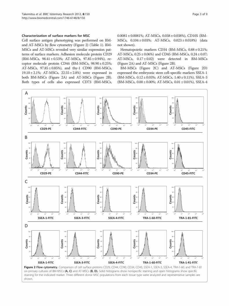
centrifugation at 800 times g for 30 min at roomtemperature The nucleated cells collected from the PBSsolution - Lymphoprep interface were then washed inPBS and transferred into T-75 cell culture flasks with10 ml of control medium consisting of Dulbeccorsquos modi-fied Eaglersquos medium (Invitrogen) 10 FBS and 1 anti-bioticndashantimycotic solution Cells were plated at adensity of 1ndash5 times 107 cellsplate and incubated at 37degC ina humidified 5 CO2 incubator and the medium waschanged twice weekly When primary cultures reached70ndash80 confluency attached cells were passaged byexposure to 025 trypsin 1 mM EDTA (Invitrogen) for3 min and replated at a density of 80 times 103 cellscm2 forsubsequent passage
AT-MSCsAdipose tissues were also harvested from each dogunder general anesthesia Subcutaneous fat pads (ap-proximately 10 g) were harvested from the inguinalarea These pads were finely minced with scissors andthen digested in 40 ml of PBS containing 015 collage-nase (Invitrogen) with vigorous shaking for 60 min at37degC Samples were then filtered using 100 μm cell strai-ners (BD Biosciences Franklin Lakes USA) and washedwith PBS The cells obtained were seeded into T-75 cellculture flasks with 10 ml of control medium and incu-bated in the same manner as bone marrow cells
In vitro differentiationFor osteogenic differentiation passage 2 BM-MSCs andAT-MSCs were plated on 6-well culture plates at a densityof 50 times 103 cellscm2 and after incubation in controlmedium for 24 h the medium was changed to osteogenicmedium The osteogenic medium was Canine OsteoblastDifferentiation Medium purchased from Cell Applications(San Diego USA) The medium was changed twiceweekly For osteogenic analysis mineral deposits werequantitatively analyzed by von Kossa (Sigma St LouisUSA) staining at 14 daysFor adipogenic differentiation passage 2 BM- and AT-
MSCs were plated on 6-well culture plates at a density
Direction Position Gen bank No
sense 453ndash472 XM_538830
anti-sense 540ndash559 XM_538830
sense 1177ndash1196 XM_545216
anti-sense 1299ndash1318 XM_545216
sense 22ndash41 XM_543828
anti-sense 156ndash175 XM_543828
sense 339ndash360 AF021873
anti-sense 446ndash468 AF021873
Takemitsu et al BMC Veterinary Research 2012 8150 Page 7 of 9httpwwwbiomedcentralcom1746-61488150
of 80 times 103 cellscm2 The cells were cultured in controlmedium until confluency and then the medium waschanged to Canine Adipocyte Differentiation Medium(Cell Applications) The medium was changed twice in aweek Oil Red O (Sigma) staining was performed toanalyze adipogenesis at 14 days
Flow cytometryPassage 2 BM-MSCs and AT-MSCs were placed inFACS tubes (BD Biosciences) at 2 times 105 cellstubewashed with FACS buffer (PBS containing 1 sodiumazide and 1 FBS pH 72) The cells were incubatedwith antibodies including CD29-PE (BioLegend SanDiego USA) CD34-PE (RampD Systems MinneapolisUSA) CD44-FITC (eBioscience San Diego USA)CD90-PE (BD Biosciences) CD45 (Abcam CambridgeUK) [44] CD73-PE (Bioss Woburn USA) CD105-FITC(Bioss) SSEA-1 (RampD Systems) SSEA-3 (RampD Systems)SSEA-4 (RampD Systems) TRA-1-60 (RampD Systems) andTRA-1-81 (Chemicon Temecula USA) [21] at roomtemperature for 1 h The cells were washed twice withFACS buffer and resuspended in 500 μl of FACS bufferThe cells incubated with CD45 SSEA-1 SSEA-3 SSEA-4 TRA-1-60 or TRA-1-81 were incubated with anti-ratIgG anti-rat IgM anti-mouse IgG and anti-mouse IgMsecondary antibodies labeled with FITC for 1 h Cellswere then washed twice with FACS buffer and resus-pended in 500 μl of FACS buffer Cell fluorescence wasevaluated by flow cytometry in a FACSCalibur instru-ment (BD Biosciences) Data were analyzed using Flowjosoftware (Tree Star Ashland USA)
Reverse transcription and quantitative real-time PCR(qRT-PCR)Total RNA was obtained from cultured BM-MSCs andAT-MSCs in passage 2 Total RNA was extracted usingTRIzol reagent (Invitrogen) according to the manufac-turerrsquos protocol Total RNA was measured by spectro-photometry Total RNA (1 μg) was reverse-transcribedat 42degC for 15 min in 20 μl with QuantiTect (QiagenDuumlsseldorf Germany) after inactivation of reverse tran-scription by heating at 95degC for 3 minThe cDNA product was subjected to real-time PCR
according to the user instructions for the Real-TimePCR System 7300 (Applied Biosystems Foster City CA)qRT-PCR was performed at 95degC for 5 s and 60degC for34 s in 20 μl buffer containing SYBR premix ExTaq IIand ROX Reference Dye (Takara Bio Shiga Japan) and02 μM each of the primers (Table 2) Quantitative meas-urement was performed by establishing a linear amplifi-cation curve from serial dilutions of plasmid DNAcontaining each cDNA
ImmunocytochemistryImmunofluorescent staining was used to assess expres-sion of the transcription factors Oct4 Sox2 and Nanogin BM- and AT-MSCs Passage 2 cells were cultured in4-well chamber slides (Nunc Roskilde Denmark) until50 confluency The cells were washed twice withPBS and fixed with 4 paraformaldehyde at roomtemperature for 30 min After washing thrice with PBSthe cells were incubated with blocking solution contain-ing 04 Triton X-100 and 4 Block Ace (DS PharmaBiomedical Osaka Japan) in PBS at room temperaturefor 1 h The cells were incubated with rabbit polyclonalprimary antibodies against Oct34 (Santa Cruz Biotech-nology Santa Cruz USA) Sox2 (Stem Cell Technolo-gies Vancouver Canada) and Nanog (Peprotech RockyHill USA) [21] diluted in blocking solution at 4degC for16 h The negative control cells were incubated withoutprimary antibody and isotype control cells were incu-bated with normal rabbit IgG antibody (RampD Systems)The cells were washed thrice with PBS and incubatedwith secondary anti-rabbit antibody labeled with AlexaFluor 488 (Invitrogen) diluted in blocking solution atroom temperature for 1 h in darkness The cells werethen washed thrice with PBS and slides were mountedin Vectashield Hard Mounting Medium with DAPI(Vector Laboratories Burlingame USA) The cells wereanalyzed under a Zeiss Axiovert 200 M fluorescencemicroscope (Carl Zeiss MicroImaging Jena Germany)and image overlay was performed using Axio VisionRel46 software (Carl Zeiss MicroImaging) (Additionalfile 1 Table S1)
Statistical analysisData were analyzed with an independent samples t-testand a P-value of less than 001 was considered signifi-cant Statistical analysis was performed using Prism soft-ware (GraphPad Software San Diego USA)
Additional file
Additional file 1 Table S1 Antibody Informations
AbbreviationsBM-MSC Bone marrow-derived mesenchymal stem cell AT-MSC Adiposetissue-derived mesenchymal stem cell ESC Embryonic stem cell qRT-PCR Quantitative real-time polymerase chain reaction SSEA Stage specificembryonic antigen TRA Keratin surface-associated antigen tumor-relatedantigen
Competing interestsNone of the authors has any financial or personal relationships that couldinappropriately influence or bias the content of the paper
Authorsrsquo contributionsHT and DZ designed the study drafted the manuscript and analyzed data IYhelped with editing and revision of the manuscript YH helped withcollection of samples and revision of the manuscript MM helped with FACS
Takemitsu et al BMC Veterinary Research 2012 8150 Page 8 of 9httpwwwbiomedcentralcom1746-61488150
analysis and revision of the manuscript and TA contributed to the studydesign and helped with editing and revision of the manuscript All authorsread and approved the final manuscript
AcknowledgmentsThis work was supported in part by the Supported Program for the StrategicResearch Foundation at Program for Private Universities from the Ministry ofEducation Culture Sports Science and Technology of Japan (MEXT) 2008ndash2012 and a Grant-in-Aid for Science Research (No 21380195 to T Arai) fromMEXT The authors would like to thank Enago (wwwenagojp) for the Englishlanguage review
Author details1Department of Veterinary Science School of Veterinary Medicine NipponVeterinary and Life Science University 1-7-1 Kyonan-cho Musashino Tokyo180-8602 Japan 2Veterinary medical teaching hospital Nippon Veterinaryand Life Science University 1-7-1 Kyonan-cho Musashino Tokyo 180-8602Japan 3Department of Veterinary Pathology Nippon Veterinary and LifeScience University 1-7-1 Kyonan-cho Musashino Tokyo 180-8602 Japan
Received 9 February 2012 Accepted 15 August 2012Published 31 August 2012
References1 Caplan AI Mesenchymal Stem Cells J Orthop Res 1991 9641ndash6502 Zuk PA Zhu M Mizuno H Huang J Futrell JW Katz AJ Benhaim AJ Lorenz
HP Hedrick MH Multi-lineage cells from human adipose tissueimplication for cell- based therapies Tissue Eng 2001 7211ndash228
3 Huang JI Beanes SR Zhu M Lorenz HP Hedrick MH Benhaim P Rat extra-medullary adipose tissue as a source of osteochondrogenic progenitorcells Plast Reconstr Surg 2002 1091033ndash1041
4 Dennis JE Caplan AI Differentiation potential of conditionallyimmortalized mesenchymal progenitor cells from adult marrow of aH-2Kb-tsA58 transgenic mouse J Cell Physiol 1996 167(3)523ndash538
5 Johnstone B Hering TM Caplan AI Goldberg VM Yoo JU In vitrochondrogenesis of bone marrow-derived mesenchymal progenitor cellsExp Cell Res 1998 238(1)265ndash272
6 Dennis JE Merriam A Awadallah A Yoo JU Johnstone B Caplan AI Aquadr- icpotential mesenchymal progenitor cell isolated from themarrow of an adult mouse J Bone Miner Res 1999 14(5)700ndash709
7 Kopen GC Prockop DJ Phinney DG Marrow stromal cells migratethroughout forebrain and cerebellum and they differentiate intoastrocytes after injection into neonatal mouse brains Proc Natl Acad SciUSA 1999 9610711ndash10716
8 Toma C Pittenger MF Cahill KS Byrne BJ Kessler PD Human mesenchymalstem cells differe- ntiate to a cardiomyocyte phenotype in the adultmurine heart Circulation 2002 10593ndash98
9 Quarto R Mastrogiacomo M Cancedda R Repair of large bone defectswith the use of autologous bone marrow stromal cells N Engl J Med2001 344385ndash386
10 Fouillard L Bensidhoum M Bories D Engraftment of allogeneicmesenchymal stem cells in the bone marrow of a patient with severeidiopathic aplastic anemia improves stroma Leukemia 2003 17474ndash476
11 Kolf CM Elizabeth C Tuan RS Biology of adult mesenchymal stem cellsregulation of niche self-renewal and differentiation Arthritis Res Ther2007 9204
12 Castro-Malaspina H Gay RE Resnick G Characterisation of human bonemarrow fibroblast colony-forming cells (CFU-F) and their progeny Blood1980 56289ndash301
13 Civin CI Trischmann T Kadan NS Highly purified CD34-positive cellsreconstitute hematopoiesis J Clin Oncol 1996 14(8)2224ndash2233
14 Zuk PA Zhu M Ashjian P Human adipose tissue is a source ofmultipotent stem cells Mol Biol Cell 2002 13(12)4279ndash4295
15 Iwata T Yamato M Zhang Z Validation of human periodontal ligament-derived cells as a reliable source for cytotherapeutic use J ClinPeriodontol 2010 37(12)1088ndash1099
16 Orciani M Mariggio MA Morabito C Di BG Di PR Functionalcharacterization of calcium-signaling pathways of human skin derivedmesenchymal stem cells Skin Pharmacol Physiol 2010 23(3)124ndash132
17 Tsai MS Lee JL Chang YJ Hwang SM Isolation of human multipotentmesenchymal stem cells from second-trimester amniotic fluid using anovel two-stage culture protocol Hum Reprod 2004 19(6)1450ndash1456
18 Dominici M Le BK Mueller I et al Minimal criteria for definingmultipotent mesenchymal stromal cells The International Society forCellular Therapy position statement Cytotherapy 2006 8(4)315ndash317
19 Wongchuensoontorn C Liebehenschel N Schwarz U Application of a newchair-side method for the harvest of mesenchymal stem cells in apatient with nonunion of a fracture of the atrophic mandiblendasha casereport J Craniomaxillofac Surg 2009 37(3)155ndash161
20 Mousavi NN Jaberipour M Razmkhah M Ghaderi A Habibagahi MMesenchymal stem cells do not suppress lymphoblastic leukemic cellline proliferation Iran J Immunol 2009 6(4)186ndash194
21 Vieira NM Brandalise V Zatz M et al Isolation Characterization andDifferentiation Potential of Canine Adipose-Derived Stem Cells CellTransplant 2010 19279ndash289
22 Dong In J Jeongim H Park H-m et al A comparison of autologous andallogenic bone marrow-derived mesenchymal stem cell transplantationin canine spinal cord injury J Neurol Sci 2009 28567ndash77
23 Vaags AK Kabkar SR Hough R Derivation and characterization of canineembryonic stem cell lines with in vitro and in vivo differentiationpotential Stem cells 2009 27329ndash340
24 Boiani M Scholer HR Regulatory networks in embryo-derived pluripotentstem cells Nat Rev Mol Cell Biol 2005 6872ndash884
25 Boyer LA Lee TI Cole MF Johnstone SE Levine SS Zucker JP Guenther MGKumar RM Murray HL Jenner RG Gifford DK Melton DA Jaenisch R YoungRA Core transcriptional regulatory circuitry in human embryonic stemcells Cell 2005 122947ndash956
26 Pittenger MF Mackay AM Beck SC Multilineage potential of adult humanmesenchymal stem cells Science 1999 284143ndash147
27 Gronthos S Franklin DM Leddy HA Robey PG Storms RW Gimble JMSurface protein characterization of human adipose tissue-derivedstromal cells J Cell Physiol 2001 189(1)54ndash63
28 Katz AJ Tholpady A Tholpady SS Shang H Ogle RC Cell surface andtranscriptional characterization of human adipose-derived adherentstromal (hADAS) cells Stem Cells 2005 23(3)412ndash423
29 Wagner W Wein F Seckinger A Frankhauser M Wirkner U Krause U Blake JSchwager C Eckstein V Ansorge W Ho AD Comparative characteristics ofmesenchymal stem cells from human bone marrow adipose tissue andumbilical cord blood Exp Hematol 2005 33(11)1402ndash1416
30 Mitchell JB McIntosh K Zvonic S Garrett S Floyd ZE Kloster A Di HalvorsenY Storms RW Goh B Kilroy G Wu X Gimble JM Immunophenotype ofhuman adipose-derived cells temporal changes in stromal-associatedand stem cell-associated markers Stem Cells 2006 24(2)376ndash385
31 Varma MJ Breuls RG Schouten TE Jurgens WJ Bontkes HJ Schuurhuis GJvan Ham SM van Milligen FJ Phenotypical and functionalcharacterization of freshly isolated adipose tissue-derived stem cellsStem Cells Dev 2007 16(1)91ndash104
32 Kolf CM Cho E Tuan RS Mesenchymal stromal cells Biology of adultmesenchymal stem cells regulation of niche self-renewal anddifferentiation Arthritis Res Ther 2007 9(1)204 Review
33 Riekstina U Cakstina I Parfejevs V Embryonic Stem Cell Marker ExpressionPattern in Human Mesenchymal Stem Cells Derived from Bone MarrowAdipose Tissue Heart and Dermis Stem Cell Rev 2009 5(4)378ndash386
34 Greco SJ Liu K Rameshwar P Functional similarities among genesregulated by OCT4 in human mesenchymal and embryonic stem cellsStem Cells 2007 25(12)3143ndash3154
35 Ratajczak MZ Kucia M Majka M Reca R Ratajczak J Heterogeneouspopulations of bone marrow stem cells - are we spotting on the samecells from the different angles Folia Histochem Cytobiol 200442(3)139ndash146
36 Collignon J Sockanathan S Hacker A Cohen-Tannoudji M Norris D RastanS Stevanovic M Goodfellow PN Lovell-Badge R A comparison of theproperties of Sox-3 with Sry and 2 related genes Sox-1 and Sox-2Development 1996 122509ndash520
37 Masui S Nakatake Y Toyooka Y Shimosato D Yagi R Takahashi K Okochi HOkuda A Matoba R Sharov AA Ko MSH Niwa H Pluripotency governedby Sox2 via regulation of Oct34 expression in mouse embryonic stemcells Nature Cell Biol 2007 9625ndash635
38 Ambrosetti DC Scholer HR Dailey L Basilico C Modulation of the activityof multiple transcriptional activation domains by the DNA binding
Takemitsu et al BMC Veterinary Research 2012 8150 Page 9 of 9httpwwwbiomedcentralcom1746-61488150
domains mediates the synergistic action of Sox2 and Oct-3 on thefibroblast growth factor-4 enhancer J Biol Chem 2000 27523387ndash23397
39 Pountos I Jones E Tzioupis C McGonagle D Giannoudis PV Growing boneand cartilage The role of mesenchymal stem cells J Bone Joint Surg Br2006 88421ndash426
40 Mitsui K Tokuzawa Y Itoh H Segawa K Murakami M Takahashi KMaruyama M Maeda M Yamanaka S The Homeoprotein Nanog isrequired for maintenance of pluripotency in mouse epiblast and ES cellsCell 2003 113631ndash642
41 Reza I Deepak K Christopher K Bruce AB et al Long-term In vitroExpansion Alters the Biology of Adult Mesenchymal Stem Cells CancerRes 2008 68(11)4229ndash4238
42 Ota M Sasaki H Mammalian Tead proteins regulate cell proliferation andcontact inhibition as transcriptional mediators of Hippo signalingDevelopment 2008 1354059ndash4069 doi101242dev027151
43 Yamazaki S Iwama A Takayanagi S Morita Y Eto K Ema H Nakauchi HCytokine signals modulated via lipid rafts mimic niche signals andinduce hibernation in hematopoietic stem cells EMBO J 200625(15)3515ndash3523 Epub 2006 Jul 20
44 Cobbold S Metcalfe S Monoclonal antibodies that define caninehomologues of human CD antigens summary of the First InternationalCanine Leukocyte Antigen Workshop (CLAW) Tissue Antigens 199443(3)137ndash154
doi1011861746-6148-8-150Cite this article as Takemitsu et al Comparison of bone marrow andadiposetissue-derived canine mesenchymalstem cells BMC Veterinary Research 2012 8150
Submit your next manuscript to BioMed Centraland take full advantage of
bull Convenient online submission
bull Thorough peer review
bull No space constraints or color figure charges
bull Immediate publication on acceptance
bull Inclusion in PubMed CAS Scopus and Google Scholar
bull Research which is freely available for redistribution
Submit your manuscript at wwwbiomedcentralcomsubmit
- Abstract
-
- Background
- Results
- Conclusion
-
- Background
- Results
-
- Cell isolation and culture
- In vitro differentiation
-
- link_Fig1
-
- Characterization of surface markers for MSC
-
- link_Fig2
-
- mRNA expression analysis using quantitative ampb_kreal-ampe_kampb_ktimeampe_k PCR
- Immunocytochemistry
-
- Discussion
- link_Tab1
- link_Fig3
- Conclusion
- link_Fig4
- Methods
-
- MSC isolation and culture
- BM-MSCs
- AT-MSCs
- In vitro differentiation
-
- link_Tab2
-
- Flow cytometry
- Reverse transcription and quantitative ampb_kreal-ampe_kampb_ktimeampe_k PCR (qRT-PCR)
- Immunocytochemistry
- Statistical analysis
-
- Additional file
- Competing interests
- Authorsacute contributions
- Acknowledgments
- References
- link_CR1
- link_CR2
- link_CR3
- link_CR4
- link_CR5
- link_CR6
- link_CR7
- link_CR8
- link_CR9
- link_CR10
- link_CR11
- link_CR12
- link_CR13
- link_CR14
- link_CR15
- link_CR16
- link_CR17
- link_CR18
- link_CR19
- link_CR20
- link_CR21
- link_CR22
- link_CR23
- link_CR24
- link_CR25
- link_CR26
- link_CR27
- link_CR28
- link_CR29
- link_CR30
- link_CR31
- link_CR32
- link_CR33
- link_CR34
- link_CR35
- link_CR36
- link_CR37
- link_CR38
- link_CR39
- link_CR40
- link_CR41
- link_CR42
- link_CR43
- link_CR44
-

Takemitsu et al BMC Veterinary Research 2012 8150 Page 2 of 9httpwwwbiomedcentralcom1746-61488150
expanding MSC cultures and maintaining the cells in theundifferentiated state [14-20]The expression profile of cell surface markers and
mRNAs is well characterized in other species In manystudies in humans and dogs CD29 CD44 and CD90were regarded as positive cell-surface markers for MSCs[14-1721] while CD34 and CD45 were regard as nega-tive surface markers [18-2022] In addition stage-specific embryonic antigen (SSEA)-1 SSEA-3 SSEA-4the keratin sulfate-associated antigen tumor-related anti-gen (TRA)-1-60 and TRA-1-81 were reported as mar-kers of canine embryonic stem cells (ESCs) [23] Thesemolecules constitute a comprehensive set of uniquestem cell markers Moreover Oct34 Sox2 and Nanogwere shown to be important transcription factors regu-lating ESC self-renewal and differentiation [2425] Thesetranscription factors interact with each other to overseea vast regulatory network that maintains pluripotencyand inhibited differentiation [24]In veterinary medicine the use of MSCs for tissue re-
pair is helpful and is likely to increase in future How-ever there have been few studies on cell surface markersand mRNA expression profiles of canine MSCs Here weevaluated the canine BM- and AT-MSC cell surface mar-kers CD29 CD44 CD90 CD34 CD45 SSEA-1 SSEA-3SSEA-4 TRA-1-60 and TRA-1-81 by flow cytometryWe also analyzed the mRNA expression profile of Oct34 Sox2 and Nanog in canine BM- and AT-MSCs byquantitative real-time PCR (qRT-PCR) Finally we usedimmunocytochemistry to examine the expression and
BM MSCs
Control Culture
OsteogenicDifferentiation
Adipogenic Differentiation
A
C
E
Figure 1 In vitro differentiation of BM-MSCs and AT-MSCs Both typesdifferentiation was identified by von Kossa staining (C D) and adipogenic
localization of Oct34 and Sox2 The aim of this studywas the biological characterization of canine MSCsisolated from bone marrow and adipose tissue
ResultsCell isolation and cultureAdherent cells were observed at the bottom of the cul-ture flasks within 2 days these cells were isolated fromboth bone marrow and adipose tissue after plating Bonemarrow-derived cells formed several colonies and prolif-erated taking various shapes including discoidal flattriangular and elongated (Figure 1A) Bone marrow-derived cells were uniformly distributed immediatelyafter passage but thereafter gradually formed coloniesand proliferated Unlike bone marrow-derived cellsadipose-derived cells were uniformly distributed and nocolony formation was observed (Figure 1B)
In vitro differentiationOsteogenic differentiation was demonstrated histo-chemically using von Kossa stain (Figure 1C D) Afteradipogenic induction culture for 3 weeks MSCs showedan adipogenic phenotype Histochemical staining withOil Red O was used to demonstrate adipogenic differen-tiation of MSCs Lipid droplets were observed at about14 days of culture in adipogenic medium and were posi-tive for Oil Red O staining (Figure 1 E F) Both differen-tiation experiments were carried out in passage 2(Figure 1C- F)
AT MSCs
B
D
F
of cell were maintained in control medium (A B) Osteogenicdifferentiation by Oil Red O staining (E F) Scale bar 200 μm
Takemitsu et al BMC Veterinary Research 2012 8150 Page 3 of 9httpwwwbiomedcentralcom1746-61488150
Characterization of surface markers for MSCCell surface antigen phenotyping was performed on BM-and AT-MSCs by flow cytometry (Figure 2) (Table 1) BM-MSCs and AT-MSCs revealed very similar expression pat-terns of surface markers Adhesion molecule protein CD29(BM-MSCs 9841plusmn053 AT-MSCs 9785plusmn094) re-ceptor molecule protein CD44 (BM-MSCs 9890plusmn025AT-MSCs 9785plusmn085) and thy-1 CD90 (BM-MSCs1910plusmn21 AT-MSCs 2255plusmn28) were expressed inboth BM-MSCs (Figure 2A) and AT-MSCs (Figure 2B)Both types of cells also expressed CD73 (BM-MSCs
Figure 2 Flow cytometry Comparison of cell surface proteins CD29 CD4on primary cultures of BM-MSCs (A C) and AT-MSCs (B D) Solid histogramstaining for the indicated marker Three different donor MSC populations frshown
00081plusmn00081 AT-MSCs 0038plusmn0038) CD105 (BM-MSCs 0104plusmn003 AT-MSCs 0023plusmn0018) (datanot shown)Hematopoietic markers CD34 (BM-MSCs 088plusmn021
AT-MSCs 025plusmn006) and CD45 (BM-MSCs 024plusmn007AT-MSCs 017plusmn002) were detected in BM-MSCs(Figure 2A) and AT-MSCs (Figure 2B)BM-MSCs (Figure 2C) and AT-MSCs (Figure 2D)
expressed the embryonic stem cell-specific markers SSEA-1(BM-MSCs 012plusmn003 AT-MSCs 140plusmn011) SSEA-3(BM-MSCs 000plusmn000 AT-MSCs 001plusmn001) SSEA-4
4 CD90 CD34 CD45 SSEA-1 SSEA-3 SSEA-4 TRA-1-60 and TRA-1-81s show nonspecific staining and open histograms show specificom each tissue type were analyzed and representative samples are
Table 1 Summary of cell surface markers on canine BM-and AT-MSCs
BM-MSCs AT-MSCs
Mesenchymal Stromal Cell Marker
CD29 ++ ++
CD44 ++ ++
CD90 + +
Hematopoietic markers
CD34 - -
CD45 - -
Embryonic stem cell markers
SSEA-1 - -
SSEA-3 - -
SSEA-4 - -
TRA-1-60 - -
TRA-1-81 - -
Canine MSCs were analyzed by FACS as shown in Figure 2 Samples werescored as ldquominusrdquo if lt9 of cells were positive ldquo+rdquo if 9ndash69 were positive andldquo++rdquo if ge70 were positive
Takemitsu et al BMC Veterinary Research 2012 8150 Page 4 of 9httpwwwbiomedcentralcom1746-61488150
(BM-MSCs 007plusmn001 AT-MSCs 002plusmn001) TRA-1-60 (BM-MSCs 001plusmn000 AT-MSCs 002plusmn001)and TRA-1-81 (BM-MSCs 000plusmn000 AT-MSCs001plusmn001)
mRNA expression analysis using quantitative real-timePCRExpression levels of canine Oct34 Sox2 and NanogmRNA in BM- and AT-MSCs were examined by qRT-PCR (Figure 3) Expression of Oct34 did not differ sig-nificantly difference between BM-MSCs and AT-MSCsHowever Sox2 expression tended to be higher in BM-MSCs than in AT-MSCs while Nanog expression in AT-MSCs was 25-fold higher than in BM-MSCs (p lt 001)
Figure 3 Quantitative RT-PCR Expression levels of mRNAs for stem cell mwas normalized to beta-actin expression Statistical comparisons were mad
ImmunocytochemistryImmunocytochemistry was used to analyze Oct34 andSox2 proteins (Figure 4) In both BM-MSCs and AT-MSCs Oct34 was detected mainly the nuclei whereasSox2 was detected in the cytoplasm
DiscussionIn this study we confirmed the potential of MSCs to dif-ferentiate into osteoblasts or adipocytes and evaluatedthe protein and mRNA expression profiles of these cellsIn addition we compared the expression patterns ofESC markers and germ layer markers in MSCs derivedfrom bone marrow and adipose tissue BM-MSCand AT-MSC populations expressed CD29 CD44 andCD90 with similar intensity In particular in both BM-and AT-MSCs more than 95 of the cell populationexpressed CD29 and CD44 Our results indicate that ex-pression of CD90 was lower than that of other markersHowever many human studies have reported strong ex-pression of CD90 This may be related to differenceswithin species Many human studies have reported posi-tive expression of CD90 whereas many studies in micehave reported negative expression of CD90 [1126] Inaddition the decline in CD90 expression may be influ-enced by the passage number of the cells In early passagecells CD90 showed variable expression [2527-32] andRiekstina et al reported that CD90 expression decreasedwith increasing passage number [33] CD34 and CD45markers are usually associated with hematopoietic stemcellsThe qRT-PCR analysis revealed expression of the stem
cell markers Oct34 Sox2 and Nanog in canine BM-and AT-MSCs In a human study mRNAs of these mar-kers were expressed in human MSCs [34] These tran-scription factors mediate self-renewal and cell-fatespecification and are downregulated when the cells arecompletely differentiated [35] Our results reflect their
arkers Oct34 Sox2 and Nanog in BM-MSCs and AT-MSCs Each valuee using Studentrsquos t test (plt 001)
Figure 4 Immunocytochemistry Expression and localization of Oct34 and Sox2 in BM-MSCs and AT-MSCs Immunofluorescent localization ofOct34 in BM-MSCs with DAPI counterstaining (C D) Immunofluorescent localization of Sox2 in BM-MSCs with DAPI counterstaining (E F)Immunofluorescent localization of Oct34 in AT-MSCs with DAPI counterstaining (I J) Immunofluorescent localization of Oct34 in BM-MSCs withDAPI counterstaining (K L) Scale bar 40 μm
Takemitsu et al BMC Veterinary Research 2012 8150 Page 5 of 9httpwwwbiomedcentralcom1746-61488150
undifferentiated phenotype and high propensity forpluripotent differentiation and support the hypothesisthat MSCs may be pluripotent stem cells deposited intissues during development Oct34 mRNA showedsimilar levels of expression in BM- and AT-MSCs How-ever expression of Sox2 in BM-MSCs tended to behigher than in AT-MSCs Sox2 belongs to the Sox sub-family whose members are defined by the relationshipof their HMG box [36] In addition Sox2 regulatesOct34 expression and maintains ESC pluripotencythrough upstream transcription factors [37] and throughcooperative binding of OCT34 to DNA [38] Howeverour results indicate Oct34 expression was similar inBM- and AT-MSCs but Sox2 expression slightly differedbetween the two cell types This is probably due to theadditional expression of other Sox family members thatact in a compensatory in MSCs [37] In addition expres-sion of Nanog in AT-MSCs was 25-fold higher than inBM-MSCs Nanog is required to maintain the undiffer-entiated state and for the self-renewal of stem cells InESCs knockout or knockdown of Nanog abolishes bothself-renewal and pluripotency and results in differenti-ation of extraembryonic endoderm [3940] In a humanstudy comparison of proliferation in BM- and AT-MSCsrevealed high proliferative ability in AT-MSCs [41]These results suggest that AT-MSCs may maintain the
undifferentiated state and that their self-renewal abilityis greater than that of BM-MSCsWe performed immunostaining with Oct34 and Sox2
in MSCs In both BM- and AT-MSCs Oct34 wasdetected in the nuclear fraction whereas Sox2 wasdetected in the cytoplasmic fraction Several transcrip-tion factors are known to be localized in the cytoplasmicfraction such as The Tead [42] and FOXO families [43]These studies showed inhibitory activity of the transcrip-tion factors Hence it is possible that they inactivateSox2 In addition Sox2 was localized in the nucleus incanine ESCs [23] There are many functional differencesbetween ESCs and MSCs For example ESCs form tera-tomas in the testis but MSCs do not We therefore sug-gest that restricted localization of Sox2 protein may leadto lack of proliferation of MSCs in vivo as well as main-tenance of pluripotency of MSCs in vitro
ConclusionOur study reveals the protein and mRNA expressionprofiles of canine BM- and AT-MSCs The two types ofcells showed similar cell surface marker profiles Quanti-tative real-time PCR revealed expression of mRNAs forOct34 Sox2 and Nanog in BM- and AT-MSCs Thelocalization of Oct34 and Sox2 was demonstratedimmunocytochemically mRNA expression of Nanog was
Takemitsu et al BMC Veterinary Research 2012 8150 Page 6 of 9httpwwwbiomedcentralcom1746-61488150
greater in AT-MSCs than in BM-MSCs Further studiesare required to characterize canine MSCs with respectto the expression of other proteins and mRNAs
MethodsMSC isolation and cultureFour young healthy female beagle dogs (1 year old 95-113 kg body weight) were used All animals wereanesthetized with propofor (Hospira Osaka Japan)(7 mgkg by intravenous injection) before tissue samplewere taken After incubation anesthesia was maintainedwith isoflurane (15ndash20) in oxygen Animal experi-ments were carried out in accordance with the NationalInstitutes of Health guidelines for the care and use oflaboratory animals The protocol of this study wasapproved by the University Committee for AnimalExperimentation
BM-MSCsUnder general anesthesia bone marrow was aspiratedfrom the proximal humerus using a general bone mar-row biopsy technique Briefly a sterilized 13-gaugeJamshidi needle (Cardinal Health McGaw Park USA)was used to aspirate 5 ml of bone marrow into a syringecontaining 5 ml of heparinized (1000 unitsml) salinesolution Perioperative analgesic management was car-ried out by the pre- and post-operative administration ofbuprenorphine which was administered twice daily for 3consecutive days after surgery In addition ampicillin(25 mgkg) was orally administered twice daily for up to7 days after surgeryThe bone marrow collected was dissociated and then
resuspended with a pipette The suspension was centri-fuged for 5 min at 300 times g and collected as a pellet Mar-row cells were then resuspended in 10 ml of 10 FBSndashPBS (FBS Invitrogen Carlsbad USA PBS Invitrogen)To obtain MSC-enriched nucleated cells density separ-ation (1077 gml) was performed using Lymphoprep(Axis-Shield Oslo Norway) A suspension of marrowcells in FBSndashPBS solution (10 ml) was carefully layeredonto 5 ml of Lymphoprep Separation was achieved by
Table 2 Primers used for qRT-PCR
Name 5rsquo ndash 3rsquo
oct34-1 GCTCCTGAAGCAGAAGAGGA
oct34-2 GCTGAACACCTTCCCAAAGA
sox2-1 CCCACCTACAGCATGTCCTA
sox2-2 GGAGTGGGAGGAGGAGGTAA
nanog-1 CCCAACTCTAGGGACCCTTC
nanog-2 CAGATCCATGGAGGAAGGAA
β-actin GCCAACCGTGAGAAGATGACT
β-actin CCCAGAGTCCATGACAATACCAG
centrifugation at 800 times g for 30 min at roomtemperature The nucleated cells collected from the PBSsolution - Lymphoprep interface were then washed inPBS and transferred into T-75 cell culture flasks with10 ml of control medium consisting of Dulbeccorsquos modi-fied Eaglersquos medium (Invitrogen) 10 FBS and 1 anti-bioticndashantimycotic solution Cells were plated at adensity of 1ndash5 times 107 cellsplate and incubated at 37degC ina humidified 5 CO2 incubator and the medium waschanged twice weekly When primary cultures reached70ndash80 confluency attached cells were passaged byexposure to 025 trypsin 1 mM EDTA (Invitrogen) for3 min and replated at a density of 80 times 103 cellscm2 forsubsequent passage
AT-MSCsAdipose tissues were also harvested from each dogunder general anesthesia Subcutaneous fat pads (ap-proximately 10 g) were harvested from the inguinalarea These pads were finely minced with scissors andthen digested in 40 ml of PBS containing 015 collage-nase (Invitrogen) with vigorous shaking for 60 min at37degC Samples were then filtered using 100 μm cell strai-ners (BD Biosciences Franklin Lakes USA) and washedwith PBS The cells obtained were seeded into T-75 cellculture flasks with 10 ml of control medium and incu-bated in the same manner as bone marrow cells
In vitro differentiationFor osteogenic differentiation passage 2 BM-MSCs andAT-MSCs were plated on 6-well culture plates at a densityof 50 times 103 cellscm2 and after incubation in controlmedium for 24 h the medium was changed to osteogenicmedium The osteogenic medium was Canine OsteoblastDifferentiation Medium purchased from Cell Applications(San Diego USA) The medium was changed twiceweekly For osteogenic analysis mineral deposits werequantitatively analyzed by von Kossa (Sigma St LouisUSA) staining at 14 daysFor adipogenic differentiation passage 2 BM- and AT-
MSCs were plated on 6-well culture plates at a density
Direction Position Gen bank No
sense 453ndash472 XM_538830
anti-sense 540ndash559 XM_538830
sense 1177ndash1196 XM_545216
anti-sense 1299ndash1318 XM_545216
sense 22ndash41 XM_543828
anti-sense 156ndash175 XM_543828
sense 339ndash360 AF021873
anti-sense 446ndash468 AF021873
Takemitsu et al BMC Veterinary Research 2012 8150 Page 7 of 9httpwwwbiomedcentralcom1746-61488150
of 80 times 103 cellscm2 The cells were cultured in controlmedium until confluency and then the medium waschanged to Canine Adipocyte Differentiation Medium(Cell Applications) The medium was changed twice in aweek Oil Red O (Sigma) staining was performed toanalyze adipogenesis at 14 days
Flow cytometryPassage 2 BM-MSCs and AT-MSCs were placed inFACS tubes (BD Biosciences) at 2 times 105 cellstubewashed with FACS buffer (PBS containing 1 sodiumazide and 1 FBS pH 72) The cells were incubatedwith antibodies including CD29-PE (BioLegend SanDiego USA) CD34-PE (RampD Systems MinneapolisUSA) CD44-FITC (eBioscience San Diego USA)CD90-PE (BD Biosciences) CD45 (Abcam CambridgeUK) [44] CD73-PE (Bioss Woburn USA) CD105-FITC(Bioss) SSEA-1 (RampD Systems) SSEA-3 (RampD Systems)SSEA-4 (RampD Systems) TRA-1-60 (RampD Systems) andTRA-1-81 (Chemicon Temecula USA) [21] at roomtemperature for 1 h The cells were washed twice withFACS buffer and resuspended in 500 μl of FACS bufferThe cells incubated with CD45 SSEA-1 SSEA-3 SSEA-4 TRA-1-60 or TRA-1-81 were incubated with anti-ratIgG anti-rat IgM anti-mouse IgG and anti-mouse IgMsecondary antibodies labeled with FITC for 1 h Cellswere then washed twice with FACS buffer and resus-pended in 500 μl of FACS buffer Cell fluorescence wasevaluated by flow cytometry in a FACSCalibur instru-ment (BD Biosciences) Data were analyzed using Flowjosoftware (Tree Star Ashland USA)
Reverse transcription and quantitative real-time PCR(qRT-PCR)Total RNA was obtained from cultured BM-MSCs andAT-MSCs in passage 2 Total RNA was extracted usingTRIzol reagent (Invitrogen) according to the manufac-turerrsquos protocol Total RNA was measured by spectro-photometry Total RNA (1 μg) was reverse-transcribedat 42degC for 15 min in 20 μl with QuantiTect (QiagenDuumlsseldorf Germany) after inactivation of reverse tran-scription by heating at 95degC for 3 minThe cDNA product was subjected to real-time PCR
according to the user instructions for the Real-TimePCR System 7300 (Applied Biosystems Foster City CA)qRT-PCR was performed at 95degC for 5 s and 60degC for34 s in 20 μl buffer containing SYBR premix ExTaq IIand ROX Reference Dye (Takara Bio Shiga Japan) and02 μM each of the primers (Table 2) Quantitative meas-urement was performed by establishing a linear amplifi-cation curve from serial dilutions of plasmid DNAcontaining each cDNA
ImmunocytochemistryImmunofluorescent staining was used to assess expres-sion of the transcription factors Oct4 Sox2 and Nanogin BM- and AT-MSCs Passage 2 cells were cultured in4-well chamber slides (Nunc Roskilde Denmark) until50 confluency The cells were washed twice withPBS and fixed with 4 paraformaldehyde at roomtemperature for 30 min After washing thrice with PBSthe cells were incubated with blocking solution contain-ing 04 Triton X-100 and 4 Block Ace (DS PharmaBiomedical Osaka Japan) in PBS at room temperaturefor 1 h The cells were incubated with rabbit polyclonalprimary antibodies against Oct34 (Santa Cruz Biotech-nology Santa Cruz USA) Sox2 (Stem Cell Technolo-gies Vancouver Canada) and Nanog (Peprotech RockyHill USA) [21] diluted in blocking solution at 4degC for16 h The negative control cells were incubated withoutprimary antibody and isotype control cells were incu-bated with normal rabbit IgG antibody (RampD Systems)The cells were washed thrice with PBS and incubatedwith secondary anti-rabbit antibody labeled with AlexaFluor 488 (Invitrogen) diluted in blocking solution atroom temperature for 1 h in darkness The cells werethen washed thrice with PBS and slides were mountedin Vectashield Hard Mounting Medium with DAPI(Vector Laboratories Burlingame USA) The cells wereanalyzed under a Zeiss Axiovert 200 M fluorescencemicroscope (Carl Zeiss MicroImaging Jena Germany)and image overlay was performed using Axio VisionRel46 software (Carl Zeiss MicroImaging) (Additionalfile 1 Table S1)
Statistical analysisData were analyzed with an independent samples t-testand a P-value of less than 001 was considered signifi-cant Statistical analysis was performed using Prism soft-ware (GraphPad Software San Diego USA)
Additional file
Additional file 1 Table S1 Antibody Informations
AbbreviationsBM-MSC Bone marrow-derived mesenchymal stem cell AT-MSC Adiposetissue-derived mesenchymal stem cell ESC Embryonic stem cell qRT-PCR Quantitative real-time polymerase chain reaction SSEA Stage specificembryonic antigen TRA Keratin surface-associated antigen tumor-relatedantigen
Competing interestsNone of the authors has any financial or personal relationships that couldinappropriately influence or bias the content of the paper
Authorsrsquo contributionsHT and DZ designed the study drafted the manuscript and analyzed data IYhelped with editing and revision of the manuscript YH helped withcollection of samples and revision of the manuscript MM helped with FACS
Takemitsu et al BMC Veterinary Research 2012 8150 Page 8 of 9httpwwwbiomedcentralcom1746-61488150
analysis and revision of the manuscript and TA contributed to the studydesign and helped with editing and revision of the manuscript All authorsread and approved the final manuscript
AcknowledgmentsThis work was supported in part by the Supported Program for the StrategicResearch Foundation at Program for Private Universities from the Ministry ofEducation Culture Sports Science and Technology of Japan (MEXT) 2008ndash2012 and a Grant-in-Aid for Science Research (No 21380195 to T Arai) fromMEXT The authors would like to thank Enago (wwwenagojp) for the Englishlanguage review
Author details1Department of Veterinary Science School of Veterinary Medicine NipponVeterinary and Life Science University 1-7-1 Kyonan-cho Musashino Tokyo180-8602 Japan 2Veterinary medical teaching hospital Nippon Veterinaryand Life Science University 1-7-1 Kyonan-cho Musashino Tokyo 180-8602Japan 3Department of Veterinary Pathology Nippon Veterinary and LifeScience University 1-7-1 Kyonan-cho Musashino Tokyo 180-8602 Japan
Received 9 February 2012 Accepted 15 August 2012Published 31 August 2012
References1 Caplan AI Mesenchymal Stem Cells J Orthop Res 1991 9641ndash6502 Zuk PA Zhu M Mizuno H Huang J Futrell JW Katz AJ Benhaim AJ Lorenz
HP Hedrick MH Multi-lineage cells from human adipose tissueimplication for cell- based therapies Tissue Eng 2001 7211ndash228
3 Huang JI Beanes SR Zhu M Lorenz HP Hedrick MH Benhaim P Rat extra-medullary adipose tissue as a source of osteochondrogenic progenitorcells Plast Reconstr Surg 2002 1091033ndash1041
4 Dennis JE Caplan AI Differentiation potential of conditionallyimmortalized mesenchymal progenitor cells from adult marrow of aH-2Kb-tsA58 transgenic mouse J Cell Physiol 1996 167(3)523ndash538
5 Johnstone B Hering TM Caplan AI Goldberg VM Yoo JU In vitrochondrogenesis of bone marrow-derived mesenchymal progenitor cellsExp Cell Res 1998 238(1)265ndash272
6 Dennis JE Merriam A Awadallah A Yoo JU Johnstone B Caplan AI Aquadr- icpotential mesenchymal progenitor cell isolated from themarrow of an adult mouse J Bone Miner Res 1999 14(5)700ndash709
7 Kopen GC Prockop DJ Phinney DG Marrow stromal cells migratethroughout forebrain and cerebellum and they differentiate intoastrocytes after injection into neonatal mouse brains Proc Natl Acad SciUSA 1999 9610711ndash10716
8 Toma C Pittenger MF Cahill KS Byrne BJ Kessler PD Human mesenchymalstem cells differe- ntiate to a cardiomyocyte phenotype in the adultmurine heart Circulation 2002 10593ndash98
9 Quarto R Mastrogiacomo M Cancedda R Repair of large bone defectswith the use of autologous bone marrow stromal cells N Engl J Med2001 344385ndash386
10 Fouillard L Bensidhoum M Bories D Engraftment of allogeneicmesenchymal stem cells in the bone marrow of a patient with severeidiopathic aplastic anemia improves stroma Leukemia 2003 17474ndash476
11 Kolf CM Elizabeth C Tuan RS Biology of adult mesenchymal stem cellsregulation of niche self-renewal and differentiation Arthritis Res Ther2007 9204
12 Castro-Malaspina H Gay RE Resnick G Characterisation of human bonemarrow fibroblast colony-forming cells (CFU-F) and their progeny Blood1980 56289ndash301
13 Civin CI Trischmann T Kadan NS Highly purified CD34-positive cellsreconstitute hematopoiesis J Clin Oncol 1996 14(8)2224ndash2233
14 Zuk PA Zhu M Ashjian P Human adipose tissue is a source ofmultipotent stem cells Mol Biol Cell 2002 13(12)4279ndash4295
15 Iwata T Yamato M Zhang Z Validation of human periodontal ligament-derived cells as a reliable source for cytotherapeutic use J ClinPeriodontol 2010 37(12)1088ndash1099
16 Orciani M Mariggio MA Morabito C Di BG Di PR Functionalcharacterization of calcium-signaling pathways of human skin derivedmesenchymal stem cells Skin Pharmacol Physiol 2010 23(3)124ndash132
17 Tsai MS Lee JL Chang YJ Hwang SM Isolation of human multipotentmesenchymal stem cells from second-trimester amniotic fluid using anovel two-stage culture protocol Hum Reprod 2004 19(6)1450ndash1456
18 Dominici M Le BK Mueller I et al Minimal criteria for definingmultipotent mesenchymal stromal cells The International Society forCellular Therapy position statement Cytotherapy 2006 8(4)315ndash317
19 Wongchuensoontorn C Liebehenschel N Schwarz U Application of a newchair-side method for the harvest of mesenchymal stem cells in apatient with nonunion of a fracture of the atrophic mandiblendasha casereport J Craniomaxillofac Surg 2009 37(3)155ndash161
20 Mousavi NN Jaberipour M Razmkhah M Ghaderi A Habibagahi MMesenchymal stem cells do not suppress lymphoblastic leukemic cellline proliferation Iran J Immunol 2009 6(4)186ndash194
21 Vieira NM Brandalise V Zatz M et al Isolation Characterization andDifferentiation Potential of Canine Adipose-Derived Stem Cells CellTransplant 2010 19279ndash289
22 Dong In J Jeongim H Park H-m et al A comparison of autologous andallogenic bone marrow-derived mesenchymal stem cell transplantationin canine spinal cord injury J Neurol Sci 2009 28567ndash77
23 Vaags AK Kabkar SR Hough R Derivation and characterization of canineembryonic stem cell lines with in vitro and in vivo differentiationpotential Stem cells 2009 27329ndash340
24 Boiani M Scholer HR Regulatory networks in embryo-derived pluripotentstem cells Nat Rev Mol Cell Biol 2005 6872ndash884
25 Boyer LA Lee TI Cole MF Johnstone SE Levine SS Zucker JP Guenther MGKumar RM Murray HL Jenner RG Gifford DK Melton DA Jaenisch R YoungRA Core transcriptional regulatory circuitry in human embryonic stemcells Cell 2005 122947ndash956
26 Pittenger MF Mackay AM Beck SC Multilineage potential of adult humanmesenchymal stem cells Science 1999 284143ndash147
27 Gronthos S Franklin DM Leddy HA Robey PG Storms RW Gimble JMSurface protein characterization of human adipose tissue-derivedstromal cells J Cell Physiol 2001 189(1)54ndash63
28 Katz AJ Tholpady A Tholpady SS Shang H Ogle RC Cell surface andtranscriptional characterization of human adipose-derived adherentstromal (hADAS) cells Stem Cells 2005 23(3)412ndash423
29 Wagner W Wein F Seckinger A Frankhauser M Wirkner U Krause U Blake JSchwager C Eckstein V Ansorge W Ho AD Comparative characteristics ofmesenchymal stem cells from human bone marrow adipose tissue andumbilical cord blood Exp Hematol 2005 33(11)1402ndash1416
30 Mitchell JB McIntosh K Zvonic S Garrett S Floyd ZE Kloster A Di HalvorsenY Storms RW Goh B Kilroy G Wu X Gimble JM Immunophenotype ofhuman adipose-derived cells temporal changes in stromal-associatedand stem cell-associated markers Stem Cells 2006 24(2)376ndash385
31 Varma MJ Breuls RG Schouten TE Jurgens WJ Bontkes HJ Schuurhuis GJvan Ham SM van Milligen FJ Phenotypical and functionalcharacterization of freshly isolated adipose tissue-derived stem cellsStem Cells Dev 2007 16(1)91ndash104
32 Kolf CM Cho E Tuan RS Mesenchymal stromal cells Biology of adultmesenchymal stem cells regulation of niche self-renewal anddifferentiation Arthritis Res Ther 2007 9(1)204 Review
33 Riekstina U Cakstina I Parfejevs V Embryonic Stem Cell Marker ExpressionPattern in Human Mesenchymal Stem Cells Derived from Bone MarrowAdipose Tissue Heart and Dermis Stem Cell Rev 2009 5(4)378ndash386
34 Greco SJ Liu K Rameshwar P Functional similarities among genesregulated by OCT4 in human mesenchymal and embryonic stem cellsStem Cells 2007 25(12)3143ndash3154
35 Ratajczak MZ Kucia M Majka M Reca R Ratajczak J Heterogeneouspopulations of bone marrow stem cells - are we spotting on the samecells from the different angles Folia Histochem Cytobiol 200442(3)139ndash146
36 Collignon J Sockanathan S Hacker A Cohen-Tannoudji M Norris D RastanS Stevanovic M Goodfellow PN Lovell-Badge R A comparison of theproperties of Sox-3 with Sry and 2 related genes Sox-1 and Sox-2Development 1996 122509ndash520
37 Masui S Nakatake Y Toyooka Y Shimosato D Yagi R Takahashi K Okochi HOkuda A Matoba R Sharov AA Ko MSH Niwa H Pluripotency governedby Sox2 via regulation of Oct34 expression in mouse embryonic stemcells Nature Cell Biol 2007 9625ndash635
38 Ambrosetti DC Scholer HR Dailey L Basilico C Modulation of the activityof multiple transcriptional activation domains by the DNA binding
Takemitsu et al BMC Veterinary Research 2012 8150 Page 9 of 9httpwwwbiomedcentralcom1746-61488150
domains mediates the synergistic action of Sox2 and Oct-3 on thefibroblast growth factor-4 enhancer J Biol Chem 2000 27523387ndash23397
39 Pountos I Jones E Tzioupis C McGonagle D Giannoudis PV Growing boneand cartilage The role of mesenchymal stem cells J Bone Joint Surg Br2006 88421ndash426
40 Mitsui K Tokuzawa Y Itoh H Segawa K Murakami M Takahashi KMaruyama M Maeda M Yamanaka S The Homeoprotein Nanog isrequired for maintenance of pluripotency in mouse epiblast and ES cellsCell 2003 113631ndash642
41 Reza I Deepak K Christopher K Bruce AB et al Long-term In vitroExpansion Alters the Biology of Adult Mesenchymal Stem Cells CancerRes 2008 68(11)4229ndash4238
42 Ota M Sasaki H Mammalian Tead proteins regulate cell proliferation andcontact inhibition as transcriptional mediators of Hippo signalingDevelopment 2008 1354059ndash4069 doi101242dev027151
43 Yamazaki S Iwama A Takayanagi S Morita Y Eto K Ema H Nakauchi HCytokine signals modulated via lipid rafts mimic niche signals andinduce hibernation in hematopoietic stem cells EMBO J 200625(15)3515ndash3523 Epub 2006 Jul 20
44 Cobbold S Metcalfe S Monoclonal antibodies that define caninehomologues of human CD antigens summary of the First InternationalCanine Leukocyte Antigen Workshop (CLAW) Tissue Antigens 199443(3)137ndash154
doi1011861746-6148-8-150Cite this article as Takemitsu et al Comparison of bone marrow andadiposetissue-derived canine mesenchymalstem cells BMC Veterinary Research 2012 8150
Submit your next manuscript to BioMed Centraland take full advantage of
bull Convenient online submission
bull Thorough peer review
bull No space constraints or color figure charges
bull Immediate publication on acceptance
bull Inclusion in PubMed CAS Scopus and Google Scholar
bull Research which is freely available for redistribution
Submit your manuscript at wwwbiomedcentralcomsubmit
- Abstract
-
- Background
- Results
- Conclusion
-
- Background
- Results
-
- Cell isolation and culture
- In vitro differentiation
-
- link_Fig1
-
- Characterization of surface markers for MSC
-
- link_Fig2
-
- mRNA expression analysis using quantitative ampb_kreal-ampe_kampb_ktimeampe_k PCR
- Immunocytochemistry
-
- Discussion
- link_Tab1
- link_Fig3
- Conclusion
- link_Fig4
- Methods
-
- MSC isolation and culture
- BM-MSCs
- AT-MSCs
- In vitro differentiation
-
- link_Tab2
-
- Flow cytometry
- Reverse transcription and quantitative ampb_kreal-ampe_kampb_ktimeampe_k PCR (qRT-PCR)
- Immunocytochemistry
- Statistical analysis
-
- Additional file
- Competing interests
- Authorsacute contributions
- Acknowledgments
- References
- link_CR1
- link_CR2
- link_CR3
- link_CR4
- link_CR5
- link_CR6
- link_CR7
- link_CR8
- link_CR9
- link_CR10
- link_CR11
- link_CR12
- link_CR13
- link_CR14
- link_CR15
- link_CR16
- link_CR17
- link_CR18
- link_CR19
- link_CR20
- link_CR21
- link_CR22
- link_CR23
- link_CR24
- link_CR25
- link_CR26
- link_CR27
- link_CR28
- link_CR29
- link_CR30
- link_CR31
- link_CR32
- link_CR33
- link_CR34
- link_CR35
- link_CR36
- link_CR37
- link_CR38
- link_CR39
- link_CR40
- link_CR41
- link_CR42
- link_CR43
- link_CR44
-
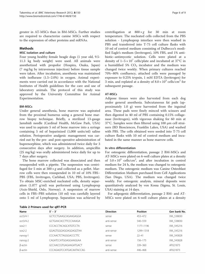