Catecholamine-induced cardiac mitochondrial dysfunction ... · Catecholamine-induced cardiac...
10
Catecholamine-induced cardiac mitochondrial dysfunction and mPTP opening: protective effect of curcumin Malika Izem-Meziane, 1 Bahia Djerdjouri, 1 Stephanie Rimbaud, 2,3 Fanny Caffin, 2,3 Dominique Fortin, 2,3 Anne Garnier, 2,3 Vladimir Veksler, 2,3 Frederic Joubert, 2,3 and Renee Ventura-Clapier 2,3 1 Faculté des Sciences Biologiques, Université des Sciences et de la Technologie Houari Boumediene Bab Ezzouar, El Alia, Alger, Algérie; 2 Institut National de la Santé et de la Recherche Médicale, U-769, Châtenay-Malabry; and 3 University Paris-Sud, IFR141, UMR-S769, Châtenay-Malabry, France Submitted 9 May 2011; accepted in final form 11 November 2011 Izem-Meziane M, Djerdjouri B, Rimbaud S, Caffin F, Fortin D, Garnier A, Veksler V, Joubert F, Ventura-Clapier R. Cat- echolamine-induced cardiac mitochondrial dysfunction and mPTP opening: protective effect of curcumin. Am J Physiol Heart Circ Physiol 302: H665–H674, 2012. First published November 18, 2011; doi:10.1152/ajpheart.00467.2011.—The present study was designed to characterize the mitochondrial dysfunction induced by catecholamines and to investigate whether curcumin, a natural anti- oxidant, induces cardioprotective effects against catecholamine- induced cardiotoxicity by preserving mitochondrial function. Because mitochondria play a central role in ischemia and oxidative stress, we hypothesized that mitochondrial dysfunction is involved in catechol- amine toxicity and in the potential protective effects of curcumin. Male Wistar rats received subcutaneous injection of 150 mg·kg 1 · day 1 isoprenaline (ISO) for two consecutive days with or without pretreatment with 60 mg · kg 1 · day 1 curcumin. Twenty four hours after, cardiac tissues were examined for apoptosis and oxidative stress. Expression of proteins involved in mitochondrial biogenesis and function were measured by real-time RT-PCR. Isolated mitochon- dria and permeabilized cardiac fibers were used for swelling and mitochondrial function experiments, respectively. Mitochondrial mor- phology and permeability transition pore (mPTP) opening were as- sessed by fluorescence in isolated cardiomyocytes. ISO treatment induced cell damage, oxidative stress, and apoptosis that were pre- vented by curcumin. Moreover, mitochondria seem to play an impor- tant role in these effects as respiration and mitochondrial swelling were increased following ISO treatment, these effects being again prevented by curcumin. Importantly, curcumin completely prevented the ISO-induced increase in mPTP calcium susceptibility in isolated cardiomyocytes without affecting mitochondrial biogenesis and mi- tochondrial network dynamic. The results unravel the importance of mitochondrial dysfunction in isoprenaline-induced cardiotoxicity as well as a new cardioprotective effect of curcumin through prevention of mitochondrial damage and mPTP opening. curcumin; isoprenaline; cardiac injury; mitochondria function; mito- chondrial morphology and permeability transition pore CIRCULATING CATECHOLAMINES closely correlate with the severity and poor prognosis in heart failure and are thus considered to play a critical role in the development of cardiovascular dis- eases (11). Excessive release of catecholamines induces myo- cardial hypertrophy, myocyte damage, and contractile dysfunc- tion, resulting in infarct-like necrosis of the heart muscle. Oxidative stress due to increased generation of reactive oxygen species (ROS) plays a recognized role in the cate- cholamine-induced cardiotoxicity (24, 41). Indeed, cat- echolamines can easily undergo oxidation with production of unstable catecholamines-O-quinones, giving rise to a subse- quent production of superoxide radicals that was proposed to be responsible for catecholamine-induced cardiotoxicity. The superoxide radicals can be reduced by superoxide dismutase to hydrogen peroxide, which damages the membrane integrity. Imbalance between ROS production and cellular antioxidant defenses initiates subcellular alterations leading to cell death, cardiomyopathy, and heart failure (12, 37, 41). Mitochondrial dysfunction is widely acknowledged as a seminal event in necrotic and apoptotic cell death and is emerging as a key mediator of ischemia-reperfusion (I/R) injury and catecholamine toxicity in the heart. Alterations in Ca 2 handling and ROS clearly contribute to mitochondrial dysfunction and to opening of the mitochondrial permeability transition pore (mPTP), a high-conductance pore of the mito- chondrial membranes. Opening of mPTP causes uncoupling of the mitochondria and swelling of the matrix, leading to rupture of the mitochondrial membranes, release of reduced equiva- lents and proapoptotic factors, and ultimately cell death (2). Increased vulnerability of the mPTP is an early event in pathological processes leading to heart failure (25). In recent years, mPTP has been considered a therapeutic target for cardioprotection during cardiac diseases, especially those as- sociated with I/R (35). Curcumin (CUR), a polyphenol responsible for the yellow color of the spice turmeric, at relatively low concentrations, is an effective anti-inflammatory agent and exhibits cardioprotec- tive effects (49). The antioxidant properties of curcumin seem to be essential for its pleiotropic biological activities. Cur- cumin inhibits lipid peroxidation and effectively scavenges superoxide anion and hydroxyl radicals. It was also shown to interact with the mPTP (28). The cardioprotective effect of curcumin against catecholamine-induced cardiotoxicity has been established for a long time (32) and involves its antiox- idant properties (24). Moreover, curcumin inhibits nuclear factor-B activation, protects cardiac cells against I/R injury (14), and stabilizes the cytoskeleton through the increased expression of the heat shock protein Hsp27 (39). However, no study has been performed yet to demonstrate the implication of mitochondria as a possible target of the cardioprotective effect of curcumin. Because mitochondria play a major role in cardiac dysfunc- tion and cell death, the aim of the present study was 1) to characterize the mitochondrial dysfunction induced by cat- echolamines and 2) to investigate whether curcumin induces cardioprotective effects against catecholamine-induced cardio- toxicity by preserving mitochondrial function. To this aim, we Address for reprint requests and other correspondence: R. Ventura-Clapier, U-769 INSERM, Univ Paris Sud, 5 rue J-B Clément, F-92296 Châtenay- Malabry, France (e-mail: [email protected]). Am J Physiol Heart Circ Physiol 302: H665–H674, 2012. First published November 18, 2011; doi:10.1152/ajpheart.00467.2011. 0363-6135/12 Copyright © 2012 the American Physiological Society http://www.ajpheart.org H665 by 10.220.33.3 on November 22, 2016 http://ajpheart.physiology.org/ Downloaded from
Transcript of Catecholamine-induced cardiac mitochondrial dysfunction ... · Catecholamine-induced cardiac...
Catecholamine-induced cardiac mitochondrial dysfunction and mPTP opening:protective effect of curcumin
Malika Izem-Meziane,1 Bahia Djerdjouri,1 Stephanie Rimbaud,2,3 Fanny Caffin,2,3 Dominique Fortin,2,3
Anne Garnier,2,3 Vladimir Veksler,2,3 Frederic Joubert,2,3 and Renee Ventura-Clapier2,3
1Faculté des Sciences Biologiques, Université des Sciences et de la Technologie Houari Boumediene Bab Ezzouar, El Alia,Alger, Algérie; 2Institut National de la Santé et de la Recherche Médicale, U-769, Châtenay-Malabry; and 3UniversityParis-Sud, IFR141, UMR-S769, Châtenay-Malabry, France
Submitted 9 May 2011; accepted in final form 11 November 2011
Izem-Meziane M, Djerdjouri B, Rimbaud S, Caffin F, FortinD, Garnier A, Veksler V, Joubert F, Ventura-Clapier R. Cat-echolamine-induced cardiac mitochondrial dysfunction and mPTPopening: protective effect of curcumin. Am J Physiol Heart CircPhysiol 302: H665–H674, 2012. First published November 18,2011; doi:10.1152/ajpheart.00467.2011.—The present study wasdesigned to characterize the mitochondrial dysfunction induced bycatecholamines and to investigate whether curcumin, a natural anti-oxidant, induces cardioprotective effects against catecholamine-induced cardiotoxicity by preserving mitochondrial function. Becausemitochondria play a central role in ischemia and oxidative stress, wehypothesized that mitochondrial dysfunction is involved in catechol-amine toxicity and in the potential protective effects of curcumin.Male Wistar rats received subcutaneous injection of 150mg·kg�1 ·day�1 isoprenaline (ISO) for two consecutive days with orwithout pretreatment with 60 mg·kg�1 ·day�1 curcumin. Twenty fourhours after, cardiac tissues were examined for apoptosis and oxidativestress. Expression of proteins involved in mitochondrial biogenesisand function were measured by real-time RT-PCR. Isolated mitochon-dria and permeabilized cardiac fibers were used for swelling andmitochondrial function experiments, respectively. Mitochondrial mor-phology and permeability transition pore (mPTP) opening were as-sessed by fluorescence in isolated cardiomyocytes. ISO treatmentinduced cell damage, oxidative stress, and apoptosis that were pre-vented by curcumin. Moreover, mitochondria seem to play an impor-tant role in these effects as respiration and mitochondrial swellingwere increased following ISO treatment, these effects being againprevented by curcumin. Importantly, curcumin completely preventedthe ISO-induced increase in mPTP calcium susceptibility in isolatedcardiomyocytes without affecting mitochondrial biogenesis and mi-tochondrial network dynamic. The results unravel the importance ofmitochondrial dysfunction in isoprenaline-induced cardiotoxicity aswell as a new cardioprotective effect of curcumin through preventionof mitochondrial damage and mPTP opening.
curcumin; isoprenaline; cardiac injury; mitochondria function; mito-chondrial morphology and permeability transition pore
CIRCULATING CATECHOLAMINES closely correlate with the severityand poor prognosis in heart failure and are thus considered toplay a critical role in the development of cardiovascular dis-eases (11). Excessive release of catecholamines induces myo-cardial hypertrophy, myocyte damage, and contractile dysfunc-tion, resulting in infarct-like necrosis of the heart muscle.
Oxidative stress due to increased generation of reactiveoxygen species (ROS) plays a recognized role in the cate-cholamine-induced cardiotoxicity (24, 41). Indeed, cat-
echolamines can easily undergo oxidation with production ofunstable catecholamines-O-quinones, giving rise to a subse-quent production of superoxide radicals that was proposed tobe responsible for catecholamine-induced cardiotoxicity. Thesuperoxide radicals can be reduced by superoxide dismutase tohydrogen peroxide, which damages the membrane integrity.Imbalance between ROS production and cellular antioxidantdefenses initiates subcellular alterations leading to cell death,cardiomyopathy, and heart failure (12, 37, 41).
Mitochondrial dysfunction is widely acknowledged as aseminal event in necrotic and apoptotic cell death and isemerging as a key mediator of ischemia-reperfusion (I/R)injury and catecholamine toxicity in the heart. Alterations inCa2� handling and ROS clearly contribute to mitochondrialdysfunction and to opening of the mitochondrial permeabilitytransition pore (mPTP), a high-conductance pore of the mito-chondrial membranes. Opening of mPTP causes uncoupling ofthe mitochondria and swelling of the matrix, leading to ruptureof the mitochondrial membranes, release of reduced equiva-lents and proapoptotic factors, and ultimately cell death (2).Increased vulnerability of the mPTP is an early event inpathological processes leading to heart failure (25). In recentyears, mPTP has been considered a therapeutic target forcardioprotection during cardiac diseases, especially those as-sociated with I/R (35).
Curcumin (CUR), a polyphenol responsible for the yellowcolor of the spice turmeric, at relatively low concentrations, isan effective anti-inflammatory agent and exhibits cardioprotec-tive effects (49). The antioxidant properties of curcumin seemto be essential for its pleiotropic biological activities. Cur-cumin inhibits lipid peroxidation and effectively scavengessuperoxide anion and hydroxyl radicals. It was also shown tointeract with the mPTP (28). The cardioprotective effect ofcurcumin against catecholamine-induced cardiotoxicity hasbeen established for a long time (32) and involves its antiox-idant properties (24). Moreover, curcumin inhibits nuclearfactor-�B activation, protects cardiac cells against I/R injury(14), and stabilizes the cytoskeleton through the increasedexpression of the heat shock protein Hsp27 (39). However, nostudy has been performed yet to demonstrate the implication ofmitochondria as a possible target of the cardioprotective effectof curcumin.
Because mitochondria play a major role in cardiac dysfunc-tion and cell death, the aim of the present study was 1) tocharacterize the mitochondrial dysfunction induced by cat-echolamines and 2) to investigate whether curcumin inducescardioprotective effects against catecholamine-induced cardio-toxicity by preserving mitochondrial function. To this aim, we
Address for reprint requests and other correspondence: R. Ventura-Clapier,U-769 INSERM, Univ Paris Sud, 5 rue J-B Clément, F-92296 Châtenay-Malabry, France (e-mail: [email protected]).
Am J Physiol Heart Circ Physiol 302: H665–H674, 2012.First published November 18, 2011; doi:10.1152/ajpheart.00467.2011.
0363-6135/12 Copyright © 2012 the American Physiological Societyhttp://www.ajpheart.org H665
by 10.220.33.3 on Novem
ber 22, 2016http://ajpheart.physiology.org/
Dow
nloaded from
investigated whether curcumin could protect the heart againstisoprenaline-induced cell death, alteration of mitochondrialrespiration, dynamics, and biogenesis, and prevent opening ofmPTP and cell death.
MATERIALS AND METHODS
Animal treatments. Male Wistar rats weighing 230–280 g wereused. All experiments were approved by the Université Paris-SudInstitutional Care Committee and conformed to the European Com-munity guiding principles in the care and use of animals (Directive2010/63/EU of the European Parliament.) Authorizations to conductanimal experiments were obtained from the French Ministère del’Agriculture, de la Pêche et de l’Alimentation (no. 92–284, June 27,2007). Rats were randomly divided into three groups of five animalseach and treated as follows: 1) the control group was given normalsaline subcutaneously one time a day for two consecutive days; 2)isoprenaline (ISO) group: 150 mg·kg�1 ·day�1 ISO was administeredsubcutaneously for 2 days to induce cardiac damage; and 3) CUR �ISO group: curcumin (CUR; 60 mg·kg�1 ·day�1) was administreredintraperitonealy for 2 days, 30 min before ISO injection. In additionalstudies, CUR (60 mg·kg�1 ·day�1) was administered alone intraperi-tonealy for 2 days (CUR group). Twenty four hours after the lasttreatment period, all rats were anesthetized by intraperitoneal injectionof pentobarbital (150 mg/kg) before death.
A supraphysiological dose of catecholamine was used to inducerapid infarct-like damages to the myocardium. The relatively low doseof curcumin was chosen in the range of the cardioprotective effectsdescribed in the literature (27, 39).
After death, hearts were quickly harvested. After left ventricleisolation, part was immediately used for mitochondrial respirationmeasurements, and other parts were rapidly frozen and kept at �80°Cfor biochemical analysis or fixed in 10% phosphate-buffered formalinfor light microscopic studies.
Preparation of tissue homogenate. Hearts were homogenized in 10volumes (wt/vol) of ice-cold phosphate buffer (50 mM, pH 7.4) usinga Polytron homogenizer (Ultra-turrax T25). The homogenates werethen centrifuged at 10,000 g for 10 min at 4°C, and the supernatantwas used for biochemical assays. Protein content was determinedusing BSA as standard (4).
Histological examinations. Formalin-fixed left ventricular tissueswere dehydrated in ascending grades of alcohol and embedded inparaffin. Five-micrometer sections were stained with hematoxylin andeosin and examined under light microscopy (Carl Zeiss). Other serial5-�m sections were stained with 1 g/ml Hoechst 33258 for 30 min atroom temperature and visualized under fluorescence microscopy (CarlZeiss, Thornwood, NY). Apoptotic morphological cells were countedin 10 visual fields of 5 different areas.
Markers of cardiac stress and metabolic enzymes. Creatine kinaseactivity was assayed using a Teco commercial kit according to themanufacturer’s instructions (CK-NAC reagents; Teco Diagnostics).Lactate dehydrogenase (LDH) activity was assayed by the method ofWahlefeld (45). The malondialdehyde (MDA) level was assessedaccording to Ohkawa et al. (34). Myeloperoxidase (MPO), a marker ofneutrophil infiltration, was measured in the heart according to Krawiszet al. (22). Catalase activity was determined by measuring the rate ofdecomposition of H2O2 according to Aebi (1). Glutathione (GSH)content in supernatant of heart homogenate was evaluated by themethod of Boyne and Ellman (3). Frozen tissue samples were weighedand homogenized in ice-cold buffer, and enzyme activities weredetermined as described previously (10). Complex I activity wasmeasured in heart homogenized in ice-cold buffer containing 10mmol/l Tris base (pH 7.2), 75 mmol/l sucrose, 225 mmol/l mannitol,100 �mol/l EDTA, and 0.1% Triton X-100. To measure NADH-coenzyme Q reductase activity with decylubiquinone as electronacceptor, samples were incubated in 25 mmol/l phosphate buffer (pH7.5), 2.5 mg/ml BSA, and 100 �mol/l decylubiquinone at 30°C.
Activity was reported as a rotenone-insensitive decrease in NADHabsorbance at 340 nm. Citrate synthase activity was measured in thepresence of acetyl-CoA, oxaloacetic acid, and DTNB according toRef. 38. Cytochrome c oxidase (COX) activity was determined bymeasuring the disappearance of reduced cytochrome c at 550 nmaccording to Ref. 48.
Real-time quantitative RT-PCR analysis. Total RNA was isolatedfrom rat left ventricle (50–100 mg) using the Trizol reagent technique(Invitrogen, Cergy Pontoise, France). Oligo(dT) first-strand cDNAwas synthesized from 5 �g total RNA using Superscript II reversetranscriptase (Invitrogen). Real-time PCR was performed using theSYBR Green technology on a Light Cycler rapid thermal cycler(Roche Diagnostics) as previously described (15). Forward and re-verse primers were each designed in a different exon of the targetsequence, eliminating the possibility of amplifying genomic DNA asdescribed previously (15, 16). Amplification was allowed to proceedfrom 30 to 40 cycles, each consisting of denaturation at 95°C for 10s, annealing at 60°C nuclear respiratory factor 2, mitochondrialtranscription factor A, COX subunits I and IV, and mitofusin 1 (Mfn1)and 2 (Mfn2), or 58°C peroxisome proliferator-activated receptorgamma coactivator 1� (PGC-1�), optic atrophy protein 1 (OPA1),and dynamin-related protein 1 (Drp1) from 5 to 9 s and extension at72°C from 5 to 19 s depending on the target gene. Cyclophilin A andacidic ribosomal protein were used for normalization with Genormsoftware.
Western blot analysis. Hearts of control, ISO, and CUR � ISOwere homogenized in a lysate buffer containing (in mM): 50 HEPES,20 KCl, 1 EDTA, 1 EGTA, 5 �-glycerolphosphate, 1 orthovanadate,1 dithiothreitol (DTT), 5 NaPPi, 2 phenylmethylsulfonyl fluoride(Sigma-Aldrich Chimie), cocktail of protease inhibitors (CalbiochemSet V EDTA free), and 0.1% Triton X-100. Protein extracts (50 �g)were loaded on a 10% SDS-polyacrylamide gel. Blots were firstincubated with a primary antibody for rat total or phospho-Drp1(dilution 1:1,000; Chemicon International) and then with an anti-rabbit IgG, horseradish peroxidase-linked antibody (dilution 1:2,500;Cell Signaling). Drp1 protein was revealed with an enhanced chemi-luminescent substrate (SuperSignal West Dura; Pierce Biotechnol-ogy).
In situ study of mitochondrial respiration. Mitochondrial respira-tion was studied in situ in freshly saponin-skinned fibers as previouslydescribed (23, 42). Briefly, fibers were separated from the endocardialsurface of the left ventricle under a binocular microscope in skinningsolution (see below) at 4°C and permeabilized in the same solutionwith 50 �g/ml of saponin for 30 min. After being placed in respirationsolution (see below) for 10 min to wash out adenine nucleotides andcreatine phosphate, permeabilized separated fibers were transferred toa 3-ml water-jacketed oxygraphic cell (Strathkelvin Instruments,Glasgow, UK) equipped with a Clark electrode under continuousstirring. The respiration and skinning solutions contained the follow-ing (in mM): 2.77 CaK2EGTA, 7.23 K2EGTA (100 nM free Ca2�), 1free Mg2�, 20 taurine, 0.5 DTT, and 20 imidazole (pH 7.1). Theskinning solution also contained (in mM) 50 potassium methanesul-fonate (160 mM ionic strength), 5.7 Na2ATP, and 15 phosphocreatinewhile the respiration solution contained 10 glutamate, 4 malate, 3phosphate, 90 potassium methanesulfonate, 10 sodium methanesulfo-nate (ionic strength 160 mM), and 2 mg/ml fatty acid-free BSA.Experiments were performed at 22°C. ADP-stimulated respiration(VADP) above basal oxygen consumption (V0) was measured bystepwise addition of ADP. Maximal respiration rate (Vmax) was VADP �VO. Oxidation-phosphorylation coupling was determined by calculat-ing the acceptor control ratio (ACR) as Vmax/V0. Respiratory chaincomplexes were successively investigated with specific substrates andinhibitors [10 mM succinate and 2 mM amytal (for complex II), 0.5mM N,N,N=,N=-tetramethyl-p-phenylenediamine � 0.5 mM ascorbate(for complex IV), and 4 mM azide (to inhibit complex IV)]. Respi-ration rates are given in �mol O2 ·min�1 ·g dry wt�1. Two or threedeterminations were performed for each heart and each protocol.
H666 CARDIOTOXICITY OF CATECHOLAMINE AND CURCUMIN
AJP-Heart Circ Physiol • doi:10.1152/ajpheart.00467.2011 • www.ajpheart.org
by 10.220.33.3 on Novem
ber 22, 2016http://ajpheart.physiology.org/
Dow
nloaded from
Isolation of mitochondria and measurement of mitochondrialswelling. Mitochondria were isolated from all experimental groups bydifferential centrifugation as previously described (19). Isolated car-diac mitochondria were resuspended in swelling buffer containing (inmM): 120 KCl, 5 KH2PO4, 20 MOPS, and 10 Tris·HCl, pH 7.4 to afinal protein concentration of 0.25 mg/ml. Mitochondrial swelling wasfollowed by changes in absorbance at 520 nm, after addition of 20 �MCa2� (46). The protein yield for isolated mitochondria was (mgprotein/g heart tissue) 6.94 � 0.32 for control, 6.55 � 0.48 for ISO,and 6.12 � 0.41 for CUR � ISO (not significant).
Preparation of adult rat ventricular myocytes. Adult rat ventricularmyocytes were dissociated by retrograde perfusion of healthy isolatedheart with collagenase as previously described (44) with slight mod-ifications. Freshly isolated cells were plated on laminin-coated culturedishes at a density of 5 � 105 cells/dish and switched to serum-freemedium for 2 h. Cells were then incubated for 48 h with ISO (5 �10�7 M) and with or without curcumin (5 � 10�6 M) or their vehicle(control). Five independent cultures were used for each condition.
Mitochondrial network analysis and mPTP experiments. Fluores-cence experiments were performed with a Carl Zeiss LSM-510confocal microscope using a �63 1.4 plan Apochromat objectivelens. For mitochondrial network analysis, fresh cardiomyocyteswere infected 48 h with an adenovirus containing green fluorescentprotein and a sequence targeted to COX IV subunit (36) (gift fromDr. G. Szabadkai). Fluorescence was excited with the 488-nm line,and images were collected through a 505- to 550-nm band passfilter. To reconstruct three-dimensional images of the cells, serialimages of individual cardiomyocytes were collected in the z-axis.Next, data were imported, and images were deconvoluted usingAutoquant X software (Media Cybernetics, Bethesda, MD). Vol-umes of cardiomyocytes and individual mitochondrion were ob-tained with IMARIS software (Bitplane, Zurich, Switzerland) us-ing the Imaris MeasurementPro module. Fluorescence emittedfrom mitochondria was analyzed by the software, which allows, bysurface segmentation methods, to identify individual objects cor-responding to individual mitochondria. Characteristics of individ-
Table 1. Anatomic data from control, ISO and CUR � ISO groups
Group Control ISO CUR � ISO CUR
n 20 19 15 5Body wt before treatment, g 241 � 6 242 � 8 251 � 9 232 � 14Body wt after treatment, g 251 � 6 229 � 8* 243 � 7 243 � 14Control, % 100 91 97 97Heart wt, g 0.83 � 0.03 1.05 � 0.05** 0.99 � 0.05* 0.99 � 0.05Control, % 100 127 119 119Heart wt/body wt, mg/g 3.3 � 0.12 4.58 � 0.14*** 4.07 � 0.21**# 3.62 � 0.13##
Control, % 100 139 123 110Protein/wet wt, % 18.1 � 0.46 17.44 � 3.43 16.44 � 2.05 ND
Values are means � SE; n, no. of rats. ISO, isoprenaline; CUR, curcumin. *P � 0.05, **P � 0.01, and ***P � 0.001 vs. the control group. #P � 0.05 and##P � 0.01 vs. the ISO group. ND, not determined.
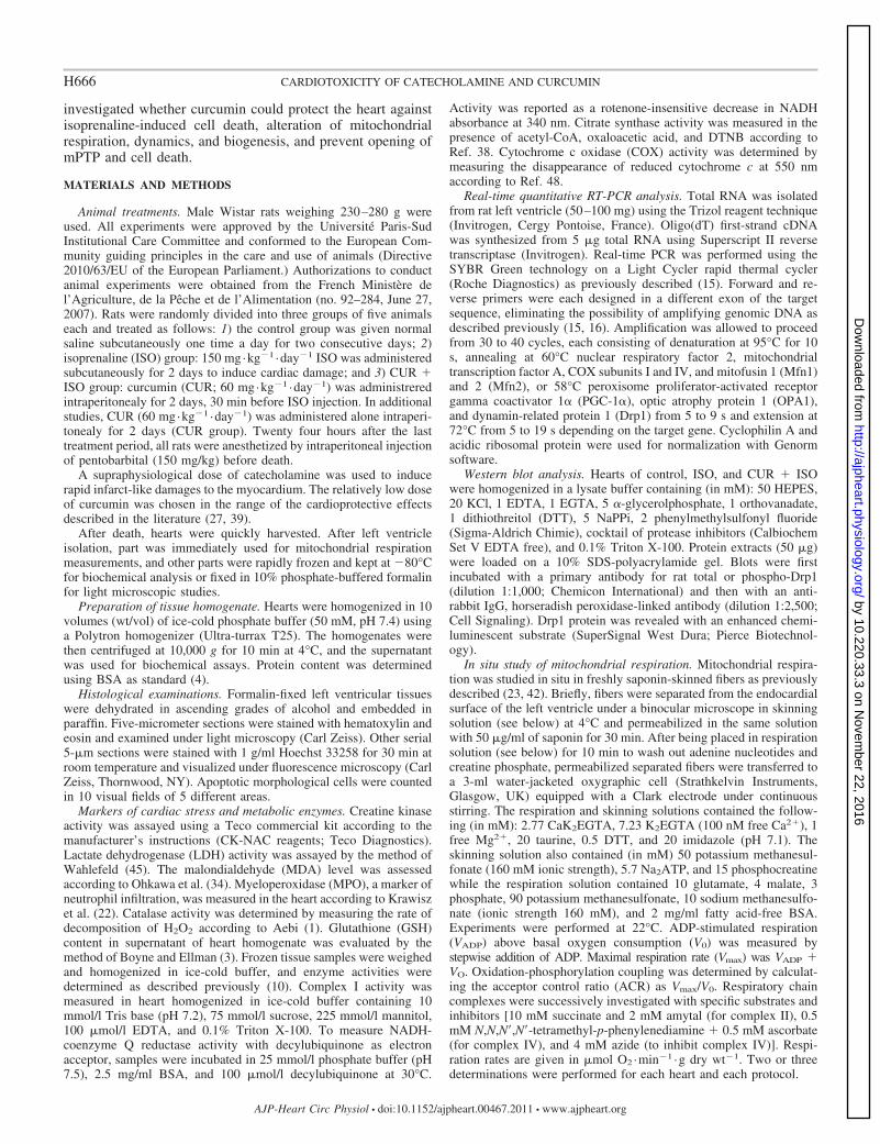
Fig. 1. Curcumin (CUR) protects the heart against apoptosis and oxidative stress. Heart tissues were processed for hematoxylin and eosin (HE) staining. A: normalaspect of myocardial tissue in control rat. B: acute extensive myofibrillar degeneration, with infiltration of neutrophil granulocytes and interstitial edema inisoprenaline (ISO)-treated hearts. C: mild degenerative changes of myocardial tissue in rats pretreated with CUR � ISO. D: effect of curcumin onmyeloperoxidase activity in ISO-treated hearts (n 5). Cell apoptosis was assessed by Hoechst staining for control (E), ISO (F)-, and CUR � ISO-treated (G)rats. H: summary data showing Hoechst-positive cells (%total counted cells) from 10 visual fields of 5 different areas. Values are means � SE. **P � 0.01 and***P � 0.001 vs. the control group. #P � 0.05 and ###P � 0.001 vs. the ISO group (n 3). Original magnification �1,000 for A–C and �400 for E–G.
H667CARDIOTOXICITY OF CATECHOLAMINE AND CURCUMIN
AJP-Heart Circ Physiol • doi:10.1152/ajpheart.00467.2011 • www.ajpheart.org
by 10.220.33.3 on Novem
ber 22, 2016http://ajpheart.physiology.org/
Dow
nloaded from
ual objects are obtained (surface, volume, etc.). By this approach,it is possible to calculate the distribution and average volume ofindividual mitochondria, as well as global mitochondrial networkand cell volume.
For mPTP experiments, fresh cardiomyocytes were coloaded inculture medium with 5 �M rhodamine (Rhod-2) and 1 �M calcein(Invitrogen) for 45 min at room temperature and then washed withculture medium. Cardiomyocytes were then permeabilized with sapo-
Table 2. Effect of curcumin on metabolic enzymes and oxidative stress
Group Control ISO CUR � ISO CUR
LDH, IU/mg protein 2.99 � 0.20 1.26 � 0.10*** 2.31 � 0.19*### NDCK, IU/mg protein 4.69 � 17 2.91 � 0.14*** 4.17 � 0.11*### NDMDA, nmol/mg protein 0.95 � 0.06 2.2 � 0.2*** 0.98 � 0.08### 0.87 � 0.08##
GSH, nmol/mg protein 24.7 � 1.1 15.5 � 0.3*** 22.6 � 1.4## 20.8 � 1.3##
Catalase, IU/mg protein 22.0 � 0.9 10.3 � 0.7*** 15.5 � 1.6**## 19.7 � 2.1##
CxI, IU/g protein 255 � 48 215 � 25 258 � 35 NDCOX, IU/g protein 2,765 � 260 2,462 � 133 2,284 � 150 NDCS, IU/g protein 587 � 12 480 � 43 556 � 29 ND
Values are means � SE; n 5 rats in each group. Cardiac data: lactate dehydrogenase (LDH), creatine kinase (CK), complex I (CxI), citrate synthase (CS),and cytochrome oxidase (COX). Levels of lipid peroxidation [malondialdehyde (MDA)], antioxidant defenses measured as reduced glutathione (GSH) or catalaseactivity. *P � 0.05, **P � 0.01, and ***P � 0.001 vs. the control group. ##P � 0.01 and ###P � 0.001 vs. the ISO group. ND, not determined.
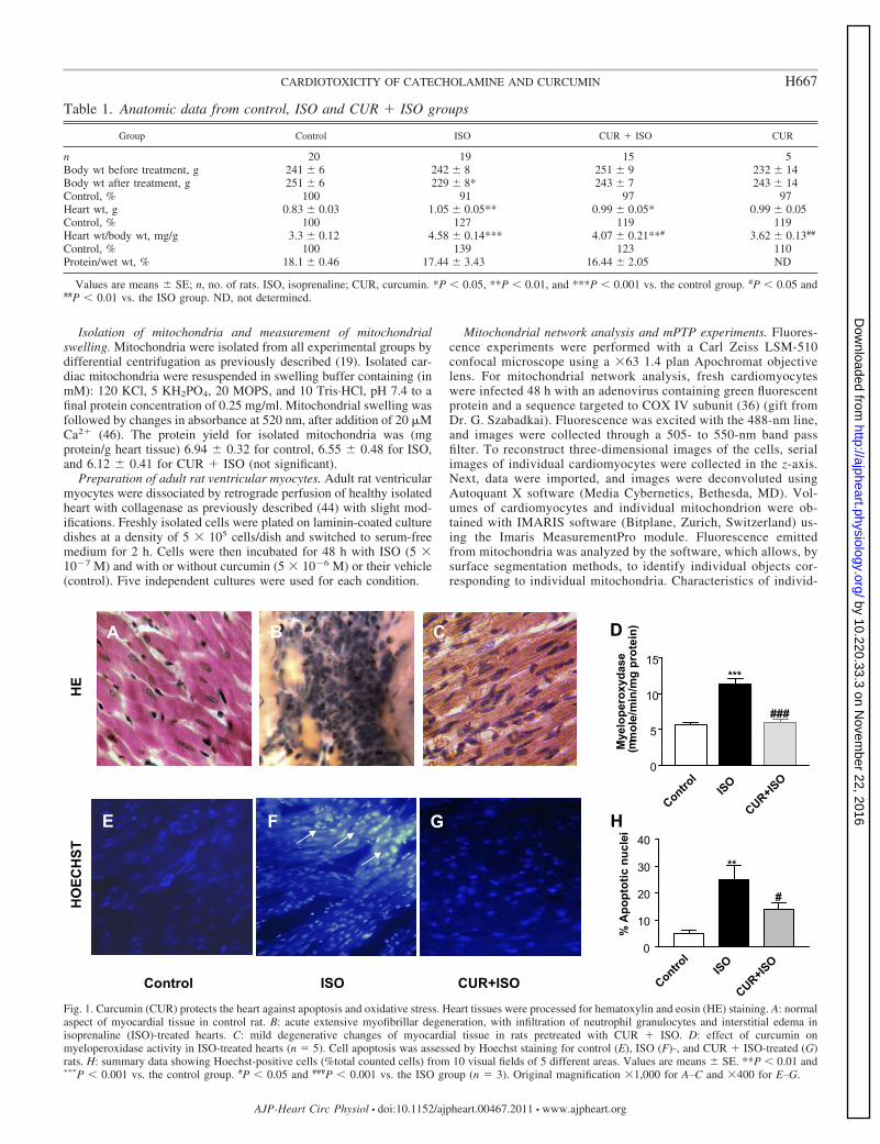
Fig. 2. Effect of curcumin on mitochondrial function and biogenesis in the heart of ISO-treated rats. A: basal respiration rate without ADP (V̇O2) and respirationrates with glutamate � malate (complex I), succinate � amytal (complex II), and N,N,N=,N=-tetramethyl-p-phenylenediamine � ascorbate (complex IV); valuesare expressed in �mol of oxygen consumed per mg dry weight per minute. B: acceptor control ratio (ACR). C: effect of curcumin on mitochondrial biogenesisin ISO-treated rats. Real-time quantitative RT-PCR analysis of mRNA expression of peroxisome proliferator-activated receptor- coactivator 1� and 1� (PGC-1�and -1�, respectively), mitochondrial transcription factor A (Tfam), PGC-1-related coactivator (PRC), nuclear respiratory factor 2 DNA binding subunit �(NRF-2�), and cytochrome oxidase I and IV (COXI and COXIV, respectively). AU, arbitrary units. **P � 0.01 and ***P � 0.001 vs. the control group.##P � 0.01 and ###P � 0.001 vs. the ISO group. Values are means � SE of 5 rats in each group.
H668 CARDIOTOXICITY OF CATECHOLAMINE AND CURCUMIN
AJP-Heart Circ Physiol • doi:10.1152/ajpheart.00467.2011 • www.ajpheart.org
by 10.220.33.3 on Novem
ber 22, 2016http://ajpheart.physiology.org/
Dow
nloaded from
nin (50 �g/ml) for 3 min and then washed in respiration solution (seeabove) for 5 min to eliminate cytosolic constituents and dyes beforeexperiments. Rhod-2 was excited with the 568-nm line of an argon/krypton laser, and images were collected through a 560-nm long passemission filter. Calcein fluorescence was excited simultaneously withthe 488-nm line of the same laser, and images were collected througha 505- to 550-nm band pass filter. Rhod-2 and calcein signals weremonitored after addition of 2 �M Ca2� in the different conditions(control, ISO, and CUR � ISO) until mPTP opening was observed. Inadditional plates, cells were superfused with 1 �M cyclosporin A(CsA) to check for specificity of the mPTP opening.
All of the data were analyzed by fitting both Rhod-2 and calceincurves with the following equation: y y� � (y0 � y�)/[1 �(t/tmax)pmax], where y� is the value of the fluorescence for time (t) �and y0 the value at t 0 (after Ca2� addition), and tmax and pmax arethe time and the slope for fluorescence decrease, respectively, whenmaximal mPTP opening occurs (where 50% of the signal has de-creased). The initial slope of signal decrease was defined as pinit. Thustmax and pinit give an index of the Ca2� sensitivity of mPTP.
Statistical analysis. All data were expressed as means � SE andwere subjected to one-way ANOVA followed by Student-Newman-Keuls’s test. All results with P � 0.05 were considered statisticallysignificant.
RESULTS
Curcumin attenuated ISO-induced cardiac hypertrophy, ap-optosis, and oxidative stress. ISO-treated rats showed a signif-icant increase in heart weight by 27% and heart-to-body weightratio by 39% compared with the control group that waspartially prevented by curcumin (Table 1). No significantdifference was observed for protein content, showing thatedema was not responsible for increased heart weight. Asexpected, histological evaluation of ISO-treated hearts showedextensive granulomas with necrotic fibers and infiltrated in-flammatory cells compared with control (Fig. 1, A and B).Curcumin pretreatment resulted in marked structural improve-ment of myonecrosis and edema compared with ISO-treatedrats (Fig. 1C) and a decrease of neutrophil infiltration asreflected by myocardial MPO activity (Fig. 1D). The nuclearmorphology was also analyzed using Hoechst 33258 stainingassay to check for apoptosis. As shown in Fig. 1, E and F, ISOincreased the number of apoptotic nuclei by 20% (P � 0.001)compared with control. Curcumin reduced nucleus abnormal-ities associated with cell death (Fig. 1, G and H). No differ-ences were detected between the control group and the cur-cumin-pretreated group. Isoprenaline significantly decreasedcardiac LDH and CK levels (P � 0.001, Table 2). ISO inducedan increase in tissue MDA content (P � 0.001) and a decrease
in GSH and catalase activities (P � 0.001) compared withcontrol (Table 2). Pretreatment with curcumin prevented all ofthe deleterious effects of ISO. Curcumin alone had no effect onanatomical parameters and oxidative stress (Tables 1 and 2).All of these data confirm the antioxidant and protective effectsof curcumin on ISO-induced cardiac injury.
Curcumin preserved cardiac mitochondrial respiration un-der ISO treatment and had no effect on mitochondrialbiogenesis. To investigate the involvement of mitochondria inISO-induced cardiotoxicity and in curcumin-induced cardio-protection, the oxygen consumption of permeabilized cardiacfibers was measured (Fig. 2). In ISO treated-rats, basal (P �0.01) and maximal (P � 0.001) respiration rates on complex Iwere significantly reduced by 47 and 41%, respectively, com-pared with control fibers. Similarly, a significant decrease (P �0.05) in the respiration on complex II (37%) or IV (38%) wasobserved (Fig. 2A), whereas the ACR was not altered (Fig. 2B).Addition of cytochrome c did not rescue maximal respirationrates, showing the intactness of the outer mitochondrial mem-brane in functioning mitochondria [Vcytc/Vmax: 1.10 � 0.03(n 11) in control vs. 1.10 � 0.02 (n 14) in ISO].Pretreatment with curcumin preserved basal as well as complexI, complex II, and complex IV respiration rates.
mRNA levels of proteins involved in mitochondrial biogen-esis (PGC-1� and its downstream targets) did not differ be-tween groups, showing that the beneficial effects of curcuminwere not related to changes in mitochondrial biogenesis (Fig.2C). These results prompted us to investigate other mitochon-drial targets such as mitochondrial remodeling, fusion andfission, and mPTP opening.
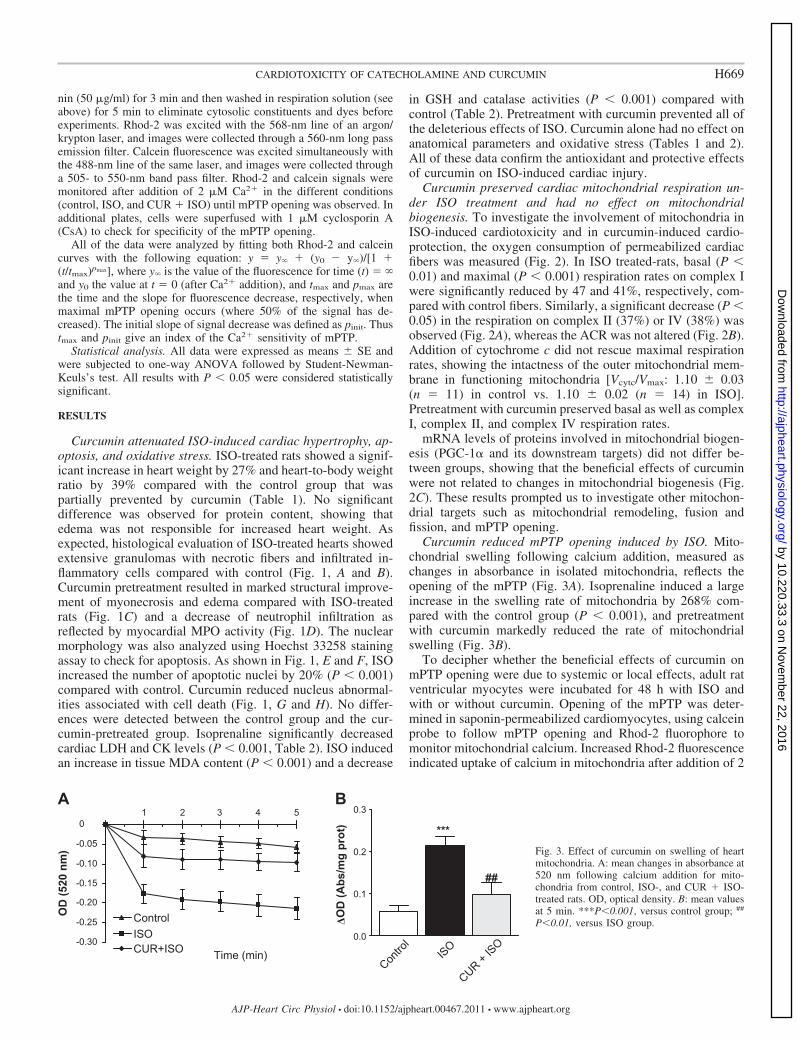
Curcumin reduced mPTP opening induced by ISO. Mito-chondrial swelling following calcium addition, measured aschanges in absorbance in isolated mitochondria, reflects theopening of the mPTP (Fig. 3A). Isoprenaline induced a largeincrease in the swelling rate of mitochondria by 268% com-pared with the control group (P � 0.001), and pretreatmentwith curcumin markedly reduced the rate of mitochondrialswelling (Fig. 3B).
To decipher whether the beneficial effects of curcumin onmPTP opening were due to systemic or local effects, adult ratventricular myocytes were incubated for 48 h with ISO andwith or without curcumin. Opening of the mPTP was deter-mined in saponin-permeabilized cardiomyocytes, using calceinprobe to follow mPTP opening and Rhod-2 fluorophore tomonitor mitochondrial calcium. Increased Rhod-2 fluorescenceindicated uptake of calcium in mitochondria after addition of 2
Fig. 3. Effect of curcumin on swelling of heartmitochondria. A: mean changes in absorbance at520 nm following calcium addition for mito-chondria from control, ISO-, and CUR � ISO-treated rats. OD, optical density. B: mean valuesat 5 min. ***P�0.001, versus control group; ##
P�0.01, versus ISO group.
H669CARDIOTOXICITY OF CATECHOLAMINE AND CURCUMIN
AJP-Heart Circ Physiol • doi:10.1152/ajpheart.00467.2011 • www.ajpheart.org
by 10.220.33.3 on Novem
ber 22, 2016http://ajpheart.physiology.org/
Dow
nloaded from
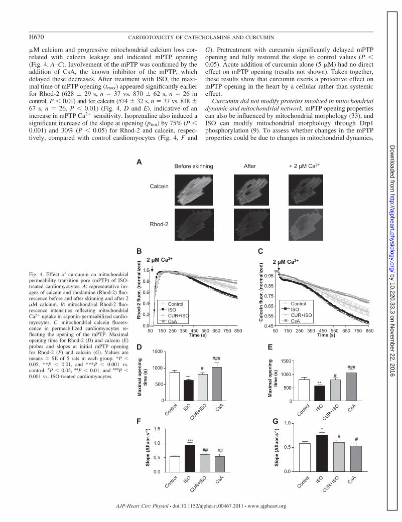
�M calcium and progressive mitochondrial calcium loss cor-related with calcein leakage and indicated mPTP opening(Fig. 4, A–C). Involvement of the mPTP was confirmed by theaddition of CsA, the known inhibitor of the mPTP, whichdelayed these decreases. After treatment with ISO, the maxi-mal time of mPTP opening (tmax) appeared significantly earlierfor Rhod-2 (628 � 29 s, n 37 vs. 870 � 62 s, n 26 incontrol, P � 0.01) and for calcein (574 � 32 s, n 37 vs. 818 �67 s, n 26, P � 0.01) (Fig. 4, D and E), indicative of anincrease in mPTP Ca2� sensitivity. Isoprenaline also induced asignificant increase of the slope at opening (pinit) by 75% (P �0.001) and 30% (P � 0.05) for Rhod-2 and calcein, respec-tively, compared with control cardiomyocytes (Fig. 4, F and
G). Pretreatment with curcumin significantly delayed mPTPopening and fully restored the slope to control values (P �0.05). Acute addition of curcumin alone (5 �M) had no directeffect on mPTP opening (results not shown). Taken together,these results show that curcumin exerts a protective effect onmPTP opening in the heart by a cellular rather than systemiceffect.
Curcumin did not modify proteins involved in mitochondrialdynamic and mitochondrial network. mPTP opening propertiescan also be influenced by mitochondrial morphology (33), andISO can modify mitochondrial morphology through Drp1phosphorylation (9). To assess whether changes in the mPTPproperties could be due to changes in mitochondrial dynamics,
Fig. 4. Effect of curcumin on mitochondrialpermeability transition pore (mPTP) of ISO-treated cardiomyocytes. A: representative im-ages of calcein and rhodamine (Rhod-2) fluo-rescence before and after skinning and after 2�M calcium. B: mitochondrial Rhod-2 fluo-rescence intensities reflecting mitochondrialCa2� uptake in saponin-permeabilized cardio-myocytes. C: mitochondrial calcein fluores-cence in permeabilized cardiomyocytes re-flecting the opening of the mPTP. Maximalopening time for Rhod-2 (D) and calcein (E)probes and slopes at initial mPTP openingfor Rhod-2 (F) and calcein (G). Values aremeans � SE of 5 rats in each group. *P �0.05, **P � 0.01, and ***P � 0.001 vs.control. #P � 0.05, ##P � 0.01, and ###P �0.001 vs. ISO-treated cardiomyocytes.
H670 CARDIOTOXICITY OF CATECHOLAMINE AND CURCUMIN
AJP-Heart Circ Physiol • doi:10.1152/ajpheart.00467.2011 • www.ajpheart.org
by 10.220.33.3 on Novem
ber 22, 2016http://ajpheart.physiology.org/
Dow
nloaded from
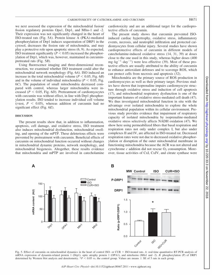
we next assessed the expression of the mitochondrial fusion/fission regulatory proteins Opa1, Drp1, and Mfns-1 and -2.Their expression was not significantly changed in the heart ofISO-treated rats (Fig. 5A). Protein kinase A (PKA)-mediatedphosphorylation of Drp1 induces sequestration of DRP1 in thecytosol, decreases the fission rate of mitochondria, and mayplay a protective role upon apoptotic stress (6, 9). As expected,ISO treatment significantly (P � 0.05) increased the phosphor-ylation of Drp1, which was, however, maintained in curcumin-pretreated rats (Fig. 5B).
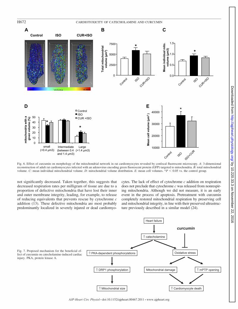
Using fluorescence imaging and three-dimensional recon-struction, we examined whether ISO treatment could alter themitochondrial network morphology (Fig. 6A). ISO induced anincrease in the total mitochondrial volume (P � 0.05, Fig. 6B)and in the volume of individual mitochondria (P � 0.05, Fig.6C). The population of small mitochondria decreased com-pared with control, whereas larger mitochondria were in-creased (P � 0.05, Fig. 6D). Pretreatment of cardiomyocyteswith curcumin was without effect, in line with Drp1 phosphor-ylation results. ISO tended to increase individual cell volume(t-test, P � 0.05), whereas addition of curcumin had nosignificant effect (Fig. 6E).
DISCUSSION
The present results show that, in addition to inflammation,apoptosis, cell damage, and oxidative stress, ISO treatmentalso induces mitochondrial dysfunction, mitochondrial swell-ing, and opening of the mPTP. These deleterious effects wereprevented by pretreatment with curcumin. Beneficial effects ofcurcumin on mitochondrial function occurred without changesin mitochondrial dynamic proteins, network morphology, andmitochondrial biogenesis. Altogether, these results evidencethat mitochondria and mPTP are involved in catecholamine
cardiotoxicity and are an additional target for the cardiopro-tective effects of curcumin.
The present study shows that curcumin prevented ISO-induced cardiac hypertrophy, oxidative stress, inflammatoryevents, necrosis, and neutrophil infiltration and protected car-diomyocytes from cellular injury. Several studies have showncardioprotective effects of curcumin in different models ofcatecholamine-induced oxidative stress (14, 31, 39) at dosesclose to the one used in this study, whereas higher doses (400mg·kg�1 ·day�1) were less effective (39). Most of these pro-tective effects are usually attributed to the ability of curcuminto enhance antioxidant defenses and to scavenge ROS, whichcan protect cells from necrosis and apoptosis (32).
Mitochondria are the primary source of ROS production incardiomyocytes as well as their primary target. Previous stud-ies have shown that isoprenaline impairs cardiomyocyte struc-ture through oxidative stress and induction of cell apoptosis(17), and mitochondrial respiratory dysfunction is one of theimportant features of oxidative stress-mediated cell death (47).We thus investigated mitochondrial function in situ with theadvantage over isolated mitochondria to explore the wholemitochondrial population within its cellular environment. Pre-vious study provides evidence that impairment of respiratorycapacity of isolated mitochondria by isoprenaline-mediatedoxidative stress selectively affects NADH oxidation (47). Weshow here using permeabilized fibers that basal respiration andrespiration rates not only under complex I, but also undercomplexes II and IV, are affected in ISO-treated rat. Decreasedrespiration rates were not due to decreased oxidative phosphor-ylation or disruption of the outer mitochondrial membrane infunctioning mitochondria because the ACR was not altered andcytochrome c addition did not rescue O2 consumption. More-over, tissue activities of CxI, CxIV, and citrate synthase were
Fig. 5. Effect of curcumin on mitochondrial dynamics in the heart of control ISO- or CUR � ISO-treated rats. A: real-time quantitative RT-PCR analysis ofmRNA expression of dynamin-related protein 1 (Drp1), optic atrophy protein 1 (OPA1), and mitofusins (Mfn1 and -2). B: phosphorylation (P) of DRP1determined by Western blot analysis and densitometry. *P � 0.05 vs. the control group. Values are means � SE of 5 rats in each group.
H671CARDIOTOXICITY OF CATECHOLAMINE AND CURCUMIN
AJP-Heart Circ Physiol • doi:10.1152/ajpheart.00467.2011 • www.ajpheart.org
by 10.220.33.3 on Novem
ber 22, 2016http://ajpheart.physiology.org/
Dow
nloaded from
not significantly decreased. Taken together, this suggests thatdecreased respiration rates per milligram of tissue are due to aproportion of defective mitochondria that have lost their innerand outer membrane integrity, leading, for example, to releaseof reducing equivalents that prevents rescue by cytochrome caddition (13). These defective mitochondria are most probablypredominantly localized in severely injured or dead cardiomyo-
cytes. The lack of effect of cytochrome c addition on respirationdoes not preclude that cytochrome c was released from nonrespir-ing mitochondria. Although we did not measure, it is an earlyevent in the process of apoptosis. Pretreatment with curcumincompletely restored mitochondrial respiration by preserving celland mitochondrial integrity, in line with their preserved ultrastruc-ture previously described in a similar model (24).
Fig. 6. Effect of curcumin on morphology of the mitochondrial network in rat cardiomyocytes revealed by confocal fluorescent microscopy. A: 3-dimensionalreconstruction of adult rat cardiomyocytes infected with an adenovirus encoding green fluorescent protein (GFP) targeted to mitochondria. B: total mitochondrialvolume. C: mean individual mitochondrial volume. D: mitochondrial volume distribution. E: mean cell volumes. *P � 0.05 vs. the control group.
Fig. 7. Proposed mechanism for the beneficial ef-fect of curcumin on catecholamine-induced cardiacinjury. PKA, protein kinase A.
H672 CARDIOTOXICITY OF CATECHOLAMINE AND CURCUMIN
AJP-Heart Circ Physiol • doi:10.1152/ajpheart.00467.2011 • www.ajpheart.org
by 10.220.33.3 on Novem
ber 22, 2016http://ajpheart.physiology.org/
Dow
nloaded from
Several mechanisms have been implicated in the regulationof PGC-1� expression and mitochondrial biogenesis, includingincreased production of reactive nitrogen species and cat-echolamines (43). We thus explored whether catecholaminecardiotoxicity was associated with alteration of mitochondrialbiogenesis by measuring the mRNA expression of PGC-1�,the major transcriptional regulator of the mitochondrial bio-genesis, and of its downstream targets. Neither ISO nor CUR �ISO significantly affected PGC-1� or -� or their downstreamtargets, suggesting that alterations in mitochondrial biogenesisare not involved.
It has been shown that a change in mitochondrial shape cancontribute to mitochondrial defects and mitochondrial mPTPopening (33). Mitochondrial network morphology is the resultof fusion and fission events that allow the cell to maintainhomogeneous mitochondrial population (7) by exchangingmitochondrial components and eliminating nonfunctional mi-tochondria (40). Mitofusins and OPA1 are involved in fusionof outer and inner mitochondrial membranes, respectively,whereas Drp1 is involved in mitochondrial fission, an earlyapoptotic event (20, 26). Catecholamine activates the PKA,and PKA-induced phosphorylation of Drp1 induces its seques-tration in the cytosol thus inhibiting mitochondrial fission, amechanism thought to be protective against apoptosis (6, 9).Whereas in ISO-treated hearts we found no change in theexpression of proteins of the mitochondrial dynamics, weobserved an increase in the number of large mitochondria andin the mean mitochondrial volume in relation with the increasein Drp1 phosphorylation. Changes in mitochondrial morphol-ogy were not prevented by curcumin, suggesting that thebeneficial effects of curcumin do not involve mitochondrialnetwork remodeling. Moreover, maintained Drp1 phosphory-lation suggests that curcumin does not interfere with thephosphorylation process induced by PKA.
Alternatively, loss of functioning mitochondria can be due tomitochondrial swelling, opening of the permeability transitionpore, and depletion of reducing equivalents. Increased mPTPsensitivity to calcium can even precede changes in respirationand can play an important role in cell death (5). mPTP openingcauses release of reducing equivalent, cytochrome c, and otherproapoptotic proteins and loss of membrane potential that maylead to apoptotic cell death (13, 18). ISO treatment acceleratedmPTP opening assessed either in mitochondria from treatedanimals or in ISO-treated isolated cardiomyocytes. Pretreat-ment with curcumin largely delayed and prevented ISO-in-duced opening of the mPTP and mitochondrial swelling. Thus,either in vivo or in vitro, curcumin was able to blunt thedeleterious effects of ISO on mPTP opening, suggesting adirect beneficial effect on cardiomyocytes. ISO-induced car-diotoxicity has also been associated with alterations in Ca2�
handling, itself being tightly related to the generation of ROS(8, 21). Oxidative stress and Ca2� mishandling induced by ISOcan favor mPTP opening. It was shown recently that heartfailure can be precipitated by Ca2� overload and mitochondria-dependent increase in myocyte necrosis and could be preventedby loss of cyclophilin D, the mitochondrial target of CsA thatprevents mPTP opening (30). The increased opening suscepti-bility of the mPTP observed in the present study may beresponsible for impairment in respiration and increased apop-tosis involved in the cardiotoxicity of catecholamine. Thecardioprotective effect of curcumin on mitochondrial respira-
tion and apoptosis could thus be mediated at least in part by itsbeneficial effect on inflammation, oxidative stress, and mPTPopening. There are limitations to the present investigation thatneed to be further addressed. For example, whether mPTPplays a major role in the cardioprotective effect of curcumincould be studied by evaluating the effects of an mPTP inhibitoror using, for example, cyclophilin D knockout mice (29).Moreover, whether curcumin can reverse the catecholamine-induced injury remains to be established.
In summary, ISO-induced cardiotoxicity involves inflamma-tion, oxidative stress, mitochondrial dysfunction, alteration ofmitochondria integrity, increase in mPTP opening, and celldamage. The cardioprotective effect of curcumin involvesdecreased oxidative stress and inflammation, and improvedresistance to mPTP opening, leading to preservation of mito-chondrial function and prevention of apoptosis (Fig. 7). Inter-estingly, increased circulating catecholamines, deregulation ofintracellular Ca2� homeostasis, unbalance between pro-oxi-dant reactions and antioxidant defenses, and modification ofthe redox state are important events involved in hypertrophiedand failing myocardium (8). Curcumin was shown to preventheart failure in rats by inhibiting p300 histone acetyltransferaseactivity (27). Alternatively, it was shown that heart failure canbe prevented by decreasing the open probability of mPTP (30).The present results show that curcumin may also protect theheart by preserving mitochondrial function and mPTP opening.Inhibition of the mPTP may thus be a new mechanism bywhich curcumin exerts its cardioprotective effects.
ACKNOWLEDGMENTS
We thank the Transcriptomic/proteomic (Valérie Nicolas) and the AnimalCare (Valérie Domergue-Dupont) platforms of IFR141 for access to instru-ments and technical advice.
GRANTS
This work was supported, in part, by the “Comité National d’Evaluation etde Programmation de la Recherche Universitaire-CNEPRU,” F00220100048,Algiers, Algeria. R. Ventura-Clapier and F. Joubert are scientists at CentreNational de la Recherche Scientifique.
DISCLOSURES
No conflicts of interest are declared by the authors.
REFERENCES
1. Aebi H. Catalase in vitro. Methods Enzymol 105: 121–126, 1984.2. Azzolin L, von Stockum S, Basso E, Petronilli V, Forte MA, Bernardi
P. The mitochondrial permeability transition from yeast to mammals.FEBS Lett 584: 2504–2509, 2010.
3. Boyne AF, Ellman GL. A methodology for analysis of tissue sulfhydrylcomponents. Anal Biochem 46: 639–653, 1972.
4. Bradford MM. A rapid and sensitive method for the quantitation ofmicrogram quantities of protein utilizing the principle of protein-dyebinding. Anal Biochem 72: 248–254, 1976.
5. Burelle Y, Khairallah M, Ascah A, Allen BG, Deschepper CF, PetrofBJ, Des Rosiers C. Alterations in mitochondrial function as a harbinger ofcardiomyopathy: lessons from the dystrophic heart. J Mol Cell Cardiol 48:310–21, 2010.
6. Cereghetti GM, Stangherlin A, Martins de Brito O, Chang CR,Blackstone C, Bernardi P, Scorrano L. Dephosphorylation by calcineu-rin regulates translocation of Drp1 to mitochondria. Proc Natl Acad SciUSA 105: 15803–15808, 2008.
7. Chan DC. Mitochondrial dynamics in disease. N Engl J Med 356:1707–1709, 2007.
8. Choudhary G, Dudley SC Jr. Heart failure, oxidative stress, and ionchannel modulation. Congest Heart Fail 8: 148–155, 2002.
H673CARDIOTOXICITY OF CATECHOLAMINE AND CURCUMIN
AJP-Heart Circ Physiol • doi:10.1152/ajpheart.00467.2011 • www.ajpheart.org
by 10.220.33.3 on Novem
ber 22, 2016http://ajpheart.physiology.org/
Dow
nloaded from
9. Cribbs JT, Strack S. Reversible phosphorylation of Drp1 by cyclicAMP-dependent protein kinase and calcineurin regulates mitochondrialfission and cell death. EMBO Rep 8: 939–944, 2007.
10. De Sousa E, Veksler V, Minajeva A, Kaasik A, Mateo P, Mayoux E,Hoerter J, Bigard X, Serrurier B, Ventura-Clapier R. Subcellularcreatine kinase alterations Implications in heart failure. Circ Res 85:68–76, 1999.
11. Dhalla NS, Dent MR, Arneja AS. Pathogenesis of catecholamine-induced cardiomyopathy. Cardiovasc Toxicol 200: 207–262, 2008.
12. Di Lisa F, Kaludercic N, Carpi A, Menabo R, Giorgio M. Mitochondriaand vascular pathology. Pharmacol Rep 61: 123–130, 2009.
13. Di Lisa F, Menabo R, Canton M, Barile M, Bernardi P. Opening of themitochondrial permeability transition pore causes depletion of mitochon-drial and cytosolic NAD� and is a causative event in the death ofmyocytes in postischemic reperfusion of the heart. J Biol Chem 276:2571–2575, 2001.
14. Fiorillo C, Becatti M, Pensalfini A, Cecchi C, Lanzilao L, Donzelli G,Nassi N, Giannini L, Borchi E, Nassi P. Curcumin protects cardiac cellsagainst ischemia-reperfusion injury: effects on oxidative stress, NF-kap-paB, and JNK pathways. Free Radic Biol Med 45: 839–846, 2008.
15. Garnier A, Fortin D, Delomenie C, Momken I, Veksler V, Ventura-Clapier R. Depressed mitochondrial transcription factors and oxidativecapacity in rat failing cardiac and skeletal muscles. J Physiol 551:491–501, 2003.
16. Garnier A, Fortin D, Zoll J, N’Guessan B, Mettauer B, Lampert E,Veksler V, Ventura-Clapier R. Coordinated changes in mitochondrialfunction and biogenesis in healthy and diseased human skeletal muscle.FASEB J 19: 43–52, 2005.
17. Goto K, Takemura G, Maruyama R, Nakagawa M, Tsujimoto A,Kanamori H, Li L, Kawamura I, Kawaguchi T, Takeyama T, Fuji-wara H, Minatoguchi S. Unique mode of cell death in freshly isolatedadult rat ventricular cardiomyocytes exposed to hydrogen peroxide. MedMol Morphol 42: 92–101, 2009.
18. Green DR, Kroemer G. The pathophysiology of mitochondrial celldeath. Science 305: 626–629, 2004.
19. Gustafsson AB, Tsai JG, Logue SE, Crow MT, Gottlieb RA. Apoptosisrepressor with caspase recruitment domain protects against cell death byinterfering with Bax activation. J Biol Chem 279: 21233–21238, 2004.
20. Heath-Engel HM, Shore GC. Mitochondrial membrane dynamics, cris-tae remodelling and apoptosis. Biochim Biophys Acta 1763: 549–560,2006.
21. Kaplan P, Babusikova E, Lehotsky J, Dobrota D. Free radical-inducedprotein modification and inhibition of Ca2�-ATPase of cardiac sarcoplas-mic reticulum. Mol Cell Biochem 248: 41–47, 2003.
22. Krawisz JE, Sharon P, Stenson WF. Quantitative assay for acuteintestinal inflammation based on myeloperoxidase activity. Assessment ofinflammation in rat and hamster models. Gastroenterology 87: 1344–1350, 1984.
23. Kuznetsov AV, Veksler V, Gellerich FN, Saks V, Margreiter R, KunzWS. Analysis of mitochondrial function in situ in permeabilized musclefibers, tissues and cells. Nat Protoc 3: 965–976, 2008.
24. Manikandan P, Sumitra M, Aishwarya S, Manohar BM, LokanadamB, Puvanakrishnan R. Curcumin modulates free radical quenching inmyocardial ischaemia in rats. Int J Biochem Cell Biol 36: 1967–1980,2004.
25. Marcil M, Ascah A, Matas J, Belanger S, Deschepper CF, Burelle Y.Compensated volume overload increases the vulnerability of heart mito-chondria without affecting their functions in the absence of stress. J MolCell Cardiol 41: 998–1009, 2006.
26. Martinou JC, Youle RJ. Which came first, the cytochrome c release orthe mitochondrial fission? Cell Death Differ 13: 1291–1295, 2006.
27. Morimoto T, Sunagawa Y, Kawamura T, Takaya T, Wada H, Na-gasawa A, Komeda M, Fujita M, Shimatsu A, Kita T, Hasegawa K.The dietary compound curcumin inhibits p300 histone acetyltransferaseactivity and prevents heart failure in rats. J Clin Invest 118: 868–878,2008.
28. Morin D, Barthelemy S, Zini R, Labidalle S, Tillement JP. Curcumininduces the mitochondrial permeability transition pore mediated by mem-brane protein thiol oxidation. FEBS Lett 495: 131–136, 2001.
29. Nakagawa T, Shimizu S, Watanabe T, Yamaguchi O, Otsu K,Yamagata H, Inohara H, Kubo T, Tsujimoto Y. Cyclophilin D-depen-dent mitochondrial permeability transition regulates some necrotic but notapoptotic cell death. Nature 434: 652–658, 2005.
30. Nakayama H, Chen X, Baines CP, Klevitsky R, Zhang X, Zhang H,Jaleel N, Chua BH, Hewett TE, Robbins J, Houser SR, Molkentin JD.Ca2�- and mitochondrial-dependent cardiomyocyte necrosis as a primarymediator of heart failure. J Clin Invest 117: 2431–2444, 2007.
31. Nazam Ansari M, Bhandari U, Pillai KK. Protective role of curcumin inmyocardial oxidative damage induced by isoproterenol in rats. Hum ExpToxicol 26: 933–938, 2007.
32. Nirmala C, Puvanakrishnan R. Protective role of curcumin againstisoproterenol induced myocardial infarction in rats. Mol Cell Biochem159: 85–93, 1996.
33. Nogueira V, Devin A, Walter L, Rigoulet M, Leverve X, Fontaine E.Effects of decreasing mitochondrial volume on the regulation of thepermeability transition pore. J Bioenerg Biomembr 37: 25–33, 2005.
34. Ohkawa H, Ohishi N, Yagi K. Assay for lipid peroxides in animal tissuesby thiobarbituric acid reaction. Anal Biochem 95: 351–358, 1979.
35. Piot C, Croisille P, Staat P, Thibault H, Rioufol G, Mewton N,Elbelghiti R, Cung TT, Bonnefoy E, Angoulvant D, Macia C, RaczkaF, Sportouch C, Gahide G, Finet G, Andre-Fouet X, Revel D, Kirko-rian G, Monassier JP, Derumeaux G, Ovize M. Effect of cyclosporineon reperfusion injury in acute myocardial infarction. N Engl J Med 359:473–481, 2008.
36. Rizzuto R, Pinton P, Carrington W, Fay FS, Fogarty KE, Lifshitz LM,Tuft RA, Pozzan T. Close contacts with the endoplasmic reticulum asdeterminants of mitochondrial Ca2� responses. Science 280: 1763–1766,1998.
37. Singal PK, Khaper N, Palace V, Kumar D. The role of oxidative stressin the genesis of heart disease. Cardiovasc Res 40: 426–432, 1998.
38. Srere PA. Citrate synthase. Methods Enzymol 13: 3–11, 1969.39. Tanwar V, Sachdeva J, Golechha M, Kumari S, Arya DS. Curcumin
protects rat myocardium against isoproterenol-induced ischemic injury:attenuation of ventricular dysfunction through increased expression ofHsp27 along with strengthening antioxidant defense system. J CardiovascPharmacol 55: 377–384, 2010.
40. Twig G, Hyde B, Shirihai OS. Mitochondrial fusion, fission and au-tophagy as a quality control axis: the bioenergetic view. Biochim BiophysActa 1777: 1092–1097, 2008.
41. Ungvari Z, Gupte SA, Recchia FA, Batkai S, Pacher P. Role ofoxidative-nitrosative stress and downstream pathways in various forms ofcardiomyopathy and heart failure. Curr Vasc Pharmacol 3: 221–229,2005.
42. Veksler VI, Kuznetsov AV, Sharov VG, Kapelko VI, Saks VA. Mito-chondrial respiratory parameters in cardiac tissue: a novel method ofassessment by using saponin-skinned fibers. Biochim Biophys Acta 892:191–196, 1987.
43. Ventura-Clapier R, Garnier A, Veksler V. Transcriptional control ofmitochondrial biogenesis: the central role of PGC-1alpha. Cardiovasc Res79: 208–217, 2008.
44. Verde I, Vandecasteele G, Lezoualc’h F, Fischmeister R. Character-ization of the cyclic nucleotide phosphodiesterase subtypes involved in theregulation of the L-type Ca2� current in rat ventricular myocytes. Br JPharmacol 127: 65–74, 1999.
45. Wahlefeld Lactate Dehydrogenase AW. UV-method with L-Lactate andNAD. In: Methods of Enzymatic Analysis, edited by Bergmeyer HU. NewYork, NY: Plenum, 1983, p. 126–132.
46. Wang G, Liem DA, Vondriska TM, Honda HM, Korge P, PantaleonDM, Qiao X, Wang Y, Weiss JN, Ping P. Nitric oxide donors protectmurine myocardium against infarction via modulation of mitochondrialpermeability transition. Am J Physiol Heart Circ Physiol 288: H1290–H1295, 2005.
47. Wang SB, Tian S, Yang F, Yang HG, Yang XY, Du GH. Cardiopro-tective effect of salvianolic acid A on isoproterenol-induced myocardialinfarction in rats. Eur J Pharmacol 615: 125–132, 2009.
48. Wharton DC, Tzagoloff A. Cytochrome oxidase from beef heart mito-chondria. Methods Enzymol 10: 245–250, 2000.
49. Wongcharoen W, Phrommintikul A. The protective role of curcumin incardiovascular diseases. Int J Cardiol 133: 145–151, 2009.
H674 CARDIOTOXICITY OF CATECHOLAMINE AND CURCUMIN
AJP-Heart Circ Physiol • doi:10.1152/ajpheart.00467.2011 • www.ajpheart.org
by 10.220.33.3 on Novem
ber 22, 2016http://ajpheart.physiology.org/
Dow
nloaded from