Case Report Primary Intrarenal Neuroblastoma with...
5
Hindawi Publishing Corporation Case Reports in Oncological Medicine Volume 2013, Article ID 684939, 4 pages http://dx.doi.org/10.1155/2013/684939 Case Report Primary Intrarenal Neuroblastoma with Hypertension and Disseminated Intravascular Coagulation Bibi Shahin Shamsian, 1,2 Mohammad Kajizadi, 1 Nima Rezaei, 3,4,5 Nozar Ghojehvand, 6 Roxana Azma, 6 Mohsen Rouzrokh, 7 Maryam Kazemi Aghdam, 8 Seyed Malek Mesbah, 6 Farid Ghazizadeh, 1 and Mohammad Taghi Arzanian 1 1 Pediatric Congenital Hematologic Disorders Research Center, Mofid Children’s Hospital, Shahid Beheshti University of Medical Sciences, Tehran 15468-15514, Iran 2 Department of Pediatric Hematology-Oncology, Mofid Children Hospital, Shariati Avenue, Tehran 15468-15514, Iran 3 Research Center for Immunodeficiencies, Children’s Medical Center, Tehran University of Medical Sciences, Tehran 14194, Iran 4 Molecular Immunology Research Center, School of Medicine, Tehran University of Medical Sciences, Tehran 14194, Iran 5 Department of Immunology, School of Medicine, Tehran University of Medical Sciences, Tehran 14194, Iran 6 Department of Pediatric Radiology, Mofid Children’s Hospital, Shahid Beheshti University of Medical Sciences, Tehran 15468-15514, Iran 7 Department of Pediatric Surgery, Mofid Children’s Hospital, Shahid Beheshti University of Medical Sciences, Tehran 15468-15514, Iran 8 Department of Pediatric Pathology, Mofid Children’s Hospital, Shahid Beheshti University of Medical Sciences, Tehran 15468-15514, Iran Correspondence should be addressed to Bibi Shahin Shamsian; [email protected] Received 14 September 2013; Accepted 3 November 2013 Academic Editors: T.-W. Chang, M. Romkes, and Y. Shimizu Copyright © 2013 Bibi Shahin Shamsian et al. is is an open access article distributed under the Creative Commons Attribution License, which permits unrestricted use, distribution, and reproduction in any medium, provided the original work is properly cited. e primary intrarenal neuroblastoma (IRNB) is a rare condition. Intrarenal neuroblastoma typically results from direct renal invasion from an adrenal neuroblastoma, but true intrarenal neuroblastoma originates either sequestered adrenal rests during the fetal life or intrarenal sympathetic ganglia. Clinical, radiological, and pathological correlation is very essential for diagnosis and appropriate management of this type of unusual cases. e distinction of this rare tumor from Wilms’ tumor is an important challenge since both tumors have major differences in prognostic and therapeutic response. We present a 3-year-old boy of primary intrarenal neuroblastoma with extensive abdominal and mediastinal mass, persistent hypertension, and disseminated intravascular coagulation (DIC). 1. Case Report A 3-year-old boy was referred to our hospital with nausea and vomiting since 2 months before admission and massive abdominal mass in the right upper quadrant of abdomen, extending up to the midline. Physical examination revealed a unilateral, palpable, firm, abdominal mass extending to more than 10 cm below the right costal margin (Figure 1). e results of initial laboratory tests were as follows: WBC = 5100, PMN = 52%, lymph = 45%, Hb = 8, platelets = 109000, LDH = 15714, ESR = 7, uric acid = 6, BUN = 15, creatinin: 0.7, SGOT = 260, SGPT = 25, bilirubin total = 2.2, bilirubin direct = 0.9, PT = 13, and PTT = 36. Bone marrow aspiration and biopsy were normal. Bone survey showed permeative lesions, some erosion in distal of right femur and lateral of middle right tibia, but bone scan was normal. Abdominal ultrasound revealed a large abdominal mass. So, computed tomography (CT) scan of abdomen and pelvis for more evaluation was performed, which showed an extensive tumoral lesion, size of 110 × 56 × 90 mm
Transcript of Case Report Primary Intrarenal Neuroblastoma with...
Hindawi Publishing CorporationCase Reports in Oncological MedicineVolume 2013, Article ID 684939, 4 pageshttp://dx.doi.org/10.1155/2013/684939
Case ReportPrimary Intrarenal Neuroblastoma with Hypertension andDisseminated Intravascular Coagulation
Bibi Shahin Shamsian,1,2 Mohammad Kajizadi,1 Nima Rezaei,3,4,5 Nozar Ghojehvand,6
Roxana Azma,6 Mohsen Rouzrokh,7 Maryam Kazemi Aghdam,8 Seyed Malek Mesbah,6
Farid Ghazizadeh,1 and Mohammad Taghi Arzanian1
1 Pediatric Congenital Hematologic Disorders Research Center, Mofid Children’s Hospital,Shahid Beheshti University of Medical Sciences, Tehran 15468-15514, Iran
2Department of Pediatric Hematology-Oncology, Mofid Children Hospital, Shariati Avenue, Tehran 15468-15514, Iran3 Research Center for Immunodeficiencies, Children’s Medical Center, Tehran University of Medical Sciences,Tehran 14194, Iran
4Molecular Immunology Research Center, School of Medicine, Tehran University of Medical Sciences, Tehran 14194, Iran5 Department of Immunology, School of Medicine, Tehran University of Medical Sciences, Tehran 14194, Iran6Department of Pediatric Radiology, Mofid Children’s Hospital, Shahid Beheshti University of Medical Sciences,Tehran 15468-15514, Iran
7Department of Pediatric Surgery, Mofid Children’s Hospital, Shahid Beheshti University of Medical Sciences,Tehran 15468-15514, Iran
8Department of Pediatric Pathology, Mofid Children’s Hospital, Shahid Beheshti University of Medical Sciences,Tehran 15468-15514, Iran
Correspondence should be addressed to Bibi Shahin Shamsian; [email protected]
Received 14 September 2013; Accepted 3 November 2013
Academic Editors: T.-W. Chang, M. Romkes, and Y. Shimizu
Copyright © 2013 Bibi Shahin Shamsian et al. This is an open access article distributed under the Creative Commons AttributionLicense, which permits unrestricted use, distribution, and reproduction in any medium, provided the original work is properlycited.
The primary intrarenal neuroblastoma (IRNB) is a rare condition. Intrarenal neuroblastoma typically results from direct renalinvasion from an adrenal neuroblastoma, but true intrarenal neuroblastoma originates either sequestered adrenal rests duringthe fetal life or intrarenal sympathetic ganglia. Clinical, radiological, and pathological correlation is very essential for diagnosisand appropriate management of this type of unusual cases. The distinction of this rare tumor from Wilms’ tumor is an importantchallenge since both tumors havemajor differences in prognostic and therapeutic response.We present a 3-year-old boy of primaryintrarenal neuroblastoma with extensive abdominal andmediastinal mass, persistent hypertension, and disseminated intravascularcoagulation (DIC).
1. Case Report
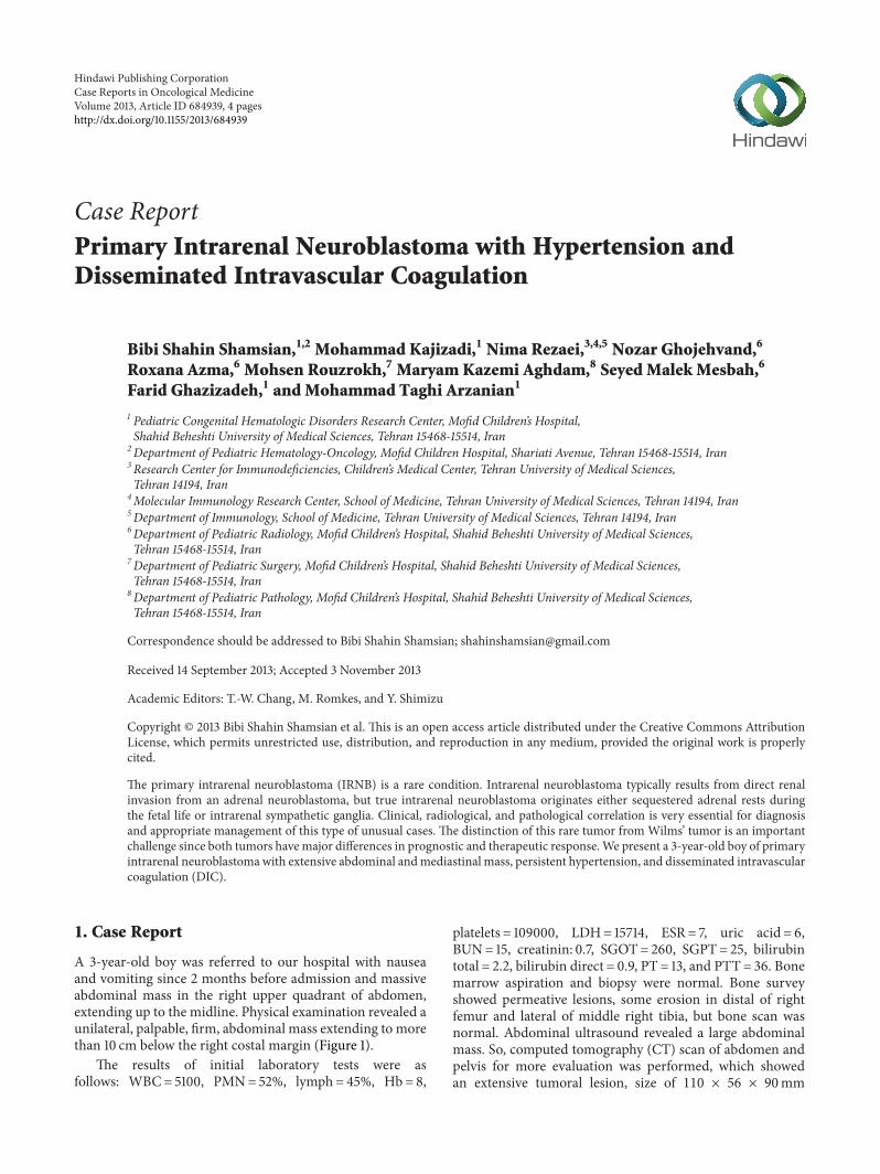
A 3-year-old boy was referred to our hospital with nauseaand vomiting since 2 months before admission and massiveabdominal mass in the right upper quadrant of abdomen,extending up to the midline. Physical examination revealed aunilateral, palpable, firm, abdominal mass extending to morethan 10 cm below the right costal margin (Figure 1).
The results of initial laboratory tests were asfollows: WBC= 5100, PMN= 52%, lymph= 45%, Hb= 8,
platelets = 109000, LDH= 15714, ESR= 7, uric acid = 6,BUN= 15, creatinin: 0.7, SGOT= 260, SGPT= 25, bilirubintotal = 2.2, bilirubin direct = 0.9, PT = 13, and PTT= 36. Bonemarrow aspiration and biopsy were normal. Bone surveyshowed permeative lesions, some erosion in distal of rightfemur and lateral of middle right tibia, but bone scan wasnormal. Abdominal ultrasound revealed a large abdominalmass. So, computed tomography (CT) scan of abdomen andpelvis for more evaluation was performed, which showedan extensive tumoral lesion, size of 110 × 56 × 90mm
2 Case Reports in Oncological Medicine
Figure 1: Large abdominal mass and petechiae, purpura due to DICin patient with neuroblastoma.
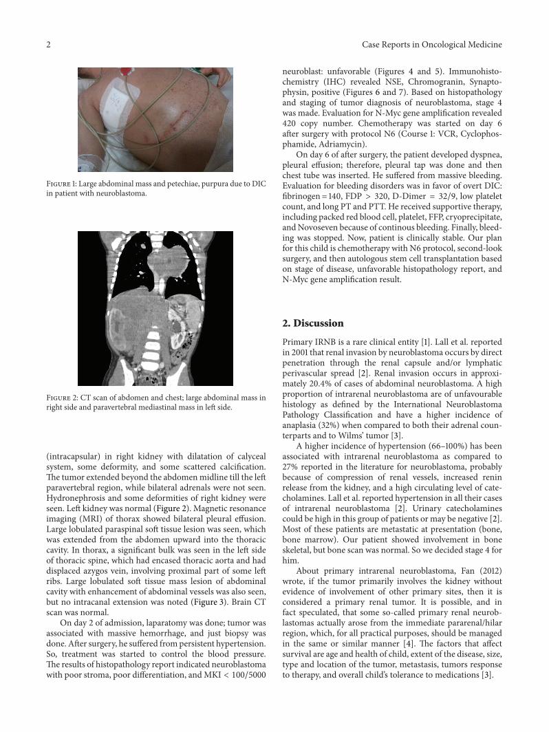
Figure 2: CT scan of abdomen and chest; large abdominal mass inright side and paravertebral mediastinal mass in left side.
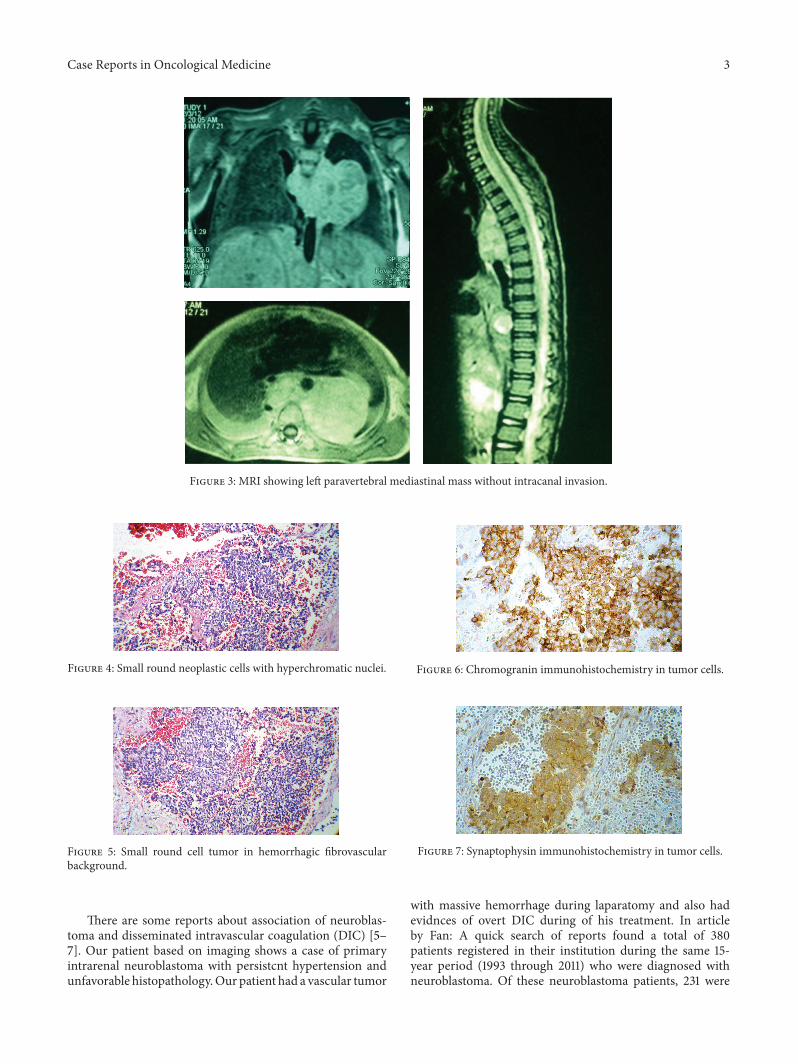
(intracapsular) in right kidney with dilatation of calycealsystem, some deformity, and some scattered calcification.The tumor extended beyond the abdomenmidline till the leftparavertebral region, while bilateral adrenals were not seen.Hydronephrosis and some deformities of right kidney wereseen. Left kidney was normal (Figure 2). Magnetic resonanceimaging (MRI) of thorax showed bilateral pleural effusion.Large lobulated paraspinal soft tissue lesion was seen, whichwas extended from the abdomen upward into the thoraciccavity. In thorax, a significant bulk was seen in the left sideof thoracic spine, which had encased thoracic aorta and haddisplaced azygos vein, involving proximal part of some leftribs. Large lobulated soft tissue mass lesion of abdominalcavity with enhancement of abdominal vessels was also seen,but no intracanal extension was noted (Figure 3). Brain CTscan was normal.
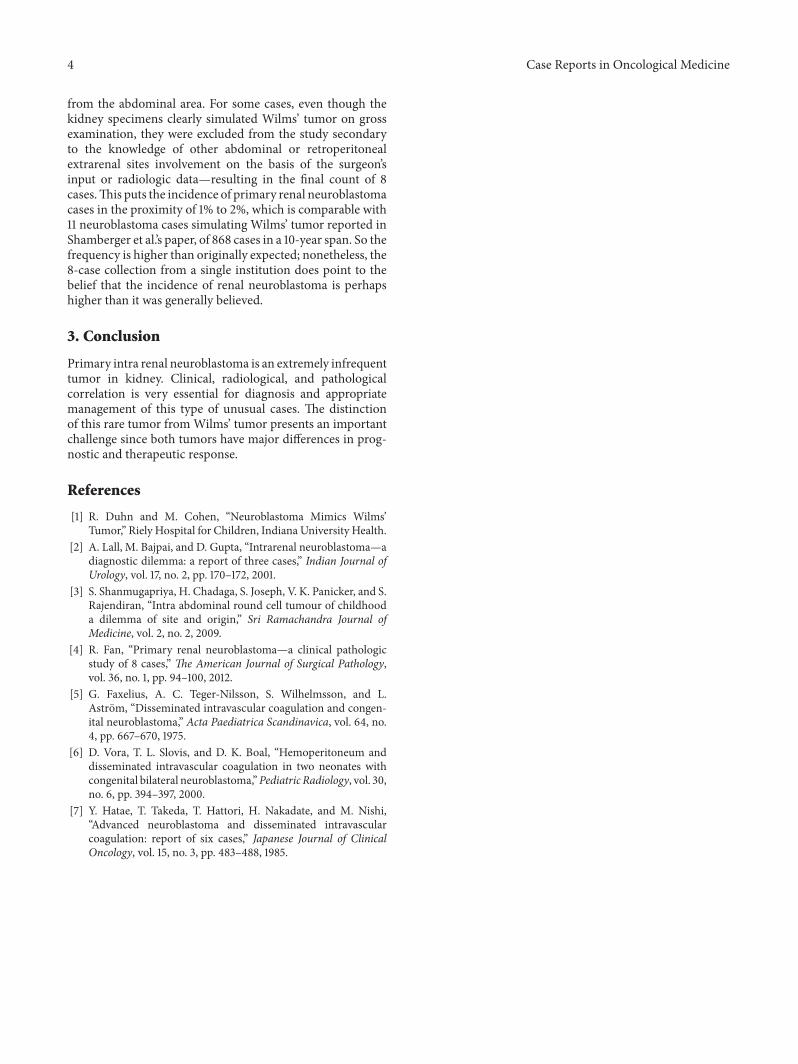
On day 2 of admission, laparatomy was done; tumor wasassociated with massive hemorrhage, and just biopsy wasdone. After surgery, he suffered frompersistent hypertension.So, treatment was started to control the blood pressure.The results of histopathology report indicated neuroblastomawith poor stroma, poor differentiation, andMKI < 100/5000
neuroblast: unfavorable (Figures 4 and 5). Immunohisto-chemistry (IHC) revealed NSE, Chromogranin, Synapto-physin, positive (Figures 6 and 7). Based on histopathologyand staging of tumor diagnosis of neuroblastoma, stage 4was made. Evaluation for N-Myc gene amplification revealed420 copy number. Chemotherapy was started on day 6after surgery with protocol N6 (Course 1: VCR, Cyclophos-phamide, Adriamycin).
On day 6 of after surgery, the patient developed dyspnea,pleural effusion; therefore, pleural tap was done and thenchest tube was inserted. He suffered from massive bleeding.Evaluation for bleeding disorders was in favor of overt DIC:fibrinogen = 140, FDP > 320, D-Dimer = 32/9, low plateletcount, and long PT and PTT. He received supportive therapy,including packed red blood cell, platelet, FFP, cryoprecipitate,andNovoseven because of continous bleeding. Finally, bleed-ing was stopped. Now, patient is clinically stable. Our planfor this child is chemotherapy with N6 protocol, second-looksurgery, and then autologous stem cell transplantation basedon stage of disease, unfavorable histopathology report, andN-Myc gene amplification result.
2. Discussion
Primary IRNB is a rare clinical entity [1]. Lall et al. reportedin 2001 that renal invasion by neuroblastoma occurs by directpenetration through the renal capsule and/or lymphaticperivascular spread [2]. Renal invasion occurs in approxi-mately 20.4% of cases of abdominal neuroblastoma. A highproportion of intrarenal neuroblastoma are of unfavourablehistology as defined by the International NeuroblastomaPathology Classification and have a higher incidence ofanaplasia (32%) when compared to both their adrenal coun-terparts and to Wilms’ tumor [3].
A higher incidence of hypertension (66–100%) has beenassociated with intrarenal neuroblastoma as compared to27% reported in the literature for neuroblastoma, probablybecause of compression of renal vessels, increased reninrelease from the kidney, and a high circulating level of cate-cholamines. Lall et al. reported hypertension in all their casesof intrarenal neuroblastoma [2]. Urinary catecholaminescould be high in this group of patients or may be negative [2].Most of these patients are metastatic at presentation (bone,bone marrow). Our patient showed involvement in boneskeletal, but bone scan was normal. So we decided stage 4 forhim.
About primary intrarenal neuroblastoma, Fan (2012)wrote, if the tumor primarily involves the kidney withoutevidence of involvement of other primary sites, then it isconsidered a primary renal tumor. It is possible, and infact speculated, that some so-called primary renal neurob-lastomas actually arose from the immediate pararenal/hilarregion, which, for all practical purposes, should be managedin the same or similar manner [4]. The factors that affectsurvival are age and health of child, extent of the disease, size,type and location of the tumor, metastasis, tumors responseto therapy, and overall child’s tolerance to medications [3].
Case Reports in Oncological Medicine 3
Figure 3: MRI showing left paravertebral mediastinal mass without intracanal invasion.
Figure 4: Small round neoplastic cells with hyperchromatic nuclei.
Figure 5: Small round cell tumor in hemorrhagic fibrovascularbackground.
There are some reports about association of neuroblas-toma and disseminated intravascular coagulation (DIC) [5–7]. Our patient based on imaging shows a case of primaryintrarenal neuroblastoma with persistcnt hypertension andunfavorable histopathology.Our patient had a vascular tumor
Figure 6: Chromogranin immunohistochemistry in tumor cells.
Figure 7: Synaptophysin immunohistochemistry in tumor cells.
with massive hemorrhage during laparatomy and also hadevidnces of overt DIC during of his treatment. In articleby Fan: A quick search of reports found a total of 380patients registered in their institution during the same 15-year period (1993 through 2011) who were diagnosed withneuroblastoma. Of these neuroblastoma patients, 231 were
4 Case Reports in Oncological Medicine
from the abdominal area. For some cases, even though thekidney specimens clearly simulated Wilms’ tumor on grossexamination, they were excluded from the study secondaryto the knowledge of other abdominal or retroperitonealextrarenal sites involvement on the basis of the surgeon’sinput or radiologic data—resulting in the final count of 8cases.This puts the incidence of primary renal neuroblastomacases in the proximity of 1% to 2%, which is comparable with11 neuroblastoma cases simulating Wilms’ tumor reported inShamberger et al.’s paper, of 868 cases in a 10-year span. So thefrequency is higher than originally expected; nonetheless, the8-case collection from a single institution does point to thebelief that the incidence of renal neuroblastoma is perhapshigher than it was generally believed.
3. Conclusion
Primary intra renal neuroblastoma is an extremely infrequenttumor in kidney. Clinical, radiological, and pathologicalcorrelation is very essential for diagnosis and appropriatemanagement of this type of unusual cases. The distinctionof this rare tumor fromWilms’ tumor presents an importantchallenge since both tumors have major differences in prog-nostic and therapeutic response.
References
[1] R. Duhn and M. Cohen, “Neuroblastoma Mimics Wilms’Tumor,” Riely Hospital for Children, Indiana University Health.
[2] A. Lall, M. Bajpai, and D. Gupta, “Intrarenal neuroblastoma—adiagnostic dilemma: a report of three cases,” Indian Journal ofUrology, vol. 17, no. 2, pp. 170–172, 2001.
[3] S. Shanmugapriya, H. Chadaga, S. Joseph, V. K. Panicker, and S.Rajendiran, “Intra abdominal round cell tumour of childhooda dilemma of site and origin,” Sri Ramachandra Journal ofMedicine, vol. 2, no. 2, 2009.
[4] R. Fan, “Primary renal neuroblastoma—a clinical pathologicstudy of 8 cases,” The American Journal of Surgical Pathology,vol. 36, no. 1, pp. 94–100, 2012.
[5] G. Faxelius, A. C. Teger-Nilsson, S. Wilhelmsson, and L.Astrom, “Disseminated intravascular coagulation and congen-ital neuroblastoma,” Acta Paediatrica Scandinavica, vol. 64, no.4, pp. 667–670, 1975.
[6] D. Vora, T. L. Slovis, and D. K. Boal, “Hemoperitoneum anddisseminated intravascular coagulation in two neonates withcongenital bilateral neuroblastoma,”Pediatric Radiology, vol. 30,no. 6, pp. 394–397, 2000.
[7] Y. Hatae, T. Takeda, T. Hattori, H. Nakadate, and M. Nishi,“Advanced neuroblastoma and disseminated intravascularcoagulation: report of six cases,” Japanese Journal of ClinicalOncology, vol. 15, no. 3, pp. 483–488, 1985.

Submit your manuscripts athttp://www.hindawi.com
Stem CellsInternational
Hindawi Publishing Corporationhttp://www.hindawi.com Volume 2014
Hindawi Publishing Corporationhttp://www.hindawi.com Volume 2014
MEDIATORSINFLAMMATION
of
Hindawi Publishing Corporationhttp://www.hindawi.com Volume 2014
Behavioural Neurology
EndocrinologyInternational Journal of
Hindawi Publishing Corporationhttp://www.hindawi.com Volume 2014
Hindawi Publishing Corporationhttp://www.hindawi.com Volume 2014
Disease Markers
Hindawi Publishing Corporationhttp://www.hindawi.com Volume 2014
BioMed Research International
OncologyJournal of
Hindawi Publishing Corporationhttp://www.hindawi.com Volume 2014
Hindawi Publishing Corporationhttp://www.hindawi.com Volume 2014
Oxidative Medicine and Cellular Longevity
Hindawi Publishing Corporationhttp://www.hindawi.com Volume 2014
PPAR Research
The Scientific World JournalHindawi Publishing Corporation http://www.hindawi.com Volume 2014
Immunology ResearchHindawi Publishing Corporationhttp://www.hindawi.com Volume 2014
Journal of
ObesityJournal of
Hindawi Publishing Corporationhttp://www.hindawi.com Volume 2014
Hindawi Publishing Corporationhttp://www.hindawi.com Volume 2014
Computational and Mathematical Methods in Medicine
OphthalmologyJournal of
Hindawi Publishing Corporationhttp://www.hindawi.com Volume 2014
Diabetes ResearchJournal of
Hindawi Publishing Corporationhttp://www.hindawi.com Volume 2014
Hindawi Publishing Corporationhttp://www.hindawi.com Volume 2014
Research and TreatmentAIDS
Hindawi Publishing Corporationhttp://www.hindawi.com Volume 2014
Gastroenterology Research and Practice
Hindawi Publishing Corporationhttp://www.hindawi.com Volume 2014
Parkinson’s Disease
Evidence-Based Complementary and Alternative Medicine
Volume 2014Hindawi Publishing Corporationhttp://www.hindawi.com