Caractérisation génomique et phénotypique de la résistance ... · croisée aux TCs de première...
241
Caractérisation génomique et phénotypique de la résistance aux antibiotiques chez Streptococcus pneumoniae Thèse Andréanne Lupien Doctorat en biologie cellulaire et moléculaire
-
Upload
phungtuyen -
Category
Documents
-
view
224 -
download
0
Transcript of Caractérisation génomique et phénotypique de la résistance ... · croisée aux TCs de première...

Caractérisation génomique et phénotypique de la résistance aux antibiotiques chez Streptococcus pneumoniae
Thèse
Andréanne Lupien
Doctorat en biologie cellulaire et moléculaire


Résumé
Streptococcus pneumoniae est le pathogène bactérien le plus important des voies
respiratoires chez les adultes et les enfants causant la pneumonie, la bronchite et l’otite de
l’oreille moyenne. Cette bactérie est responsable d’une morbidité et d’une mortalité
importante, entre autres, chez les jeunes enfants. La prévalence générale des souches de
pneumocoques résistants et multirésistants est en hausse dans le monde compliquant la
thérapie antimicrobienne vis-à-vis cette bactérie. Face à cette problématique, nous avons
voulu caractériser les mécanismes de résistance à la ciprofloxacine (CIP), la tétracycline
(TC) et la tigécycline (TGC) chez des mutants sélectionnés en laboratoire et des souches
cliniques afin de potentiellement découvrir de nouvelles cibles diagnostiques de la
résistance vis-à-vis ces molécules.
L’approche génomique utilisée dans cette thèse (séquençage de génome et
transformation) a permis de faire la caractérisation génotypique et phénotypique des
mutants résistants aux trois antibiotiques utilisés dans l’étude. Cette approche, en plus de
préciser le rôle des mutations dans les gènes parC et gyrA dans la résistance à la CIP, a
permis de déterminer que le pneumocoque peut mettre en place des mécanismes de
résistance secondaires le rendant résistant à la CIP et à la TC. En effet, l’acquisition d’une
mutation dans le gène spr1902 protège la bactérie contre les dérivés réactifs à l’oxygène
(ROS) induit par la CIP. De plus, la surexpression de PatA/PatB ainsi que la présence de
mutations dans l’opéron du transporteur PatA/PatB induit de faibles niveaux de résistance à
la CIP et à la TC, en absence de tetM et tetO. Un lien a également été établi entre la
résistance à la TC et la surexpression de gènes de la voie de biosynthèse de la thiamine
chez des souches de S. pneumoniae non-sensibles à la TC. Finalement, les mécanismes de
résistance à la TGC ont été décrit, pour la première fois, chez le pneumocoque (mutations
dans la protéine ribosomale S3 (rpsC ; spr0195), S10 (rpsJ; spr0187), l’ARNr 16S et une
méthyltransférase de l’ARNr 16S hypothétique (spr1784). Ceuxi-ci causent une résistance
iii

croisée aux TCs de première et deuxième génération. Dans cette thèse, nous avons mis en
lumière de nouveaux marqueurs de la résistance aux antibiotiques chez S. pneumoniae.
iv
Abstract
Streptococcus pneumoniae is a Gram-positive pathogen responsible for pneumonia,
bronchitis and otitis media leading to considerable morbidity and mortality among children
and adults. The prevalence of resistant and multi-resistant strains increases worldwide
impairing antimicrobial treatments toward this bacterium. We characterised resistance to
ciprofloxacin (CIP), tetracycline (TC) and tigecycline (TGC) in laboratory-derived resistant
mutants and unsusceptible clinical isolates to further our comprehension of resistance
mechanisms and potentially uncover new therapeutic and diagnostic targets toward these
drugs.
The genomic approaches used in this thesis (genome sequencing and DNA
transformation) allowed the phenotypic and genotypic characterisation of mutants resistant
to three antibiotics. By this approach, the role of parC and gyrA mutations in CIP resistance
was confirmed and even extended and it was also possible to determine that S. pneumoniae
may select secondary mechanisms of resistance to CIP and TC besides target-site
mutations. Acquisition of a mutation in spr1902 is shown to protect the bacteria against
oxygen-reactive species induced by CIP. Furthermore, overexpression of the ABC
transporter PatA/PatB and mutations in the coding region of this transporter confer low-
level resistance to CIP and TC. A link was also established between TC resistance and
overexpression of genes involved in the thiamine biosynthesis and salvage pathway in S.
pneumoniae TC non-susceptible isolates. Finally, for the first time, the mechanisms of
resistance to TGC in S. pneumoniae were described (mutations in ribosomal protein S3
(rpsC; spr0195), S10 (rpsJ: spr0187), 16S ribosomal RNA (rRNA) and a putative 16S
rRNA methyltransferase). These confer cross-resistance to first and second generation TCs.
This work highlights new markers of antibiotic resistance in S. pneumoniae.
v


Table des matières
Résumé .................................................................................................................................. iii
Abstract ................................................................................................................................... v
Table des matières ............................................................................................................... vii
Liste des tableaux ................................................................................................................... xi
Liste des figures .................................................................................................................. xiii
Liste des abréviations ............................................................................................................ xv
Remerciements ................................................................................................................... xvii
Avant-Propos ....................................................................................................................... xix
Chapitre I Streptococcus pneumoniae .................................................................................... 1
1.1 Bactériologie ............................................................................................................ 1
1.2 Identification ............................................................................................................ 2
1.3 Manifestations cliniques ........................................................................................... 3
1.4 Facteurs de virulence .................................................................................................... 4
1.4.1 Capsule ................................................................................................................... 5
1.4.3 Paroi bactérienne .................................................................................................... 6
1.4.3 Pneumolysine ......................................................................................................... 6
1.4.4 Protéines de surface ............................................................................................... 6
1.4 Traitements ................................................................................................................... 8
1.5 Vaccins .......................................................................................................................... 9
1.5.1 Vaccin polysaccharidique (VP) ............................................................................. 9
1.5.2 Vaccin polysaccharidique conjugué (VPC) ........................................................... 9
Chapitre II : La diversité du génome de Streptococcus pneumoniae ................................... 11
2.1 Acquisition d’ADN exogène ...................................................................................... 12
2.1.1 Transformation ..................................................................................................... 12
2.1.2 Conjugaison ......................................................................................................... 14
2.1.3 Transduction ........................................................................................................ 15
Chapitre III : Antibiothérapie ............................................................................................... 17
3.1 Mécanismes d’action des antibiotiques .................................................................. 17
vii

3.1.1 Inhibition de la synthèse de la paroi bactérienne ................................................ 18
3.1.2 Inhibition de la transcription ............................................................................... 19
3.1.3 Inhibition de la réplication de l’ADN ................................................................. 19
3.1.4 Inhibition de la synthèse protéique ...................................................................... 19
3.1.5 Inhibition du métabolisme des folates ................................................................. 20
3.1.6 Nouveaux mécanismes d’action des antibiotiques; productions de dérivés réactifs à l’oxygène ................................................................................................................... 20
3.2 Mécanismes de résistance aux antibiotiques .............................................................. 22
3.2.5 Réduction de la perméabilité membranaire ......................................................... 23
3.2.6 Efflux de l’antibiotique ....................................................................................... 24
3.2.1 Modification de la cible ....................................................................................... 25
3.2.2 Inactivation de l’antibiotique .............................................................................. 25
3.2.2 Amplification de la cible ..................................................................................... 26
3.3 Résistance aux antibiotiques chez S. pneumoniae ..................................................... 26
3.3.1 Résistance aux β-lactamines................................................................................ 27
3.3.2 Résistance aux macrolides et aux kétolides ........................................................ 27
3.3.3 Résistance à la LZD ............................................................................................ 28
Chapitre IV : La famille des fluoroquinolones ..................................................................... 29
4.2 Mode d’action des fluoroquinolones .......................................................................... 29
4.3 Mécanismes de résistance .......................................................................................... 31
4.3.1 Mutations dans l’ADN gyrase et la topoisomérase IV ........................................ 31
4.3.2 Efflux ................................................................................................................... 32
4.3.3 Résistance aux quinolones médiée par des plasmides ........................................ 34
Chapitre V : La famille des TCs et des glycylcyclines ........................................................ 35
5.2 Mode d’action des TCs et des glycylcyclines ............................................................ 36
5.3 Mécanismes de résistance .......................................................................................... 38
5.3.1 La famille des TCs .............................................................................................. 38
5.3.2 La TGC ................................................................................................................ 40
Chapitre VI : Étude de la résistance aux antibiotiques par approche « omique » ................ 43
6.1 Séquençage de génome entier .................................................................................... 43
6.1.1 Séquençage à haut débit (NGS) .......................................................................... 44
viii

6.2 Étude du transcriptome ............................................................................................... 47
6.2.1 Biopuces ............................................................................................................... 47
6.2.2 Séquençage de l’ARN .......................................................................................... 49
6.3 Étude de la résistance chez le pneumocoque par des techniques de protéomique ..... 50
6.4 Étude du métabolome chez S. pneumoniae ................................................................ 50
Chapitre VII: Problématique, hypothèses et objectifs .......................................................... 53
7.1 Problématique ............................................................................................................. 53
7.2 Hypothèses .................................................................................................................. 53
7.3 Objectifs ...................................................................................................................... 54
Chapitre VIII: Caractérisation génomique de la résistance à la ciprofloxacine chez un mutant dérivé d’une souche de laboratoire et d’une souche clinique de Streptococcus pneumoniae ........................................................................................................................... 57
8.1 Résumé ........................................................................................................................ 57
8.2 Article ......................................................................................................................... 59
Chapitre IX: La présence de mutations et l’augmentation de l’expression d’ARN chez des souches de Streptococcus pneumoniae résistantes à la tétracycline causent la résistance tel que démontré par séquençage de génome et séquençage de l’ARN messager. .................... 97
9.1 Résumé ........................................................................................................................ 97
9.2 Article ......................................................................................................................... 99
Chapitre X: L’induction de la résistance à la tigécycline chez Streptococcus pneumoniae révèle la sélection de mutations dans des protéines ribosomales et de l’ARN ribosomal .. 151
10.1 Résumé .................................................................................................................... 151
10.2 Article ..................................................................................................................... 152
Chapitre XI : Discussion générale ...................................................................................... 181
11.1 Les mutants résistants à la CIP, TC et TGC ........................................................... 181
11.1.1 Les mutants résistants à la CIP ........................................................................ 182
11.1.2 Les mutants résistants à la TC ......................................................................... 183
11.1.3 Les mutants résistants à la TGC ...................................................................... 184
11.2 Mutations dans les gènes parC et gyrA .................................................................. 185
11.3 Glycérol-3-phosphate déshydrogénase dépendante du NADPH et la protection contre les ROS induits par la CIP chez S. pneumoniae .................................................. 186
11.4 Le transporteur PatA/PatB et la multirésistance aux antibiotiques chez S. pneumoniae ..................................................................................................................... 187
ix

11.4.1 La surexpression du transporteur PatA/PatB .................................................. 188
11.4.2 Mutations dans la région codante de patA et patB .......................................... 190
11.5 Utilisation de l’ARN-seq pour l’étude de la résistance chez le pneumocoque ...... 193
11.6 Mécanismes de résistance à la TGC chez le pneumocoque ................................... 195
Conclusion .......................................................................................................................... 199
Bibliographie ...................................................................................................................... 201
x

Liste des tableaux
Tableau 3.1 Mécanismes d’action des principales familles d’antibiotiques Tableau 4.1 Classification des pompes d’efflux causant la résistance chez les bactéries Tableau 5.1 Classification des gènes tet et otr
xi


Liste des figures
Figure 1.1 Coloration de Gram d’une culture de pneumocoque Figure 1.2 Mode de dispersion du pneumocoque chez l’humain Figure 1.3 Principaux facteurs de virulence chez S. pneumoniae Figure 2.1 L’induction de la compétence chez S. pneumoniae Figure 3.1 Modèle du mode d’action des antibiotiques bactéricides Figure 3.2 Représentation schématique de différents types de pompes d’efflux
d’antibiotiques chez les bactéries Figure 4.1 Structure des fluoroquinolones Figure 4.2 Représentation du site de clivage du complexe de la topoisomérase IV Figure 5.1 Structure des tétracyclines et glycylcyclines Figure 5.2 Sites de liaison de la tétracycline (TC) à l’ARN ribosomal 30S Figure 5.3 Site de liaison de la tigécycline (TGC) à l’ARN ribosomal 30S Figure 6.1 Principes de la technologie de séquençage 454 Figure 6.2 La technologie Illumina Figure 6.3 Principe des biopuces à deux fluorochromes Figure 11.1 Schéma des mutations dans l’opéron patA-patB impliquées dans la résistance
dans cette étude
xiii


Liste des abréviations
ABC de l’anglais « ATP-binding cassette » ADN Acide désoxyribonucléique ADNc Acide désoxyribonucléique complémentaire ARN Acide ribonucléique ARNm Acide ribonucléique messager ARN-seq séquençage de l’ARN ATP Adénosine-5'-triphosphate CIP Ciprofloxacine CM Chloramphénicol CMI Concentration minimale inhibitrice CSP de l’anglais « Competence-stimulating peptide » Cy3 Cyanine 3 Cy5 Cyanine 5 DOX Doxycycline dTMP Désoxythymidine monophosphate dTTP Désoxythymidine triphosphate dUMP Désoxyuridine monophosphate GTP Guanosine triphosphate EM Érythromycine Etbr de l’anglais « Ethidium bromide » H Hélice HMP 4-amino-5-hydroxymethyl-2-methylpyrimidine Ig Immunoglobuline KAN Kanamycine Kb Kilobases kDa Kilodalton lytA Autolysine A LZD Linézolide MFS de l’anglais « Major facilitator superfamilly » MI Minocycline MOX Moxifloxacine NADH Nicotinamide adénine dinucléotide NADPH Nicotinamide adénine dinucléotide phosphate NGS de l’anglais « Next-generation sequencing » oxyTC Oxytétracycline PCR de l’anglais « Polymerase chain reaction » PG Pénicilline G PLP Protéine liant la pénicilline ply Pneumolysine QRDR de l’anglais « quinolone-resistance-determining region » RND de l’anglais « Resistance-nodulation-division »
xv
ROS de l’anglais « Reactive oxygen species » RSS de l’anglais « Recombinaison segment sequence » SMR de l’anglais « Small multidrug resistance family » srt Sortase TC Tétracycline TCs Famille des Tétracyclines TGC Tigécycline TMHMM de l’anglais « TransMembrane prediction using Hidden Markov Models » VA Vancomycine VP Vaccin polysaccharidique VPC Vaccin polysaccharidique conjugué WGT de l’anglais « Whole-genome transformation »
xvi

Remerciements J’aimerais tout d’abord remercier mon directeur de recherche le Dr Marc Ouellette
de m’avoir permis de faire mes études graduées dans son laboratoire. Sa passion pour la
recherche et sa rigueur scientifique sont des qualités qui m’ont grandement inspirées durant
mes études. Il a été un excellent mentor et j’aimerais sincèrement le remercier pour sa
patience, son support, sa disponibilité et l’autonomie qu’il m’a laissée afin de mener à bien
cette thèse.
J’aimerais également remercier mon co-directeur de recherche, le Dr Philippe
Leprohon. Il a été un mentor hors pair. Sa passion pour la science transparaît par son désir
de transmettre ses connaissances. J’aimerais le remercier pour sa grande disponibilité et
pour les nombreuses discussions qui ont permis de faire avancer le projet.
J’aimerais remercier les membres du comité évaluateur de cette thèse, le Dr Gerard
D. Wright, de l’Université McMaster, le Dr Steve Charette et le Dr Daniel Grenier pour le
temps accordé à l’évaluation de cette thèse. Également j’aimerais remercier la Dre Josée N.
Lavoie, directrice du programme de Biologie cellulaire et moléculaire, ainsi que Mme
Chantal Joubert, Agente de gestions des études, pour le dépôt de cette thèse.
Durant mes études graduées au Centre de Recherche en Infectiologie, j’ai eu la
chance de côtoyer des gens formidables. Je désire sincèrement remercier chacun des
membres de l’équipe du Dr Marc Ouellette (MOU) passés et présents, et particulièrement
deux personnes piliers du laboratoire, Suzanne Avoine et Gaétan Roy, pour leurs conseils et
leur aide. Un gros merci également à Danielle Légaré pour ses nombreux conseils et les
partys d’équipe à son chalet. Également, la réalisation de mon projet de thèse n’aurait pas
été possible sans les membres présents du groupe Strep et trois anciens membres de
l’équipe, Fereshteh Fani, Dewan Billal et Jie Feng qui ont su me conseiller au cours de mon
parcours académique et avec qui j’ai eu beaucoup de plaisirs à travailler.
xvii

Mon passage au CRI m’a permis de rencontrer des gens extraordinaires avec qui j’ai
développé de précieuses amitiés. J’aimerais spécialement remercier ma collègue et amie
Hélène Gingras avec qui j’ai eu la chance de travailler sur les projets TC et TGC.
L’achèvement de ses travaux aurait été très difficile sans son aide. Elle est une personne
exceptionnelle à qui j’ai pu me confier aussi bien sur le plan personnel que professionnel.
Également, je veux remercier mes amies Marie Plourde, Andrée Maheux, Marie-Christine
Brotherton, Jessica El-Khoury et Jade-Éva Potvin qui m’ont supporté et ont su agrémenter
ces dernières années.
Finalement j’aimerais remercier mes amis, Agnès, Stéphane et Isabelle, mes
parents, Jean-François et Christiane, mon frère, Jean-Philippe, ainsi que tous les membres
de ma famille pour leurs encouragements, leur soutien et leur amour inconditionnel.
xviii
Avant-Propos
Le manuscrit présenté au Chapitre 8, intitulé « Genomic characterisation of
ciprofloxacin resistance in a laboratory derived mutant and clinical isolate of S.
pneumoniae. » a été publié dans le journal Antimicrobial Agents and Chemotherapy. J’ai
effectué les inactivations géniques, les reconstructions phénotypiques, les quantifications
des ROS et rédigé le manuscrit. Dewan S. Billal a généré les mutants R6M2B, T5-R6M2B
et T1-60827 et a effectué les expériences de qRT-PCR. Fereshteh Fani a produit le
plasmide pFF6. Hafid Soualhine a contribué au design expérimental. George G. Zhanel a
fourni la souche 60827. Philippe Leprohon a analysé les résultats de séquençage et révisé le
manuscrit. Marc Ouellette a supervisé le projet.
Le manuscrit présenté au Chapitre 9 intitulé « Multiple mutations and increased
RNA expression in tetracycline resistant Streptococcus pneumoniae as determined by
genome wide DNA and mRNA sequencing. » a été soumis pour publication dans le journal
Journal of Antimicrobial Chemotherapy. Hélène Gingras a sélectionné les souches
R6M1TC-5 and R6M2TC-4. J’ai déterminé le profil de résistance aux antibiotiques des
mutants. L’analyse du séquençage des génomes des mutants TC a été effectuée par moi-
même avec la contribution de Philippe Leprohon. Les reconstructions génotypiques par
fragments PCR ont été effectuées par moi-même (introduction des mutations) et Hélène
Gingras (réversion des mutations introduites). J’ai effectué avec l’aide de Philippe
Leprohon l’analyse des données obtenues par ARN-seq en utilisant l’algorithme Cufflinks.
J’ai effectué l’analyse Blast2Go des gènes dont l’expression est altérée chez nos mutants.
J’ai effectué les études d’accumulation de TC tritié. J’ai effectué en majorité, avec l’aide
d’Hélène Gingras, les expériences de qRT-PCR pour les gènes patA, patB, spr0632, tenA,
thiD, thiE et spr1021 ainsi que leur inactivation chez les mutants et les souches cliniques.
J’ai effectué le génotypage des souches cliniques (rpsJ, tetM et tetO). Michel G. Bergeron a
fourni les souches cliniques utilisées dans cette étude. Le manuscrit a été écrit par moi-
même et Philippe Leprohon. Marc Ouellette a supervisé le projet.
xix
Le manuscrit présenté au Chapitre 10, intitulé « Induced tigecycline resistance in
Streptococcus pneumoniae mutants reveals mutations in ribosomal proteins and rRNA. »
sera soumis sous peu pour publication. Hélène Gingras a sélectionné les souches
R6M1TGC-6 et R6M2TGC-6. J’ai déterminé le profil de résistance des mutants aux
antibiotiques. L’analyse du séquençage des génomes des mutants TC a été effectuée par
moi-même avec la contribution de Philippe Leprohon. J’ai effectué les reconstructions
génotypiques par fragments PCR. L’article a été écrit par moi-même et Philippe Leprohon.
Marc Ouellette a supervisé le projet.
xx

Introduction
Chapitre I Streptococcus pneumoniae
La bactérie pathogène S. pneumoniae fût isolée en 1881 par le chimiste français
Louis Pasteur et le médecin américain George Sternberg. En 1926, la bactérie a été
classifiée sous le nom de Diplococcus pneumoniae due à sa forme de diplocoque lorsqu’on
l’observe au microscope. Cependant, c’est en 1974 qu’elle sera finalement classée dans le
genre Streptococcus, en raison de sa croissance en courtes chaînettes en milieu liquide
(253).
S. pneumoniae, également connu sous le nom commun de pneumocoque, fait partie de
l’embranchement taxonomique des Firmicutes, de la classe des Bacilli, de l’ordre des
Lactobacillales et de la famille des Streptococcaceae (253). Le genre Streptococcus
regroupe plusieurs espèces pathogènes chez l’être humain, en plus de S. pneumoniae, dont
Streptococcus pyogenes, Streptococcus agalactiae, Streptococcus suis et Streptococcus
bovis.
1.1 Bactériologie
S. pneumoniae est une coque à Gram positif d’environ 0,5 à 1,25 micromètre de
diamètre qui possède une forme légèrement allongée (cocci de lancet) (Figure 1.1). On le
retrouve le plus souvent en paires (diplocoques), toutefois il peut être observé sous forme
de courtes chaînettes, la culture en milieu liquide favorisant cette organisation des cocci
(253). Cette bactérie mésophile est tolérante à l’oxygène. Néanmoins, environ 20% des
souches cliniques nécessiteraient des conditions d’anaérobie strictes pour leur croissance.
Comme d’autres Streptococci, S. pneumoniae ne possède pas de catalase d’où la nécessité
de le cultiver en milieu contenant une source de cette enzyme afin de neutraliser les
1

quantités importantes de peroxyde d’hydrogène produites par la bactérie (113). Lors de la
culture sur gélose au sang, un halo verdâtre est visible autour des colonies (α-hémolytique),
cependant la culture en condition d’anaérobie peut induire une hémolyse complète du sang
(β-hémolytique). La bactérie ne forme pas de spores et n’est pas motile, bien que la
présence de pili ait été rapportée (89, 192). La principale source d’énergie du pneumocoque
provient de la fermentation du glucose en acide lactique (113). En effet, plusieurs enzymes
essentielles au cycle des acides tricarboxyliques (cycle de Krebs) sont absentes chez la
bactérie (113).
Figure 1.1 : Coloration de Gram d’une culture de pneumocoque en milieu contenant du sang. Les bactéries apparaissent principalement sous la forme de diplocoques de couleur mauve. Tiré de (43)
1.2 Identification
L’identification du pneumocoque est essentielle afin de le différencier des autres
espèces pathogènes et commensales vivant dans sa niche écologique principale, le
nasopharynx. En effet, la présence de certaines espèces bactériennes, telles les bactéries
commensales du groupe des Streptococcus viridans, peuvent compliquer l’identification du
pneumocoque.
2

En premier lieu, les techniques de microbiologie classiques, telle la coloration de Gram,
la présence d’hémolyse sur gélose au sang, la sensibilité à l’optochine et la solubilisation
par les sels biliaires sont généralement utilisées pour l’identification de S. pneumoniae (72).
L’apparition d’une hémolyse de type α en condition d’aérobie, l’obtention d’une coloration
de Gram positif où l’on observe des diplocoques ou des coques en chaînettes ainsi qu’un
test de catalase négatif permettent de confirmer la présence d’un Streptococcus spp. dans un
échantillon. Par la suite, la sensibilité en présence d’un disque contenant 5 mg d’optochine
permet de discriminer si nous sommes bien en présence du pneumocoque. En effet, une
zone d’inhibition d’un diamètre >14 mm confirme l’identification du pneumocoque.
Toutefois, une zone d’inhibition de 9-13 mm doit être contrevérifiée par le test de solubilité
en présence de sels biliaires, car des souches résistantes à l’optochine ont déjà été observées
(134). Tous ces tests sont très spécifiques (portée de la spécificité 85%-95%) mais peuvent
produire des résultats positifs avec d’autres bactéries du groupe des streptocoques viridans
(134, 178). D’autres tests peuvent être utilisés, comme la réactivité d’antigènes anti-
pneumococciques spécifiques à la capsule, ou réaction de Quellung (16, 172). De plus,
l’amplification par PCR des gènes de l’autolysine A (lytA) et de la pneumolysine (ply) a
déjà été utilisée afin d’identifier la bactérie S. pneumoniae dans un échantillon de patient
ayant une pneumonie (243).
1.3 Manifestations cliniques
Le pneumocoque est une bactérie pathogène stricte de l’être humain qui se transmet
d’un hôte colonisé au niveau de la muqueuse rhinopharyngée à un autre individu par
l’intermédiaire d’aérosols (105). Cette première colonisation est essentielle à toutes les
manifestations cliniques du pneumocoque (Figure 1.2). Il est important de noter que la
colonisation du nasopharynx par le pneumocoque est fréquemment asymptomatique. En
effet, jusqu’à 60% des jeunes enfants et moins de 10% des adultes sains sont porteurs de S.
pneumoniae au niveau du nasopharynx et ceux-ci ne développeront pas de symptômes liés
à cette colonisation (105). Ce taux peut varier et est affecté par plusieurs facteurs de risque
dont l’âge (diminue avec l’âge chez les personnes saines), le statut socio-économique,
3

l’exposition à la fumée de cigarette, les saisons (plus élevé lors de la saison froide) et la
fréquentation d’enfants (83). Néanmoins, une dispersion du pneumocoque au niveau des
voies respiratoires supérieures (infection de la muqueuse) ou de sites normalement stériles,
comme le sang et le système nerveux central (infection invasive), peut entraîner des otites,
des sinusites, des pneumonies, des bactériémies et des méningites (Figure 1.2). Dans des
cas plus rares, S. pneumoniae peut causer l’arthrite septique, l’ostéomyélite, la polymyosite,
la fasciite nécrosante, l’endocardite, la péricardite, un abcès du système nerveux central, la
péritonite, l’infection des voies urinaires et génitales, la parotidite, l’épiglottite, la
mastoïdite, l’endophtalmite et la conjonctivite, selon le site colonisé par la bactérie (253).
Figure 1.2 : Mode de dispersion du pneumocoque chez l’humain et les manifestations cliniques principales associées. Adapté de (105).
1.4 Facteurs de virulence
Afin de pouvoir coloniser un hôte, le pneumocoque utilise plusieurs facteurs de
virulence essentiels à l’infection par la bactérie en empêchant l’opsonisation par les cellules
présentatrices d’antigènes et en permettant l’adhésion du pneumocoque à la muqueuse.
Aérosols
Colonisation
Colonisation asymptomatique
Otite moyenne
Pneumonie
Bactériémie
Méningite
4

Dans cette section, les principaux facteurs de virulence, représentés à la Figure 1.3 seront
abordés.
Figure 1.3 : Principaux facteurs de virulence chez S. pneumoniae. Adapté de (130).
1.4.1 Capsule
La capsule est le principal facteur de virulence chez S. pneumoniae puisqu’elle
empêche la phagocytose en bloquant la liaison de la région Fc des Immunoglobulines G ou
la composante iC3b du complément de l’hôte (180, 268). La capsule est juxtaposée à la
paroi bactérienne et est composée de polysaccharides. La charge négative de la capsule
prévient la capture du pneumocoque dans le mucus du nasopharynx lors de l’infection
(105). Les gènes de la capsule sont codés en un seul opéron situé entre les gènes dexB et
aliA sur le génome bactérien (à l’exception des sérotypes 3 et 37) et la diversité chimique
Transporteurs ABC
Protéines liant les métaux
Protéines liant la choline
Protéines à motif LPxTG
Capsule
Paroi cellulaire
Membrane
Pneumolysine PsaA PiaA PiuA
Sortases Eno PavA
LytA
5

des sucres qui peuvent être synthétisés donne lieu à plus de 90 types capsulaires (sérotypes)
connus à ce jour pour le pneumocoque (24).
1.4.3 Paroi bactérienne
La paroi bactérienne est composée principalement de peptidoglycane, d’acides
teichoiques, d’acides lipoteichoiques, et de phosphorylcholines. La phosphorylcholine est
un phospholipide hydrophobe caractéristique des bactéries colonisant les voies respiratoires
supérieures telles Haemophilus influenzae, Actinobacillus actinomycetemcomitans et des
bactéries commensales et pathogènes du genre Neisseria (130). Cette composante, en plus
de permettre l’ancrage des protéines liant la choline, joue un rôle important dans
l’inflammation (24).
1.4.3 Pneumolysine
La pneumolysine est une toxine faisant partie de la famille des cytolysines
dépendantes du cholestérol. Cette protéine monomérique de 52 kDa a la capacité, une fois
excrétée, de s’assembler en pore de 30-50 unités sur la membrane d’une cellule cible (247).
La séquence de la pneumolysine varie très peu d’un sérotype à l’autre ce qui en fait un
excellent candidat pour le développement d’un vaccin anti-pneumococcique (247).
1.4.4 Protéines de surface
S. pneumoniae produit plusieurs protéines de surface lui permettant d’interagir avec
les composantes de l’hôte lors de la colonisation et de sa dissémination. Les analyses des
génomes de R6, une souche non capsulée qui est dérivée de la souche D39 (sérotype 2), et
de la souche TIGR4 (sérotype 4) ont permis de regrouper les protéines de surface en trois
grands groupes selon leurs motifs: les lipoprotéines (42 chez R6 et 47 chez TIGR4), les
protéines liant la choline (10 chez R6 et 16 chez TIGR4) et les protéines contenant un motif
6
LPxTG (13 chez R6 et 10 chez TIGR4) (25). Un quatrième groupe, formé de protéines non
classiques est également présent.
1.4.4.1 Protéines liant la choline
La phosphorylcholine est une composante importante de la paroi bactérienne de S.
pneumoniae, car elle permet l’ancrage de manière non covalente des protéines liant la
choline (25). Le pneumocoque produit en moyenne 13 à 16 protéines liant la choline, dont
quatre hydrolases dégradant la paroi bactérienne (N-acetyl-muramoyl-L-alanine amidase
(LytA), β-N-acétylglucosamidase (LytB), β-N-acetylmuraminidase (LytC; lysosyme) et une
phosphorylcholine estérase (Pce)) qui sont toutes importantes dans la virulence de la
bactérie (25). La protéine de surface PspA, qui est retrouvée virtuellement chez tous les
sérotypes cliniques et qui protège contre la réponse immunitaire innée, et la protéine de
surface PspC qui est importante pour la colonisation du pneumocoque au niveau du
nasopharynx de par son interaction avec l’ectodomaine des récepteurs des
immunoglobulines polymériques font également parties de cette famille (25, 275).
1.4.4.2 Protéines de surface LPxTG
L’ancrage covalent au peptidoglycane de la paroi bactérienne des protéines de
surface à motif LPxTG s’effectue par des sortases (srt). Chez TIGR4, quatre srt ont été
répertoriées et leur rôle diffère selon les protéines de surface auxquelles elles permettent
l’ancrage. Plusieurs protéines à motifs LPxTG ont été répertoriées dont les neuraminidases
(NanA et NanB). Tous les pneumocoques produisent au moins une neuraminidase et une
étude faite chez le chinchilla a démontré que l’absence de cette enzyme chez la bactérie
diminuait sa persistance dans le nasopharynx et l’oreille moyenne (249). Également,
plusieurs protéases à motifs LPxTG sont produites par le pneumocoque, telle la chaperone
HtrA qui permettrait selon des études chez le rat de résister au stress oxydatif lors de la
colonisation du nasopharynx. De plus, le pneumocoque peut produire jusqu’à quatre
métalloprotéases à motifs LPxTG, dont IgA1, ZmpB, ZmpC et ZmpD. La protéase IgA est
7

produite virtuellement par tous les pneumocoques et est importante lors de l’infection des
poumons et lors des cas de bactériémies (203).
1.4.4.3 Lipoprotéines
Chez le pneumocoque, les lipoprotéines présentes au niveau de la paroi sont
essentielles pour le transport de substrat et l’état général de la bactérie. On retrouve dans
cette famille plusieurs transporteurs de type ABC dont PsaA, une lipoprotéine importante
dans l’adhérence du pneumocoque et qui constitue la partie soluble du système de transport
de manganèse. De plus, on retrouve deux lipoprotéines, PiaA et PuiA faisant partie de deux
systèmes d’import du fer (36). Également, la peptidyl-prolyl isomérase permet la sécrétion
et l’activation des molécules de surface. De plus, PpmA, ayant une homologie avec les
membres de la famille des parvulines et SlrA, un membre de la famille des cyclophilines,
font partie de ce groupe (6).
1.4.4.4 Autres protéines
D’autres protéines ne possèdent pas de séquence « signal » reconnue par les
complexes de sécrétion ni de domaines retrouvés chez les groupes décrits précédemment.
On retrouve l’adhésine de la fibronectine PavA qui est un important déterminant de la
virulence dans l’infection à pneumocoque (206). De plus, la glycéraldéhyde-3-phosphate
déshydrogénase (GAPDH) lie le plasminogène et l’énolase Eno augmenterait la
dégradation de la matrice extracellulaire et dissoudrait la fibrine ce qui aiderait à la
transmigration du pneumocoque (25).
1.4 Traitements
Le traitement des infections aux pneumocoques repose principalement sur
l’administration d’antibiotiques. Les principaux antibiotiques utilisés sont les β-lactamines,
8

les macrolides et les fluoroquinolones. Également, la vancomycine (VA), les tétracyclines
(TCs) et la linézolide (LZD) peuvent être utilisées pour traiter les infections aux
pneumocoques ne répondant pas aux antibiotiques de première ligne. Le mode d’action de
ces antibiotiques sera traité plus en détail à la section 3.1.
1.5 Vaccins
La vaccination est un moyen simple et efficace de prophylaxie contre un grand
nombre de sérotypes infectieux du pneumocoque qui sont potentiellement porteurs de gènes
de résistance aux antibiotiques. Deux formulations vaccinales sont disponibles, soit le
vaccin polysaccharidique Pneumovax® et le vaccin conjugué polysaccharidique Prevnar®.
1.5.1 Vaccin polysaccharidique (VP)
Ce vaccin contient des antigènes polysaccharidiques purifiés dérivés de la capsule
de 23 sérotypes (1, 2, 3, 4, 5, 6B, 7F, 8, 9N, 10A, 11A, 12F, 14, 15B, 17F, 18C, 19A, 19F,
20, 22F, 23F et 33F). Les sérotypes contenus dans la formulation vaccinale procureraient
environ 90% de protection contre les isolats provenant du sang et 85% de protection contre
les isolats de pneumocoques colonisant des sites stériles (217). La vaccination avec le
Pneumovax 23® induit une réponse immunitaire indépendante des cellules T généralement
faible et inconsistante chez les enfants de moins de deux ans et les patients
immunosupprimés. Le VP est recommandé chez les enfants ayant plus de deux ans ainsi
que les adultes sains. Le VP aurait un effet protecteur chez les groupes à risque, entre
autres, chez les patients âgés ayant des maladies respiratoires obstructives chroniques, les
patients aspléniques ou les patients à risque de contracter une infection à pneumocoque
(43).
1.5.2 Vaccin polysaccharidique conjugué (VPC)
9

Le VPC Prevnar® consiste en des polysaccharides de capsule conjugués à la
protéine diphtérique CRM197 en suspension dans une solution contenant de l’aluminium
sous forme de phosphate, à titre d’adjuvant, ce qui induit une réponse immunitaire
dépendante des cellules T (4). Toutefois, l’immunogénicité de la liaison des
polysaccharides à l’adjuvant fait en sorte que le nombre de sérotypes couverts par le vaccin
est limité. Aujourd’hui, des valences de 4-13 sérotypes sont disponibles couvrant, dans le
cas du Prevnar-7, 85% des sérotypes causant des infections aux pneumocoques aux États-
Unis, 60-70% en Europe et 55% en Asie (194). Ce vaccin est, entre autres, recommandé
pour les enfants de moins de deux ans (43).
10
Chapitre II : La diversité du génome de Streptococcus
pneumoniae
La bactérie S. pneumoniae a joué un rôle critique dans la démonstration que l’ADN
est le matériel génétique héréditaire. En effet, en 1944, Avery, MacLeod, et McCarthy ont
démontré que l’ADN était l’élément transformé identifié par Griffith quelques années plus
tôt (17, 92). Depuis ce jour, le pneumocoque a été un organisme modèle afin d’étudier la
transformation chez les bactéries dû à son système de compétence inductible qui lui permet
d’acquérir de l’ADN de son environnement et de l’intégrer par recombinaison dans son
génome. Cette caractéristique du pneumocoque lui apporte ainsi une certaine plasticité
génomique caractéristique des bactéries naturellement compétentes (63, 120). En effet,
l’analyse comparative des génomes des souches TIGR4 (2 161 kb) et de R6 (2 039 kb) a
permis de confirmer que 10% de leur contenu génomique diffère (38, 246). L’analyse de
soixante-douze isolats invasifs et non invasifs de pneumocoques (23 de sérotype 6A, 29 de
sérotype 6B et 20 sérotype 14) par la méthode d’hybridation de génomes sur puce à ADN a
permis de déterminer que l’équivalent de 1553 gènes (73% du génome de TIGR4 présent
sur la puce) correspondrait au génome de base du pneumocoque, suggérant qu’environ 27%
du génome serait variable parmi les isolats (185). Une étude comparative similaire, mais
utilisant dans ce cas le pyroséquençage 454 chez huit isolats cliniques combinés aux
résultats de séquences de neuf autres souches a permis de déterminer que 21 à 32% du
génome de chaque souche serait variable (109). Ces observations sur la très grande
diversité du génome du pneumocoque entre les souches supportent en effet l’hypothèse de
la distribution du génome qui stipule que pour une bactérie, la totalité des gènes disponibles
existe sous forme de « supragénome » et que chacun des membres de la population qui sont
naturellement transformables contribue à la sélection des gènes disponibles. Chez le
pneumocoque, le supragénome serait estimé à plus de 5000 groupes orthologues, dont
environ 3000 seraient retrouvés à une fréquence ≥ 0,1% dans la population de S.
pneumoniae (109).
11
2.1 Acquisition d’ADN exogène
Chez les bactéries, l’acquisition d’ADN exogène se fait principalement par
transferts horizontaux impliquant la transformation, la conjugaison et la transduction. Ces
mécanismes seront plus amplement détaillés dans cette section.
2.1.1 Transformation
La transformation est l'intégration d'un fragment d'ADN étranger dans une cellule,
ce qui peut entraîner une modification héréditaire du phénotype de l'organisme receveur.
Cette section abordera le mécanisme de transformation chez S. pneumoniae.
Le pneumocoque, comme plusieurs autres bactéries telles H. influenzae, Neisseria
gonorrhoeae, Neisseria meningitidis et Bacillus subtilis ont la possibilité de devenir
naturellement compétent à la transformation. Cet état requiert un consortium de gènes
présent dans le génome bactérien. Contrairement à la transformation artificielle, la
transformation naturelle permet d’acquérir de grandes quantités d’ADN pouvant
correspondre à 10% de la taille du génome dans le cas du pneumocoque (253).
L’induction de la compétence est faite par un mécanisme dit à deux composantes
(176). Cet état temporaire utilise un mécanisme de communication cellule-cellule
hautement régulé dépendant d’un peptide de stimulation de la compétence (CSP :
« competence-stimulating peptide») (Voir Figure 2.1). Le CSP est synthétisé sous forme de
précurseur, ComC, qui est clivé par ComA, une protéine de la famille des transporteurs
ABC, qui avec ComB, sécrète le CSP mature dans l’espace extracellulaire. Il existe deux
isoformes de CSP chez le pneumocoque, soit le CSP-1 et le CSP-2 qui diffèrent de huit
acides aminés (121). Environ la moitié des souches cliniques possèdent une séquence comC
codant pour la variante 1 du CSP (121). L’accumulation de CSP à l’extérieur de la cellule
permet sa liaison avec son récepteur ComD, une histidine kinase qui s’autophosphoryle et
12

active le régulateur transcriptionnel ComE. L’activation de ComE aura pour effet
d’augmenter la transcription des opérons comAB et comCDE par la reconnaissance d’une
séquence répétée (« com-box ») en amont de ceux-ci. Cette même séquence est également
présente en amont des deux copies du gène comX et du gène comW. Un autre système à
deux composantes, CiaH-CiaR, peut empêcher le développement de la compétence en
réprimant l’expression de l’opéron comCDE (96). L’utilisation de CSP synthétique a le
même effet sur l’activation de la compétence, ce qui permet l’induction de la compétence
dans des conditions de laboratoire. Les gènes comAB, comCDE, comX, comW sont
considérés comme des gènes précoces de la compétence.
Figure 2.1 : L’induction de la compétence chez S. pneumoniae est régulée par un système de détection du quorum dépendant du peptide de compétence CSP. La sécrétion de CSP dans l’espace extracellulaire et la liaison à son récepteur ComD causera l’activation de ComE, puis ultimement ComX, permettant l’expression des gènes tardifs requis pour la translocation de l’ADN exogène dans la bactérie et l’intégration du fragment d’ADN dans le génome. Adapté de (56).
Gènes tardifs
RNA pol spécifique de Com
13

ComX est un facteur sigma alternatif qui est responsable, avec l’ARN polymérase,
de la transcription des gènes tardifs (158). La quasi-totalité des gènes codant pour les
protéines constituant le translocasome, un complexe protéique qui permet l’entrée de
l’ADN dans la cellule, contiennent un motif « com-box » reconnu par ComX (176).
Plusieurs de ces protéines jouent un rôle dans le transport de l’ADN (cglABCDEFG) à
l’intérieur de la bactérie, alors que d’autres permettent la préparation de l’ADN avant son
internalisation. À partir d’un ADN double brin, l’activité 5’-3’ endonucléase de EndA clive
l’un des brins d’ADN causant l’entrée d’un ADN simple brin (171). La liaison de la
protéine SsB protège l’ADN simple brin de la dégradation par les nucléases intracellulaires.
L’homologie de la séquence internalisée avec une région de l’ADN génomique bactérien
permettra la formation d’une synapse de recombinaison et l’intégration du fragment grâce à
la recombinase RecA (164).
La compétence joue un rôle crucial dans la plasticité du génome du pneumocoque.
En plus de permettre l’acquisition de gènes de résistance aux antibiotiques, elle joue un rôle
important dans la virulence de la bactérie, soit en facilitant l’acquisition d’îlots de
pathogénicité (122) ou en permettant d’échapper au vaccin anti-pneumococcique par le
changement de sérotype de la capsule (122).
2.1.2 Conjugaison
Chez le pneumocoque, l’acquisition par transformation d’éléments conjugatifs, tels
les plasmides et les transposons peut causer la résistance aux agents antimicrobiens. Cette
section traitera des plasmides et des transposons conjugatifs reconnus pour causer la
résistance aux antibiotiques chez S. pneumoniae.
Chez le pneumocoque, la présence naturelle de plasmide est rare. À ce jour,
seulement des plasmides dits cryptiques, dont pDP1, ont été retrouvés. Ces plasmides
codant seulement des gènes importants pour leur propre réplication seraient présents chez
environ 4% des isolats cliniques de pneumocoques (228). Cependant, S. pneumoniae a la
14
capacité d’acquérir des plasmides conjugatifs « R » normalement retrouvés chez les autres
streptococci (234), ce qui a permis d’utiliser des dérivés de ces plasmides pour des études
fonctionnelles (239). Une multitude de plasmides réplicatifs et non-réplicatifs sont
disponibles afin de respectivement exprimer ou inactiver des gènes chez le pneumocoque
(98, 144). Ces plasmides peuvent contenir des gènes de résistance à l’érythromycine (EM),
la tétracycline (TC), le chloramphénicol (CM), la kanamycine (KAN) et la spectinomycine
ce qui permet la sélection de ceux-ci chez le pneumocoque (98). Dans les études présentées
dans cette thèse, nous avons utilisé une série de plasmides nommés pFF. Ceux-ci ont la
capacité de se répliquer chez Escherichia coli, mais pas chez S. pneumoniae en raison de
l’absence d’origine de réplication. Les marqueurs de résistance CM (pFF3), KAN (pFF6) et
EM (pFF4) sont sous le contrôle du promoteur du gène amiC. Ces plasmides, en plus d’être
de petite taille, permettent d’obtenir un nombre élevé de copies chez E. coli et ils
contiennent un site EamII05I qui permet un clonage TA.
Les transposons conjugatifs de la famille Tn916/Tn1545 sont les principaux
éléments mobiles responsables de la multirésistance clinique chez S. pneumoniae (48, 49).
Les transposons de cette famille peuvent contenir des gènes de résistance aux TCs, aux
macrolides, aux lincosamides, aux streptogramines, à la KAN et au mercure. Tn916 diffère
de Tn1545 par l’insertion du gène ermB, causant le phénotype de résistance MLS
(résistance aux macrolides-lincosamides et streptogramine) et aphA responsable de la
résistance à la KAN (48, 49). Plusieurs autres éléments dérivés de Tn916 (Tn3872),
Tn2010, Tn2017 et Tn5253) et de Tn1545 (Tn6002 et Tn6003) ont été répertoriés et chacun
peut causer la résistance chez S. pneumoniae.
2.1.3 Transduction
L’échange de matériel génétique par transduction est médié par les bactériophages,
qui peuvent être classés en deux groupes distincts soit les phages lytiques ou tempérés. Les
phages lytiques détournent le cycle cellulaire de la bactérie afin de produire de nouvelles
particules virales infectieuses, alors que les phages tempérés ont une phase lysogénique où
15

ils s’intègrent au génome de la bactérie pour former un prophage. Il est intéressant de noter
que chez le pneumocoque environ 70% des souches contiendraient des prophages ou des
vestiges de ceux-ci (210). Chez le pneumocoque, certains prophages peuvent être induits
par la présence de fluoroquinolones (CIP et lévofloxacine (LVX)) (156). La fréquence des
prophages fonctionnels et inductibles à la CIP serait plus élevée chez les souches sensibles
en comparaison aux souches résistantes, ce qui affecterait le développement de la résistance
à cet antibiotique chez la bactérie (156). Deux phages lytiques ont été isolés et séquencés
chez S. pneumoniae, soit Cp-1, un phage de la famille des Podoviridae, et Dp-1, un
Siphoviridae (84). Les phages lytiques contribueraient chez le pneumocoque à l’expansion
du réservoir d’ADN exogène disponible par leur incorporation dans le génome de S.
pneumoniae. (157, 210).
16

Chapitre III : Antibiothérapie
La découverte du premier antibiotique (la pénicilline (PG)) par Sir Alexander
Flemming, en 1928, et sa production dans les années 1940 a révolutionné le traitement des
infections causées par les bactéries. Un antibiotique (du grec anti : « contre », et bios : « la
vie ») est une molécule naturelle ou synthétique qui détruit ou bloque la croissance des
bactéries. Dans le premier cas, on qualifie l’antibiotique de bactéricide et dans le second cas
on dira plutôt que l’antibiotique est bactériostatique. La majorité des antibiotiques sont
produits par les bactéries du genre Streptomyces et ont pour but premier d’éliminer les
bactéries concurrentes dans leur biotope (207). Plusieurs molécules aujourd'hui sur le
marché sont des molécules de synthèse, dérivées ou non d'antibiotiques naturels.
3.1 Mécanismes d’action des antibiotiques
Les antibiotiques agissent de manière spécifique sur les bactéries en bloquant une
étape essentielle de leur développement. On peut classer ces molécules selon les
mécanismes qu’elles inhibent. Cinq mécanismes sont majoritairement ciblés, soit
l’inhibition de la synthèse de la paroi bactérienne, de la transcription, de la réplication de
l’ADN, de la traduction et finalement le métabolisme des folates.
17
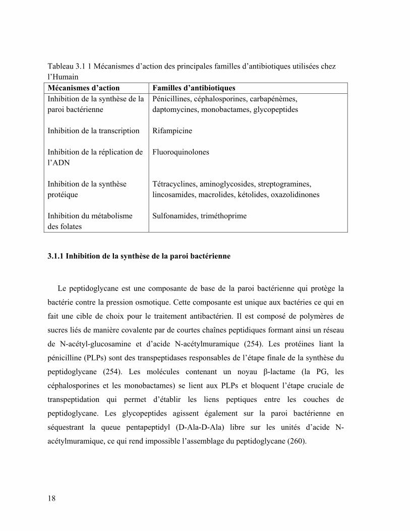
Tableau 3.1 1 Mécanismes d’action des principales familles d’antibiotiques utilisées chez l’Humain Mécanismes d’action Familles d’antibiotiques Inhibition de la synthèse de la paroi bactérienne
Pénicillines, céphalosporines, carbapénèmes, daptomycines, monobactames, glycopeptides
Inhibition de la transcription
Rifampicine
Inhibition de la réplication de l’ADN
Fluoroquinolones
Inhibition de la synthèse protéique
Tétracyclines, aminoglycosides, streptogramines, lincosamides, macrolides, kétolides, oxazolidinones
Inhibition du métabolisme des folates
Sulfonamides, triméthoprime
3.1.1 Inhibition de la synthèse de la paroi bactérienne
Le peptidoglycane est une composante de base de la paroi bactérienne qui protège la
bactérie contre la pression osmotique. Cette composante est unique aux bactéries ce qui en
fait une cible de choix pour le traitement antibactérien. Il est composé de polymères de
sucres liés de manière covalente par de courtes chaînes peptidiques formant ainsi un réseau
de N-acétyl-glucosamine et d’acide N-acétylmuramique (254). Les protéines liant la
pénicilline (PLPs) sont des transpeptidases responsables de l’étape finale de la synthèse du
peptidoglycane (254). Les molécules contenant un noyau β-lactame (la PG, les
céphalosporines et les monobactames) se lient aux PLPs et bloquent l’étape cruciale de
transpeptidation qui permet d’établir les liens peptiques entre les couches de
peptidoglycane. Les glycopeptides agissent également sur la paroi bactérienne en
séquestrant la queue pentapeptidyl (D-Ala-D-Ala) libre sur les unités d’acide N-
acétylmuramique, ce qui rend impossible l’assemblage du peptidoglycane (260).
18

3.1.2 Inhibition de la transcription
Un seul antibiotique de cette classe est utilisé cliniquement dans le traitement des
infections à Mycobacterium tuberculosis; il s’agit de la rifampicine (rifampine). Cette
drogue agit en tant qu’inhibiteur de l’ARN polymérase. Elle lie la sous-unité β de l’ARN
polymérase bactérien à un site allostérique, ce qui bloque l’élongation de la chaine d’ARN
(260).
3.1.3 Inhibition de la réplication de l’ADN
Les quinolones (fluoroquinolones) constituent une importante famille
d’antibiotiques qui agit en inhibant la réplication de l’ADN. Cette famille bloque l’action
des enzymes ADN gyrase et ADN topoisomérase IV (104) . Les fluoroquinolones feront
l’objet du chapitre IV de cette thèse.
3.1.4 Inhibition de la synthèse protéique
La composition du ribosome bactérien diffère de celle des cellules eucaryotes, ce
qui en fait une excellente cible pour les antibiotiques. En effet, le ribosome bactérien
(coefficient de sédimentation : 70 svedbergs (S)) est constitué d’une grande sous-unité 50S
(60S cellules eucaryotes) et d’une petite sous-unité 30S (40S cellules eucaryotes), chacune
composée d’ARNr et de protéines ribosomales. La plupart des antibiotiques qui inhibent la
synthèse protéique se lient à l’une ou l’autre des sous-unités du ribosome. Ainsi, les
aminoglycosides et les TCs fixent la petite sous-unité du ribosome (30S). Les TCs inhibent
l’élongation de la chaîne polypeptidique alors que les aminoglycosides agissent en
induisant des erreurs dans le décodage des codons effectués par le ribosome ce qui entraîne
l'accumulation d’erreurs dans les protéines synthétisées. L’accumulation de protéines
aberrantes est responsable de la létalité induite par les aminoglycosides. Les composés
phénicols (CM), les macrolides, les lincosamides et les streptogramines se lient à la grande
sous-unité 50S et bloquent la formation du lien peptidique (CM) ainsi que l’élongation de
19

la chaîne polypeptidique (macrolides, lincosamides et streptogramines). Les
oxazolidinones, quant à eux, empêchent la formation du complexe d’initiation sur le 70S
(260).
3.1.5 Inhibition du métabolisme des folates
Le métabolisme des folates permet la conversion du dUMP en dTMP, molécule
essentielle à la synthèse du dTTP utilisé lors de la synthèse d’ADN. Cette étape de
conversion nécessite le métabolite terminal de la voie de biosynthèse des folates, l’acide
tétrahydrofolique (26). Chez les bactéries, l’obtention de ce métabolite utilise une voie de
synthèse de novo ayant comme substrat primaire le GTP et le para-aminobenzoate. Les
sulfonamides empêchent la synthèse du dihydroptéorate, un intermédiaire de la voie de
synthèse de novo, en inhibant l’activité de la dihydroptéorate synthase, alors que la
thriméthoprime inhibe la synthèse du tétrahydrofolate en inhibant l’activité de la
dihydrofolate réductase (260).
3.1.6 Nouveaux mécanismes d’action des antibiotiques; productions de dérivés réactifs
à l’oxygène
La classification du mode d’action des antibiotiques repose principalement sur la
cible affectée (Voir section 3.1) et la conséquence phénotypique sur la croissance. Les
antibiotiques peuvent être désignés comme bactéricides s’ils tuent plus de 99,9% des
bactéries, alors que les antibiotiques bactériostatiques causent une inhibition de la
croissance. En 2007, un modèle fut proposé quant aux mécanismes expliquant le caractère
létal des antibiotiques bactéricides, celui-ci impliquant la production de dérivés réactifs à
l’oxygène (ROS) causant la mort cellulaire aussi bien chez les bactéries à Gram positif que
Gram négatif (141). Selon ce modèle, peu importe la cible de l’antibiotique bactéricide (β-
lactamines : PLPs, fluoroquinolones : ADN gyrase et aminoglycoside : ribosome) la mort
cellulaire résulterait de la production de radicaux libres impliquant l’activation du cycle de
Krebs, une déplétion transitoire en NADH, une déstabilisation des groupements fer-soufre
20

et la stimulation de la réaction de Fenton. Récemment, cette hypothèse a été
vigoureusement contestée et des éclaircissements seront nécessaires afin de mieux
comprendre le rôle des ROS dans le mode d’action des antibiotiques bactéricides (135,
154). Néanmoins, des études subséquentes semblent démontrer que la létalité des
antibiotiques bactéricides nécessite leurs mécanismes primaires drogue-spécifiques, en plus
de la production de ROS (68).
La production de ROS survient également chez S. pneumoniae suite à l’exposition à
certains antibiotiques bactéricides (PG, CIP et KAN). En effet, une diminution potentielle
de la quantité de fer intracellulaire subséquente à la sélection d’une mutation non-sens dans
un importeur de fer fut impliquée dans la diminution de ROS induite par ces drogues (74).
21

Figure 3.1 Modèle du mode d’action des antibiotiques bactéricides tel que renouvelé par (140).
3.2 Mécanismes de résistance aux antibiotiques
Chaque année aux États-Unis au moins deux millions de personnes sont infectées
par des bactéries résistantes aux antibiotiques et environ 23 000 personnes en décèdent
β-lactamine
PLP
Topoisomérase
Cycle de Krebs
Systèmes à deux composantes Cpx et Arc
Formation des liaisons disulfures
Réaction de Fenton
Mort cellulaire
Rétroaction métabolique
Hyperactivation de la chaîne de transport des
électrons Formation de superoxyde
Dommages aux groupements Fe-S
Dommages à l’ADN, aux lipides et aux protéines
Formation de radicaux
hydroxyles
Dommage
22
(43). Le fardeau associé à la résistance aux antibiotiques augmente depuis plusieurs années
et une accélération a pu être observée dans les dix dernières années (103). En effet, l'usage
généralisé, voire abusif de certains antibiotiques, y compris en traitement préventif, curatif,
en complément alimentaire ou encore comme pesticide a introduit une pression de sélection
qui a conduit au développement de populations de micro-organismes antibiorésistants et à
une baisse générale de l'efficacité thérapeutique. Également, l’absence de nouvelles
molécules thérapeutiques depuis plus de dix ans n’a fait qu’augmenter le fardeau causé par
la résistance, rendant dans certains cas le traitement des infections complexes, par exemple
dans le cas de souches multi-résistantes d’Acinetobacter baumannii, d’Enterobacteriaceae
résistants aux carbapénèmes, de Neisseria résistants résistants à la ceftriaxone et
d’Enterococcus résisants à la vancomycine (173, 237, 244).
Chez les bactéries, la résistance à un antibiotique peut être innée (« naturelle ») ou
acquise. En effet, la plupart des bactéries du genre Streptomyces produisant des
antibiotiques sont résistantes aux molécules qu’elles produisent. Cependant, l’acquisition
de gènes de résistance par transformation, conjugaison et transduction peut induire la
résistance à un antibiotique chez la bactérie qui a acquis cet élément d’ADN exogène. Les
mécanismes de résistance chez les bactéries peuvent être classés en cinq types qui sont
décrits dans les prochaines sous-sections.
3.2.5 Réduction de la perméabilité membranaire
Afin d’être efficace, un antibiotique doit pouvoir s’accumuler en quantité suffisante
pour avoir l’effet escompté. Ainsi, tout mécanisme visant à diminuer l’entrée de
l’antibiotique dans la bactérie causera la résistance envers cette molécule. Chez les
bactéries à Gram négatif, l’entrée des antibiotiques tels les β-lactamines (273), le CM et les
fluoroquinolones (181) nécessite la présence de porines dans la membrane externe. Un
changement dans le nombre de copies de porines, de la taille ou de la sélectivité de celles-ci
peut causer un changement de la perméabilité membranaire envers l’antibiotique (101,
182).
23

3.2.6 Efflux de l’antibiotique
La concentration intracellulaire de l’antibiotique peut également être diminuée par
l’efflux de celui-ci à l’extérieur de la cellule. L’augmentation de l’efflux actif des
antibiotiques est préoccupante, car certains transporteurs peuvent produire la résistance à
plusieurs antibiotiques, rendant les souches multi-résistantes (11, 106, 151, 204).
Également, l’efflux combiné avec d’autres mécanismes peut conduire à des niveaux de
résistance élevés chez des souches cliniques, compromettant l’antibiothérapie (151). Pour
fonctionner, les pompes d’efflux utilisent l’énergie fournie par dissipation d’un gradient de
protons (familles MFS, RND et SMR) ou d’ions sodium (famille MATE) ou encore par
l’hydrolyse d’ATP (famille ABC). Des exemples types reliés à chacune des cinq familles
de pompe d’efflux causant la résistance sont illustrés à la Figure 3.2.
Chez le pneumocoque, l’efflux est un mécanisme de résistance aux
fluoroquinolones et aux macrolides (151). La résistance aux fluoroquinolones et aux
macrolides, incluant les kétolides, peut être causée par des pompes de types MFS comme
PmrA (fluoroquinolones), ainsi que MefA et MefE (macrolides) (128, 138, 151, 267). La
résistance à la bacitracine (23) et aux fluoroquinolones (70, 86, 163, 220) peut également
être médiée par des pompes de type ABC chez S. pneumoniae.
24

Figure 3.2 Représentation schématique de différents types de pompes d’efflux d’antibiotiques chez les bactéries. Les pompes NorA (MFS) de Staphylococcus aureus, EmrE (SMR) d’E. coli, NorM (MATE) de Vibrio parahaemolyticus, AcrAB-TolC (RND) d’E. coli et LmrA (ABC) de Lactococcus lactis expulsent leurs substrats (Ab) par l’utilisation d’énergie provenant d’un gradient d’ions (proton (H+) ou sodium (Na+)) ou d’ATP. Adapté de (143).
3.2.1 Modification de la cible
Une troisième stratégie afin de contrer l’effet d’un antibiotique est la modification
de la cible de celui-ci. En effet, la présence de mutations peut modifier le site actif de la
cible de l’antibiotique et ainsi empêcher sa liaison. La résistance aux β-lactamines est le
résultat de mutations au niveau des PLPs (100). De plus, la résistance aux fluoroquinolones
est médiée par l’acquisition de mutations dans les gènes codant pour l’ADN gyrase et la
topoisomérase IV, qui sont les cibles des fluoroquinolones (71). Également, une
modification chimique de la cible peut causer la résistance en empêchant l’antibiotique de
s’y lier. Par exemple, la méthylation de l’ARNr 23S au site de liaison des macrolides par
une ARN méthyltransférase peut causer la résistance à cette famille (260).
3.2.2 Inactivation de l’antibiotique
Membrane cytoplasmique
Cytoplasme
Membrane externe
Ab
Ab
Ab
Ab Ab
Ab
Ab
Ab
Ab
Ab
25

Une autre stratégie consiste en la destruction de la drogue. Certaines souches
bactériennes peuvent acquérir des gènes codant pour des enzymes dégradant le noyau β-
lactame des pénicillines et des céphalosporines (200). Également, d’autres classes
d’antibiotiques comme les aminoglycosides peuvent être neutralisées par des adénylyl-
transférases, des phosphoryl-transférase ou des acétyl-transférases, ce qui réduit leur
affinité pour leurs cibles (227).
3.2.2 Amplification de la cible
En réponse à la présence d’un antibiotique, l’amplification génique constitue une
réponse adaptative observée chez les bactéries et les cellules eucaryotes. Chez S.
agalactiae, la résistance aux sulfonamides et au triméthoprime est médiée par
l’amplification génique d’une région de 13,5 kb contenant les gènes folCEPBK impliqués
dans le métabolisme des folates (34). Récemment, l’amplification génique d’une région de
9,2 kb contenant les gènes patA et patB a été répertoriée chez une souche clinique de S.
pneumoniae résistante aux fluoroquinolones (21).
3.3 Résistance aux antibiotiques chez S. pneumoniae
En 2005, l’OMS a évalué que près de 1,6 million de personnes décèdent chaque
année suite à une infection au pneumocoque, la majorité étant des enfants vivant dans des
pays sous-développés (0,7 à 1 million) (1). Bien que la vaccination ait diminué la
prévalence des infections causées par S. pneumoniae, la résistance à la PG, aux macrolides,
aux TCs et au CM serait attribuée à certains clones de pneumocoques (PG : 6A, 6B, 9V,
14,19A, 19F et 23F; Multi-résistance : 6B, 19A, 19F et 23F) (169, 202). Dans cette partie
du texte, la résistance aux β-lactamines, aux macrolides, à la VA et à la LZD sera abordée.
Les fluoroquinolones, ainsi que les TCs feront l’objet des chapitres IV et V,
respectivement.
26

3.3.1 Résistance aux β-lactamines
Aux États-Unis, plus de 25% des souches seraient insensibles à la PG, mais ce
pourcentage pourrait atteindre jusqu’à 60 et même 80% en Amérique latine et dans
quelques pays d’Asie (13). Chez les souches cliniques de pneumocoque, la résistance
résulte principalement de l’acquisition de versions mutées des PLPs suite à des transferts
intra- ou inter-espèces (214). Plusieurs mutations dans une PLP peuvent être nécessaires
afin de causer une diminution de l’affinité de la β-lactamine. Également, des mutations au
niveau de plus d’une PLP peuvent être nécessaires afin que la souche acquière une
résistance élevée à l’antibiotique (91, 212). L’acquisition de haut niveau de résistance à la
PG chez des souches cliniques serait due à des mutations au niveau des PLP1a, PLP2b et
PLP2x, alors que la résistance aux céphalosporines nécessiterait des mutations dans les
PLP1a et PLP2x (51). Plusieurs mécanismes n’impliquant pas les PLPs ont également été
décrits chez le pneumocoque (73, 99, 233).
3.3.2 Résistance aux macrolides et aux kétolides
Aux États-Unis, environ 31% des souches de S. pneumoniae seraient résistantes aux
macrolides (13). Les molécules les plus utilisées de cette famille en Amérique du Nord sont
l’EM, la clarythromycine, l’azithromycine et la télithromycine, une kétolide
structurellement similaire à l’EM et à la clarythromycine (64). La résistance aux macrolides
est principalement causée par l’altération de la cible et par efflux. L’expression du gène
ermB codant pour une méthylase ribosomale confère une haute résistance aux macrolides
(Concentration minimale inhibitrice (CMI) ≥ 64 µg/mL), aux lincosamides et à la
streptogramine B par méthylation de la position 2058 de l’ARNr 23S. Également, l’efflux
causé par MefE et MefA confère un bas niveau de résistance aux macrolides (CMI 1-32
µg/mL) uniquement. La présence de mutations au niveau des protéines L4 and L22, toutes
deux importantes dans l’assemblage précoce de la sous-unité 50S du ribosome, pourrait
contribuer à la résistance aux macrolides, ainsi qu’aux kétolides (64).
27

3.3.3 Résistance à la LZD
La résistance à la LZD chez les bactéries à Gram positif résulte le plus souvent de
l’acquisition de mutations dans l’ARNr 23S (208). Bien que la résistance clinique à la LZD
n’ait pas été rapportée pour le moment chez le pneumocoque, des souches ayant une
sensibilité diminuée (MIC 4 µg/mL) suite à une délétion dans le gène codant pour la
protéine ribosomale L4 ont été décrites (269). L’étude de souches sélectionnées en
laboratoire a démontré que la résistance à la LZD implique également des mutations dans
une méthyltransférase de l’ARNr 23S et des mutations causant la surexpression du
transporteur ABC PatA/PatB (77).
28

Chapitre IV : La famille des fluoroquinolones
Les fluoroquinolones sont des dérivés synthétiques de l’acide nalidixique ayant un
noyau 4-quinolone (Figure 4.1). Cette famille d’antibiotiques comporte aujourd’hui quatre
générations dont les diverses modifications ont permis d’augmenter leur spectre d’activité.
Les quinolones de troisième (exemples : LVX, sparfloxacine, témafloxacine, MOX et
gatifloxacine) et quatrième (exemples : clinafloxacine, trovafloxacine) générations sont
utilisées pour traiter les pneumonies acquises en communauté (125), dont S. pneumoniae
est un important agent étiologique.
Figure 4.1 : Structure des fluoroquinolones. Adapté de (66).
4.2 Mode d’action des fluoroquinolones
Les quinolones inhibent la synthèse de l’ADN en ciblant deux topoisomérases de
type II, l’ADN gyrase et la topoisomérase IV. Les deux enzymes permettent la
Acide nalidixique
Ciprofloxacine Norfloxacine
Moxifloxacine Gatifloxacine
29

concaténation d’ADN double brin en superenroulement positif ou négatif en créant une
cassure double brin au site de réarrangement. Malgré la similarité de structure entre les
deux enzymes, celles-ci ont des fonctions cellulaires différentes. L’ADN gyrase est un
tétramère de type A2B2 codé par les gènes gyrA et gyrB et est une enzyme exclusive aux
bactéries. Elle utilise l’ATP afin d’introduire des supertours négatifs dans l’ADN, élément
essentiel à la condensation du chromosome et à l’initiation de la transcription (150, 262).
La topoisomérase IV a deux fonctions dans la cellule. L’une d’elle consiste en la
déconcaténation des deux chromosomes filles, permettant ainsi leur ségrégation. La
seconde fonction consiste en la relaxation des supertours positifs. La topoisomérase IV est
également un tétramère de type A2B2 codé par les gènes parC et parE. Les sous-unités
ParC et ParE partagent environ 35 % d’identité avec les sous-unités GyrA et GyrB (71).
L’action inhibitrice des quinolones sur les topoisomérases de type II se produit via
la formation d’un complexe topoisomérase-ADN en présence de deux molécules
d’antibiotique tel que proposé par le modèle crystalographique obtenu pour la
topoisomérase IV en présence de MOX et d’ADN (Figure 4.2) (148).
Figure 4.2 : Représentation du site de clivage du complexe de la topoisomérase IV. La moxifloxacine est représentée en rouge. L’ADN est représenté en vert. Le site actif tyrosine (Tyr118) est représenté en orange. Les résidus Ser-70 et Asp83 sont représentés en jaune. Adapté de (148).
30

4.3 Mécanismes de résistance
La prévalence mondiale de la résistance aux fluoroquinolone est inférieure à 1 %,
toutefois celle-ci est beaucoup plus élevée dans certaines régions telles que Hong Kong
(14%), le Sri Lanka (9.5%), les Philippines (9.1%) et la Corée (6.5%) (177, 235). Au
Canada, l’utilisation de la CIP a causé une augmentation de la prévalence des souches
résistantes de 1% en 1997 à 4,2% en 2005 (5). Cependant, la résistance à la CIP semble
s’être stabilisée dû, entre autres, au remplacement de la CIP par des fluoroquinolones
respiratoires de nouvelles générations (193).
4.3.1 Mutations dans l’ADN gyrase et la topoisomérase IV
La résistance aux fluoroquinolones est, entre autres, provoquée par l’apparition
successive de mutations au niveau des cibles des fluoroquinolones, l’ADN gyrase et l’ADN
topoisomérase IV. Chez S. pneumoniae, plusieurs fluoroquinolones (CIP, LVX,
norfloxacine, et perfloxacine) sélectionnent préférentiellement des changements au niveau
du gène parC, alors que d’autres molécules comme la sparfloxacine et la MOX acquièrent
plutôt des changements au niveau de gyrA (37, 190). Des expériences in vitro chez S.
pneumoniae exposé à la CIP ont démontré que les mutations parC apparaissent avant les
mutations gyrA, ce qui supporte l’hypothèse que ParC est la cible primaire de la CIP chez
cette bactérie (118, 179, 189). Cette situation est différente chez les bactéries à Gram
négatif pour lesquelles gyrA est la cible primaire des fluoroquinolones (272). Chez S.
pneumoniae, la présence de mutations uniquement au niveau de parC est souvent associée
à un faible niveau de résistance aux fluoroquinolones, alors que l’acquisition de hauts
niveaux de résistance nécessite des mutations dans parC et dans gyrA (118, 240). Les
mutations sont le plus souvent retrouvées dans des régions conservées nommées QRDRs
(« Quinolones-resistance-determining-regions »), représentées par les acides aminés entre
la position 67 et 106 de GyrA chez E. coli (224). Des mutations dans les régions QRDRs de
gyrB et parE peuvent aussi diminuer la sensibilité aux fluoroquinolones, mais leur
prévalence est faible comparativement aux mutations dans parC et gyrA (126). Ces
31

mutations apparaissent généralement de façon spontanée, telles que décrites pour la LVX et
la CIP chez le pneumocoque (82). Néanmoins, la recombinaison d’allèles mutés codants
pour les topoisomérases a déjà été décrite comme mécanisme d’acquisition de mutations
(255). L’analyse génétique de souches de pneumocoques résistantes aux fluoroquinolones a
démontré que la présence de mutations dans les régions QRDR de parC, à la position 79 et
83, et de gyrA, à la position 81 et 85, sont responsables majoritairement de la résistance aux
fluoroquinolones (20, 28, 40). Cependant, bien que les mutations soient généralement
présentes au niveau des régions QRDRs, l’apparition des mutations semble hétérogène
puisque des mutations à l’extérieur de cette région peuvent réduire la sensibilité aux
fluoroquinolones chez S. pneumoniae (127, 263).
4.3.2 Efflux
Plusieurs bactéries à Gram négatif sont protégées de l’action des fluoroquinolones
par des pompes de type MFS et RND exprimées de façon constitutive. Chez Pseudomonas
aeruginosa et A. baumannii, les pompes de type RND MexAB-OprM et AdeABC sont
responsables de la résistance multiple aux drogues (205, 266). Toutefois, chez plusieurs
bactéries, la surexpression de pompes de type RND, MFS et MATE est nécessaire au
phénotype de résistance aux fluoroquinolones (Tableau 4.1).
Chez le pneumocoque, la résistance aux fluoroquinolones peut être médiée par
l’expression de deux pompes d’efflux, soit la pompe de type MFS PmrA et le transporteur
ABC PatA/PatB. La pompe PmrA a été le premier mécanisme d’efflux décrit chez le
pneumocoque et a été identifié en raison de son degré d’identité en acides aminés à la
pompe NorA de S. aureus et Bmr de B. subtilis (24%) (90). Sa surexpression peut causer
une augmentation de la CMI à la norfloxacine, la CIP et au colorant bromure d’éthidium
(EtBr). En général, la surexpression de PmrA est retrouvée chez des souches résistantes
contenant des mutations dans les régions QRDRs et son rôle dans la résistance aux
fluoroquinolones pourrait être limité (18, 201)
32

Le transporteur ABC PatA/PatB fut révélé lors de la caractérisation de treize
systèmes de transport de la famille des ABC chez S. pneumoniae TIGR4 (220). Les sous-
unités PatA (SP2075;TIGR4 Spr1887; R6) et PatB (SP2073;TIGR4 Spr1885;R6) possèdent
chacune un domaine transmembranaire et un domaine liant l’ATP et s’assemblent en un
hétérodimère afin de constituer le transporteur ABC PatA/PatB (31). Ce transporteur est
homologue au transporteur LmrCD de L. lactis (248) et est capable d’induire la résistance à
plusieurs agents, dont l’Etbr et les fluoroquinolones. La surexpression du transporteur
PatA/PatB est associée à la résistance clinique du pneumocoque aux fluoroquinolones (86).
En effet, l’expression du transporteur semble être modulée par la présence de
concentrations sous-inhibitrices de CIP et de norfloxacine, deux fluoroquinolones
considérées comme de bons substrats de ce transporteur. L’expression de patA est
également modulée par la présence de la LVX, de MOX et de gemifloxacine, trois
fluoroquinolones hydrophobes qui ne sont pas de bons substrats du transporteur, et
également par la mitomycine C laissant présager que la surexpression du transporteur
PatA/PatB serait le résultat d’une réponse générale au stress chez S. pneumoniae (18, 70).
Tableau 4.1 : Classification des pompes d’efflux causant la résistance aux fluoroquinolones chez les bactéries. Adapté de (71)
Microorganism Popmes d’efflux RND MFS MATE ABC SMR Escherichia coli AcrAB, AcrEF MdfA YdhE Salmonella enterica AcrAB, AcrEF Klebsiella pneumoniae
AcrAB KmrA, KdeA
Pseudomonas aeruginosa
MexAB-OprM, MexCD-OprJ, MexEF-OprN, MexXY, MexVW, MexHI-OpmD, MexPQ-OpmD
PmpM
Stenotrophomonas maltophilia
SmeDEF, SmeABC
Acinetobacter baumannii
AdeABC, AdeIJK AbeM
Campylobacter jejuni CmeABC, CmeDEF Staphylococcus aureus
NorA, NorB, NorC, SdrM
MepA
Streptococcus pneumoniae
PmrA PatA, PatB
33

4.3.3 Résistance aux quinolones médiée par des plasmides
La résistance aux quinolones médiée par les plasmides fut originellement décrite
chez une souche clinique de Klebsiella pneumoniae de l’Alabama (165). Les déterminants
les plus caractérisés de ce groupe sont sans contredit les gènes qnr. Ces gènes codent pour
des protéines pentapeptidiques répétées qui diminuent la sensibilité aux quinolones en
protégeant le complexe formé de l’ADN gyrase ou de la topoisomérase IV avec l’ADN de
l’effet inhibiteur des quinolones. Deux autres mécanismes codés par des plasmides ont
également été décrits, soit une version mutée du gène aac(6’)-lb-cr codant pour une
aminoglycoside transférase permettant l’inactivation de la CIP par l’acétylation de cette
dernière ainsi que l’efflux médié par les gènes oqxAB et qepA, découvert tous les deux chez
des souches d’E. coli (241).
34

Chapitre V : La famille des TCs et des glycylcyclines
À la fin des années 1940, la découverte de la chlortétracycline et l’oxytétracycline
(oxyTC), synthétisées naturellement par Streptomyces aureofaciens et Steptomyces
rimosus, a ouvert la voie à une nouvelle famille d’antibiotiques, les TCs. Aujourd’hui,
d’autres TCs naturellement synthétisées par des bactéries du genre Streptomyces (TC,
démethyltétracycline) ont été découvertes, mais des produits de synthèse dérivés des TCs
naturelles (méthacycline, doxycycline (DOX) et minocycline(MI)) sont également
disponibles. Plus récemment, des dérivés synthétiques de la MI, les glycylcyclines, ont été
introduits sur le marché.
Les TCs sont des agents antimicrobiens à large spectre ayant une bonne activité
contre les bactéries à Gram positif et à Gram négatif, les bactéries atypiques (chlamydia,
mycoplasmes et rickettsies) ainsi que certains parasites protozoaires. Cette famille
d’antibiotiques fut largement utilisée pour le traitement des infections bactériennes et
également en prophylaxie contre le Plasmodium falciparum résistant à la melfoquine (57).
Toutefois, bien que les TCs aient un rôle important en médecine humaine et vétérinaire,
l’utilisation abusive de celles-ci en clinique et en agriculture a sélectionné un niveau de
résistance élevé chez plusieurs espèces bactériennes (219).
35

Figure 5.1 : Structure des TCs et glycylcyclines Adapté de (123).
5.2 Mode d’action des TCs et des glycylcyclines
Les TCs et les glycylcyclines sont des antibiotiques généralement bactériostatiques
(à l’exception de la tigécycline (TGC)) chez S. pneumoniae (199) qui agissent en bloquant
l’entrée de l’aminoacyl-ARNt au site A de l’ARNr 70S lors de la traduction (62). Cette
inhibition s’effectue par l’interaction de l’antibiotique à des sites précis sur l’ARNr 30S.
Aucune protéine ribosomale ne semble impliquée dans la liaison des TCs et glycylcyclines
au ribosome. Selon le modèle d’interaction de Brodersen et al. (35), la TC interagit
principalement avec deux sites sur la sous-unité 30S. Le premier, appelé site primaire, est
localisé près du site accepteur de l’aminoacyl-ARNt (le site A). La TC interagit
principalement avec les bases 1054 et 1196 de l’hélice (H) 34 ainsi qu’avec la guanine 966
de H31 (Figure 5.2). Un ion magnésium divalent, essentiel à la liaison de la TC avec le
ribosome, interagit avec les guanines 1197 et 1198. Le second site (site secondaire) est
localisé près de H44 et entre le domaine central de H27 et le haut du domaine H11, où la
TC interagit principalement avec les nucléotides U244, C893 et A892 (Figure 5.2).
Tétracycline Doxycycline
Minocycline DMG-DMDOT
DMG-MINO Tigécycline
36

Figure 5.2 : Sites de liaison (A) primaire et (B) secondaire de la TC avec l’ARNr 30S. Adapté de (35).
La TGC partage le même site d’interaction primaire que la TC, toutefois, la liaison
de la partie 9-tert-butylaminoacétamido de la TGC avec la base C1054 en plus de la liaison
d’un deuxième ion magnésium avec C966 seraient responsables de l’augmentation de
l’affinité de la TGC pour le ribosome (Figure 5.3). En effet, la TGC a jusqu’à vingt fois
plus d’affinité pour le ribosome comparativement à la TC (186). Aucun site d’interaction
secondaire avec le ribosome n’a été observé dans le cas de la TGC (119).
B
A
37

Figure 5.3. Schéma de la liaison de la TGC (A) avec l’ARNr 16s (B) Comparaison de la liaison de la TC et la TGC à l’ARNr 16s. Adapté de (119).
5.3 Mécanismes de résistance
5.3.1 La famille des TCs
Chez les bactéries, la résistance aux TCs est principalement causée par l’acquisition
de gène tet (« tetracycline resistance »). Toutefois, chez certaines espèces, l’acquisition de
mutations chromosomiques peut également diminuer la sensibilité aux TCs.
5.3.1.1 Gènes tet et otr
Les protéines Tet et Otr sont des déterminants de la résistance à la TC et à l’oxyTC
qui sont principalement acquis par conjugaison de plasmides et de transposons conjugatifs.
À ce jour, plus de quarante-cinq gènes tet et otr ont été répertoriés (Tableau 5.1). La
classification des gènes tet et otr dépend principalement de l’homologie de séquences entre
BA
38
les gènes faisant partie d’une même classe : un gène doit avoir au moins 80% d’homologie
pour être répertorié dans une classe connue. Trois mécanismes ont été répertoriés à ce jour,
soit l’efflux, la protection ribosomale et l’inactivation enzymatique de la TC. Toutefois, les
deux premiers mécanismes sembleraient avoir davantage d’implications cliniques. En effet,
seulement trois gènes tet causeraient l’inactivation de la TC. Le gène tet(X), isolé de
Bacteroides, ne semble avoir d’effet que chez ce dernier (238). Également, les gènes tet(37)
codant pour une oxydoréductase dépendante du NADPH et tet(34) codant potentiellement
pour une xanthine-guanine phosphoribosyl transférase n’ont pour le moment pas été
observés chez des bactéries pathogènes. Dans le cas du déterminant tet(U), isolé chez
Enterococcus et Staphylococcus, la fonction reste à être confirmée.
Vingt-neuf gènes tet et otr codent pour des protéines d’efflux de la famille MFS à
douze ou quatorze domaines transmembranaires conduisant ainsi à une diminution de la
concentration intracellulaire de TC. La plupart de ces protéines d’efflux confèrent la
résistance à la TC uniquement (exception de tet(B)). À l’inverse, les mécanismes de
protection du ribosome peuvent causer la résistance à la TC, DOX et MI. Douze gènes tet et
otr codent pour des protéines de protection du ribosome. Ces protéines Tet ont une
homologie avec le facteur d’élongation de la traduction EF-Tu, particulièrement au niveau
de sa région amino-terminale, qui permet le rapatriement de l’aminoacyl-ARNt au site A.
La présence de protéines de protection du ribosome (comme TetM) cause la résistance en
déplaçant la TC de son site de liaison ce qui donne accès au facteur EF-Tu lié à
l’aminoacyl-ARNt au site A du ribosome (65).
La distribution des gènes tet varie grandement selon les genres bactériens. Chez les
Enterobacteriaceae, les gènes tet(A) et tet(E) seraient les principaux responsables de la
résistance aux TCs. Chez S. aureus, les gènes tet(K) et tet(M) sont généralement
rencontrés, alors que chez S. pneumoniae, les gènes tet(M) et tet(O) sont pour le moment
les seuls gènes tet à être impliqués dans la résistance aux TCs (79).
39

Tableau 5.1 Classification des gènes tet et otr. Adapté de (218) Efflux (29) Protection du ribosome (12) Inactivation
enzymatique (3)
Inconnu
tet(A), tet(B), tet(C), tet(D), tet(E) tet(G), tet(H), tet(J), tet(V), tet(Y) tet(Z), tet(30), tet(31), tet(33) tet(35) tet(39), tet(41) tet(K), tet(L), tet(38), tet(45) tetA(P), tet(40) otr(B), otr(C) tcr tet(42) tet(43) tetAB(46)
tet(M), tet(O), tet(S), tet(W), tet(32), tet(Q), tet(T), tet(36) otr(A), tetB(P), tet tet(44)
tet(X) tet(37) tet(34)
tet(U)
5.3.1.2 Autres mécanismes
Des mutations chromosomiques au niveau de gènes codant pour des constituants du
complexe ribosomal peuvent également causer la résistance aux TCs chez certaines
espèces. Chez Helicobacter pylori, la mutation du triplet AGA926-928→TTC dans les deux
copies des gènes codant pour l’ARNr 16S cause un haut niveau de résistance à la TC, alors
qu’un changement d’une ou deux bases induit un niveau de résistance plus faible (58, 270).
Chez N. gonorrhoeae, des mutations ponctuelles au niveau du gène rpsJ, codant pour la
protéine ribosomale S10, du gène mtrR codant pour un régulateur transcriptionnel et du
gène penB, codant pour une porine ont été impliquées dans la résistance à la TC (115). De
plus, la surexpression de pompes d’efflux de type ABC peut contribuer à la résistance à la
TC chez certaines bactéries à Gram négatif (9, 116, 167).
5.3.2 La TGC
Bien que la résistance à la TGC soit rare chez le pneumocoque, elle fut
décrite chez certaines espèces de bactéries à Gram négatif (14, 19, 32, 111, 131, 184) pour
40

lesquelles la résistance résulte principalement de l’expression de pompes d’efflux de type
RND. Chez Pseudomonas spp. et Proteus mirabilis, deux espèces qui ont une résistance
innée à la TGC, l’expression des pompes MexXY-OmpR et AcrAB est responsable de la
résistance (61, 258). Également, chez A. baumannii et Serratia marcescens, la
surexpression des pompes RND AdeABC et SdeXY-HasF diminue la sensibilité à la TGC
(50, 112). Il a été démontré que la surexpression des facteurs de transcription MarA chez E.
coli, de RamA chez K. pneumoniae et l’inactivation du répresseur RamR chez Salmonella
enterica et chez K. pneumoniae peut altérer l’expression de la pompe AcrAB en faveur de
la résistance à la TGC (132, 222, 257). Également, l’activation du système à deux
composantes BaeSR, régulant l’expression de AdeABC chez A. baumannii, peut causer la
résistance chez des isolats cliniques et des souches sélectionnées in vitro (152).
Mis à part l’efflux, des mutations dans les gènes rpsJ codant pour la protéine
ribosomale S10, trm codant pour une méthyltransférase dépendante de s-
adenosylméthionine (SAM) et plusieurs gènes liés à la biosynthèse des lipopolysaccharides
ont été associées à la résistance à la TGC chez K. pneumoniae, A. baumannii et E. coli (44,
153, 257). La TGC n’est pas affectée par la présence de gènes tet sauf dans le cas de tet(A)
qui semble augmenter la résistance à la TGC chez S. enterica (8). La dégradation de la
TGC par le produit du gène tet(X) a également été proposée comme mécanisme de
résistance à la TGC (175). Chez les bactéries à Gram positif, la résistance n’a été rapportée
que chez le genre Enterococcus et chez S. aureus (80). L’analyse génomique de souches S.
aureus résistantes à la TGC sélectionnées in vitro a démontré que la diminution de la
sensibilité est due à la pompe de type MATE MepA (168).
41

Chapitre VI : Étude de la résistance aux antibiotiques par
approche « omique »
6.1 Séquençage de génome entier
Le premier génome bactérien publié a été celui de H. influenzae en 1995 par la
méthode d’approche globale ou « shotgun » (78). Chez le pneumocoque, cette technique a
également été utilisée pour le séquençage des premiers génomes de S. pneumoniae (R6 et
TIGR4), au début des années 2000. Néanmoins, déjà au début des années 1990 des
séquences partielles du génome du pneumocoque (localisés sur des fragments SmaI et
ApaI) ont été utilisées pour construire la carte du génome de cette bactérie (88).
Étudier la résistance aux antibiotiques par une approche génomique nécessite des
techniques permettant de résoudre la séquence du génome de l’organisme que l’on étudie.
En effet, le génome bactérien est exposé à plusieurs événements génétiques, incluant les
mutations, les duplications, les inversions, les transpositions, les délétions, les
recombinaisons et les insertions qui peuvent conférer une nouvelle capacité métabolique à
l’organisme qui a subi ces modifications. Pour repérer ces modifications impliquées dans la
résistance, l’analyse comparative de souches résistantes et sensibles aux antibiotiques s’est
avérée être une méthode de choix pour étudier les mécanismes impliqués dans la résistance
et pour élucider le mode d’action des antibiotiques. L’étude comparative des génomes par
hybridation sur puce a été initialement utilisée pour déterminer les mutations contenues
dans des souches résistantes aux antibiotiques (10, 77). De nos jours, la caractérisation
génomique repose principalement sur des techniques de nouvelles générations (454 Life
Sciences, Illumina, ABI-SOLiD) qui ont révolutionné le séquençage de génome entier (55,
73-75, 77, 108, 114).
43
6.1.1 Séquençage à haut débit (NGS)
Les techniques de séquençage de nouvelles générations, aussi appelées séquençage
à haut débit, ont révolutionné l’étude de la résistance aux antibiotiques en permettant de
séquencer des génomes entiers plus rapidement et à un coût moindre. Ces techniques de
séquençage en parallèle regroupent plusieurs technologies dont Ion Torrent de Life
Technologies, le pyroséquençage 454 de Roche, la plateforme Illumina, le système SOLiD
de Applied BioSystems et le système de séquençage PacBio de Pacific Biosystems. Le
pyroséquençage 454 et le séquençage Illumina qui ont été utilisés dans cette thèse seront
abordés dans cette section.
6.1.1.1 Le séquençage 454 (Roche)
La technologie GS-FLX de Roche permet d’obtenir une couverture détaillée des
génomes séquencés en plus d’une observation complète des variations génétiques en
utilisant des lectures (« reads ») longues (jusqu’à 1Kb pour le GS-FLX +) et de haute
qualité (221). Ceci permet de faire l’assemblage de novo de nouveaux génomes et de re-
séquencer les génomes connus. Cette technologie de séquençage en parallèle permet de
générer plus de vingt-cinq millions de bases à une exactitude d’au moins 99% (phred 20) en
4 heures (161). Le pyroséquençage 454 a déjà été utilisé pour séquencer des souches de S.
pneumoniae résistantes à la LZD (77), au cefotaxime (73) et à la PG (74).
Le séquençage 454 comporte deux étapes principales, soit la préparation des
échantillons par émulsion et le pyroséquençage (Figure 6.1) (229). En résumé, le génome
entier est fragmenté mécaniquement et des adaptateurs (A et B) sont ligués aux extrémités
franches des fragments. L’émulsion des billes de streptavidine, contenant un seul fragment
d’ADN biotinylé, permet l’individualisation de chaque bille pour la polymérisation en
chaîne du fragment d’ADN qu’elle contient. Chaque microgoutelette d’émulsion devient un
microréacteur dans lequel l’unique fragment d’ADN fixé sur la bille va pouvoir être
44

amplifié. Après l’amplification, les billes sont déposées sur une plaque PTP (Pico Titer
Plate) contenant 1 600 000 puits dont le diamètre ne permet la récupération que d’une seule
bille par puits. C’est au sein de chacun de ces puits que va se réaliser la réaction de
pyroséquençage. Les nucléotides (A, T, C, G) seront ajoutés de manière séquentielle, et ce,
plusieurs fois de suite. Si le nucléotide ajouté correspond à celui permettant l’élongation du
brin complémentaire, il y a libération de pyrophosphate inorganique. Celui-ci est alors
utilisé par l’ATP sulphurylase pour produire de l’ATP. La luciférase utilise cet ATP pour
produire de l’oxyluciférine dont la lumière sera captée par un capteur CCD (Charge-
Coupled Device). La détermination de la séquence repose donc sur la mise en évidence
d’une émission de lumière résultant de l’incorporation d’un ou plusieurs nucléotides lors de
la polymérisation de l’ADN.
Figure 6.1 : Principes de la technologie de séquençage 454. Adapté de (229).
6.1.1.2 Illumina (Solexa)
Préparation des fragments d’ADN
45
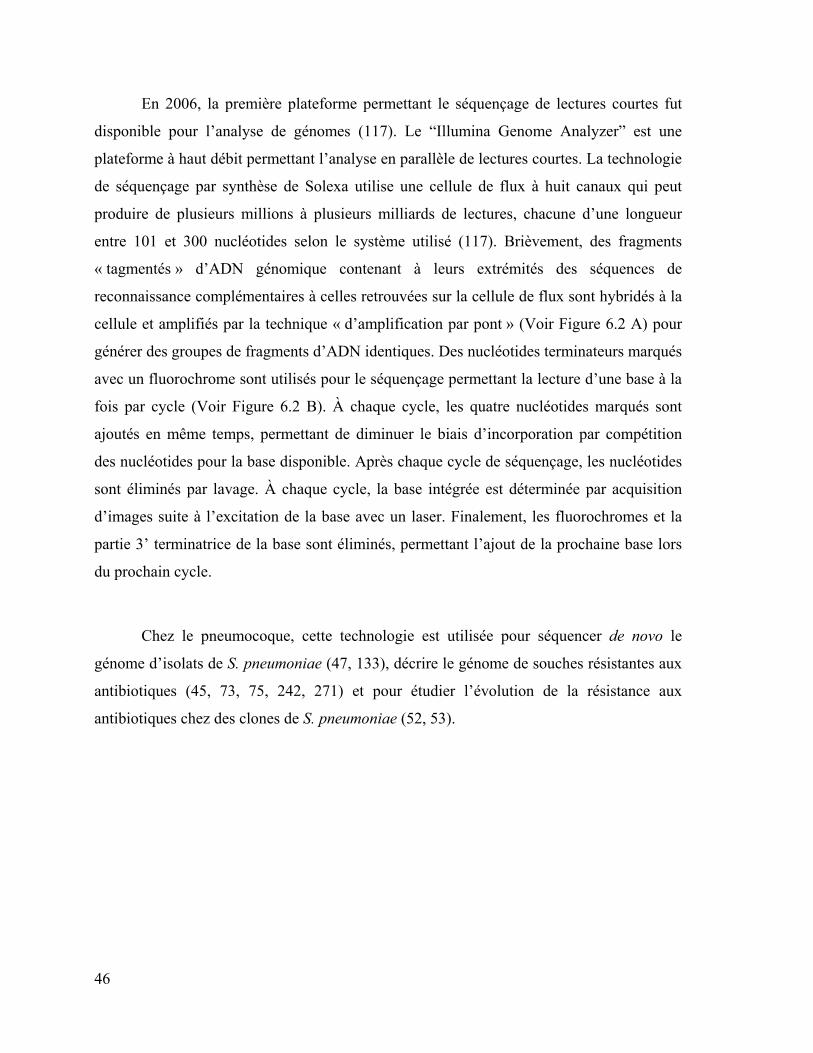
En 2006, la première plateforme permettant le séquençage de lectures courtes fut
disponible pour l’analyse de génomes (117). Le “Illumina Genome Analyzer” est une
plateforme à haut débit permettant l’analyse en parallèle de lectures courtes. La technologie
de séquençage par synthèse de Solexa utilise une cellule de flux à huit canaux qui peut
produire de plusieurs millions à plusieurs milliards de lectures, chacune d’une longueur
entre 101 et 300 nucléotides selon le système utilisé (117). Brièvement, des fragments
« tagmentés » d’ADN génomique contenant à leurs extrémités des séquences de
reconnaissance complémentaires à celles retrouvées sur la cellule de flux sont hybridés à la
cellule et amplifiés par la technique « d’amplification par pont » (Voir Figure 6.2 A) pour
générer des groupes de fragments d’ADN identiques. Des nucléotides terminateurs marqués
avec un fluorochrome sont utilisés pour le séquençage permettant la lecture d’une base à la
fois par cycle (Voir Figure 6.2 B). À chaque cycle, les quatre nucléotides marqués sont
ajoutés en même temps, permettant de diminuer le biais d’incorporation par compétition
des nucléotides pour la base disponible. Après chaque cycle de séquençage, les nucléotides
sont éliminés par lavage. À chaque cycle, la base intégrée est déterminée par acquisition
d’images suite à l’excitation de la base avec un laser. Finalement, les fluorochromes et la
partie 3’ terminatrice de la base sont éliminés, permettant l’ajout de la prochaine base lors
du prochain cycle.
Chez le pneumocoque, cette technologie est utilisée pour séquencer de novo le
génome d’isolats de S. pneumoniae (47, 133), décrire le génome de souches résistantes aux
antibiotiques (45, 73, 75, 242, 271) et pour étudier l’évolution de la résistance aux
antibiotiques chez des clones de S. pneumoniae (52, 53).
46

Figure 6.2 : La technologie Illumina. (A) Génération des groupes de lectures sur la cellule de flux par amplification par pont (« bridge-amplification »). (B) Séquençage par synthèse par ajout de nucléotide terminateur. Adapté de (117).
6.2 Étude du transcriptome
Le transcriptome est défini comme l’ensemble des transcrits présents dans une
cellule à un moment donné et dans une condition donnée. Deux technologies nous
permettent aujourd’hui de déterminer l’état du transcriptome : les biopuces et le séquençage
de l’ARN.
6.2.1 Biopuces
Les biopuces, ou puces à ADN, sont des collections de points microscopiques
d’ADN monocaténaires (sondes) sur une surface solide qui permettent de mesurer
A
B
47

simultanément l’expression d’un grand nombre de gènes. Le principe des biopuces repose
sur la propriété que possède l’ADN dénaturé de reformer spontanément sa double hélice
lorsqu’il est porté face à un brin complémentaire. Plusieurs dizaines de milliers de
fragments d'ADN peuvent être fixés sur une puce, et chacun peut correspondre au brin
complémentaire d’un ARNm d’un organisme interrogé.
Brièvement, les ARNm provenant des échantillons à comparer (ex. : souche
résistante et souche sensible) sont extraits et des fluorochromes différents sont fixés aux
bases de chacune des conditions (fluorochrome : Cyanine 3, Cyanine 5 ou Alexa) (Figure
6.3). Les ADNc marqués sont ensuite hybridés au brin monocaténaire d'ADN présent sur la
puce. Une image est ensuite générée par excitation des fluorochromes par un laser et le
niveau d’expression est directement proportionnel à la quantité d’ARNm hybridé à la
séquence sur la puce (245). Dans ce type d’étude, la normalisation des résultats est
essentielle. En effet, les résultats peuvent différer d’une puce à l’autre, entre autres, en
raison du bruit de fond de la puce, et également selon l’endroit où la sonde a été déposée
sur la puce. De plus, l’intensité des points des échantillons peut différer selon le
fluorochrome utilisé (surtout dans des études à deux fluorochrome où le Cy3 et Cy5 sont
utilisés, puisque la fluorescence du Cy3 est plus élevée que le Cy5); il est donc important
de conduire l’expérience en inversant le fluorochrome associé à chaque échantillon (209,
245).
Chez le pneumocoque, cette technique a été utilisée afin de détecter la réponse au
stress causée par la VA chez TIGR4 (94), les gènes responsables de la résistance aux
macrolides, streptogramines et lincosamides chez vingt différentes espèces dont S.
pneumoniae (42) et pour caractériser la réponse causée par l’efflux chez S. pneumoniae en
réponse aux fluoroquinolones (163). Également, cette technique a été utilisée pour l’étude
de l’expression des facteurs de virulence en fonction du stade de croissance (139).
48


aussi la caractérisation et la comparaison de l’expression de transcriptome avec ou sans
génome de référence. Chez le pneumocoque, l’ARN-seq a été utilisé afin de découvrir les
petits ARN non codants impliqués dans la virulence (160) et pour décrire les modifications
du transcriptome survenant lors de l’adhésion du pneumocoque aux cellules D562 (cellules
épithéliales du nasopharynx d’origine humaine) (137).
6.3 Étude de la résistance chez le pneumocoque par des techniques de
protéomique
Chez le pneumocoque, comme chez d’autres organismes, il a été démontré que la
corrélation entre les niveaux d’expression des protéines et de l’ARNm est imparfait (236).
En effet, les analyses protéomiques et transcriptomiques de souches de pneumocoque
résistantes à la PG a démontré que la quantité d’une protéine ne corrèle pas nécessairement
avec l’abondance de son transcrit dans la cellule (236). Les études protéomiques
représentent donc une approche complémentaire à la caractérisation du profil d’expression
génique associé à la résistance. Chez S. pneumoniae, l’analyse du niveau d’expression des
protéines par spectrométrie de masse demeure l’approche protéomique la plus utilisée dans
l’étude des mécanismes de résistance aux antibiotiques. Cette technique a été utilisée avec
succès pour décrire le protéome et l’intéractome du phage Cp-1 (102), du phage Dp-1
(225), le protéome membranaire de la membrane cytoplasmique de S. pneumoniae R6 (46)
et afin de caractériser le protéome de souches résistantes à la LZD (76), aux macrolides
(41) et à la PG(236).
6.4 Étude du métabolome chez S. pneumoniae
La métabolomique permet l’analyse intégrative et fonctionnelle du phénotype
métabolique d’un système biologique. Au même titre que les autres données obtenues par
les techniques « omiques », le métabolome est une partie essentielle de tout système
biologique. L’analyse globale du métabolome utilise principalement des techniques de
bioanalyses de chromatographie en phase liquide ou gazeuse (LC-MS ou GC-MS) couplées
50

avec des approches de spectrométrie de masse et de résonance magnétique nucléaire, ce qui
permet d’obtenir une idée globale de l'ensemble des métabolites (sucres, acides aminés,
acides gras, etc.) présents dans une cellule, un organe ou un organisme à un moment précis.
Chez le pneumocoque, l’utilisation de la métabolomique est pour le moment
présentie comme outil diagnostic pour la détection du pneumocoque dans les liquides
biologiques chez les hôtes infectés (110, 145, 159, 231, 232) et également pour la
découverte de nouvelles cibles vaccinales (226). Cette méthode relativement récente n’a
pas été utilisée, pour le moment, pour caractériser la résistance aux antibiotiques chez S.
pneumoniae. Cependant, elle a été utilisée avec succès chez S. aureus, afin de décrire le
profil métabolique de souches sensibles et résistantes à la méthicilline (12) et chez des
souches de M. tuberculosis résistantes à la rifampicine (30), ce qui suggère que cette
approche pourrait être utilisée afin de caractériser les composantes du métabolome
associées à la résistance aux antibiotiques chez S. pneumoniae.
51


Chapitre VII: Problématique, hypothèses et objectifs
7.1 Problématique
La bactérie S. pneumoniae est l’un des plus importants agents étiologiques de
pneumonies acquises en communauté, de méningites, de bactériémies et d’otites et est
responsable d’un haut taux de mortalité et de morbidité (264). Chez le pneumocoque,
l’émergence de souches cliniques résistantes et multi-résistantes est devenue
problématique. Ces souches peuvent présenter une résistance à la PG de même qu’au
céfotaxime, aux fluoroquinolones, à la TC, au thriméthoprime/sulfamethaxone, aux
macrolides, au CM et à la ceftriaxone (95, 136, 216) . L’étude des mécanismes de
résistance devient alors un moyen de détecter les isolats résistants et de contrecarrer
l’émergence de la résistance aux antibiotiques par le développement de nouvelles
molécules échappant aux mécanismes connus.
7.2 Hypothèses
L’apparition de mutations peut donner un avantage sélectif à une bactérie, la rendant
capable de résister plus aisément à un stress (195). Cette adaptation peut être critique lors
d’un traitement antimicrobien, car il peut amener à l’inefficacité d’un traitement dirigé
contre une bactérie pathogène. À ce jour, les mécanismes de résistance à la CIP rapportés
chez le pneumocoque se limitent à l’acquisition de mutations dans les topoisomérases de
type II, ainsi que l’efflux (86, 124, 191). Dans le cas de la TC, l’acquisition des gènes tetM
et tetO sont les principaux mécanismes responsables de la résistance clinique (79).
Cependant, d’autres mécanismes intervenant dans la résistance aux fluoroquinolones et à la
TC ont été décrits chez d’autres espèces, laissant présager que des mécanismes de
résistance additionnels peuvent également intervenir dans le cas du pneumocoque (58, 115,
116, 167, 170, 205, 223, 251, 257, 270). La résistance à la TGC n’a pas encore été observée
53

chez le pneumocoque en raison de son usage restreint. Cependant, la résistance à cette
molécule a déjà été observée chez différentes bactéries à Gram négatif et à Gram positif
(19, 85, 131). Déterminer les mécanismes de résistance à la TGC chez le pneumocoque
avant son apparition en clinique par la caractérisation de souches résistantes générées en
laboratoire nous permettrait de connaître la dynamique de résistance à cet antibiotique chez
le pneumocoque et d’aider à générer des outils pour faciliter la détection de la résistance à
la TGC chez S. pneumoniae.
7.3 Objectifs
L’objectif général des projets présentés dans cette thèse consiste en la
caractérisation génomique et phénotypique des mécanismes de résistance à la CIP, la TC et
la TGC chez S. pneumoniae.
L’objectif principal du chapitre VIII est la caractérisation fonctionnelle des
mécanismes de résistance à la CIP à l’échelle génomique chez des mutants S. pneumoniae
sélectionnés en laboratoire et chez des souches cliniques non sensibles à la CIP. Nous
avons voulu plus spécifiquement (1) évaluer le rôle des mutations dans la résistance par
reconstruction génotypique, et (2) caractériser les nouveaux mécanismes impliqués dans la
résistance à la CIP chez les mutants.
L’objectif principal du chapitre IX porte sur la caractérisation fonctionnelle des
mécanismes de résistance à la TC à l’échelle génomique chez des mutants S. pneumoniae
sélectionnés en laboratoire par (1) l’identification et la validation des mutations associées à
la résistance à la TC chez les mutants, et (2) l’étude du profil d’expression génique associé
à la résistance à la TC.
L’objectif du chapitre X est d’effectuer la caractérisation fonctionnelle des
mécanismes de résistance à la TGC à l’échelle génomique chez des mutants S. pneumoniae
54

sélectionnés en laboratoire en déterminant le rôle des mutations associées à la résistance à
la TGC par reconstruction génotypique.
55


Chapitre VIII: Caractérisation génomique de la résistance à la
ciprofloxacine chez un mutant dérivé d’une souche de
laboratoire et d’une souche clinique de Streptococcus
pneumoniae
Ce chapitre contient un article qui a été publié dans la revue Antimicrobial Agents and
Chemotherapy 2013 Oct;57(10):4911-9.
8.1 Résumé
La ciprofloxacine, une fluoroquinolone à large spectre, est un antibiotique
bactéricide ayant pour cible l’ADN topoisomérase IV et l’ADN gyrase. La résistance à la
ciprofloxacine chez le pneumocoque est principalement causée par l’acquisition de
mutations dans les régions dites QRDRs (« quinolone resistance-determining region ») de
ParC et GyrA. Également, un faible niveau de résistance a été attribué à l’efflux de la
drogue. Afin d’étudier la résistance à la ciprofloxacine à l’échelle génomique et
possiblement découvrir de nouvelles mutations impliquées dans la résistance, nous avons
effectué le séquençage du génome entier d’une souche de S. pneumoniae sélectionnée in
vitro pour la résistance à la ciprofloxacine (128 µg/ml) et d’une souche clinique ayant un
faible niveau de résistance (2 µg/ml). L’inactivation génique et la transformation de
fragments PCR contenant les mutations détectées chez le mutant in vitro ont révélé que la
résistance est principalement causée par des mutations dans les régions QRDRs de parC et
gyrA, ainsi que par la surexpression du transporteur ABC PatA/PatB. Aucune mutation
QRDR ne fut identifiée dans le génome de la souche clinique de S. pneumoniae ayant un
faible niveau de résistance à la ciprofloxacine. Des essais de sensibilité à la ciprofloxacine
effectués en présence d’un inhibiteur d’efflux, la réserpine, suggèrent que la résistance chez
cette souche est médiée par l’efflux. Il est intéressant de noter que le séquençage de la
souche clinique a également permis de révéler des mutations dans les régions codantes de
57

patA et patB qui ont été impliquées dans la résistance. De plus, une mutation dans une
glycérol-3-phosphate déshydrogénase dépendante du NAD(P)H identifiée chez la souche
clinique serait impliquée dans la protection contre les dérivés réactifs de l’oxygène induits
par la ciprofloxacine.
58

8.2 Article
Genomic characterisation of ciprofloxacin resistance in a laboratory
derived mutant and clinical isolate of S. pneumoniae.
Andréanne Lupien1, Dewan S. Billal1¶, Fereshteh Fani1, Hafid Soualhine1¶, George G.
Zhanel2, Philippe Leprohon1 and Marc Ouellette1#
1 Centre de recherche en Infectiologie du Centre de recherche du CHUL and Département
de Microbiologie, Infectiologie et Immunologie, Faculté de Médecine, Université Laval,
Québec, QC, Canada. 2 Department of Medical Microbiology, Faculty of Medicine, University of Manitoba,
Winnipeg, MB, Canada.
Corresponding author: Marc Ouellette Ph.D.
Centre de Recherche en Infectiologie
2705 Boul. Laurier
Québec, QC
Canada
G1V 4G2
Tel : 1-418-654-2705
Fax : 1-418-654-2715
E-mail : [email protected]
¶Present address: Department of Biological Science at University of Calgary (DSB);
Laboratoire de Santé Publique du Québec (HS).
Running title: Ciprofloxacin resistance in Streptococcus pneumoniae
59

Abstract
The second-generation fluoroquinolone ciprofloxacin is a bactericidal antibiotic
targeting DNA topoisomerase IV and DNA gyrase encoded by the parC and gyrA genes.
Resistance to ciprofloxacin in Streptococcus pneumoniae mainly occurs through the
acquisition of mutations in the quinolone-resistance-determining-region (QRDR) of the
ParC and GyrA targets. A role in low-level ciprofloxacin resistance has also been attributed
to efflux systems. To look into ciprofloxacin resistance at a genome wide scale and to
discover additional mutations implicated in resistance, we performed whole genome
sequencing of a S. pneumoniae isolate selected for resistance to ciprofloxacin in vitro (128
µg/mL) and of a clinical isolate displaying low-level ciprofloxacin resistance (2 µg/mL).
Gene disruption and DNA transformation experiments with PCR fragments harboring the
mutations identified in the in vitro S. pneumoniae mutant revealed that resistance is mainly
due to QRDR mutations in parC and gyrA and by the overexpression of the ABC
transporters PatA and PatB. In contrast, no QRDR mutations were identified in the genome
of the S. pneumoniae clinical isolate with low level resistance to ciprofloxacin. Assays
performed in the presence of the efflux pump inhibitor reserpine suggested that resistance is
likely mediated by efflux. Interestingly, the genome sequence of this clinical isolate also
revealed mutations in the coding region of patA and patB that we implicated in resistance.
Finally, a mutation in the NAD(P)H-dependent glycerol-3-phosphate dehydrogenase
identified in the S. pneumoniae clinical strain was shown to protect against ciprofloxacin-
mediated reactive oxygen species.
60

Introduction
Streptococcus pneumoniae is a major Gram-positive pathogen responsible for
pneumonia, bacteremia, otitis media and meningitis leading to considerable morbidity and
mortality among children and elderly (1). Penicillin, a ß-lactam antibiotic, has long been
the mainstay against pneumococcal infections (2, 3) but the worldwide spread of antibiotic-
resistant clones over the past decades has impaired its usefulness for dealing with S.
pneumoniae infections (4-6). The rates of resistance against β-lactams and macrolides
amongst S. pneumoniae isolates have translated into an increased usage of fluoroquinolone
antibiotics in the treatment of respiratory diseases (7-10).
Fluoroquinolones are part of a class of synthetic broad spectrum antibiotics that
inhibit DNA synthesis in bacteria by targeting DNA gyrase (GyrA and B subunits) and
topoisomerase IV (ParC and E subunits), two enzymes that are vital for DNA supercoiling
and chromosome segregation, respectively (11, 12). Although the worldwide prevalence of
fluoroquinolone-resistant S. pneumoniae remains low in relation to β-lactam resistance (≤
1%) (13-15), the dissemination of successful resistant clones has nonetheless increased the
prevalence in some countries (16, 17). Resistance to fluoroquinolones in S. pneumoniae
arises in a stepwise fashion and results from alterations in the target binding site due to the
acquisition of spontaneous mutations in the quinolone resistance determinant regions
(QRDR) of topoisomerase IV and DNA gyrase (18, 19). Although mutations usually occur
in the QRDRs of parC and gyrA (20, 21), a role for mutations in the parE and gyrB
subunits in low-level resistance has been reported (22-24). S. pneumoniae isolates with a
mutation only in ParC usually remain susceptible or display only a modest increase in
resistance (19, 24) but these first-step mutants are associated with an increased risk for
secondary mutations that may enhance resistance (18, 19, 24-27). Higher levels of
fluoroquinolone resistance require mutations in both parC and gyrA (18, 19, 24).
Resistance to fluoroquinolones can also occur through the overexpression of efflux
systems, a phenotype frequently observed among resistant clinical pneumococci (28-30).
61

While active efflux only accounts for a moderate increase in resistance (31), efflux positive
strains exhibit an increased likelihood of acquiring QRDR mutations and the combination
of efflux and first step mutations can lead to minimum inhibitory concentrations (MIC)
associated with treatment failure (32). To date, two efflux systems were found to confer
resistance to fluoroquinolones in S. pneumoniae. The major facilitator PmrA was the first
efflux system described during in vitro work on norfloxacin resistance in S. pneumoniae
(33) but subsequent studies challenged the clinical significance of this transporter (34, 35).
The second efflux system was revealed while characterizing thirteen putative efflux
systems of the ATP-binding cassette (ABC) superfamily in S. pneumoniae TIGR4 (36).
The ABC transporters SP2075 (PatA) and SP2073 (PatB) were shown to act as
heterodimers (37) to enable resistance to several antimicrobial agents including ethidium
bromide (EtBr), dyes and fluoroquinolones, and the overexpression of their genes is
associated with fluoroquinolone resistance in S. pneumoniae clinical isolates (30).
Studying resistance at the genome scale can reveal additional insights into
mechanisms having more subtle roles like in facilitating resistance (38, 39) or in
compensating for fitness cost (40). Here, we characterized by genome sequencing one
laboratory-derived S. pneumoniae mutant and a derived transformant highly resistant to
ciprofloxacin (CIP). We also characterised a low-level CIP-resistant transformant obtained
by whole genome DNA transformation of a clinical isolate into a susceptible strain in order
gain insights into mechanisms leading to a decreased CIP susceptibility in the absence of
QRDR mutations. We report the functional analysis of QRDR mutations in CIP resistance,
in addition to provide new knowledge about the role in resistance for mutations in the
coding regions of patA and patB and for a mutation preventing the generation of
ciprofloxacin-induced oxidants.
62

Materials and Methods
Bacterial strains and growth conditions
Strains used in this study are listed in Supplementary Table 1. Pneumococci were
grown in brain heart infusion broth (BHI; Difco), or in blood agar containing 5%
defibrinated sheep’s blood. Cultures were incubated for 16-24 hours in 5% CO2 atmosphere
at 35oC. All strains were maintained frozen at -80oC in BHI containing 15% glycerol. The
mutant R6M2B was generated from S. pneumoniae R6 by a stepwise fashion on plates
containing increasing concentration of CIP as described previously (39). Eight selection
cycles were required to obtain R6M2B. The transformants of R6M2B (T1- to T5-R6M2B)
were generated by transforming genomic DNA (gDNA) derived from R6M2B into S.
pneumoniae R6 recipients in presence of CIP. This gDNA transformation scheme was done
four times in presence of increasing concentration of CIP to obtain the final transformant
T5-R6M2B. The clinical isolate 60827 was isolated from a respiratory sample collected
from a 66 years old female at Mount Sinai Hospital in Toronto, Canada. The T1-60827
transformant was produced by a single round of gDNA transformation in S. pneumoniae R6
WT.
MIC determination
MICs to CIP (Sigma-Aldrich), ethidium bromide (EtBr; Fluka) and the efflux
inhibitor reserpine (Sigma-Aldrich) were determined by microdilution according to the
guidelines of the Clinical and Laboratory Standards Institute (CLSI). All compounds listed
were prepared according to the manufacturer’s instructions. The MICs for CIP and EtBr
were also measured in the presence of a range of reserpine concentrations (0-20 µg/mL).
The MICs were recorded as the lowest dilution showing no growth. All MIC measurements
were done at least in triplicates.
63

RNA extraction and Quantitative real-time RT-PCR
Total RNA was isolated from mid-log cells grown in BHI using the Qiagen RNeasy
Mini Kit (Qiagen) according to the manufacturer’s instructions. The RNAs were treated
with DNase I (Ambion) to avoid any DNA contamination. The quality and integrity of the
RNAs was assessed using a 2100 BioAnalyzer and RNA6000 Nano chips (Agilent). The
cDNAs were generated from total RNAs using the Superscript II reverse transcriptase
(Invitrogen) and random hexamers according to the manufacturer’s instruction. Real-time
quantitative RT-PCR assays were carried out in a BioRad Cycler using SYBR Green I
(Molecular Probes). A final volume of 20 µl was used for each reaction containing specific
primers (Supplementary Table 2) and iQ SYBR Green Supermix (Bio-Rad). All qRT-PCR
data were normalized according to the amplification signals of the 16S rRNA.
Gene inactivation
The genes patA, patB and spr0043 (coding for the ABC protein ComA) were
inactivated by insertional duplication mutagenesis using the non-replicative plasmids pFF3
or pFF6. The pFF3 vector is a derivative from pGEM-T-Easy plasmid (Promega) from
which an Eam1105I restriction site was cloned in the multicloning site and the ampicillin
resistance marker was replaced by the chloramphenicol resistance marker of the pEVP3
vector (41). The pFF6 plasmid is a derivative of pFF3 in which the chloramphenicol
resistance marker was replaced by the kanamycin marker of the pDL289 plasmid (42).
Fragments internal to the patA, patB and spr0043 genes were amplified from the gDNA of
S. pneumoniae R6 and 60827 using the primers patAF-KO, patAR-KO, patBF-KO, patBR-
KO, spr0043F-KO and spr0043R-KO, respectively (Supplementary Table 2), prior to be
cloned into the Eam1105I site restriction site of pFF6 or pFF3. Transformants were
selected on casein tryptone medium (CAT) agar plates supplemented with 5% defibrinated
sheep’s and either 500µg/mL kanamycin (pFF6) or 5µg/mL chloramphenicol (pFF3).
DNA transformation
64

Genetic transformation was performed as previously described (38, 40). When
needed, a rpsL+ fragment conferring resistance to streptomycin (Lys57Thr) was co-
transformed as a surrogate selection marker along with the DNA fragment of interest (38).
Whole-genome sequencing (WGS)
Genomic DNA was prepared from mid-log phase cultures of S. pneumoniae using
the Wizard Genomic DNA Purification Kit (Promega) according to the manufacturer’s
instructions. The genomes of R6M2B, T5-R6M2B and T1-60827 were sequenced using a
454 Life Sciences GS-FLX system (Roche). Genome sequencing, assemblies and
comparative analyses were performed at the McGill University Genome Québec Innovation
Center (http://gqinnovationcenter.com/index.aspx). R6M2B, T5-R6M2B and T1-60827
generated aggregated genome size of 2016866bps, 2016652bps and 2011560bps,
respectively at a mean 20X coverage. Whenever possible, the order and orientation of
assembled contigs were done in accordance with the genome assembly of S. pneumoniae
R6 (accession number NC_003098). Mutations deduced from massively parallel
sequencing were confirmed by PCR amplification and conventional DNA sequencing using
the primers listed in Supplementary Table 2. The sequencing data has been deposited at the
EBI SRA database (http://www.ebi.ac.uk/) under the study accession number ERP002062,
samples accessions ERS199507, ERS199508, ERS199509 corresponding to S. pneumoniae
R6M2B, T5-R6M2B and T1-60827, respectively.
Reactive oxygen species (ROS) accumulation
The intracellular accumulation of ROS was measured using dichlorofluorescein
diacetate (DCF-DA; Invitrogen) as described (38). CIP was added to exponential growth
phase bacteria (OD600 0.12) according to the MIC of the tested S. pneumoniae strains. ROS
accumulation was thus tested in presence of 32 µg/mL CIP for strain R6parC,gyrA-R6M2B and
65

R6parC,gyrA,spr1902-T5R6M2B, smR and 128 µg/mL CIP for strain T5-R6M2B and T5-
R6M2Bspr0335R6, smR. One milliliter aliquots were collected at baseline and up to 3 hour after
exposure to CIP, a time point at which ROS were previously shown to be induced by CIP
in S. pneumoniae R6 (38). Aliquots were washed once with 500 µL of PBS 1X and
incubated in the presence of 5 µg/mL DCF-DA in the dark for 30 minutes. The cells were
washed once with 1X PBS and a 200 µl aliquot was analysed using a Victor fluorometer
(485 nm/535 nm) in 96 wells Nunc-Immuno™ Plates (Thermo scientific). The fluorescence
signals were normalized according to the bacterial count obtained by plating on trypticase
soy agar supplemented with 5% sheep blood (BD).
66
Results
Selection of S. pneumoniae mutants and DNA transformants resistant to ciprofloxacin
To investigate CIP resistance at the genome scale, we produced a highly resistant
mutant derived from S. pneumoniae R6 (CIP MIC 0.5 µg/mL) by stepwise CIP increments.
The resulting S. pneumoniae mutant R6M2B had a final CIP MIC of 128 µg/mL and
displayed cross-resistance to the third-generation fluoroquinolone levofloxacin
(MIC=32µg/mL). We also studied the basis of CIP resistance in a serotype 11A
S. pneumoniae clinical isolate named 60827 that displayed a CIP MIC characteristic of
single-step CIP-resistant mutants (2 µg/mL). S. pneumoniae 60827 remained susceptible to
levofloxacin (MIC=1 µg/mL). No cross resistance to other antibiotics was observed in any
of the strains (erythromycin, tetracycline, tigecycline, chloramphenicol, vancomycin,
penicillin, clindamycin and amikacin tested). Owing to the extensive polymorphism
occurring between S. pneumoniae clinical isolates (43), we reconstructed the resistance
phenotype of both the in vitro mutant and the clinical isolate by transforming their genomic
DNAs (gDNA) into the well-characterized S. pneumoniae R6 WT genetic background and
selected transformants under CIP pressure. A total of five rounds and a single round of
transformation were required to fully reconstruct the CIP resistance levels of R6M2B and
60827, the resulting T5-R6M2B and T1-60827 transformants having a CIP MIC of
128 µg/mL and 2 µg/mL, respectively (Table 1).
Genome sequence of S. pneumoniae ciprofloxacin-resistant strains
The genome sequence of R6M2B revealed only four mutations that were either
within the QRDRs of parC (S79F) and gyrA (E85K) or within intergenic regions upstream
of spr0129 and downstream of spr0952 (Table 2). Of these, the only mutations that
transferred to T5-R6M2B were those within the QRDRs of parC and gyrA. Their
contribution to CIP resistance was further assessed by transforming S. pneumoniae R6 WT
67

recipients with PCR products amplified from R6M2B. As CIP usually selects for mutations
within parC prior to those in gyrA (24-26), parC was first amplified from gDNA derived
from R6M2B using the primers listed in Supplementary Table 2 and the PCR products
transformed into S. pneumoniae R6 WT. The selection of transformants using a CIP
concentration of 1 µg/mL enabled recovering the transformant R6parC-R6M2B which had a
four-fold increase in CIP resistance (2 µg/mL) compared to S. pneumoniae R6 WT (Table
3). PCR-sequencing of parC confirmed that the mutation found in the mutant was
transferred and selected during transformation. A second round of transformation using
R6parC-R6M2B as recipients and PCR products covering the mutated gyrA of R6M2B further
increased CIP resistance to a MIC of 32µg/ml (Table 3). In contrast, no role in resistance
could be attributed to the A to G transition observed three nucleotides upstream of spr0129
in R6M2B as its reversion to a wild type sequence had no effect on the CIP MIC of the
mutant (Table 3). This was done by transforming a PCR fragment derived from the
upstream region of spr0129 of S. pneumoniae R6 WT along with a PCR fragment covering
the rpsL+ allele of S. pneumoniae CP1296 conferring resistance to streptomycin that was
used as a surrogate resistance marker (see Materials and Methods). The rpsL+ PCR
fragment had no impact on CIP susceptibility levels when transformed alone (Table 3). The
lack of phenotype in CIP resistance for the mutation upstream of spr0129 is consistent with
the fact that this SNP was not transferred into T5-R6M2B and since the 12 nucleotides
deletion downstream of spr0952 in R6M2B was not transferred either (Table 2), its role in
resistance was not further studied.
Despite our efforts to prevent the selection of de novo mutations while selecting for
the T1- to T5-R6M2B transformants (see Materials and Methods), the genome sequence of
T5-R6M2B revealed three additional mutations not found in the genome of its R6M2B
parent mutant (Table 2). These occurred upstream of spr1544 and within the coding regions
of the 6-phosphogluconate dehydrogenase spr0335 and the NAD(P)H-dependent glycerol-
3-phosphate dehydrogenase spr1902 (Table 2). Albeit they were an artefact of
transformation, we were still interested in assessing the role of these mutations in resistance
to ciprofloxacin, especially due to the fact that we observed an identical T83I mutation in
spr1902 of the T1-60827 transformant derived from clinical isolate 60827 (Table 2 and see
68

below). Interestingly, co-transformation (rpsL+) of the spr1902 T83I substitution conferred
a two-fold increase in CIP resistance when transformed in the presence of both parC and
gyrA mutations, but not when expressed in a WT background or only in the presence of an
altered parC (Table 3). Similarly, the reversion of the spr0335 mutation to a wild type
allele in S. pneumoniae R6M2B (using a co-transformation with the rpsL+ PCR fragment)
decreased the CIP MIC of the mutant by two-fold, from 128 µg/ml to 64 µg/ml (Table 3). It
should be noted that it proved impossible to transform the spr0335 mutation directly into
S. pneumoniae R6 WT or R6parC,gyrA-R6M2B backgrounds. No role in resistance could be
observed for the mutation upstream of spr1544 when transformed alone (data not shown) or
in the presence of QRDR mutations (Table 3).
The genome sequence of T1-60827 revealed several single nucleotide
polymorphisms (SNPs) compared to the S. pneumoniae R6 WT reference. We found a total
of 7 stretches of SNPs that clustered in neighbor genes and that we named multi-gene
recombination sequence segments (RSSs) (Table 2 and Fig. S1). Multi-genes RSS were
comprised of 2 to 4 genes. An additional eight RSSs were also transferred from strain
60827 but these were restricted to single genes (Table 2 and Fig. S1). None of the RSSs
covered the QRDRs of parC or gyrA (Tables 1 and 2), which is consistent with the low
level of resistance of strain 60827 and with the absence of cross-resistance to levofloxacin
(data not shown). Targeted sequencing of parC and gyrA in the 60827 clinical isolates also
revealed the absence of mutations (data not shown).
ABC transporters and ciprofloxacin resistance
While the T5-R6M2B transformant is similarly resistant to CIP as its parent R6M2B
mutant, the introduction of the mutations found in the latter by transformation of PCR
fragments failed to fully reconstruct resistance. Resistance to the fluoroquinolones CIP and
norfloxacin (but not to newer fluoroquinolones) is often associated with a reserpine-
sensitive efflux mediated by the ABC proteins PatA and PatB (30, 36, 44) whose
expression is frequently increased in strains showing a reserpine-sensitive phenotype in
69
fluoroquinolone resistance (30). The CIP MIC of the S. pneumoniae R6M2B and 60827
mutants and of their transformants was thus measured in the presence and absence of the
efflux pump inhibitor reserpine to assess the role of efflux in resistance. The resistance of
R6M2B and T5-R6M2B was partly reverted in the presence of reserpine (Tables 4 and S3)
to a level attributable to the mutations in QRDRs (32 µg/mL) (Table 3). The inactivation of
either patA or patB mirrored the phenotype conferred by reserpine (Table 4) and confirmed
that these were fully responsible for the reserpine-sensitive CIP resistance of R6M2B and
T5-R6M2B. In R6M2B a massive overexpression of patA was confirmed by quantitative
RT-PCR (Table 4), a phenotype specifically transferred to its last-level transformant (Table
1). This was also true for a panel of clinical isolates with varying levels of susceptibility to
ciprofloxacin for which the expression level of patA correlated with resitance (Table S4).
The high expression of patA in R6M2B and T5-R6M2B could not be correlated with a
point mutation in the promoter region of patA/patB locus (Table 2). Overexpression was a
prerequisite for patA to confer resistance since neither reserpine nor gene inactivation
impacted on the CIP susceptibility of S. pneumoniae R6 WT (Table 4). Reserpine also
altered the susceptibility levels to EtBr of S. pneumoniae R6 WT, R6M2B and T5-R6M2B,
a phenotype that could be attributed only in part to PatA and PatB (Table 4). The gene
spr0043 codes for the ATP-binding protein ComA, an ABC transporter unrelated to
resistance, and was used as a negative control for ABC gene inactivation (Table 4).
Consistent with previous reports about the role of efflux in low-level CIP resistance
in the absence of QRDR mutations, the resistance of T1-60827 and of its parent mutant was
almost exclusively the result of reserpine-sensitive efflux (Tables 4 and S3). The expression
of patA was only slightly increased in the 60827 mutant and its T1-60827 transformant
compared to a S. pneumoniae R6 reference (Table 4). The role of patA/patB in resistance
was less clear with only the inactivation of patB having a minor effect on the level of CIP
resistance in T1-60827 (Table 4). This lack of a clear phenotype might be explainable by
the fact that overexpression of patA seems primordial for resistance (see above) and by the
probable presence of another reserpine-sensitive efflux system (Table 4). Notwithstanding,
the genome sequence of T1-60827 revealed that multi-gene RRS10 transferred from isolate
60827 conveyed non-synonymous mutations in the coding regions of patA (spr1887) and
70
patB (spr1885) in addition to an A to C transversion 33 nucleotides upstream of patA
(Tables 1 and 2). To assess the role of these mutations in CIP resistance, PCR fragments of
patA and patB derived from T1-60827 were co-transformed in S. pneumoniae R6 WT along
with an rpsL+ fragment for the selection of recombinant clones. The resulting S.
pneumoniae transformants R6patA,T1-60827, smR and R6patB,T1-60827, smR displayed a two-fold
increase in CIP MIC compared to S. pneumoniae R6 WT and confirmed the role for the
patA and patB mutations in resistance (Table 3). While mutations in the promoter region of
patA and patB were previously shown to be responsible for their increased expression in
linezolid-resistant S. pneumoniae (40), we could not attribute a phenotype to the mutation
upstream of the patA loci in T1-60827 by targeted transformation.
Ciprofloxacin resistance and protection against reactive oxygen species
As previously mentioned, we were intrigued that a mutation identical to the one
found at position 248 of spr1902 in T5-R6M2B was also acquired by T1-60827 (Table 1)
as part of RSS11 (Table 2). The same mutation could also be found in the parent clinical
isolate 60827 (data not shown). The spr1902 gene product correspond to a glycerol 3-
phosphate dehydrogenase which participates in the maintenance of the intracellular redox
potential by reducing NADP+ to NADPH while catalyzing the conversion of sn-glycerol 3-
phosphate to glycerone phosphate (45). Given that the mode of action of bactericidal
antibiotics such as CIP was proposed to involve the production of reactive oxygen species
(ROS) (46), we hypothesized that the basis for the selection of the mutation in spr1902 in
both T5-R6M2B and strain 60827 involved protection against reactive oxygen species,
hence contributing to resistance. The accumulation of ROS upon exposure to CIP according
to the status of spr1902 was analysed in a S. pneumoniae background of altered parC and
gyrA (R6parC,gyrA-R6M2B and R6parC,gyrA,spr1902-T5R6M2B, smR) using DCF-DA, a dye whose
fluorescence intensity is indicative of the intracellular ROS levels. S. pneumoniae
R6parC,gyrA-R6M2B cells subjected to 32 µg/mL of CIP (the MIC of R6parC,gyrA-R6M2B)
demonstrated a time-dependent increase in DCF-DA fluorescence signals (Fig. 1),
confirming an increase in ROS production upon exposure to inhibitory CIP concentrations.
In contrast, the same treatment failed to induce the accumulation of oxidants in the S.
71

pneumoniae R6parC,gyrA,spr1902-T5R6M2B, smR transformant harboring the T83I substitution of
T5-R6M2B or T1-60827 in spr1902 (Fig. 1). While NADPH is also produced by the 6-
phosphogluconate dehydrogenase spr0335, an enzyme catalyzing the decarboxylation of 6-
phosphogluconate into ribulose 5-phosphate in the presence of NADP during the third step
of the pentose phosphate pathway, the role in resistance for the mutation identified in T5-
R6M2B (Table 3) could not be linked to ROS protection (data not shown).
72

Discussion
Fluoroquinolone antibiotics act by interfering with DNA topoisomerase and DNA
gyrase which ultimately lead to inhibition of DNA replication and to bacterial death (11,
12). Prior work on S. pneumoniae laboratory-derived CIP-resistant mutants and
unsusceptible clinical isolates has underlined the major role that QRDR mutations have in
resistance in addition to highlight efflux proteins as contributors to resistance (21, 28).
Genetic analyses into the basis of resistance found that conventional QRDR mutations
usually target positions 79 and 83 of ParC and positions 81 and 85 of GyrA (47-49).
Nonetheless, resistance appears to be heterogeneous since mutations at other QRDR sites
also decreased susceptibility to fluoroquinolones (22, 50). The effect of specific mutations
on the level of resistance depends on the fluoroquinolone molecule however, and the
combination of amino acid changes within the four QRDR genes probably has more impact
than the number of mutations (22). Here, the genome sequences of T5-R6M2B and of its
parent mutant revealed mutations in QRDRs of both parC and gyrA. The Ser79Phe
substitution in ParC was transferred early into the S. pneumoniae R6 WT recipients, at the
first step of transformation, while the Glu85Lys change in GyrA occurred during the
second round of transformation (Table 1). This is consistent with mutations in parC usually
preceding those in gyrA during the selection for CIP resistance (25). Through targeted
transformation of mutated PCR fragments, a role in CIP resistance was further confirmed
for both mutations, the ParC Ser79Phe conferring low-level resistance (CIP MIC 2 µg/mL)
while the GyrA Glu85Lys led to higher resistance levels in the presence of an altered ParC
(CIP MIC 32 µg/mL). This is in agreement with previous work having shown that
mutations in both genes were necessary for higher levels of fluoroquinolone resistance (18,
19, 24). Mutations affecting the Glu85 of GyrA have previously been observed in
S. pneumoniae isolates unsusceptible to CIP and levofloxacin from the United States and
Canada (22, 29, 48, 49, 51) and to be associated with a decreased susceptibility to
fluoroquinolones in the presence of other QRDR mutations (52).
73

CIP resistance in the S. pneumoniae R6M2B mutant and in the clinical isolate
60827 was reversible by reserpine, suggesting that efflux may be implicated. The gene
coding for the ABC transporters PatA is overexpressed in R6M2B and its transformant
(Table 4). No mutations in the promoter region of patA/patB could explain this
overexpression and the mechanism of overexpression remains unknown. Whatever the
mechanism, it is intriguing that it is transferable by transformation and work is now
ongoing to look into this. It is salient to point out that this phenomenon is likely to have
clinical implications given that a similar lack of mutations upstream of patA or its patB
partner was reported for S. pneumoniae clinical isolates resistant to fluoroquinolones with
overexpression of patA and patB (30). Inactivation of patA and patB in R6M2B fully
accounted for the reserpine-reversible CIP resistance. The inactivation of patA and patB
also confirmed their role in resistance to EtBr, although this time an additional reserpine-
sensitive efflux pump is also likely to intervene since the sensitization to EtBr conferred by
reserpine was higher than that observed in the absence of functional patA or patB. The T1-
60827 transformant acquired a multi-gene RSS derived from clinical isolate 60827 that
conveyed non-synonymous mutations to the patA and patB loci and a twofold increase in
resistance to CIP was conferred by these mutations when transformed into a WT
background (Table 3). While increased expression of patA/B has been shown to lead to
resistance, this is the first example that point mutations can also lead to CIP resistance. The
inactivation of patA and patB had only a minor effect on the level of CIP resistance in
strain 60827 however, and susceptibilities assays in the presence of reserpine revealed that
another efflux system may also be implicated in resistance (Table 4). This efflux system
was probably transferred from the clinical isolate 60827 to the transformants during the
DNA transformation process which suggested that it may be encoded by one of the 15
RSSs present in T1-60827. While clearly enhancing resistance to CIP, it is possible that the
contribution of the mutations in the coding regions of patA and patB is masked in the
clinical isolate by the presence of this additional reserpine-efflux system. The single
mutation in PatA (R112L) is predicted to be located in the first cytoplasmic loop located
between transmembrane helixes 2 and 3. The crystal structure of the homologous Sav1866
ABC protein purified from Staphylococcus aureus indicated that the first intracellular loop
is important for the activity of the protein by participating in conformational changes upon
74

ATP binding and hydrolysis that are transmitted from the nucleotide-binding to the
transmembrane domains through non-covalent interactions at the shared interface (53). For
PatB, the S48A substitution is predicted to occur in the first transmembrane helix while the
A503T change is located in the C-terminal hydrophilic domain of the protein.
Unfortunately, the lack of crystal structures for PatA and PatB precludes hypothesizing
about the precise role of the PatA and PatB mutations in resistance.
Bactericidal antibiotics, regardless of their primary targets, are thought to kill
bacteria by inducing alterations in iron homeostasis which ultimately lead to the
accumulation of hydroxyl radicals through the Fenton reaction (46). In that context, non-
enzymatic antioxidant molecules such as NADPH can help maintaining a reduced
intracellular environment by scavenging ROS (54). Two of the enzyme mutated in our
mutants or transformants (spr0335 and spr1902) are responsible for the reduction of
NADP+ and should affect the pool of intracellular NADPH (45). Although the effect of the
mutation on the activity of the protein remains unclear, ROS accumulation assay indicated
that the T83I substitution in the glycerol-3-phosphate dehydrogenase spr1902 prevents the
accumulation of ROS after exposure to CIP. Similarly to a previously described mutation in
a putative iron importer that protected penicillin-resistant S. pneumoniae against the
antibiotic-induced production of ROS (38), the T83I substitution in spr1902 appeared early
during the acquisition of CIP resistance. This suggests that monitoring for similar mutations
in S. pneumoniae clinical isolates might become warranted since these may possibly favors
the acquisition of additional mutations (e.g. in QRDRs) by providing an early survival
benefit against one of the main mode of action of bactericidal antibiotics. In contrast, the
role of the spr0335 V416L substitution in CIP resistance appears to be unrelated to ROS.
In conclusion, resistance reconstruction by whole genomic DNA transformation
combined with WGS had previously proved useful for pinpointing known and new
mutations implicated in resistance to β-lactams (38) and linezolid (40) in S. pneumoniae
and we have now applied this approach to CIP resistance. In addition to confirm the role of
efflux and of QRDR mutations in resistance, we provide new knowledge into the more
subtle but relevant roles for mutations in drug transporters and in redox enzymes. As these
75

could prove important in facilitating resistance, it will be interesting to assess their
prevalence in additional S. pneumoniae isolates resistant to fluoroquinolones.
76
Acknowledgements
This work was supported by a CIHR grant (# MOP-81266) to M.O. A.L. received a
studentship from the CIHR, D.S.B. received a postdoctoral fellowship from the
CIHR/Rx&D - Wyeth Pharmaceuticals Research Program and M.O. holds the Canada
Research Chair in Antimicrobial Resistance. We thank the Genome Quebec Innovation
Centre at McGill University for performing the sequencing.
77

References
1. Levine OS, O'Brien KL, Knoll M, Adegbola RA, Black S, Cherian T, Dagan R, Goldblatt D, Grange A, Greenwood B, Hennessy T, Klugman KP, Madhi SA, Mulholland K, Nohynek H, Santosham M, Saha SK, Scott JA, Sow S, Whitney CG, Cutts F. 2006. Pneumococcal vaccination in developing countries. Lancet 367:1880-1882.
2. Linares J, Ardanuy C, Pallares R, Fenoll A. 2010. Changes in antimicrobial resistance, serotypes and genotypes in Streptococcus pneumoniae over a 30-year period. Clin Microbiol Infect 16:402-410.
3. Zhanel GG, Karlowsky JA, Palatnick L, Vercaigne L, Low DE, Hoban DJ. 1999. Prevalence of antimicrobial resistance in respiratory tract isolates of Streptococcus pneumoniae: results of a Canadian national surveillance study. The Canadian Respiratory Infection Study Group. Antimicrob Agents Chemother 43:2504-2509.
4. Richter SS, Heilmann KP, Dohrn CL, Riahi F, Beekmann SE, Doern GV. 2009. Changing epidemiology of antimicrobial-resistant Streptococcus pneumoniae in the United States, 2004-2005. Clin Infect Dis 48:e23-33.
5. Imai S, Ito Y, Ishida T, Hirai T, Ito I, Maekawa K, Takakura S, Iinuma Y, Ichiyama S, Mishima M. 2009. High prevalence of multidrug-resistant Pneumococcal molecular epidemiology network clones among Streptococcus pneumoniae isolates from adult patients with community-acquired pneumonia in Japan. Clin Microbiol Infect 15:1039-1045.
6. Sadowy E, Kuch A, Gniadkowski M, Hryniewicz W. 2010. Expansion and evolution of the Streptococcus pneumoniae Spain9V-ST156 clonal complex in Poland. Antimicrob Agents Chemother 54:1720-1727.
7. Patel SN, McGeer A, Melano R, Tyrrell GJ, Green K, Pillai DR, Low DE. 2011. Susceptibility of Streptococcus pneumoniae to fluoroquinolones in Canada. Antimicrob Agents Chemother 55:3703-3708.
8. Zhanel GG, Ennis K, Vercaigne L, Walkty A, Gin AS, Embil J, Smith H, Hoban DJ. 2002. A critical review of the fluoroquinolones: focus on respiratory infections. Drugs 62:13-59.
9. Adam HJ, Hoban DJ, Gin AS, Zhanel GG. 2009. Association between fluoroquinolone usage and a dramatic rise in ciprofloxacin-resistant Streptococcus pneumoniae in Canada, 1997-2006. Int J Antimicrob Agents 34:82-85.
10. Bhavnani SM, Hammel JP, Jones RN, Ambrose PG. 2005. Relationship between increased levofloxacin use and decreased susceptibility of Streptococcus pneumoniae in the United States. Diagn Microbiol Infect Dis 51:31-37.
11. Hooper DC. 1999. Mechanisms of fluoroquinolone resistance. Drug Resist Updat 2:38-55.
12. Drlica K, Zhao X. 1997. DNA gyrase, topoisomerase IV, and the 4-quinolones. Microbiol Mol Biol Rev 61:377-392.
13. Morrissey I, Farrell DJ, Bakker S, Buckridge S, Felmingham D. 2003. Molecular characterization and antimicrobial susceptibility of fluoroquinolone-
78
resistant or -susceptible Streptococcus pneumoniae from Hong Kong. Antimicrob Agents Chemother 47:1433-1435.
14. Felmingham D, Reinert RR, Hirakata Y, Rodloff A. 2002. Increasing prevalence of antimicrobial resistance among isolates of Streptococcus pneumoniae from the PROTEKT surveillance study, and compatative in vitro activity of the ketolide, telithromycin. J Antimicrob Chemother 50 Suppl S1:25-37.
15. Hoban DJ, Doern GV, Fluit AC, Roussel-Delvallez M, Jones RN. 2001. Worldwide prevalence of antimicrobial resistance in Streptococcus pneumoniae, Haemophilus influenzae, and Moraxella catarrhalis in the SENTRY Antimicrobial Surveillance Program, 1997-1999. Clin Infect Dis 32 Suppl 2:S81-93.
16. Fuller JD, Low DE. 2005. A review of Streptococcus pneumoniae infection treatment failures associated with fluoroquinolone resistance. Clin Infect Dis 41:118-121.
17. de la Campa AG, Balsalobre L, Ardanuy C, Fenoll A, Perez-Trallero E, Linares J. 2004. Fluoroquinolone resistance in penicillin-resistant Streptococcus pneumoniae clones, Spain. Emerg Infect Dis 10:1751-1759.
18. Janoir C, Zeller V, Kitzis MD, Moreau NJ, Gutmann L. 1996. High-level fluoroquinolone resistance in Streptococcus pneumoniae requires mutations in parC and gyrA. Antimicrob Agents Chemother 40:2760-2764.
19. Stewart BA, Johnson AP, Woodford N. 1999. Relationship between mutations in parC and gyrA of clinical isolates of Streptococcus pneumoniae and resistance to ciprofloxacin and grepafloxacin. J Med Microbiol 48:1103-1106.
20. Jones ME, Sahm DF, Martin N, Scheuring S, Heisig P, Thornsberry C, Kohrer K, Schmitz FJ. 2000. Prevalence of gyrA, gyrB, parC, and parE mutations in clinical isolates of Streptococcus pneumoniae with decreased susceptibilities to different fluoroquinolones and originating from Worldwide Surveillance Studies during the 1997-1998 respiratory season. Antimicrob Agents Chemother 44:462-466.
21. Broskey J, Coleman K, Gwynn MN, McCloskey L, Traini C, Voelker L, Warren R. 2000. Efflux and target mutations as quinolone resistance mechanisms in clinical isolates of Streptococcus pneumoniae. J Antimicrob Chemother 45 Suppl 1:95-99.
22. Weigel LM, Anderson GJ, Facklam RR, Tenover FC. 2001. Genetic analyses of mutations contributing to fluoroquinolone resistance in clinical isolates of Streptococcus pneumoniae. Antimicrob Agents Chemother 45:3517-3523.
23. Perichon B, Tankovic J, Courvalin P. 1997. Characterization of a mutation in the parE gene that confers fluoroquinolone resistance in Streptococcus pneumoniae. Antimicrob Agents Chemother 41:1166-1167.
24. Pan XS, Ambler J, Mehtar S, Fisher LM. 1996. Involvement of topoisomerase IV and DNA gyrase as ciprofloxacin targets in Streptococcus pneumoniae. Antimicrob Agents Chemother 40:2321-2326.
25. Fukuda H, Hiramatsu K. 1999. Primary targets of fluoroquinolones in Streptococcus pneumoniae. Antimicrob Agents Chemother 43:410-412.
26. Fisher LM, Gould KA, Pan XS, Patel S, Heaton VJ. 2003. Analysis of dual active fluoroquinolones in Streptococcus pneumoniae. J Antimicrob Chemother 52:312-313; author reply 313-314.
79

27. Pan XS, Fisher LM. 1997. Targeting of DNA gyrase in Streptococcus pneumoniae by sparfloxacin: selective targeting of gyrase or topoisomerase IV by quinolones. Antimicrob Agents Chemother 41:471-474.
28. Brenwald NP, Gill MJ, Wise R. 1998. Prevalence of a putative efflux mechanism among fluoroquinolone-resistant clinical isolates of Streptococcus pneumoniae. Antimicrob Agents Chemother 42:2032-2035.
29. Zhanel GG, Palatnick L, Nichol KA, Bellyou T, Low DE, Hoban DJ. 2003. Antimicrobial resistance in respiratory tract Streptococcus pneumoniae isolates: results of the Canadian Respiratory Organism Susceptibility Study, 1997 to 2002. Antimicrob Agents Chemother 47:1867-1874.
30. Garvey MI, Baylay AJ, Wong RL, Piddock LJ. 2011. Overexpression of patA and patB, which encode ABC transporters, is associated with fluoroquinolone resistance in clinical isolates of Streptococcus pneumoniae. Antimicrob Agents Chemother 55:190-196.
31. Zhanel GG, Hoban DJ, Schurek K, Karlowsky JA. 2004. Role of efflux mechanisms on fluoroquinolone resistance in Streptococcus pneumoniae and Pseudomonas aeruginosa. Int J Antimicrob Agents 24:529-535.
32. Jumbe NL, Louie A, Miller MH, Liu W, Deziel MR, Tam VH, Bachhawat R, Drusano GL. 2006. Quinolone efflux pumps play a central role in emergence of fluoroquinolone resistance in Streptococcus pneumoniae. Antimicrob Agents Chemother 50:310-317.
33. Gill MJ, Brenwald NP, Wise R. 1999. Identification of an efflux pump gene, pmrA, associated with fluoroquinolone resistance in Streptococcus pneumoniae. Antimicrob Agents Chemother 43:187-189.
34. Piddock LJ, Johnson MM, Simjee S, Pumbwe L. 2002. Expression of efflux pump gene pmrA in fluoroquinolone-resistant and -susceptible clinical isolates of Streptococcus pneumoniae. Antimicrob Agents Chemother 46:808-812.
35. El Garch F, Lismond A, Piddock LJ, Courvalin P, Tulkens PM, Van Bambeke F. 2010. Fluoroquinolones induce the expression of patA and patB, which encode ABC efflux pumps in Streptococcus pneumoniae. J Antimicrob Chemother 65:2076-2082.
36. Robertson GT, Doyle TB, Lynch AS. 2005. Use of an efflux-deficient Streptococcus pneumoniae strain panel to identify ABC-class multidrug transporters involved in intrinsic resistance to antimicrobial agents. Antimicrob Agents Chemother 49:4781-4783.
37. Boncoeur E, Durmort C, Bernay B, Ebel C, Di Guilmi AM, Croize J, Vernet T, Jault JM. 2012. PatA and PatB Form a Functional Heterodimeric ABC Multidrug Efflux Transporter Responsible for the Resistance of Streptococcus pneumoniae to Fluoroquinolones. Biochemistry 51:7755-7765.
38. Fani F, Leprohon P, Legare D, Ouellette M. 2011. Whole genome sequencing of penicillin-resistant Streptococcus pneumoniae reveals mutations in penicillin-binding proteins and in a putative iron permease. Genome Biol 12:R115.
39. Feng J, Lupien A, Gingras H, Wasserscheid J, Dewar K, Legare D, Ouellette M. 2009. Genome sequencing of linezolid-resistant Streptococcus pneumoniae mutants reveals novel mechanisms of resistance. Genome Res 19:1214-1223.
80
40. Billal DS, Feng J, Leprohon P, Legare D, Ouellette M. 2011. Whole genome analysis of linezolid resistance in Streptococcus pneumoniae reveals resistance and compensatory mutations. BMC Genomics 12:512.
41. Claverys JP, Dintilhac A, Pestova EV, Martin B, Morrison DA. 1995. Construction and evaluation of new drug-resistance cassettes for gene disruption mutagenesis in Streptococcus pneumoniae, using an ami test platform. Gene 164:123-128.
42. Buckley ND, Lee LN, LeBlanc DJ. 1995. Use of a novel mobilizable vector to inactivate the scrA gene of Streptococcus sobrinus by allelic replacement. J Bacteriol 177:5028-5034.
43. Hakenbeck R, Balmelle N, Weber B, Gardes C, Keck W, de Saizieu A. 2001. Mosaic genes and mosaic chromosomes: intra- and interspecies genomic variation of Streptococcus pneumoniae. Infect Immun 69:2477-2486.
44. Marrer E, Schad K, Satoh AT, Page MG, Johnson MM, Piddock LJ. 2006. Involvement of the putative ATP-dependent efflux proteins PatA and PatB in fluoroquinolone resistance of a multidrug-resistant mutant of Streptococcus pneumoniae. Antimicrob Agents Chemother 50:685-693.
45. Purich DLA, R.D. 2000. Handbook of Biochemical Kinetics: A Guide to Dynamic Processes in the Molecular Life Sciences, 1st Edition ed.
46. Kohanski MA, Dwyer DJ, Hayete B, Lawrence CA, Collins JJ. 2007. A common mechanism of cellular death induced by bactericidal antibiotics. Cell 130:797-810.
47. Biedenbach DJ, Toleman MA, Walsh TR, Jones RN. 2006. Characterization of fluoroquinolone-resistant beta-hemolytic Streptococcus spp. isolated in North America and Europe including the first report of fluoroquinolone-resistant Streptococcus dysgalactiae subspecies equisimilis: report from the SENTRY Antimicrobial Surveillance Program (1997-2004). Diagn Microbiol Infect Dis 55:119-127.
48. Bast DJ, Low DE, Duncan CL, Kilburn L, Mandell LA, Davidson RJ, de Azavedo JC. 2000. Fluoroquinolone resistance in clinical isolates of Streptococcus pneumoniae: contributions of type II topoisomerase mutations and efflux to levels of resistance. Antimicrob Agents Chemother 44:3049-3054.
49. Canton R, Morosini M, Enright MC, Morrissey I. 2003. Worldwide incidence, molecular epidemiology and mutations implicated in fluoroquinolone-resistant Streptococcus pneumoniae: data from the global PROTEKT surveillance programme. J Antimicrob Chemother 52:944-952.
50. Jorgensen JH, Weigel LM, Ferraro MJ, Swenson JM, Tenover FC. 1999. Activities of newer fluoroquinolones against Streptococcus pneumoniae clinical isolates including those with mutations in the gyrA, parC, and parE loci. Antimicrob Agents Chemother 43:329-334.
51. Davies TA, Goldschmidt R, Pfleger S, Loeloff M, Bush K, Sahm DF, Evangelista A. 2003. Cross-resistance, relatedness and allele analysis of fluoroquinolone-resistant US clinical isolates of Streptococcus pneumoniae (1998-2000). J Antimicrob Chemother 52:168-175.
52. Richter SS, Heilmann KP, Beekmann SE, Miller NJ, Rice CL, Doern GV. 2005. The molecular epidemiology of Streptococcus pneumoniae with quinolone resistance mutations. Clin Infect Dis 40:225-235.
81

53. Dawson RJ, Locher KP. 2006. Structure of a bacterial multidrug ABC transporter. Nature 443:180-185.
54. Cabiscol E, Tamarit J, Ros J. 2000. Oxidative stress in bacteria and protein damage by reactive oxygen species. Int Microbiol 3:3-8.
82

Tables
Table 1. Chronological appearance of the mutations functionally characterized in
S. pneumoniae R6M2B and 60827 and their transformants.
Strains CIP MIC
(µg/ml)
Alleles
parC gyrA patAa,b patBc spr0335 spr1544d spr1902 R6 0.5 WT WT WT WT WT WT WT
R6M2B 128 C245T G253A WT* WT WT WT WT T1-R6M2B 16 C245T WT WT WT WT A-28G C248T T2-R6M2B 64 C245T G253A WT WT WT A-28G C248T T3-R6M2B 64 C245T G253A WT WT WT A-28G C248T T4-R6M2B 64 C245T G253A WT WT WT A-28G C248T T5-R6M2B 128 C245T G253A WT* WT C1246T A-28G C248T T1-60827 2 WT WT RSS10 RSS10 WT WT C248T
a PatA mutations identified in RSS10: A-33C, G335T, T537C, A540G, C660T. b An asterisk indicates an increased expression of patA as measured by quantitative real-
time PCR. c PatB mutations identified in RSS10: T142G , T357C, T618C, T1260C, G1507A,
A1620G. d In non-coding sequence, the number preceded by “-” indicates the position upstream of the ATG.
83

Table 2. List of mutations identified by WGS in ciprofloxacin resistant S. pneumoniae
Gene IDa Gene Function (gene name)
Mutationsb,c,d,e R6M2B T5-R6M2B T1-60827f
spr0084 conserved hypothetical protein RSS1 spr0085 Hypothetical protein RSS1 spr0086 Hypothetical protein RSS1 spr0129 hypothetical protein G-3T spr0291 PTS system IIA component RSS2 spr0309 Hypothetical protein RSS3 spr0310 Alpha, 1-6-glucosidase RSS3 spr0335 6-phosphogluconate dehydrogenase G1246C
V416L
spr0757 DNA topoisomerase IV subunit A C245T S79F
C245T S79F
spr0931 Conserved hypothetical protein RSS4 spr0952 hypothetical protein 12 nucleotides del
downstream
spr0974 Phosphoenolpyruvate carboxylase RSS5 spr0981 Glycosyltransferase RSS6 spr1039 Second subunit of major exonuclease RSS7 spr1099 DNA gyrase subunit A G247A
E85K G247A E85K
spr1248 Conserved hypothetical protein RSS8 spr1249 Alpha-acetolactate decarboxylase RSS8 spr1250 Conserved hypothetical protein RSS8 spr1251 ABC transporter substrate-binding protein -
glutamine-binding protein RSS8
spr1544 preprotein translocase subunit SecA A-28G spr1545 hypothetical protein RSS9 spr1885 ABC transporter ATP-binding/membrane
spanning protein - unknown substrate (patB) RSS10
spr1886 degenerate transposase RSS10 spr1887 ABC transporter ATP-binding/membrane
spanning protein - unknown substrate (patA) RSS10
spr1902 NAD(P)H-dependent glycerol-3-phosphate dehydrogenase
C248T T83I
RSS11
spr1919 ABC transporter membrane-spanning permease - maltose/maltodextrin
RSS12
spr1945 Choline-binding protein RSS13 spr1946 Degenerate transposase (orf1) RSS13 spr1979 D-alanine transfer from undecaprenol-
phosphate to the poly(glycerophosphate) chain RSS14
spr1981 D-alanine transfer from Dcp to undecaprenol-phosphate
RSS14
spr1982 D-alanine-D-alanyl carrier protein ligase RSS14 spr1983 hypothetical protein RSS14 spr1988 Glycerol uptake facilitator protein RSS15
84

spr1989 Glycerol-3-phosphate dehydrogenase, truncation
RSS15
spr1990 Glycerol-3-phosphate dehydrogenase, truncation
RSS15
a Nomenclature according to the genome annotation of S. pneumoniae R6. b When mutations are within coding regions, the change in amino acids is also indicated in italics. c In non-coding sequence, the number preceded by “-” indicates the position upstream of the ATG. d Abbreviations: del, deletion; RSS, recombinant sequence segment e Mutations in bold were further studied to determine their potential role in CIP resistance. f Mutations in bolded RSSs have been confirmed in the parental isolate 60827.
85

Table 3. Functional analysis of mutations found by whole genome sequencing in R6M2B,
T5-R6M2B and T1-60827
Strainsa Alleles CIP MIC
(µg/ml)b parC gyrA spr0129 spr0335 spr1544 spr1902 patA patB
R6 WT WT WT WT WT WT WT WT 0.5
R6smR WT WT WT WT WT WT WT WT 0.5
R6spr1902-T5R6M2B,smR WT WT WT WT WT T5-R6M2B WT WT 0.5
R6parC-R6M2B R6M2B WT WT WT WT WT WT WT 2
R6parC,spr1902-T5R6M2B,smR R6M2B WT WT WT WT T5-R6M2B WT WT 2
R6parC,gyrA-R6M2B R6M2B R6M2B WT WT WT WT WT WT 32 R6parC,gyrA,spr1544-
T5R6M2B,smR R6M2B R6M2B WT WT T5-R6M2B WT WT WT 32
R6parC,gyrA,spr1902-
T5R6M2B,smR R6M2B R6M2B WT WT WT T5-R6M2B WT WT 64
R6patA,T1-60827,smR WT WT WT WT WT WT T1-60827 WT 1
R6patB,T1-60827,smR WT WT WT WT WT WT WT T1-60827 1
T1-60827 WT WT WT WT WT WT T1-60827
T1-60827 2
T5-R6M2B R6M2B R6M2B WT T5-R6M2B
T5-R6M2B
T5-R6M2B WT WT 128
T5-R6M2Bspr0335-
WT,smR R6M2B R6M2B WT WT R6M2B R6M2B WT WT 64
R6M2B R6M2B R6M2B R6M2B WT WT WT WT WT 128
R6M2Bspr0129-WT,smR R6M2B R6M2B WT WT WT WT WT WT 128 a smR indicates that a rpsL+ allele conferring resistance to streptomycin was co-transformed along with the PCR fragment of interest for selection purposes. These strains are resistant to streptomycin. b MICs in bold are significantly different from the parent strain without the mutation.
86

Table 4. ABC transporters and ciprofloxacin resistance
Genetic background
QRDR mutationsa patA
expressionb Gene
inactivated MIC (µg/ml)c
gyrA parC CIP CIP+R EtBr EtBr+R R6 WT WT 1.0 None 0.5 0.5 2 0.25 WT WT patB 0.5 0.5 1 0.25 WT WT patA 0.5 0.5 1 0.25 WT WT spr0043 0.5 0.5 2 0.25 R6M2B E85K S79F 66±12.44 None 128 32 16 0.25 E85K S79F patB 32 32 4 0.25 E85K S79F patA 32 32 4 0.25 E85K S79F spr0043 64 32 16 0.25 T5-R6M2B E85K S79F 170±9.25 None 128 32 16 0.25 E85K S79F patB 32 32 4 0.25 E85K S79F patA 32 32 1 0.25 E85K S79F spr0043 128 32 16 0.25 T1-60827 WT WT 1.82±0.28 None 2 0.5 16 0.25 WT WT patB 1 0.5 8 0.25 WT WT patA 2 0.5 8 0.25 WT WT spr0043 2 0.5 16 0.25 60827 WT WT 4.78±0.88 None 2 0.5 16 0.25 WT WT patB 2 1 16 0.25 WT WT patA 2 0.5 16 025 WT WT spr0043 2 0.5 16 0.25
a gyrA: DNA gyrase; parC: topoisomerase IV. b As measured by qRT-PCR. c Gene inactivations conferring a ≥ 2 fold-change in MICs compared to the parental strain are shown in bold. CIP, ciprofloxacin; R, Reserpine (20 µg/mL); Etbr, ethidium bromide.
87

Figure 1. Mutations in spr1902 and ciprofloxacin-induced production of reactive
oxygen species.
Intracellular levels of reactive oxygen species in the absence of ciprofloxacin were
monitored in S. pneumoniae R6parC,gyrA-R6M2B (dark gray bars) and R6parC,gyrA,spr1902-T5R6M2B,
smR (white bars) by measuring DCF-DA fluorescence signals for 3 h. Ciprofloxacin-induced
reactive oxygen species were monitored in S. pneumoniae R6parC,gyrA-R6M2B (black bars) and
R6parC,gyrA,spr1902-T5R6M2B, smR (light gray bars) at baseline and up to 3 h after exposure to 32 μg/ml
of ciprofloxacin. Results are the averages of three independent experiments. The
significance of the differences in DCF-DA signal between conditions was confirmed by
Student's t test. The level of significance was set to a P value of ≤0.05.
88

Supplementary Table 1. Strains and plasmids used in this study
Description Source Strains R6 Wild type ATCC BAA 255 R6M2B R6 clone selected from CIP resistance This work T5-R6M2B R6 clone transform with gDNA from R6M2B selected
for CIP resistance This work
T1-60827 R6 clone transformed with gDNA from 60827 selected for CIP resistance
This work
14599 14635 14636 64933 45089 51597 59774 60827 60911 45693
clinical isolate clinical isolate clinical isolate clinical isolate clinical isolate clinical isolate clinical isolate clinical isolate clinical isolate clinical isolate
Dr. GG Zhanel Dr. GG Zhanel Dr. GG Zhanel Dr. GG Zhanel Dr. GG Zhanel Dr. GG Zhanel Dr. GG Zhanel Dr. GG Zhanel Dr. GG Zhanel Dr. GG Zhanel
60827 clinical isolate Dr. GG Zhanel R6 pFF60043
R6 transformed with plasmid pFF60043, KANr This work
R6M2B pFF6patA
R6 transformed with plasmid pFF6patA, KANr This work
R6M2B pFF6patB
R6 transformed with plasmid pFF6patB, KANr This work
R6M2B pFF60043
R6M2B transformed with plasmid pFF60043, KANr This work
R6M2B pFF6patA
R6M2B transformed with plasmid pFF6patA, KANr This work
R6M2B pFF6patB
R6M2B transformed with plasmid pFF6patB, KANr This work
T5-R6M2B pFF60043
T5-R6M2B transformed with plasmid pFF60043, KANr This work
T5-R6M2B pFF6patA
T5-R6M2B transformed with plasmid pFF6patA, KANr This work
T5-R6M2B pFF6patB
T5-R6M2B transformed with plasmid pFF6patB, KANr This work
T1-60827 pFF30043
T1-60827 transformed with plasmid pFF30043,CMr This work
T1-60827 pFF3patA
T1-60827 transformed with plasmid pFF3patA, CMr This work
T1-60827 pFF3patB
T1-60827 transformed with plasmid pFF3patA, CMr This work
89

60827 pFF30043
60827 transformed with plasmid pFF30043, CMr This work
60827 pFF3patA
60827 transformed with plasmid pFF3patA, CMr This work
60827 pFF3patB
60827 transformed with plasmid pFF3patB, CMr This work
R6parC-R6M2B R6 transformed with parC PCR fragment from R6M2B This work R6spr1902-
T5R6M2B,smR R6 transformed with spr1902 PCR fragment from T5-R6M2B and rpsL+ streptomycin resistance marker, SMr
This work
R6parC, gyrA-
R6M2B R6parC transformed with gyrA PCR fragments from R6M2B
This work
R6parC, gyrA,
spr1902-
T5R6M2B,smR
R6parC,gyrA-R6M2B co-transformed with spr1902 PCR fragment from T5-R6M2B and rpsL+, SMr
This work
R6parC, gyrA,
spr1544-
T5R6M2B,smR
R6parC,gyrA-R6M2B co-transformed with spr1544 PCR fragment from T5-R6M2B and rpsL+, SMr
This work
R6patB,T1-
60827,smR R6 co-transformed with patB fragment from T1-60827 and rpsL+ ( contain mutations T142G;T357C; T618C;C1260T), SMr
This work
R6patA,T1-
60827, smR R6 co-transformed with patB fragment from T1-60827 and rpsL+ ( contain mutations G335T; T537C; A540C; C660T) SMr
This work
T5-R6M2B spr0335R6, smR
T5-R6M2B co-transformed with spr0335 PCR fragment from R6 and rpsL+, SMr
This work
R6M2B spr0129R6, smR
R6M2B co-transformed with spr0129 PCR fragment from R6 and rpsL+, SMr
This work
Plasmids pFF3 S. pneumoniae non-replicative vector that contains a CMr
resistance marker Unpublished data
pFF6 pFF3 in which the CMr resistance marker was replaced with a KMr resistance marker.
Unpublished data
pFF60043 plasmid pFF6(KANr) containing a fragment of spr0043 amplified from R6
This work
pFF6patA pFF6(KANr) containing a fragment of patA (spr1887) amplified from R6
This work
pFF6patB pFF6(KANr) containing a fragment of patB (spr1885) amplified from R6
This work
pFF30043 pFF3(CMr) containing a fragment of patA (spr1887) amplified from 60827
This work
pFF3patA plasmid pFF3(CMr) containing a fragment of patA (spr1887) amplified from 60827
This work
pFF3patB Plasmid pFF3(CMr) containing a fragment of patB (spr1885) amplified from 60827
This work
Abbreviations: CIP: Ciprofloxacin; KAN: Kanamycin; CM: Chloramphenicol; SM: streptomycin; CRI; Centre de Recherche en Infectiologie
90

Supplementary Table 2. Primers used in this study
Primers Primer sequences Gene inactivations patBF-KO CCAACCTCCAGCAGAAAGAG patBR-KO CCTGACTAGCATCTGGCACA patAF-KO GGGGTACCGTTGGTTCCATCGCTTCTTT patAR-KO CGGGATCCATCCTTTGTTTTTGTCCACC spr0043F-KO GGGAGATATGACCTTCAAGC spr0043R-KO AAACTGCTAGTCGCCTCATC
Mutant reconstruction parC-F TGGGTTGAAGCCGGTTCA parC-R CAAGACCGTTGGTTCTTTC gyrA-F TTCTCTACGGAATGAATG gyrA-R GATATCACGAAGCATTTCCAG spr0129(L)-F TGTTGCTTGCGTTTATGGAG spr0129(L)-R TGATAGAGAAATTTTTATGA spr1902-F ATGGAAAAACAAACCGTCGCCGT spr1902-R TTAAGACCACTCATTTTCTGC spr1902(L)-F CCATATTGAAGGCCAAGTCCT spr1902(L)-R GTTGCAGCTGCCTTTGATATGG spr0335(L)-F TGACCGTTTTGGCGCTTGTATC spr0335(L)-R ATTTTGTAGCTGACCATTGA spr1544(L)-F TTCAGTTCTGTTTTTCAATATCG spr1544(L)-R TGGTACGACCTGTAAATTGGT uppatA-F AAACCAAGACTCACTAGTTA patB-R TTATTCGAAAACAAATTGATTGTG
qRT-PCR RT16sRNA-F CCTATTGTTAGTTGCCATCATTCAG RT16srRNA-R GACTCGTTGTACCAGCCATTGT RTpatA-F GGTGGCAAGGATATTCGAGA RTpatA-R AGAATGGCACGTTGGAGAAC RTpatB-F GCCTTTACTGGAGCTGAACG RTpatB-R AGGTTGGAGCCTTTTCAGGT RTspr0081-F CCTACTACGAAGCAGCGACAG RTspr0081-R TACAAGCGGAGTCAACTGAGG RTspr1884-F CGGTGTCCAACTTGTCAATG RTspr1884-R GAGTGAGATGTCCCGAAAGG Abbreviation: F: Forward; R: Reverse;(L): Long fragment; up: upstream; qRT-PCR: quantitative real-time PCR.
91

Supplementary Table 3. Reversion of the efflux phenotype by reserpine.
Strains CIP MIC µg/mL
µg/mL reserpine 0 2.5 5 10 20
R6 0.5 0.5 0.5 0.5 0.5
T1-60827 2 0.5 0.5 0.5 0.5
60827 2 1 1 0.5 0.5
R6M2B 128 64 64 32 32
T5-R6M2B 128 64 64 32 32
92
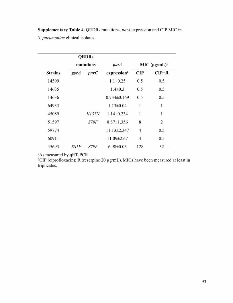
Supplementary Table 4. QRDRs mutations, patA expression and CIP MIC in
S. pneumoniae clinical isolates.
QRDRs
mutations patA MIC (µg/mL)b
Strains gyrA parC expressiona CIP CIP+R
14599 1.1±0.25 0.5 0.5
14635 1.4±0.3 0.5 0.5
14636 0.734±0.169 0.5 0.5
64933 1.13±0.04 1 1
45089 K137N 1.14±0.234 1 1
51597 S79F 8.87±1.356 8 2
59774 11.13±2.347 4 0.5
60911 11.09±2.67 4 0.5
45693 S81F S79F 6.98±0.03 128 32 aAs measured by qRT-PCR bCIP (ciprofloxacin); R (reserpine 20 µg/mL). MICs have been measured at least in triplicates.
93

Figure S1. Schematic representation (in CG view) of mutated locus present in each CIP resistant strains sequenced R6M2B (A) T5-R6M2B (B) and T1-60827 (C). In this figure, genes indicates in red are encoded on the forward DNA strand and the genes in blue are encoded on the reverse strand. Genes with (*) have mutation(s) in the region upstream of the ATG and genes with (+) have mutation(s) in the downstream region of the gene. Abbreviations: parC: DNA topoisomerase IV subunit A, gyrA: DNA gyrase subunit A,
94

gnd: 6-phosphogluconate dehydrogenase, secA: preprotein translocase subunit SecA, gpsA: NAD(P)H-dependent glycerol-3-phosphate dehydrogenase, PTS-EIIA: PTS system IIA component, dexB: glucan 1,6-alpha-glucosidase, ppc: phosphoenolpyruvate carboxylase, cpoA: glycosyl transferase CpoA, rexB: second subunit of major exonuclease, aldB: alpha-acetolactate decarboxylase, glnH: amino acid ABC transporter amino acid-binding protein, patB: ABC transporter ATP-binding protein/permease, patA: ABC transporter ATP-binding protein/permease, pcpA: choline binding protein PcpA, dltD: D-alanine transfer from undecaprenol-phosphate to the poly(glycerophosphate) chain, dltC: D-alanine--poly(phosphoribitol) ligase subunit 2, dltB: D-alanine transfer from Dcp to undecaprenol-phosphate, dltA: D-alanine--poly(phosphoribitol) ligase subunit 1, glpF: glycerol uptake facilitator protein.
95


Chapitre IX: La présence de mutations et l’augmentation de
l’expression d’ARN chez des souches de Streptococcus
pneumoniae résistantes à la tétracycline causent la résistance tel
que démontré par séquençage de génome et séquençage de
l’ARN messager.
Ce chapitre contient un article soumis pour publication dans le journal Journal of
Antimicrobial Chemotherapy.
9.1 Résumé
Contexte
Le but de cette étude est de caractériser les mutations chromosomiques associées à
la résistance à la tétracycline chez Streptococcus pneumoniae.
Résultats
Le séquençage du génome de deux mutants S. pneumoniae R6 sélectionnés pour la
résistance à la tétracycline a révélé des mutations dans le gène rpsJ, codant pour la protéine
S10, et dans les régions promotrice et codante du transporteur ABC PatA/PatB. Les
mutants présentent une résistance croisée à la ciprofloxacine. La reconstruction de la
résistance par transformation d’allèles contenant les mutations dans la région codante de
patA a confirmé le rôle de ces mutations dans la résistance. La surexpression du
transporteur ABC PatA/PatB contribue à la résistance à la tétracycline, à la ciprofloxacine
et au bromure d’éthidium et est associée à une diminution de l’accumulation de la
tétracycline tritiée. La réintroduction des mutations détectées par le séquençage du génome
97

n’a pas permis de reconstruire le niveau de résistance observé chez les souches parentes.
L’analyse comparative du transcriptome par profilage de l’ARN des mutants résistants a
permis de révéler qu’en plus de la surexpression de patA et patB, l’expression de plusieurs
gènes de la voie de biosynthèse et de récupération de la thiamine est augmentée chez les
deux mutants. L’inactivation de ces gènes a confirmé leur rôle dans la résistance à la
tétracycline.
Conclusion
La combinaison de l’analyse génomique et transcriptomique couplée à l’étude
fonctionnelle a permis la découverte de nouvelles mutations causant la résistance à la
tétracycline chez S. pneumoniae.
98

9.2 Article
Multiple mutations and increased RNA expression in tetracycline
resistant Streptococcus pneumoniae as determined by genome wide DNA
and mRNA sequencing.
Andréanne Lupien1, Hélène Gingras1, Michel G. Bergeron1, Philippe Leprohon1, Marc
Ouellette1§
1 Centre de recherche en Infectiologie du Centre de recherche du CHU de Québec and
Département de Microbiologie, Infectiologie et Immunologie, Faculté de Médecine,
Université Laval, Québec, QC, Canada.
§Corresponding author: Marc Ouellette Ph.D. Centre de Recherche en Infectiologie 2705 Boul. Laurier Québec, QC Canada G1V 4G2 Tel : 1-418-654-2705 Fax : 1-418-654-2715 E-mail :[email protected]
Running title: Tetracycline resistance in Streptococcus pneumoniae.
Emails
Andréanne Lupien: [email protected]
Hélène Gingras : [email protected]
Michel G. Bergeron : [email protected]
Philippe Leprohon: [email protected]
99

Synopsis
Objectives
This study aimed at characterizing chromosomal mutations associated to resistance to
tetracycline in Streptococcus pneumoniae.
Methods
Chronological appearance of mutations in two S. pneumoniae R6 mutants (R6M1TC-5 and
R6M2TC-4) selected for resistance to tetracycline was determined by next-generation
sequencing. A role for the mutations identified was confirmed by reconstructing resistance
to tetracycline in a S. pneumoniae R6 wild-type background. RNA sequencing was
performed on R6M1TC-5 and R6M2TC-4 and the relative expression of genes was
reported according to R6. Differentially expressed genes were classified according to their
ontology.
Results
Whole-genome sequencing of R6M1TC-5 and R6M2TC-4 revealed mutations in the gene
rpsJ coding for the ribosomal protein S10 and in the promoter region and coding sequences
of the ABC genes patA and patB. These cells were cross-resistant to ciprofloxacin.
Resistance reconstruction confirmed a role in resistance for the mutations in rpsJ and patA.
Overexpression of the ABC transporter PatA/PatB or mutations in the coding sequence of
patA contributed to resistance to tetracycline, ciprofloxacin and ethidium bromide, and was
associated with a decreased accumulation of H3-tetracycline. Comparative transcriptome
profiling of the resistant mutants further revealed that, in addition to the overexpression of
patA and patB, several genes of the thiamine biosynthesis and salvage pathway were
100
increased in the two mutants but also in clinical isolates resistant to tetracycline. This
overexpression most likely contributes to the tetracycline resistance phenotype.
Conclusions
The combination of genomic and transcriptomic analysis coupled to functional studies has
allowed the discovery of novel tetracycline resistance mutations in S. pneumoniae.
Keywords
Streptococcus pneumoniae, tetracycline, ABC transporter, rpsJ, thiamine, genomic,
RNAseq.
101

Introduction
Streptococcus pneumoniae is a Gram positive bacterium responsible for several
diseases such as otitis media, meningitis, community-acquired pneumonia and bacteremia 1
and for which resistance to antibiotics has become a worldwide concern. The prevalence of
resistance varies between countries but is globally high for β-lactams, macrolides,
chloramphenicol and tetracycline (TC) mainly due to the spread of multidrug-resistant
clones. 2 Since the first report in 1967 of penicillin and TC non-susceptible isolates, 3-7
resistance has increased to a point where 24%, 35%, 42%, and even >80% of disease-
causing pneumococci are now resistant to TC in the USA, France, Spain and some regions
of Asia, respectively. 8-10 Doxycycline, a TC mostly use for the treatment of intracellular
bacterial infections, have been proposed for the treatment of community-acquired
pneumonia. 11 However, cross-resistance between the TC family compounds is observable
in pneumococci, 12, 13 except for the new glycylcycline tigecycline (TGC).
Tetracycline inhibits protein synthesis by blocking the attachment of charged
aminoacyl-tRNA to the A site on the ribosome, which prevent the introduction of new
amino acids to the nascent peptide chain. 14 Resistance to TC in bacteria occurs through
enzymatic inactivation, 15 by active efflux or by ribosome protection from the acquisition of
tet genes. 16, 17 In pneumococci, the acquisition of the gene tet(M) is the most common
mechanism of resistance to TC, 6, 18, 19 while resistance mediated by tet(O) have only been
reported sporadically. 20 Both genes are located on Tn916-like mobile genetic elements and
encode ribosomal protection proteins that have homology to elongation factors G. 21 The
GTPase activity of tet(M) and tet(O) appears to be important for the displacement of
tetracycline from the ribosome. 19 Resistance to TC is now so frequent in S. pneumoniae
that it is seldomly used for treating this bacterium.
Besides mobile elements, chromosomal mutations have also been shown to
participate in resistance to TC. In Helicobacter pylori, the triple base pair mutation AGA926-
928→TTC in both copies of the 16S rRNA gene caused high level resistance to TC, whereas
102
strains with low-level resistance harbored only single or double base pair mutations. 22, 23 In
Neisseria gonorrhoeae, point mutations in the rpsJ gene coding for ribosomal protein S10,
the gene mtrR coding for a transcriptional regulator or the gene penB coding for a porin
have also been implicated in resistance to TC. 24
Overexpression of multidrug efflux pumps of the ABC superfamily can contribute
to resistance to structurally unrelated molecules, including TC in Gram negative bacteria
and fluoroquinolones in S. pneumoniae. 25-27 Fluoroquinolones still have an excellent
activity against S. pneumoniae but resistance in in vitro-selected or clinical strains was
indeed shown to involve the overexpression of the multidrug ABC transporter PatA/PatB in
addition to mutations in gyrase and topoisomerase genes. 28-32 Here, we studied resistance
to TC in the absence of tet(M) in S. pneumoniae by DNA and RNA sequencing and found
several novel mutations and gene overexpression, including one ribosomal protein, the
ABC proteins PatA/PatB and thiamine biosynthetic enzymes that are associated for the first
time with TC resistance in S. pneumoniae.
103

Methods
Bacteria culture and strains
Strains used in this study are listed in Table S1. Pneumococci were grown in brain
heart infusion broth (BHI, Difco), or on tryptic-soy agar containing 5% defibrinated
sheep’s blood. Cultures were incubated for 16-24 hours in a 5% CO2 atmosphere at 35oC.
TC resistant mutants of S. pneumoniae were obtained by successive passages of S.
pneumoniae R6 WT on plates containing increasing concentrations of TC, as previously
described. 32-35 Briefly, the selection for tetracycline resistance was conducted on Szybalski
plates containing concentration gradients of tetracycline. Mutants were selected by
scrapping colonies growing at the front of the highest concentration of tetracycline at each
stage. This pool of colonies was then streaked onto plates containing either the same
concentration of antibiotic or a gradient of increased antibiotic concentrations. End-point
mutants corresponded to colonies growing on plates with the highest concentration of
antibiotic and for which an additional increase in tetracycline concentration failed to yield
resistant colonies. A total of five and four selection cycles were required to obtain the
endpoint S. pneumoniae R6M1TC-5 and R6M2TC-4 mutants, respectively.
Next-generation DNA sequencing
Genomic DNAs were extracted using the Wizard Genomic DNA Purification Kit
(Promega) according to the manufacturer’s instructions. The genomes of R6M1TC-5 and
R6M2TC-4 were sequenced using a 454 Life Sciences GS-FLX system (Roche). Genome
sequencing, assemblies and comparative analyses were performed at the McGill University
Genome Québec Innovation Center (http://gqinnovationcenter.com/index.aspx). R6M1TC-
5 and R6M2TC-4 generated aggregated genome size of 2016699 bps, 2017241 bps,
respectively, at a mean 21X coverage. Whole genome sequencing was also performed on
clones of strains R6M1TC-1, R6M1TC-2, R6M1TC-3, R6M1TC-4, R6M2TC-1, R6M2TC-
2 and R6M2TC-3 using an Illumina MiSeq system (Centre de Recherche du CHU de
104

Québec) and a 250-nucleotides paired-end reads protocol. This generated genome
assemblies covering at least 99% of the S. pneumoniae R6 genome at a mean 120X
coverage. Sequence reads from each strain were filtered based on quality score using
Trimmomatic 36 and aligned to the genome of S. pneumoniae R6 using the software bwa
(bwa aln, version 0.5.9) with default parameters. 37 The maximum number of mismatches
was 4, the seed length was 32 and 2 mismatches were allowed within the seed. The
detection of single nucleotide polymorphisms was performed using samtools (version
0.1.18), bcftools (distributed with samtools) and vcfutils.pl (distributed with samtools), 38
with a minimum of three reads to call a potential variation prior to further analysis.
Mutations deduced from massively parallel sequencing were confirmed by PCR
amplification and conventional DNA sequencing.
Sequencing data accession numbers
All sequencing data described in this paper has been deposited at the EBI SRA
database (http://www.ebi.ac.uk/) under the study accession number PRJEB6539 and the
sample accession numbers ERS480579 (R6M1TC-1); ERS480580 (R6M1TC-2);
ERS480581 (R6M1TC-3a); ERS480582 (R6M1TC-3b); ERS480583 (R6M1TC-4a);
ERS480584 (R6M1TC-4b); ERS480567 (R6M1TC-5); ERS480585 (R6M2TC-1a);
ERS480586 (R6M2TC-1b); ERS480587 (R6M2TC-2a); ERS480588 (R6M2TC-2b);
ERS480589 (R6M2TC-3a); ERS480590 (R6M2TC-3b) and ERS480568 (R6M2TC-4).
Genetic transformation and inactivation
Long PCR fragments (~5kb) containing the mutations of interest were amplified
using primers listed in Table S2 and transformed in S. pneumoniae R6 as described
previously. 32, 34 For introducing mutations that were less amenable to selection, short
(500bp) PCR fragments amplified from ribosomal protein S12 (rpsL) or dihydrofolate
reductase (dhfr) gene variants conferring resistance to streptomycin and trimethoprim,
respectively, were co-transformed and used as surrogate selection markers as previously
105

described. 32 Fragments containing the mutations from the entire patA-patB operon from
R6M1TC-5 and R6M2TC-4 were produced by overlap PCR from two distinct fragments to
increase the length of the PCR product.
Gene inactivation was performed using the plasmid pFF6 (KANR) or pFF3 (CHLR). 32 PCR fragments covering the middle region of the genes patA, patB, spr0632, spr0634,
spr0637 and spr0638 were amplified using the primers listed in Table S2 and cloned into
EamII05I-digested pFF6 or pFF3 to produce plasmids pFF6patA, pFF6patB,
pFF6spr0632(R6), pFF3spr0632(CCRI22087), pFF6spr0634, pFF6spr0637 and
pFF6spr0638. These were transformed into S. pneumoniae as previously described. 34, 39
MIC determination
MICs to TC (Sigma), CIP (Sigma) and Etbr (Fluka) in the absence and the presence
of 20ug/mL reserpine (Sigma) were determined by microdilution. All MICs measurements
were performed at least in triplicates.
RNA sequencing
Total RNA was isolated from S. pneumoniae R6M1TC-5, R6M2TC-4 and S.
pneumoniae R6 WT grown to mid-log phase in BHI using the Qiagen RNeasy Mini Kit
(Qiagen) according to the manufacturer’s instructions. RNAs were quantified using 2100
BioAnalyzer RNA6000 Nano chips (Agilent) and 1µg of total RNA was treated with Ribo-
Zero™ rRNA Removal Kits (Bacteria) (Epicentre). RNAseq libraries were produced from
50 ng of rRNA-depleted samples using the ScriptSeq™ v2 RNA-Seq Library Preparation
Kit (Epicentre). The libraries were analyzed using 2100 BioAnalyser High Sensitivity DNA
chips and quantified by PicoGreen. The libraries were pooled, diluted to 8pM and
sequenced on an Illumina MiSeq system using a 250bps paired-ends reads protocol.
106

Reads were mapped to the S. pneumoniae R6 reference genome as described above.
Transcripts were assembled from the alignment files by using the Cufflinks pipeline. 40
Differential gene expression was computed with CuffDiff, and genes with a p-value ≤
0.015 were considered for further analysis.
Quantitative real-time PCR
Total RNAs were extracted as described above and treated with DNase I (Ambion)
to avoid any DNA contamination. The quality and integrity of the RNAs was assessed
using a 2100 BioAnalyzer and RNA6000 Nano chips (Agilent). cDNAs were generated
from 250ng of total RNAs using the Superscript II reverse transcriptase (Invitrogen) and
random hexamers according to the manufacturer’s instruction. Real-time quantitative RT-
PCR assays were carried out with a BioRad Cycler using SYBR Green I (Molecular
Probes). A final volume of 10μl was used for each reaction containing specific primers
(Table S2) and iQ SYBR Green Supermix (Bio-Rad). All qRT-PCR data were normalized
according to the amplification signals of 16S rRNA.
Gene ontology (GO)
Differentially expressed genes were classified according to their ontology by using
the Blast2GO software. 41 If not mentioned, the default setups were used during the
analysis. S. pneumoniae R6 ORFs were first blasted against the NCBI genome database and
GO mapping performed on the blasted genomes to assign cellular component (CC),
molecular function (MF) and biological process (BP) ontology levels. Mapped genes where
then screened using KEGG. GO Enrichment analysis (Fisher exact two-tailed t-test) was
then performed for genes identified as overexpressed or down-regulated (p-value ≤0.015) in
R6M1TC-5 and R6M2TC-4 by the CuffLinks pipeline, and also on the S. pneumoniae R6
genome as a control. GO terms with a value p≤0.015 were considered significantly
107

enriched in the group tested. Results are expressed as the proportion of sequences
associated with a particular GO term in the tested group.
TC accumulation
S. pneumoniae was grown to exponential phase (O.D.600nm 0.2-0.3) in BHI. Cells
were centrifuged and washed once with 0.1M Phosphate buffer pH 7. Cells were then
concentrated 40X (approximately 109cells/mL) in 0.1M Phosphate buffer containing 1mM
MgSO4 pH 7 and incubated at 37oC in the presence 1% glucose for 15 min. After pre-
incubation with glucose, 0.05µM of [7-3H(N)]-tetracycline (H3-TC) (17.9 Ci/mmol,
Moravek Biochemicals and Radiochemicals) was added to a final concentration of 5µM of
TC. H3-TC was used within one week upon arrival to avoid possible degradation. 42 When
required, 20 µg/ml of the efflux pump inhibitor reserpine was added 15 minutes after the
addition of H3-TC. Aliquots (0.1mL) were withdrawn at specific intervals and
accumulation was stopped by diluting with 1mL of ice-cold 0.1M Phosphate buffer pH 7
containing 100mM LiCl. Samples were centrifuged immediately at 4oC for 2 minutes and
washed once with 0.1M ice-cold Phosphate buffer, 100mM LiCl, pH 7. Pelleted cells were
resuspended in 0.1mL 1X Phosphate buffered saline, and 5mL Ecolite was added before
scintillation counts were measured using a LS6000TA Scintillation counter (Beckman).
Results were normalised to bacterial count obtained prior to the addition of TC at 0oC and
37oC, since no growth was observed during the course of the experiment after the addition
of the drug.
108

Results
Tetracycline resistant mutants
Two S. pneumoniae mutants named R6M1TC-5 and R6M2TC-4 were
independently selected for resistance to TC from S. pneumoniae R6 wild-type (WT) (MIC
TC 0.125µg/mL) by serial passages in the presence of increasing TC concentrations until
they reached a MIC of 8µg/mL (Table 1). This is considered resistant according to recently
revised breakpoints for TC. 43 Five and four passages on TC-supplemented agar plates were
required for the selection of R6M1TC-5 and R6M2TC-4, respectively. The R6M1TC-5 and
R6M2TC-4 mutants were also cross-resistant to the fluoroquinolone CIP and to the
intercalating dye ethidium bromide (EtBr) (Table 1). A low level cross-resistance was also
observed in R6M1TC-5 and R6M2TC-4 to the new glycylcycline tigecycline (TGC).
Interestingly, the efflux pump inhibitor reserpine decreased the CIP and EtBr MICs of both
mutants to WT levels and also substantially increased their sensitivity to TC (Table 1).
Reserpine had no effect on the small cross-resistance to TGC (Table 1). While confirming a
role for drug efflux in the multidrug resistance phenotype of S. pneumoniae R6M1TC-5
and R6M2TC-4, the reserpine-insensitive residual TC resistance suggests that additional
mechanisms are also likely to contribute to resistance in these mutants.
Whole-genome sequencing (WGS) of TC mutants
The genomes of each selection steps leading to both R6M1TC-5 and R6M2TC-4
mutants were sequenced to understand the genomics of TC resistance and the order of
appearance of mutations linked to resistance. The genomes of S. pneumoniae R6M1TC-5
and R6M2TC-4 both contained 9 mutations which were distributed over 7 and 8 genomic
loci, respectively (Table 2). Two of the mutated loci were common to both mutants, i.e. the
gene spr0187 coding for ribosomal protein S10 and the gene spr1887 coding for the
multidrug ABC transporter PatA, although mutations occurred at different positions in each
109

mutant (Table 2). For the gene spr0187 (also known as rpsJ), the R6M1TC-5 mutant
harboured a C157T transition (leading to a R53C mutation) in addition to a small deletion
covering positions 175-180 (Table 2). Interestingly, intermediate mutants R6M1TC-1 to
R6M1TC-4 presented instead a single G178T transversion in spr0187 (Table 2). The
increasing TC pressure thus appears to have selected for several distinct events in
R6M1TC, the first one being selected early at position 178 and the others occurring late at
positions 157 and 175-180 of rpsJ (which encompasses the initial G178T mutation) (Table
2). In contrast, a unique A169G transition leading to a K57E substitution was observed in
spr0187 in R6M2TC-4, which was selected early at the first selection step (Table 2). For
the patA locus, both mutants harbored mutations upstream of the start codon (possibly in
the promoter region) and within the coding region (Table 2). Interestingly, in both cases the
mutations in the promoter region of patA preceded those within the gene (Table 2). The
number of patA mutations also correlated with the level of resistance to TC (Table 2).
Reconstruction of TC resistance
Recurrent mutations are the most likely candidates to contribute to resistance and
the role of the rpsJ and patA mutations was thus further assessed by resistance
reconstruction. This was done by transforming S. pneumoniae R6 WT with variant alleles
amplified from RM1TC-5 or R6M2TC-4 either alone or along with a PCR fragment
covering the rpsL+ allele of S. pneumoniae CP1296 and conferring resistance to
streptomycin that was used as a surrogate marker for the selection of transformants (see
Materials and Methods). The rpsL+ PCR fragment had no impact on TC susceptibility
levels when transformed alone (Table 3). Each reconstructed strain was then tested for
resistance to TC, CIP and EtBr in the presence and absence of reserpine. The introduction
of the rpsJ alleles from R6M1TC-1 (harboring a G178T mutation) or R6M2TC-4
(harboring a A169G mutation) into S. pneumoniae R6 WT increased the TC MIC four-fold
in the S. pneumoniae R6rpsJ(R6M1TC-1) and S. pneumoniae R6rpsJ(R6M2TC-1) transformants
(Table 3). Interestingly, the rpsJ allele from R6M1TC-5 was more potent at conferring
resistance to TC than the one from R6M1TC-1 (compare R6rpsJ(R6M1TC-1) and R6rpsJ(R6M1TC-5)
in Table 3), which is consistent with their chronological order of appearance during the
110

selection of the R6M1TC series of mutants (Table 3). To exclude the possibility of second-
site mutations introduced during the transformation, we reintroduced the wild-type rpsJ
allele in the S. pneumoniae R6rpsJ(R6M1TC-1) and R6rpsJ(R6M1TC-5) transformants and these cells
became more sensitive to TC (see R6rpsJ(R6M1TC-1)-rev(R6WT)-rpsL+ and R6rpsJ(R6M1TC-5)-rev(R6WT)-
rpsL+ in Table 3).
The patA allele from R6M1TC-5 also accounted for a four-fold increase in
resistance to TC in the S. pneumoniae R6patA(R6M1TC) transformant (Table 3), of which half
is attributable to the G-35A mutation in the promoter region (R6p_patA(R6M1TC)-rpsL+ in Table
3) and the other half to the G510A mutation within the gene (compare R6CDSpatA(R6M1TC)-
rpsL+, R6CDSpatA(G510A)(R6M1TC)-rpsL+ and R6CDSpatA(C883A)(R6M1TC)-rpsL+ in Table 3). The patA
allele from R6M2TC-4 was less effective at increasing TC resistance however, as it could
only doubled the TC MIC of the S. pneumoniae R6patA(R6M2TC) transformant (Table 3). As
expected, TC resistance conferred by the patA mutations, but not by the rpsJ alleles, was
reversible by reserpine (Table 3). Resistance reconstruction experiments also implicated
mutations upstream and within patA in resistance to CIP and EtBr for both mutants (Table
3). As a final proof of the role of patA in resistance we introduced the wild-type version of
patA in S. pneumoniae R6patA-patB(R6M2TC) which became more sensitive to TC (see R6patA-
patB(R6M2TC)-rev(patA)-rpsL+ in Table 3).
Gene inactivation experiments further confirmed the role of the variant patA alleles
in the resistance of R6M1TC-5 and R6M2TC-4 (Table 4). Inactivation of the gene coding
for the ABC transporter PatB also sensitized both mutants to TC and CIP, which is
consistent with the finding that PatA and PatB act as heterodimers to extrude toxic
compounds in S. pneumoniae. 44 In contrast to patA however, no role in resistance could be
attributed to the mutation selected in R6M2TC-4 in the coding region of patB (spr1885) by
transformation into a TC susceptible S. pneumoniae background (Table 3).
Other strain specific mutations were observed in the mutants (Table 2). These were
often, but not always, detected in the most resistant mutants but we could not, by gene
transformation along with the rpsL+ allele, link any of these strain specific mutations with
TC resistance (result not shown).
111

Decreased accumulation of TC in S. pneumoniae resistant mutants
Mutations upstream of patA have previously been linked to drug resistance in S.
pneumoniae by inducing the overexpression of the transporter, 35, 45 although never for TC.
Quantitative RT-PCR also confirmed that patA was overexpressed in the S. pneumoniae
R6M1TC-5 and R6M2TC-4 mutants and introducing the G-35A and G-48A mutations in a
S. pneumoniae R6 wild-type background confirmed that these were indeed responsible for
patA overexpression (Table 4). As mutations in the coding regions of patA were also shown
to contribute to TC (Table 3) and CIP (Table 3 and by Lupien and coworkers 32) resistance,
TC accumulation experiments were conducted to directly address to role of both types of
mutations (i.e. promoter vs coding region) on the transport capacity of the PatA/PatB
heterodimer. S. pneumoniae R6M1TC-5 and R6M2TC-4 accumulated less H3-TC than the
parental S. pneumoniae R6 WT (p-value ≤ 0.05) (Figure 1A). This is directly linked to the
presence of patA variants as S. pneumoniae R6patA(R6M1TC) and R6patA-patB(R6M2TC) harboring
promoter and coding region mutations derived from R6M1TC-5 and R6M2TC-4,
respectively, displayed the same reduced accumulation of H3-TC as the original mutants
(Figure 1B, C). Interestingly, while mutations in the promoter of patA and in its coding
region conferred resistance to TC when introduced on their own in S. pneumoniae R6 WT
(Table 3), only the mutations in the promoter region were responsible for the decreased
accumulation of TC in the mutants (p-value ≤ 0.05) (Figure 1B, C). The addition of
reserpine, an inhibitor of efflux pumps, 29, 46 restored the accumulation of TC in the S.
pneumoniae R6patA(R6M1TC) and R6patA-patB(R6M2TC) transformants (Figure 1D).
RNA expression profiling in TC mutants
The rpsJ and patA mutations could only partly explain the levels of TC resistance of
S. pneumoniae R6M1TC-5 and R6M2TC-4 (Table 3) and none of the other mutations
identified in the genome of the mutants (Table 2) conferred resistance to TC when
introduced into S. pneumoniae R6 WT (not shown). We therefore hypothesized that
112

resistance to TC could also involve differences in gene expression (as in the case of
patA/patB) and performed comparative gene expression profiling between the R6M1TC-5
and R6M2TC-4 mutants and their susceptible S. pneumoniae R6 WT parent by RNAseq.
Overall, 43 and 60 genes were found to be significantly overexpressed (p-value ≤ 0.015) in
the R6M1TC-5 and R6M2TC-4 mutants compared to R6 WT, respectively (Figure 2 and
Table S3). Moreover, 31 and 26 genes had their expression significantly decreased (p-value
≤ 0.015) in R6M1TC-5 and R6M2TC-4 compared to R6 WT, respectively (Figure 2 and
Table S3). Overall, 13 overexpressed and 14 downregulated genes were common to both
mutants (Table S3).
Clustering differentially expressed genes according to their ontology revealed that
genes overexpressed in S. pneumoniae R6M1TC-5 were enriched for biological processes
involving thiamine metabolism (Figure 3). For S. pneumoniae R6M2TC-4, genes identified
as overexpressed by RNAseq were enriched mainly for processes implicated in
carbohydrates metabolism and transport functions (Figure 3) and were not directly linked to
the metabolism of thiamine. Nonetheless, a thorough examination of the RNAseq data
revealed that the expression of most genes related to thiamine metabolism overexpressed in
R6M1TC-5 were also increased in R6M2TC-4 when the cutoff p-value for filtering
differentially expressed genes was loosened to p≤0.05. Quantitative RT-PCR later
confirmed that genes spr0632, spr0634, spr0637 and spr0638, all involved in thiamine
metabolism (See Figure S1) were indeed overexpressed in both mutants (Table 4). The
expression of the spr0632 gene was further tested by qRT-PCR in 10 clinical isolates of S.
pneumoniae (5 sensitive and 5 resistant). Interestingly, an increased expression of spr0632
was observed in two resistant clinical isolates and in none of the sensitive ones (Table 5).
Downregulated genes also clustered in GO terms shared by the two TC-resistant mutants
(biological processes implicated in alcohol/polyol/glycerol or organophosphate
metabolism) (Figure 3).
In addition to its central role in intermediary metabolism, vitamin B1 (i.e. thiamine)
has also been reported to act as an important stress-response molecule against oxidative or
osmotic stress in bacteria, 47, 48 plants 49, 50 and fungi. 51 Interestingly, a triphosphorylated
113
derivative of vitamin B1, namely thiamine triphosphate, was shown to act as an alarmone
initiating a reaction cascade involved in the adaptation of bacteria to stringent conditions
such as amino acid starvation. 52 As the inhibition of translation by tetracycline should
mimic amino acid starvation, we hypothesized that the overexpression of genes linked to
thiamine metabolism in R6M1TC-5 and R6M2TC-4 could also have a role in favoring
resistance to TC. The gene spr0632 is overexpressed in R6M1TC-5 and R6M2TC-4 and
codes for the ATPase subunit of an ABC transporter putatively involved in the transport of
HMP from the extracellular milieu (Figure S1). The inactivation of spr0632 by insertion-
duplication mutagenesis decreased the level of TC resistance of R6M1TC-5 and R6M2TC-
4 by two and four fold, respectively (Table 4). A two-fold sensitization to TC was also
observed upon inactivation of spr0632 in the clinical isolate CCRI 22087 initially
overexpressing the gene, but not in the clinical isolate CCRI 18414 not overexpressing
spr0632 (Table 5). We were incapable to transform strain CCRI 14774 which also had
overexpression of spr0632 (Table 5). This is consistent with the lack of TC sensitization
upon inactivation of spr0632 in S. pneumoniae R6 WT (Table 4 and Table5) and suggests
that the overexpression of the gene is required for its contribution in resistance. In the case
of R6M2TC-4, the inactivation of spr0632 also sensitized to CIP (data not shown). The
inactivation of spr0634 and spr0638, coding for a thiaminase and for a
phosphomethylpyrimidine kinase in the thiamine biosynthesis pathway (Figure S1), also
affected the level of TC susceptibility but this time specifically in R6M2TC-4 (Table 4),
which suggests that overexpression of some genes of the HMP salvage pathway is
associated with TC resistance.
114

Discussion
The selection of the S. pneumoniae TC-resistant mutants R6M1TC-5 and R6M2TC-
4 from S. pneumoniae R6 WT (which lacks tet(M) or tet(O)) by incremental increases in
TC concentrations revealed the possibility for S. pneumoniae to become resistant to TC
through genomic alterations. For both mutants, the first event selected occurred at the level
of the gene rpsJ coding for ribosomal protein S10. Based on the crystal structure of the 30S
ribosomal subunit from Thermus thermophilus cross-linked with TC, 53 the mutations in the
R6M1TC-5 and R6M2TC-4 mutants are all located in the vertex loop of ribosomal protein
S10. This loop is composed of 50–60 amino acids and is located near the primary site of
action of TC, i.e. the aminoacyl-tRNA site. 17 As previously proposed, amino acid changes
located in the vicinity of this site could decrease the affinity for the antibiotic by altering
the rRNA structure near the TC-binding site. 53 Interestingly, the amino acid from
ribosomal protein S10 the nearest to the primary site in T. thermophilus (lysine-55) was
found to be within 8 to 9 Ä of bound TC 24, 53 and the equivalent residue is mutated in
R6M2TC-4 (K57E). Substitutions at homologous residues were also previously observed
and implicated in TC resistance in high-level resistant Neisseria gonorrhoeae, 24
confirming the importance of endogenous mutations near the ribosomal aminoacyl-tRNA
site for TC resistance in the absence of tet genes.
Multidrug efflux pumps are important contributors of resistance to antimicrobials
and the S. pneumoniae multidrug ABC transporter PatA/B had already been implicated in
resistance to EtBr, berberine, novobiocine, acriflavin, erythromycin, fluoroquinolones,
chloramphenicol and linezolid. 35, 54 A phenotypic microarray screening for 240 compounds
against a panel of S. pneumoniae mutants inactivated for putative efflux pumps also
identified PatA/PatB as a possible contributor to oxytetracycline resistance, but this was not
further validated by an additional quantitative method like microdilution MIC. 55 Here we
provide such evidence with S. pneumoniae transformants for which the introduction of a
mutation upstream of patA facilitates the transcription of the PatA/PatB genes to foster
resistance to TC by decreasing its accumulation, most likely by increased efflux. While
several other bacterial ABC transporters have been shown to confer resistance to
115

structurally dissimilar compounds, including TC and fluoroquinolones, 25-27, 56 this is the
first time that a reduced accumulation of TC mediated by such transporter is directly
measured. The mutation in the promoter region of patA most likely increases transcription
rates by loosening a terminator-like structure upstream of the gene. It was recently
proposed that mutations upstream of patA could lead to its overexpression by altering the
strength of a terminator-like stem-loop structure in the 5’ UTR of patA. 45, 57 Also,
constitutive overexpression of patAB in fluoroquinolones resistants isolates may be caused
by mutations in a Rho-independent transcriptional terminator structure located upstream of
patA gene.57 An analysis of the DNA sequence upstream of patA using RibEx 58 indeed
suggested the presence of three stem-loop structures (a transcriptional terminator, an
antiterminator and an anti-antiterminator structure) in S. pneumoniae R6 (Figure S2).
Interestingly, the strength (ΔG) of the transcriptional terminator stem-loop structure is
decreased by the presence of the mutations detected upstream of patA in R6M1TC-5 and
R6M2TC-4 mutants (Figure S2) suggesting that these alterations are responsible for the
increase in patA transcription (Table 4). Similarly, small deletions impeding with the
formation of a transcriptional attenuator upstream of tet(M) have been shown to increase its
transcription rate in S. pneumoniae clinical isolates unsusceptible to TC. 59 In S.
pneumoniae, TC resistance conferred by ribosomal protection proteins such as Tet(M)
would lead to an excess of unbound TC and overexpressing the chromosomally encoded
PatA/PatB efflux pump may be exploited by the pneumococcus to efflux the excess TC. It
would thus be interesting to assess whether patA/patB overexpression also occur in S.
pneumoniae clinical isolates resistant to TC due to the presence of mobile elements of the
Tn916 family.
Mutations in the coding region of patA also contributed to resistance to TC and CIP
(Table 2), possibly by altering the specificity of the transporter. A TMHMM analysis of the
PatA sequence revealed that every mutation observed in R6M1TC-5 and R6M2TC-4 was
located in transmembrane domains (TM). A comparison with the crystal structure of the
bacterial ABC exporter SAV1866 from Staphylococcus aureus 60 also supported the
location of mutation M170I within TM4 and the location of mutations G287E and R295S
within TM6. Residues from transmembrane helices TM1, TM4–TM6 and TM10–TM12 in
116

the homologous human MDR1 have been shown to interact with the substrates of the
transporter. 61 Unfortunately, no well-defined substrate binding sites have been identified
for bacterial ABC exporters 62, 63 to hypothesize more precisely about the role of these
mutations on the activity of the transporter. Nonetheless, it is intriguing that for both
R6M1TC-5 and R6M2TC-4 mutants, a single mutation in the coding region of patA
enhanced resistance against structurally different molecules. Mutations affecting substrate
specificity have also been reported previously for other multidrug efflux pumps. 64, 65 These
mutations can possibly alter the tertiary structure of the transporter and thereby modulate
the substrate profile or provide additional substrate binding sites relevant for the
recognition of a given substrate.
The expression profile of R6M1TC-5 and R6M2TC-4 has highlighted that gene
expression alterations others than the overexpression of patA/patB can foster resistance to
TC (Figure 2 and Table S3). Comparative expression profiling by RNAseq and qRT-PCR
indeed revealed that key genes involved in the salvage of HMP in the thiamine biosynthesis
pathway were overexpressed in the R6M1TC-5 and R6M2TC-4 mutants (Tables 4 and S3
and Figure S1). This appears to be coherent with resistance to TC since the inactivation of
spr0632, which is responsible for the uptake of HMP from the extracellular milieu,
increased TC susceptibility in both mutants. Interestingly, increased expression of thiamine
metabolism gene was also observed in clinical isolates resistant to TC. An increased
expression of spr0632 could be detected in two TC-resistant pneumococcal isolates but in
none of the sensitive isolates tested (Table 5). While the number of isolates tested is small,
these results indicates that the expression of thiamine metabolism genes can be increased in
clinical isolates and further work will undoubtedly help refining our understanding of the
clinical significance between thiamine metabolism and resistance to TC. The inactivation of
the spr0632 gene in one TC resistant clinical isolate reduced its resistance to TC. The
activity of several enzymes of carbohydrate metabolism such as pyruvate dehydrogenase,
2-oxoglutarate dehydrogenase and transketolase requires thiamine pyrophosphate as a co-
factor. 66 Resistance to TC may require increased energetic needs which would translate
into a higher thiamine demand from these enzymes. Alternatively, thiamine derivatives
have been shown to act as signals involved in the adaptation of bacteria to stressful
117

conditions 52, 67 and it will be interesting to conduct metabolomics studies with extracts
derived from mutants R6M1TC-5 and R6M2TC-4 to fully understand the changes
happening in the thiamine biosynthesis pathway.
Overall, the combined use of whole genome sequencing, RNAseq and functional
analyses revealed that endogenous TC resistance mechanisms can be selected in S.
pneumoniae in the absence of tet(M) and tet(O). One early mutation is the S10 ribosomal
protein but the clinical relevance of antibiotic efflux mediated by the ABC transporter
PatA/PatB in S. pneumoniae was reiterated by the demonstration that the overexpression of
this transporter can foster resistance to TC in addition to resistance to fluoroquinolones 30-32,
44 and linezolid. 35 A novel link between thiamine metabolism and resistance to TC was
also revealed.
118
Acknowledgements
We thank the McGill University Genome Quebec Innovation Centre for performing
the sequencing of R6M1TC-5 and R6M2TC-4 mutants. Les bourses d’Andréanne et mon
CRC.
Funding
This work was supported by a Canadian Institutes of Health Research grant to M. O.
Transparency declarations
The authors declare that they have no competing interests.
Author contributions
AL, PL and MO conceived and designed the experiments. AL and HG performed
the experiments. AL and PL conducted the bioinformatics analyses of the data. AL and PL
wrote the paper (with contributions from all of the authors). All authors have read and
approved the manuscript for publication.
119

Supplementary data
Table S1: list of strains and plasmids used in this study Table S2: list of primers used in this study Table S3: genes lists of differentially expressed genes (p-value ≤0.015) in R6M1TC-5 and R6M1TC-4 compared to R6 according to RNAseq Figure S1: schematic representation of the thiamine biosynthesis pathway genes altered in R6 TC resistant mutants. Figure S2: schematic representation of the predicted structure upstream of patA by RibEx.
120

References
1. Levine OS, O'Brien KL, Knoll M et al. Pneumococcal vaccination in developing countries. Lancet 2006; 367: 1880-2. 2. McGee L, McDougal L, Zhou J et al. Nomenclature of major antimicrobial-resistant clones of Streptococcus pneumoniae defined by the pneumococcal molecular epidemiology network. Journal of clinical microbiology 2001; 39: 2565-71. 3. Hansman D. BMM. A resistant pneumococcus. Lancet 1967; 290: Pages 264–5. 4. Parra EL, Ramos V, Sanabria O et al. Serotype and Genotype Distribution among Invasive Streptococcus pneumoniae Isolates in Colombia, 2005-2010. PloS one 2014; 9: e84993. 5. Zhou L, Ma X, Gao W et al. Molecular characteristics of erythromycin-resistant Streptococcus pneumoniae from pediatric patients younger than five years in Beijing, 2010. BMC microbiology 2012; 12: 228. 6. Montanari MP, Cochetti I, Mingoia M et al. Phenotypic and molecular characterization of tetracycline- and erythromycin-resistant strains of Streptococcus pneumoniae. Antimicrobial agents and chemotherapy 2003; 47: 2236-41. 7. Sa-Leao R, Tomasz A, Sanches IS et al. Carriage of internationally spread clones of Streptococcus pneumoniae with unusual drug resistance patterns in children attending day care centers in Lisbon, Portugal. The Journal of infectious diseases 2000; 182: 1153-60. 8. Jones RN, Sader HS, Moet GJ et al. Declining antimicrobial susceptibility of Streptococcus pneumoniae in the United States: report from the SENTRY Antimicrobial Surveillance Program (1998-2009). Diagnostic microbiology and infectious disease 2010; 68: 334-6. 9. Wyres KL, van Tonder A, Lambertsen LM et al. Evidence of antimicrobial resistance-conferring genetic elements among pneumococci isolated prior to 1974. BMC genomics 2013; 14: 500. 10. Kim SH, Song JH, Chung DR et al. Changing trends in antimicrobial resistance and serotypes of Streptococcus pneumoniae isolates in Asian countries: an Asian Network for Surveillance of Resistant Pathogens (ANSORP) study. Antimicrobial agents and chemotherapy 2012; 56: 1418-26. 11. Johnson JR. Doxycycline for treatment of community-acquired pneumonia. Clinical infectious diseases : an official publication of the Infectious Diseases Society of America 2002; 35: 632; author reply -3. 12. Izdebski R, Sadowy E, Fiett J et al. Clonal diversity and resistance mechanisms in tetracycline-nonsusceptible Streptococcus pneumoniae isolates in Poland. Antimicrobial agents and chemotherapy 2007; 51: 1155-63. 13. Lederman ER, Gleeson TD, Driscoll T et al. Doxycycline sensitivity of S. pneumoniae isolates. Clinical infectious diseases : an official publication of the Infectious Diseases Society of America 2003; 36: 1091. 14. Connell SR, Tracz DM, Nierhaus KH et al. Ribosomal protection proteins and their mechanism of tetracycline resistance. Antimicrobial agents and chemotherapy 2003; 47: 3675-81.
121

15. Yang W, Moore IF, Koteva KP et al. TetX is a flavin-dependent monooxygenase conferring resistance to tetracycline antibiotics. The Journal of biological chemistry 2004; 279: 52346-52. 16. Nguyen F, Starosta AL, Arenz S et al. Tetracycline antibiotics and resistance mechanisms. Biological chemistry 2014. 17. Chopra I, Roberts M. Tetracycline antibiotics: mode of action, applications, molecular biology, and epidemiology of bacterial resistance. Microbiology and molecular biology reviews : MMBR 2001; 65: 232-60 ; second page, table of contents. 18. Doherty N, Trzcinski K, Pickerill P et al. Genetic diversity of the tet(M) gene in tetracycline-resistant clonal lineages of Streptococcus pneumoniae. Antimicrobial agents and chemotherapy 2000; 44: 2979-84. 19. Widdowson CA, Klugman KP. The molecular mechanisms of tetracycline resistance in the pneumococcus. Microb Drug Resist 1998; 4: 79-84. 20. Widdowson CA, Klugman KP, Hanslo D. Identification of the tetracycline resistance gene, tet(O), in Streptococcus pneumoniae. Antimicrobial agents and chemotherapy 1996; 40: 2891-3. 21. Donhofer A, Franckenberg S, Wickles S et al. Structural basis for TetM-mediated tetracycline resistance. Proceedings of the National Academy of Sciences of the United States of America 2012; 109: 16900-5. 22. Wu JY, Kim JJ, Reddy R et al. Tetracycline-resistant clinical Helicobacter pylori isolates with and without mutations in 16S rRNA-encoding genes. Antimicrobial agents and chemotherapy 2005; 49: 578-83. 23. Dailidiene D, Bertoli MT, Miciuleviciene J et al. Emergence of tetracycline resistance in Helicobacter pylori: multiple mutational changes in 16S ribosomal DNA and other genetic loci. Antimicrobial agents and chemotherapy 2002; 46: 3940-6. 24. Hu M, Nandi S, Davies C et al. High-level chromosomally mediated tetracycline resistance in Neisseria gonorrhoeae results from a point mutation in the rpsJ gene encoding ribosomal protein S10 in combination with the mtrR and penB resistance determinants. Antimicrobial agents and chemotherapy 2005; 49: 4327-34. 25. Huda N, Lee EW, Chen J et al. Molecular cloning and characterization of an ABC multidrug efflux pump, VcaM, in Non-O1 Vibrio cholerae. Antimicrobial agents and chemotherapy 2003; 47: 2413-7. 26. Al-Hamad A, Upton M, Burnie J. Molecular cloning and characterization of SmrA, a novel ABC multidrug efflux pump from Stenotrophomonas maltophilia. The Journal of antimicrobial chemotherapy 2009; 64: 731-4. 27. Matsuo T, Chen J, Minato Y et al. SmdAB, a heterodimeric ABC-Type multidrug efflux pump, in Serratia marcescens. Journal of bacteriology 2008; 190: 648-54. 28. Avrain L, Garvey M, Mesaros N et al. Selection of quinolone resistance in Streptococcus pneumoniae exposed in vitro to subinhibitory drug concentrations. The Journal of antimicrobial chemotherapy 2007; 60: 965-72. 29. Garvey MI, Piddock LJ. The efflux pump inhibitor reserpine selects multidrug-resistant Streptococcus pneumoniae strains that overexpress the ABC transporters PatA and PatB. Antimicrobial agents and chemotherapy 2008; 52: 1677-85. 30. El Garch F, Lismond A, Piddock LJ et al. Fluoroquinolones induce the expression of patA and patB, which encode ABC efflux pumps in Streptococcus pneumoniae. The Journal of antimicrobial chemotherapy 2010; 65: 2076-82.
122
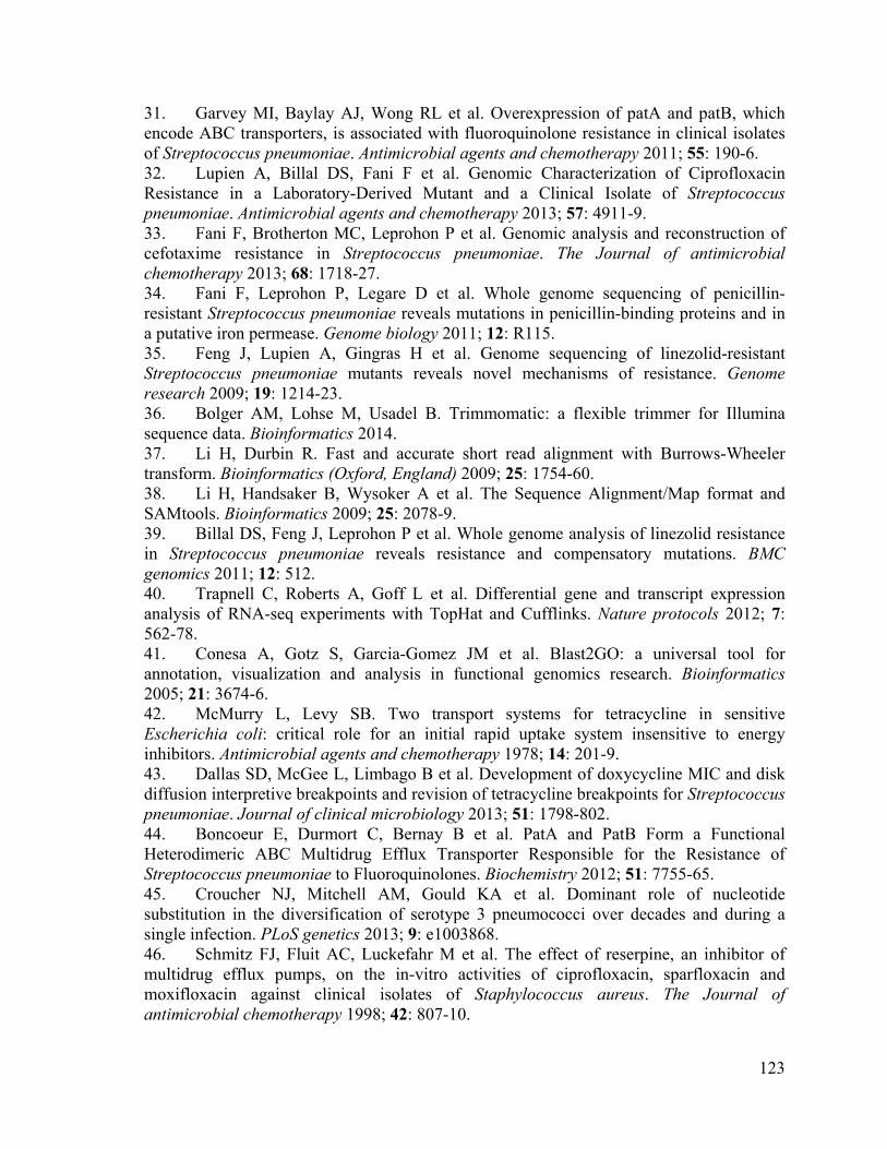
31. Garvey MI, Baylay AJ, Wong RL et al. Overexpression of patA and patB, which encode ABC transporters, is associated with fluoroquinolone resistance in clinical isolates of Streptococcus pneumoniae. Antimicrobial agents and chemotherapy 2011; 55: 190-6. 32. Lupien A, Billal DS, Fani F et al. Genomic Characterization of Ciprofloxacin Resistance in a Laboratory-Derived Mutant and a Clinical Isolate of Streptococcus pneumoniae. Antimicrobial agents and chemotherapy 2013; 57: 4911-9. 33. Fani F, Brotherton MC, Leprohon P et al. Genomic analysis and reconstruction of cefotaxime resistance in Streptococcus pneumoniae. The Journal of antimicrobial chemotherapy 2013; 68: 1718-27. 34. Fani F, Leprohon P, Legare D et al. Whole genome sequencing of penicillin-resistant Streptococcus pneumoniae reveals mutations in penicillin-binding proteins and in a putative iron permease. Genome biology 2011; 12: R115. 35. Feng J, Lupien A, Gingras H et al. Genome sequencing of linezolid-resistant Streptococcus pneumoniae mutants reveals novel mechanisms of resistance. Genome research 2009; 19: 1214-23. 36. Bolger AM, Lohse M, Usadel B. Trimmomatic: a flexible trimmer for Illumina sequence data. Bioinformatics 2014. 37. Li H, Durbin R. Fast and accurate short read alignment with Burrows-Wheeler transform. Bioinformatics (Oxford, England) 2009; 25: 1754-60. 38. Li H, Handsaker B, Wysoker A et al. The Sequence Alignment/Map format and SAMtools. Bioinformatics 2009; 25: 2078-9. 39. Billal DS, Feng J, Leprohon P et al. Whole genome analysis of linezolid resistance in Streptococcus pneumoniae reveals resistance and compensatory mutations. BMC genomics 2011; 12: 512. 40. Trapnell C, Roberts A, Goff L et al. Differential gene and transcript expression analysis of RNA-seq experiments with TopHat and Cufflinks. Nature protocols 2012; 7: 562-78. 41. Conesa A, Gotz S, Garcia-Gomez JM et al. Blast2GO: a universal tool for annotation, visualization and analysis in functional genomics research. Bioinformatics 2005; 21: 3674-6. 42. McMurry L, Levy SB. Two transport systems for tetracycline in sensitive Escherichia coli: critical role for an initial rapid uptake system insensitive to energy inhibitors. Antimicrobial agents and chemotherapy 1978; 14: 201-9. 43. Dallas SD, McGee L, Limbago B et al. Development of doxycycline MIC and disk diffusion interpretive breakpoints and revision of tetracycline breakpoints for Streptococcus pneumoniae. Journal of clinical microbiology 2013; 51: 1798-802. 44. Boncoeur E, Durmort C, Bernay B et al. PatA and PatB Form a Functional Heterodimeric ABC Multidrug Efflux Transporter Responsible for the Resistance of Streptococcus pneumoniae to Fluoroquinolones. Biochemistry 2012; 51: 7755-65. 45. Croucher NJ, Mitchell AM, Gould KA et al. Dominant role of nucleotide substitution in the diversification of serotype 3 pneumococci over decades and during a single infection. PLoS genetics 2013; 9: e1003868. 46. Schmitz FJ, Fluit AC, Luckefahr M et al. The effect of reserpine, an inhibitor of multidrug efflux pumps, on the in-vitro activities of ciprofloxacin, sparfloxacin and moxifloxacin against clinical isolates of Staphylococcus aureus. The Journal of antimicrobial chemotherapy 1998; 42: 807-10.
123

47. Jung IL, Kim IG. Thiamine protects against paraquat-induced damage: scavenging activity of reactive oxygen species. Environmental toxicology and pharmacology 2003; 15: 19-26. 48. Fukui K, Wakamatsu T, Agari Y et al. Inactivation of the DNA repair genes mutS, mutL or the anti-recombination gene mutS2 leads to activation of vitamin B1 biosynthesis genes. PloS one 2011; 6: e19053. 49. Tunc-Ozdemir M, Miller G, Song L et al. Thiamin confers enhanced tolerance to oxidative stress in Arabidopsis. Plant physiology 2009; 151: 421-32. 50. Rapala-Kozik M, Wolak N, Kujda M et al. The upregulation of thiamine (vitamin B1) biosynthesis in Arabidopsis thaliana seedlings under salt and osmotic stress conditions is mediated by abscisic acid at the early stages of this stress response. BMC plant biology 2012; 12: 2. 51. Kowalska E, Kujda M, Wolak N et al. Altered expression and activities of enzymes involved in thiamine diphosphate biosynthesis in Saccharomyces cerevisiae under oxidative and osmotic stress. FEMS yeast research 2012; 12: 534-46. 52. Lakaye B, Wirtzfeld B, Wins P et al. Thiamine triphosphate, a new signal required for optimal growth of Escherichia coli during amino acid starvation. The Journal of biological chemistry 2004; 279: 17142-7. 53. Brodersen DE, Clemons WM, Jr., Carter AP et al. The structural basis for the action of the antibiotics tetracycline, pactamycin, and hygromycin B on the 30S ribosomal subunit. Cell 2000; 103: 1143-54. 54. Robertson GT, Doyle TB, Lynch AS. Use of an efflux-deficient Streptococcus pneumoniae strain panel to identify ABC-class multidrug transporters involved in intrinsic resistance to antimicrobial agents. Antimicrobial agents and chemotherapy 2005; 49: 4781-3. 55. Tocci N, Iannelli F, Bidossi A et al. Functional analysis of pneumococcal drug efflux pumps associates the MATE DinF transporter with quinolone susceptibility. Antimicrobial agents and chemotherapy 2013; 57: 248-53. 56. Fernandez-Moreno MA, Carbo L, Cuesta T et al. A silent ABC transporter isolated from Streptomyces rochei F20 induces multidrug resistance. Journal of bacteriology 1998; 180: 4017-23. 57. Baylay AJ, Piddock LJ. Clinically relevant fluoroquinolone resistance due to constitutive overexpression of the PatAB ABC transporter in Streptococcus pneumoniae is conferred by disruption of a transcriptional attenuator. The Journal of antimicrobial chemotherapy 2014. 58. Abreu-Goodger C, Merino E. RibEx: a web server for locating riboswitches and other conserved bacterial regulatory elements. Nucleic acids research 2005; 33: W690-2. 59. Grohs P, Trieu-Cuot P, Podglajen I et al. Molecular basis for different levels of tet(M) expression in Streptococcus pneumoniae clinical isolates. Antimicrobial agents and chemotherapy 2012; 56: 5040-5. 60. Dawson RJ, Locher KP. Structure of a bacterial multidrug ABC transporter. Nature 2006; 443: 180-5. 61. Loo TW, Clarke DM. Recent progress in understanding the mechanism of P-glycoprotein-mediated drug efflux. The Journal of membrane biology 2005; 206: 173-85. 62. Higgins CF, Linton KJ. The ATP switch model for ABC transporters. Nature structural & molecular biology 2004; 11: 918-26.
124

63. Higgins CF. Multiple molecular mechanisms for multidrug resistance transporters. Nature 2007; 446: 749-57. 64. Bohnert JA, Schuster S, Fahnrich E et al. Altered spectrum of multidrug resistance associated with a single point mutation in the Escherichia coli RND-type MDR efflux pump YhiV (MdtF). The Journal of antimicrobial chemotherapy 2007; 59: 1216-22. 65. Vettoretti L, Plesiat P, Muller C et al. Efflux unbalance in Pseudomonas aeruginosa isolates from cystic fibrosis patients. Antimicrobial agents and chemotherapy 2009; 53: 1987-97. 66. Du Q, Wang H, Xie J. Thiamin (vitamin B1) biosynthesis and regulation: a rich source of antimicrobial drug targets? International journal of biological sciences 2011; 7: 41-52. 67. Bettendorff L, Wirtzfeld B, Makarchikov AF et al. Discovery of a natural thiamine adenine nucleotide. Nature chemical biology 2007; 3: 211-2.
125

Tables
Table 1. Minimal inhibitory concentration (MICs) of TC-resistant strains used in this
study.
MICs (ug/mL)a TC TC+R CIP CIP+R EtBr EtBr+R TGC TGC+R
R6 0.125 0.125 0.5 0.5 2 0.125 0.031 0.031 R6M1TC-1 2 1 1 0.5 8 0.25 0.063 0.063 R6M1TC-2 2 1 2 0.5 8 0.25 0.063 0.063 R6M1TC-3 4 1 4 0.5 8 0.25 0.063 0.063 R6M1TC-4 4 1 4 0.5 16 0.25 0.063 0.063 R6M1TC-5 8 2 4 0.5 16 0.25 0.25 0.25 R6M2TC-1 2 1 1 0.5 2 0.25 0.063 0.063 R6M2TC-2 2 1 1 0.5 2 0.25 0.063 0.063 R6M2TC-3 4 1 4 0.5 16 0.25 0.125 0.125 R6M2TC-4 8 1 8 0.5 16 0.25 0.125 0.125 aCIP, ciprofloxacin; Etbr, ethidium bromide; R, reserpine (20ug/mL); TC, tetracycline; TGC, tigecycline. MICs are the results of three independents triplicates.
126

Table 2. Chronological appearance of mutations in TC mutants.
Locus IDa
Functiona Mutationsb,c,d
R6M1TC R6M2TC 1e 2f 3g 4h 5 1i 2j 3k 4 spr0187 30S ribosomal
protein S10 G178T D60Y
G178T D60Y
G178T D60Y
G178T D60Y
C157T R53C ∆175-180
A169G K57E
A169G K57E
A169G K57E
A169G K57E
spr0219 phosphoglycerate mutase
WT WT WT WT T-52C WT WT WT WT
spr0291 PTS system IIA component
WT WT WT WT WT C-101G
C-101G
C-101G
C-101G T-30C
spr0691 biotin synthase WT WT WT WT WT G356T A119D
G356T A119D
G356T A119D
G356T A119D
spr0822 agmatine deiminase
WT WT WT WT WT WT WT WT A95C Stop►S position 32
spr0825 hypothetical protein
G-61A G-61A G-61A G-61A G-61A WT WT WT WT
spr0960 positive transcriptional regulator MutR
WT WT WT WT A-151C WT WT WT WT
spr1262 transcriptional regulator Spx
WT WT T-11C T-11C T-11C WT WT WT WT
spr1377 cystathionine gamma-synthase
WT WT WT WT WT WT WT WT T290 CA97V
spr1777 DNA-directed RNA polymerasesubunit beta
WT WT WT WT C1442T Syn
WT WT WT WT
spr1885 ABC transporter ATP-binding protein/permease
WT WT WT WT WT WT G794A G265E
G794A G265E
G794A G265E
spr1887 ABC transporter ATP-binding protein/permease
G-35A G-35A C883A (R295S)
G-35A G510A M170I C883A R295S
G-35A G510A M170I C883A R295S
G-35A G510A M170I C883A R295S
WT WT G-48A G-48A G860A G287E
spr1991 Glycerol kinase WT WT WT WT WT WT WT WT ∆G position 75
a ID, identity. Nomenclature according to the genome annotation of S. pneumoniae R6. b When mutations are within coding regions, the change in amino acids is also indicated in italics. c In noncoding sequence, the number preceded by a hyphen indicates the position upstream of the ATG. d Mutations in bold were further studied to determine their potential role in TC resistance and mutations underlined are involved in drugs resistances (TC, CIP, Etbr) e1 clone was sequenced. Unique mutations: spr1138(C443A) f1 clone was sequenced. Unique mutations: spr0428(G250A); spr0857(G1298T)
127

g: 2 clones were sequenced. Unique mutations: spr0313(A195G); spr1032(G1216T); spr1367(C511A); spr1865(G224A); spr2035(G-72A) h: 2clones were sequenced. Unique mutations: spr0294(T111C) i: 2 clones were sequenced. Unique mutations: spr0661(G896T)spr0797: 458bp downstream j 2 clones were sequenced. Unique mutations: spr0661(G896T,A); spr1250(spr1250) k 2 clones were sequences. Unique mutations: spr0232(G191A)
128

Table 3. Functional analysis of S.pneumoniae R6 containing mutations in R6M1TC-5 and
R6M2TC-4.
Mutated alleles MIC (ug/mL)b,c
Strainsa rpsJ patA patB TC TC+R CIP CIP+R Etbr Etbr+R promotor CDS
R6 WT WT WT WT 0.125 0.125 0.5 0.5 2 0.25 R6rpsL+ WT WT WT WT 0.125 0.125 0.5 0.5 2 0.25
R6M1TC-5 R6M1TC-5
R6M1TC-5
R6M1TC-5 WT 8 2 4 0.5 16 0.25
R6M2TC-4 R6M2TC-4
R6M2TC-4
R6M2TC-4 R6M2TC-4
8 1 8 0.5 16 0.25
R6rpsJ(R6M1TC-1) R6M1TC-1
WT WT WT 0.5 0.5 0.5 0.5 2 0.25
R6rpsJ(R6M1TC-1)-rpsL+ R6M1TC-1
WT WT WT 0.5 ND 0.5 ND ND ND
R6rpsJ(R6M1TC-1)-
rev(R6WT)-rpsL+ WT WT WT WT 0.125 ND 0.5 ND ND ND
R6rpsJ(R6M1TC-5) R6MITC-5
WT WT WT 1 1 0.5 0.5 2 0.25
R6rpsJ(R6M1TC-5)- rpsL+ R6MITC-5
WT WT WT 1 ND 0.5 ND ND ND
R6rpsJ(R6M1TC-5)-
rev(R6WT)- rpsL+ WT WT WT WT 0.125 ND 0.5 ND ND ND
R6patA(R6M1TC) WT R6M1TC-5
R6M1TC-5 WT 0.5 0.125 2 0.5 16 0.25
R6p_patA(R6M1TC)-rpsL+ WT R6M1TC-5
WT WT 0.25 0.125 1 0.5 8 0.25
R6CDSpatA(R6M1TC)-rpsL+ WT WT R6M1TC-5 WT 0.25 0.125 1 0.5 8 0.25
R6CDSpatA(G510A)(R6M1TC)-
rpsL+ WT WT R6M1TC(G510A) WT 0.25 0.125 1 0.5 4 0.25
R6CDSpatA(C883A)(R6M1TC)-
rpsL+ WT WT R6M1TC(C883A) WT 0.125 0.125 1 0.5 8 0.25
R6rpsJ(R6M1TC)-
patA(R6M1TC) R6M1TC-
5 R6M1TC R6M1TC-5 WT 2 0.5 2 0.5 16 0.25
R6rpsJ(R6M2TC) R6M2TC-4
WT WT WT 0.5 0.5 0.5 0.5 2 0.25
R6patA-patB(R6M2TC) WT R6M2TC-4
R6M2TC-4 R6M2TC-4
0.25 0.125 1 0.5 16 0.25
R6patA-patB(R6M2TC)-rpsL+ WT R6M2TC-4
R6M2TC-4 R6M2TC-4
0.25 ND 1 ND ND ND
R6patA-patB(R6M2TC)-
rev(patA)-rpsL+ WT WT WT R6M2TC-
4 0.125 ND 0.5 ND ND ND
R6p_patA(R6M2TC)-rpsL+ WT R6M2TC-4
WT WT 0.25 0.125 1 0.5 8 0.25
R6CDSpatA(R6M2TC)-rpsL+ WT WT R6M2TC-4 WT 0.25 0.125 1 0.5 4 0.25
R6CDSpatB(R6M2TC)-rpsL+ WT WT WT R6M2TC-4
0.125 0.125 0.5 0.5 2 0.25
R6CDSpatA-patB(R6M2TC)-
dhfr+ WT WT R6M2TC-4 R6M2TC-
4 0.25 0.125 1 0.5 8 0.25
129

arpsL+and dhfr+ indicates an allele conferring resistance to streptomycin and trimethoprim, respectively, that was co-transformed along with the PCR fragment of interest for selection purposes. b MICs in bold are significantly different from the parent strain without the mutation. cND, Not done.
R6rpsJ(R6M2TC)-
patA(R6M2TC) R6M2TC-
4 R6M2TC-
4 R6M2TC-4 WT 0.5 0.25 2 0.5 16 0.25
R6rpsJ(R6M2TC)-
patA(R6M2TC)-patB(R6M2TC) R6M2TC-
4 R6M2TC-
4 R6M2TC-4 R6M2TC-
4 0.5 0.25 2 0.5 16 0.25
130

Table 4 Genes relative expression and functional inactivation
Strains Genea
Mean fold expression compare to R6b
Gene inactivateda
MIC TC (µg/mL)c
R6
- None 0.125 patA - patA 0.125 patB - patB 0.125 spr0632 - spr0632 0.125 tenA - tenA 0.125 thiD - thiD 0.125 thiE - thiE 0.125 R6M1TC-5
- None 8
patA 6,031±3.647 patA 2 patB - patB 4 spr0632 4,334±1,048 spr0632 4 tenA 5,597±0,997 tenA 8 thiD 5,165±1,374 thiD 8 thiE 3,952±0,880 thiE 8 spr1021 1,685±0,247 spr1021 - R6p_patA(R6M1TC)-
rpsL+ - None
0.25 patA 7,782±1,157 - - R6M2TC-4
- None 8
patA 15,268±5,841 patA 2 patB - patB 2 spr0632 8,325±2,587 spr0632 2 tenA 6,616±1,513 tenA 2 thiD 6,955±0,541 thiD 4
thiE 4,381±0,570 thiE - spr1021 1,548±0,199 spr1021 - R6p_patA(R6M1TC)-
rpsL+ - None
0.25 patA 11,426±3,034 - - apatA, spr1887; patB, spr1885; tenA, spr0634; thiD, spr0638: thiE; spr0637. bMean fold expression was measured using quantitative real-time PCR (qRT-PCR) Gene expressions were normalized on the 16srRNA expression. The gene spr1021was used as a control. cInactivation of patA and patB were done as control. The MICs are the results of three independent replicates. MICs in bold are significantly different from the parent strain without the inactivated gene.
131
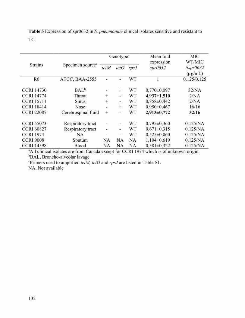
Table 5 Expression of spr0632 in S. pneumoniae clinical isolates sensitive and resistant to
TC.
Strains
Specimen sourcea
Genotypec Mean fold expression
MIC WT/MIC ∆spr0632 (µg/mL)
tetM tetO rpsJ spr0632
R6 ATCC, BAA-2555 - - WT 1 0.125/0.125 CCRI 14730 BALb - + WT 0,770±0,097 32/NA CCRI 14774 Throat + - WT 4,937±1,510 2/NA CCRI 15711 Sinus + - WT 0,858±0,442 2/NA CCRI 18414 Nose - + WT 0,950±0,467 16/16 CCRI 22087 Cerebrospinal fluid + - WT 2,913±0,772 32/16 CCRI 55073 Respiratory tract - - WT 0,795±0,360 0.125/NA CCRI 60827 Respiratory tract - - WT 0,671±0,315 0.125/NA CCRI 1974 NA - - WT 0,525±0,060 0.125/NA CCRI 9008 Sputum NA NA NA 1,104±0,619 0.125/NA CCRI 14598 Blood NA NA NA 0,581±0,322 0.125/NA
aAll clinical isolates are from Canada except for CCRI 1974 which is of unknown origin. bBAL, Broncho-alveolar lavage cPrimers used to amplified tetM, tetO and rpsJ are listed in Table S1. NA, Not available
132

133

Figure 1 Accumulation of TC in S. pneumoniae. (A) Accumulation of labeled TC was
measured in S. pneumoniae R6 WT (circles), R6M1TC-5 (diamonds) and R6M2TC-4
(squares) at baseline and at 5 minutes intervals for a total of 20 minutes. (B) The role of
mutations selected at the patA locus in R6M1TC-5 on accumulation of TC was assessed by
comparing the accumulation of labeled TC in R6 WT (circles) R6patA(R6M1TC) (empty
diamonds), R6p_patA(R6M1TC)-rpsL+(upward triangles) and R6CDSpatA(R6M1TC)-rpsL+ (downward
triangles) for up to 20 minutes. (C) The role of mutations selected at the patA locus in
R6M2TC-4 on accumulation of TC was assessed by comparing the accumulation of labeled
TC in R6 WT (circles), R6patA-patB(R6M2TC) (empty squares), R6p_patA(R6M2TC)-rpsL+ (upward
triangles) and R6CDSpatA-patB(R6M2TC)-dhfr+ (downward triangles) for up to 20 minutes. Results
represent the mean of three independent replicates. T-test was performed on normalized
data at 10 and 20 minutes and the significance threshold set to p≤ 0.05. (D) Accumulation
of TC in the presence of reserpine in R6 WT (circles), R6patA(R6M1TC) (empty diamonds) and
R6patA-patB(R6M2TC) (empty squares). The arrow indicates the time point at which 20 mg/mL
reserpine was added.
134

Figure 2 Gene expression alterations in S. pneumoniae TC resistant mutants. Gene
expression was compared between S. pneumoniae R6 WT and (A) R6M1TC-5 or (B)
R6M2TC-4 by RNAseq. The dots represent the 2043 genes from the S. pneumoniae R6
genome sorted in numerical order on the abscissa. Dots coloured in black, blue and grey
represent overexpressed, downregulated and not significantly altered genes (p≤ 0.01),
respectively.
135

Figure 3. GO classification of genes whose expression is significantly altered in S.
pneumoniae TC resistant mutants. Genes differentially expressed (p-value≤0.015) in (A)
R6M1TC-5 and (B) R6M2TC-4 compared to S. pneumoniae R6 WT were clustered
136

according to their ontology. Go terms surround by a dark grey, medium grey and light grey
correspond to cellular component, molecular function and biological process associated
GO, respectively. The percentage of sequences associated with a specific GO term is shown
for overexpressed genes (green), downregulated genes (blue) and overall S. pneumoniae R6
genes (red).
137

Suplementary data
Table S1 – Strain used in this study
Strains Description References R6 Wild-type ATCC BAA
255 CCRI 14730 clinical isolates CCRI 14774 clinical isolates CCRI 15711 clinical isolates CCRI 18414 clinical isolates CCRI 22087 clinical isolates CCRI 55073 clinical isolates CCRI 60827 clinical isolates CCRI 1974 clinical isolates CCRI 9008 clinical isolates CCRI 14598 clinical isolates R6M1TC 1-5 R6 laboratory-derived mutants resistant to
TC This work
R6M2TC 1-4 R6 laboratory derived mutants resistant to TC
This work
R6rpsJ(R6M1TC-1) R6 transformed with rpsJ (spr0187) PCR fragment amplified from R6M1TC-1
This work
R6rpsJ(R6M1TC-1)-rev(R6WT)-
rpsL+ R6rpsJ(R6M1TC-1) co-transformed with rpsJ (spr0187) PCR fragment amplified from R6 and streptomycin resistance marker rpsL+, SMr
R6rpsJ(R6M1TC-5) R6 transformed with rpsJ (spr0187) PCR fragment amplified from R6M1TC-5
This work
R6rpsJ(R6M1TC-5)-rev(R6WT)-
rpsL+ R6rpsJ(R6M1TC-5) co-transformed with rpsJ (spr0187) PCR fragment amplified from R6 and rpsL+, SMr
R6spr0825(R6M1TC)- rpsL+ R6 co-transformed with spr0825 PCR fragment amplified from R6M1TC-5 and rpsL+, SMr
This work
R6patA(R6M1TC) R6 transformed with PCR fragment containing mutations in coding region and promoter region of patA from R6M1TC-5
This work
R6p_patA(R6M1TC)-rpsL+ R6 co-transformed with a PCR fragment containing mutations in promoter region of patA from R6M1TC-5 and rpsL+, SMr
This work
R6CDSpatA(R6M1TC)-rpsL+ R6 co-transformed with PCR fragment containing mutations in coding region of patA from R6M1TC-5 and rpsL+, SMr
This work
138

R6CDSpatA(G510A)(R6M1TC)-
rpsL+ R6 co-transformed with PCR fragment containing mutations in coding region of patA from R6M1TC-5 and rpsL+, SMr
This work
R6CDSpatA(C883A)(R6M1TC)-
rpsL+ R6 co-transformed with PCR fragment containing mutations in coding region of patA from R6M1TC-5 and rpsL+, SMr
This work
R6rpsJ(R6M1TC)-spr0825(R6M1TC)-
rpsL+ R6rpsJ-R6M1TC co-transformed with spr0825 PCR fragment amplified from R6M1TC-5 and rpsL+,SMr
This work
R6rpsJ(R6M1TC)-patA(R6M1TC) R6rpsJ-R6M1TC transformed with PCR fragment containing mutations in the coding region of patA from R6M1TC-5
This work
R6rpsJ(R6M1TC)-patA(R6M1TC)-
spr0825(R6M1TC)-rpsL+ R6rpsJ-R6M1TC-patA-M1TC co-transformed with spr0825 PCR fragment amplified from R6M1TC-5
This work
R6rpsJ(R6M1TC)-patA(R6M1TC)-
spr0825(R6M1TC)-spr1262(R6M1TC)-
dhfr+
R6rpsJ-R6M1TC-patA-R6M1TC-spr0825-R6M1TC rpsL+ co-transformed with spr1262 PCR fragment amplified from R6M1TC and dhfr+, thrimethoprim resistance marker, TMPr
This work
R6rpsJ(R6M2TC) R6 transformed with rpsJ (spr0187) PCR fragment amplified from R6M2TC-4
This work
R6patA-patB(R6M2TC) R6 transformed with a PCR fragment containing mutations in promoter and coding region of patA and patB from R6M2TC-4
This work
R6patA-patB(R6M2TC)-rev(patA)-
rpsL+ R6patA-patB(R6M2TC) co-transformed with patA PCR fragment amplified from R6 and rpsL+, SMr
R6p_patA(R6M2TC)-rpsL+ R6 co-transformed with PCR fragment containing mutations in promoter region of patA from R6M2TC-4 and rpsL+, SMr
This work
R6CDSpatA(R6M2TC)-rpsL+ R6 co-transformed with fragment containing mutations in coding region of patA from R6M2TC-4 and rpsL+, SMr
This work
R6CDSpatB(R6M2TC)-rpsL+ R6 co-transformed with fragment containing mutations in coding region of patB from R6M2TC-4 and rpsL+, SMr
This work
R6CDSpatA-patB(R6M2TC)-dhfr+ R6CDSpatB-R6M2TC rpsL+ co-transformed with fragment containing mutations in patA from R6M2TC-4 and dhfr+, TMPr
This work
R6rpsJ(R6M2TC)-patA(R6M2TC) R6rpsJ-R6M1TC transformed with patA PCR fragment containing mutations in promoter and coding regions of R6M2TC-5
This work
R6rpsJ(R6M2TC)-patA(R6M2TC)-
patB(R6M2TC) R6rpsJ-R6M1TC transformed with PCR fragment containing mutations in promoter region of patA and coding regions of patA and patB from R6M1TC-5
This work
R6rpsJ(R6M2TC)-patA(R6M2TC)- R6rpsJ- R6M2TC-patAR6M2TC-patBR6M2TC co- This work
139

patB(R6M2TC)-spr0291(R6M2TC)-
rpsL+ transformed with spr0291 PCR fragment amplified from R6M2TC-4 and rpsL+, SMr
R6rpsJ(R6M2TC)-patA(R6M2TC)-
patB(R6M2TC)-spr0822(R6M2TC)-
rpsL+
R6rpsJ- R6M2TC-patAR6M2TC-patBR6M2TC co-transformed with spr0822 PCR fragment amplified from R6M2TC-4 and rpsL+, SMr
This work
R6rpsJ(R6M2TC)-patA(R6M2TC)-
patB(R6M2TC)-spr1377(R6M2TC)-
rpsL+
R6rpsJ- R6M2TC-patAR6M2TC-patBR6M2TC co-transformed with spr1377 PCR fragment amplified from R6M2TC-4 and rpsL+, SMr
This work
R6rpsJ(R6M2TC)-patA(R6M2TC)-
patB(R6M2TC)-spr0691(R6M2TC)-
rpsL+
R6rpsJ- R6M2TC-patAR6M2TC-patBR6M2TC co-transformed with spr0691 PCR fragment amplified from R6M2TC-4 and rpsL+, SMr
This work
CCRI 22087 pFF3spr0632(CCRI22087)
CCRI 22087 transformed with pFF3spr0632(CCRI22087), CMr
This work
R6pFF6patB Lupien et al 2013
R6pFF6patA Lupien et al.2013
R6pFF6spr0632 R6 transformed with plasmid pFF6spr0632, KANr
This work
R6M1TC-5pFF6patB R6M1TC-5 transformed with plasmid pFF6patB, KANr
This work
R6M1TC-5pFF6patA R6M1TC-5 transformed with plasmid pFF6patA, KANr
This work
R6M1TC-5pFF6spr0632 R6M1TC-5 transformed with plasmid pFF6spr0632, KANr
This work
R6M1TC-5pFF6spr0634 R6M1TC-5 transformed with plasmid pFF6spr0634, KANr
This work
R6M1TC-5pFF6spr0637 R6M1TC-5 transformed with plasmid pFF6spr0637, KANr
This work
R6M1TC-5pFF6spr0638 R6M1TC-5 transformed with plasmid pFF6spr0638, KANr
This work
R6M2TC-4pFF6patB R6M2TC-2 transformed with plasmid pFF6patB, KANr
This work
R6M2TC-4pFF6patA R6M2TC-2 transformed with plasmid pFF6patA, KANr
This work
R6M2TC-4pFF6 spr0632 R6M2TC-2 transformed with plasmid pFF6spr0632, KANr
This work
R6M2TC-4pFF6 spr0634 R6M2TC-2 transformed with plasmid pFF6spr0634, KANr
This work
R6M2TC-4pFF6 spr0638 R6M2TC-2 transformed with plasmid pFF6spr0638, KANr
This work
Plasmid pFF6 Lupien et
al.2013 pFF6patA Lupien et
al.2013 pFF6patB Lupien et
140

al.2013 pFF6spr0632(R6) plasmid pFF6(KANr) containing a fragment
of spr0632 amplified from R6 This work
pFF3spr0632(CCRI22087) plasmid pFF3(CMr) containing a fragment of spr0632 amplified from CCRI 22087
This work
pFF6spr0634 plasmid pFF6(KANr) containing a fragment of spr0634 amplified from R6
This work
pFF6spr0637 plasmid pFF6(KANr) containing a fragment of spr0637 amplified from R6
This work
pFF6spr0638 plasmid pFF6(KANr) containing a fragment of spr0638 amplified from R6
This work
141
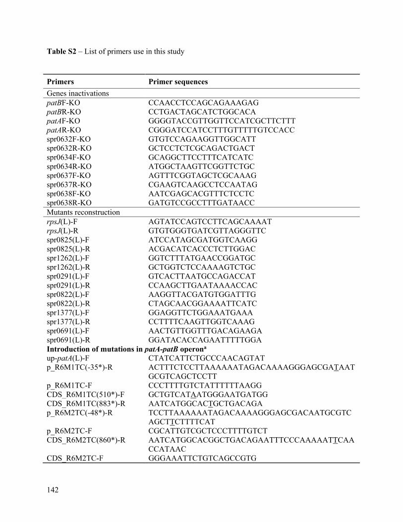
Table S2 – List of primers use in this study
Primers Primer sequences Genes inactivations patBF-KO CCAACCTCCAGCAGAAAGAG patBR-KO CCTGACTAGCATCTGGCACA patAF-KO GGGGTACCGTTGGTTCCATCGCTTCTTT patAR-KO CGGGATCCATCCTTTGTTTTTGTCCACC spr0632F-KO GTGTCCAGAAGGTTGGCATT spr0632R-KO GCTCCTCTCGCAGACTGACT spr0634F-KO GCAGGCTTCCTTTCATCATC spr0634R-KO ATGGCTAAGTTCGGTTCTGC spr0637F-KO AGTTTCGGTAGCTCGCAAAG spr0637R-KO CGAAGTCAAGCCTCCAATAG spr0638F-KO AATCGAGCACGTTTCTCCTC spr0638R-KO GATGTCCGCCTTTGATAACC Mutants reconstruction rpsJ(L)-F AGTATCCAGTCCTTCAGCAAAAT rpsJ(L)-R GTGTGGGTGATCGTTAGGGTTC spr0825(L)-F ATCCATAGCGATGGTCAAGG spr0825(L)-R ACGACATCACCCTCTTGGAC spr1262(L)-F GGTCTTTATGAACCGGATGC spr1262(L)-R GCTGGTCTCCAAAAGTCTGC spr0291(L)-F GTCACTTAATGCCAGACCAT spr0291(L)-R CCAAGCTTGAATAAAACCAC spr0822(L)-F AAGGTTACGATGTGGATTTG spr0822(L)-R CTAGCAACGGAAAATTCATC spr1377(L)-F GGAGGTTCTGGAAATGAAA spr1377(L)-R CCTTTTCAAGTTGGTCAAAG spr0691(L)-F AACTGTTGGTTTGACAGAAGA spr0691(L)-R GGATACACCAGAATTTTTGGA Introduction of mutations in patA-patB operona up-patA(L)-F CTATCATTCTGCCCAACAGTAT p_R6M1TC(-35*)-R ACTTTCTCCTTAAAAAATAGACAAAAGGGAGCGATAAT
GCGTCAGCTCCTT p_R6M1TC-F CCCTTTTGTCTATTTTTTAAGG CDS_R6M1TC(510*)-F GCTGTCATAATGGGAATGATGG CDS_R6M1TC(883*)-R AATCATGGCACTGCTGACAGA p_R6M2TC(-48*)-R TCCTTAAAAAATAGACAAAAGGGAGCGACAATGCGTC
AGCTTCTTTTCAT p_R6M2TC-F CGCATTGTCGCTCCCTTTTGTCT CDS_R6M2TC(860*)-R AATCATGGCACGGCTGACAGAATTTCCCAAAAATTCAA
CCATAAC CDS_R6M2TC-F GGGAAATTCTGTCAGCCGTG
142

patB-R TTATTCGAAAACAAATTGATTGTG
tet(M) and tet(O) PCR tet(M)-F GTGGACAAAGGTACAACGAG tet(M)-R CGGTAAAGTTCGTCACACAC tet(O)-F AACTTAGGCATTCTGGCTCAC tet(O)-R TCCCACTGTTCCATATCGTCA qRT-PCR RT16sRNA-F CCTATTGTTAGTTGCCATCATTCAG RT16srRNA-R GACTCGTTGTACCAGCCATTGT RTpatA -F GGTGGCAAGGATATTCGAGA RTpatA-R AGAATGGCACGTTGGAGAAC RTspr0632-F GTGTCCAGAAGGTTGGCATT RTspr0632-R ATTGCTGGACAACCTCTGCT RTspr0634-F CAGGGTTTAGTGGAGGGTGA RTspr0634-R GGATGTTGCTCCATTTCCTG RTspr0637-F AGTTTCGGTAGCTCGCAAAG RTspr0637-R AAATGGCTCCTGTACCCAAG RTspr0638-F GGTTATCAAAGGCGGACATC RTspr0638-R CTGCAGCAAAGGTACATCCA Abbreviation: F: Forward; R: Reverse; (L): Long fragment; up: upstream; qRT-PCR: quantitative real-time PCR. a Modify nucleotides, compare to R6 sequence, are underlines
143

Table S3: Significantly expressed genes (p-value ≤0,015) in R6M1TC-5 compared to R6
according to RNAseq experiment
R6M1TC-5/R6 R6M2TC-4/R6
gene function log2(fold-change)
p_value q_value log2(fold_change)
p_value q_value
overexpressed
Spr0057
beta-N-acetyl hexosaminidase 2,6601 0,00046337 0,0583966
Spr0559 Beta-galactosidase 3 2,38199 0,00261514 0,138373
Spr0661 PTS system IIC component 2,69512 0,0004137 0,0562084
Spr0664 sugar isomerase domain protein AgaS
2,52529 0,00108121 0,0786964
Spr0065 aldose 1-epimerase 2,36627 0,00264796 0,138373
Spr0081 ABC transporter permease 2,88557 0,00014387 0,0325785
Spr0082 ABC transporter permease 3,22419 3,10E-05 0,0104996
Spr0083 ABC transporter substrate-binding protein
2,72874 0,00031731 0,0497448
spr0098 bacteriocin 2,25696 0,00700 0,28540 2,07978 0,00847928 0,250446
Spr0115 hypothetical protein 2,05847 0,0106456
0,285469
Spr0118 hypothetical protein 2,27423 0,00344075
0,163076
Spr0161 6,7-dimethyl-8-ribityllumazine synthase
1,94427 0,0122371 0,311739
spr0299 hypothetical protein 2,67247 0,00699 0,28540
Spr0440 endo-beta-N-acetylglucosaminidase
2,63361 0,00048712 0,0583966
spr0470 hypothetical protein 2,83064 0,00149 0,11237
spr0482 ribosome-binding factor A 2,44772 0,00146 0,11237
spr0484 Putative uncharacterized protein 4,52046 0,00004 0,00893
spr0550 hypothetical protein 2,47757 0,00806 0,31256
Spr0563 hypothetical protein 2,68373 0,00088272 0,0764286
Spr0567 hypothetical protein 3,06705 0,00022324 0,0379143
spr0586 hypothetical protein 2,00283 0,01338 0,40130 2,12146 0,00576149 0,225806
Spr0587 hypothetical protein 2,07933 0,00463764 0,18903
Spr0600 hypothetical protein 2,8081 0,00021249 0,0379143
Spr0602 sodium ABC transporter ATP-binding protein
2,5702 0,00062315 0,0634993
Spr0617 hypothetical protein 2,13386 0,00713938 0,236314
Spr0620 ABC transporter substrate-binding protein - unknown substrate, truncation, partial
2,38426 0,00291874 0,14871
Spr0621 ABC transporter substrate-binding protein - unknown substrate, truncation, partial
2,32539 0,00605917 0,232629
Spr0622 amino acid ABC transporter ATP-binding protein
2,63268 0,0007733 0,0749924
144
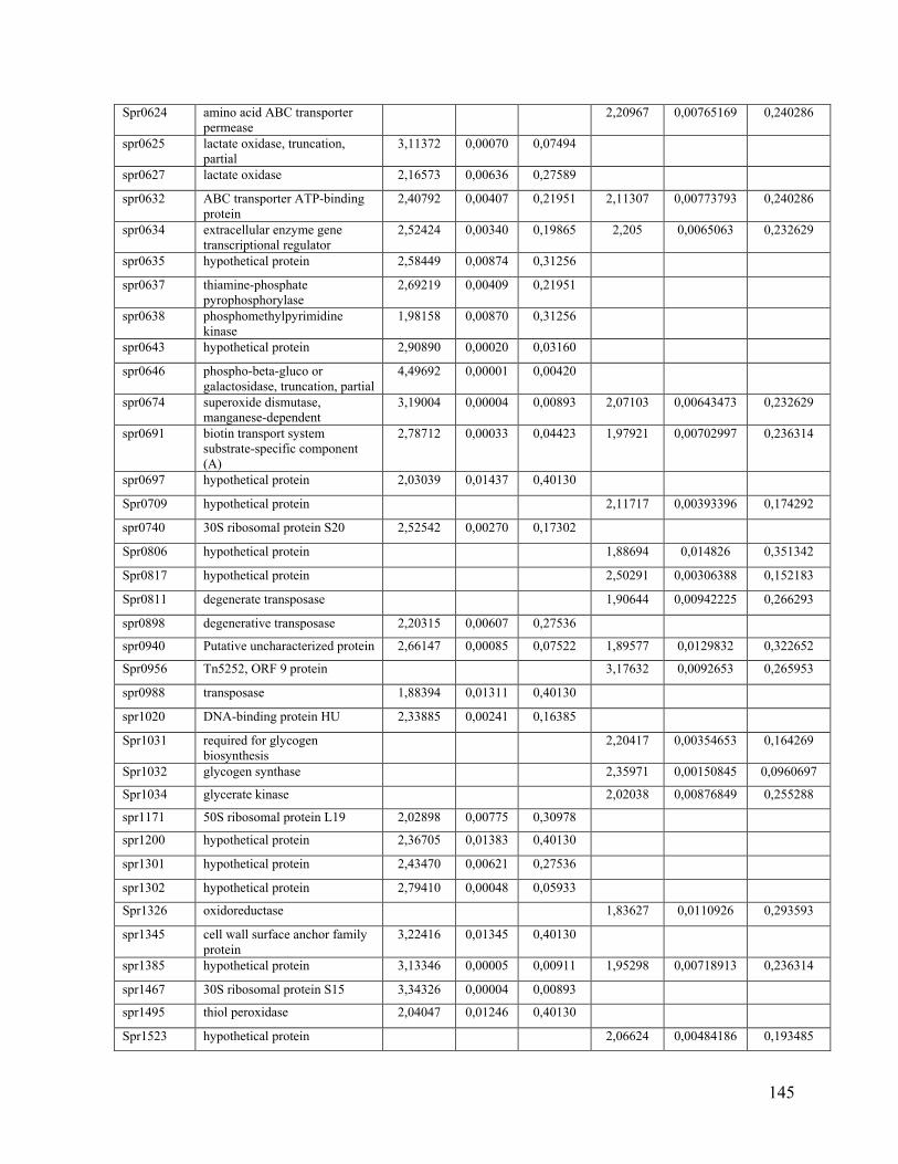
Spr0624 amino acid ABC transporter permease
2,20967 0,00765169 0,240286
spr0625 lactate oxidase, truncation, partial
3,11372 0,00070 0,07494
spr0627 lactate oxidase 2,16573 0,00636 0,27589
spr0632 ABC transporter ATP-binding protein
2,40792 0,00407 0,21951 2,11307 0,00773793 0,240286
spr0634 extracellular enzyme gene transcriptional regulator
2,52424 0,00340 0,19865 2,205 0,0065063 0,232629
spr0635 hypothetical protein 2,58449 0,00874 0,31256
spr0637 thiamine-phosphate pyrophosphorylase
2,69219 0,00409 0,21951
spr0638 phosphomethylpyrimidine kinase
1,98158 0,00870 0,31256
spr0643 hypothetical protein 2,90890 0,00020 0,03160
spr0646 phospho-beta-gluco or galactosidase, truncation, partial
4,49692 0,00001 0,00420
spr0674 superoxide dismutase, manganese-dependent
3,19004 0,00004 0,00893 2,07103 0,00643473 0,232629
spr0691 biotin transport system substrate-specific component (A)
2,78712 0,00033 0,04423 1,97921 0,00702997 0,236314
spr0697 hypothetical protein 2,03039 0,01437 0,40130
Spr0709 hypothetical protein 2,11717 0,00393396 0,174292
spr0740 30S ribosomal protein S20 2,52542 0,00270 0,17302
Spr0806 hypothetical protein 1,88694 0,014826 0,351342
Spr0817 hypothetical protein 2,50291 0,00306388 0,152183
Spr0811 degenerate transposase 1,90644 0,00942225 0,266293
spr0898 degenerative transposase 2,20315 0,00607 0,27536
spr0940 Putative uncharacterized protein 2,66147 0,00085 0,07522 1,89577 0,0129832 0,322652
Spr0956 Tn5252, ORF 9 protein 3,17632 0,0092653 0,265953
spr0988 transposase 1,88394 0,01311 0,40130
spr1020 DNA-binding protein HU 2,33885 0,00241 0,16385
Spr1031 required for glycogen biosynthesis
2,20417 0,00354653 0,164269
Spr1032 glycogen synthase 2,35971 0,00150845 0,0960697
Spr1034 glycerate kinase 2,02038 0,00876849 0,255288
spr1171 50S ribosomal protein L19 2,02898 0,00775 0,30978
spr1200 hypothetical protein 2,36705 0,01383 0,40130
spr1301 hypothetical protein 2,43470 0,00621 0,27536
spr1302 hypothetical protein 2,79410 0,00048 0,05933
Spr1326 oxidoreductase 1,83627 0,0110926 0,293593
spr1345 cell wall surface anchor family protein
3,22416 0,01345 0,40130
spr1385 hypothetical protein 3,13346 0,00005 0,00911 1,95298 0,00718913 0,236314
spr1467 30S ribosomal protein S15 3,34326 0,00004 0,00893
spr1495 thiol peroxidase 2,04047 0,01246 0,40130
Spr1523 hypothetical protein 2,06624 0,00484186 0,193485
145
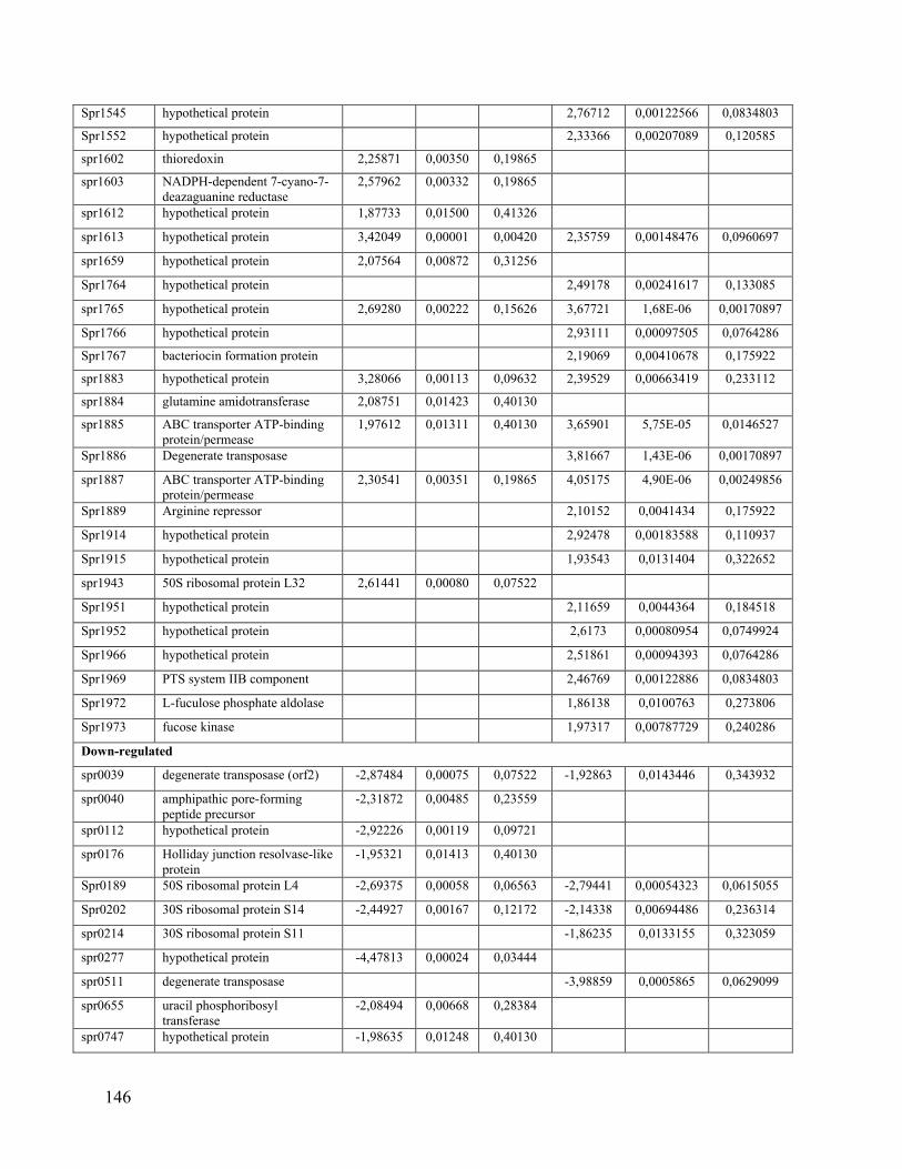
Spr1545 hypothetical protein 2,76712 0,00122566 0,0834803
Spr1552 hypothetical protein 2,33366 0,00207089 0,120585
spr1602 thioredoxin 2,25871 0,00350 0,19865
spr1603 NADPH-dependent 7-cyano-7-deazaguanine reductase
2,57962 0,00332 0,19865
spr1612 hypothetical protein 1,87733 0,01500 0,41326
spr1613 hypothetical protein 3,42049 0,00001 0,00420 2,35759 0,00148476 0,0960697
spr1659 hypothetical protein 2,07564 0,00872 0,31256
Spr1764 hypothetical protein 2,49178 0,00241617 0,133085
spr1765 hypothetical protein 2,69280 0,00222 0,15626 3,67721 1,68E-06 0,00170897
Spr1766 hypothetical protein 2,93111 0,00097505 0,0764286
Spr1767 bacteriocin formation protein 2,19069 0,00410678 0,175922
spr1883 hypothetical protein 3,28066 0,00113 0,09632 2,39529 0,00663419 0,233112
spr1884 glutamine amidotransferase 2,08751 0,01423 0,40130
spr1885 ABC transporter ATP-binding protein/permease
1,97612 0,01311 0,40130 3,65901 5,75E-05 0,0146527
Spr1886 Degenerate transposase 3,81667 1,43E-06 0,00170897
spr1887 ABC transporter ATP-binding protein/permease
2,30541 0,00351 0,19865 4,05175 4,90E-06 0,00249856
Spr1889 Arginine repressor 2,10152 0,0041434 0,175922
Spr1914 hypothetical protein 2,92478 0,00183588 0,110937
Spr1915 hypothetical protein 1,93543 0,0131404 0,322652
spr1943 50S ribosomal protein L32 2,61441 0,00080 0,07522
Spr1951 hypothetical protein 2,11659 0,0044364 0,184518
Spr1952 hypothetical protein 2,6173 0,00080954 0,0749924
Spr1966 hypothetical protein 2,51861 0,00094393 0,0764286
Spr1969 PTS system IIB component 2,46769 0,00122886 0,0834803
Spr1972 L-fuculose phosphate aldolase 1,86138 0,0100763 0,273806
Spr1973 fucose kinase 1,97317 0,00787729 0,240286
Down-regulated
spr0039 degenerate transposase (orf2) -2,87484 0,00075 0,07522 -1,92863 0,0143446 0,343932
spr0040 amphipathic pore-forming peptide precursor
-2,31872 0,00485 0,23559
spr0112 hypothetical protein -2,92226 0,00119 0,09721
spr0176 Holliday junction resolvase-like protein
-1,95321 0,01413 0,40130
Spr0189 50S ribosomal protein L4 -2,69375 0,00058 0,06563 -2,79441 0,00054323 0,0615055
Spr0202 30S ribosomal protein S14 -2,44927 0,00167 0,12172 -2,14338 0,00694486 0,236314
spr0214 30S ribosomal protein S11 -1,86235 0,0133155 0,323059
spr0277 hypothetical protein -4,47813 0,00024 0,03444
spr0511 degenerate transposase -3,98859 0,0005865 0,0629099
spr0655 uracil phosphoribosyl transferase
-2,08494 0,00668 0,28384
spr0747 hypothetical protein -1,98635 0,01248 0,40130
146

spr0751 degenerate transposase (orf1) -2,87366 0,00611 0,27536
spr0798 hypothetical protein -2,11898 0,0117194 0,30233
spr0861 translation initiation factor IF-3 -2,03179 0,00849 0,31256 -1,92438 0,0112523 0,294003
spr0922 thymidine kinase -2,13159 0,01028 0,35532 -2,08244 0,00782243 0,240286
spr0961 UDP-N-acetyl-D-mannosaminuronic acid dehydrogenase
-4,75566 0,00464 0,23559
spr1075 galactose-6-phosphate isomerase subunit LacB
-2,67414 0,00049 0,05933
spr1080 hypothetical protein -2,35048 0,01088 0,36983
spr1199 hypothetical protein -3,75566 0,00597 0,27536
spr1239 cytoplasmic alpha-amylase -1,86341 0,00993652 0,273657
spr1241 hypothetical protein -1,86572 0,01419 0,40130
spr1396 30S ribosomal protein S6 -2,1709 0,00313625 0,152183
spr1440 ATP-dependent RNA helicase -2,08664 0,00789948 0,240286
spr1481 hypothetical protein -2,53445 0,00889 0,31256 -2,60577 0,00223728 0,126655
spr1573 tranposase (orf2) -2,00762 0,01286 0,40130
spr1744 degenerate transposase (orf1) -2,87212 0,00873 0,31256
spr1817 ABC transporter ATP-bindingprotein
-2,82623 0,00037121 0,0540381
spr1818 hypothetical protein -3,19923 0,00018185 0,0370606
spr1859 hypothetical protein -2,47308 0,00366759 0,166101
Spr1863 competence protein CglB -2,17497
0,00617806 0,232629
spr1936 transketolase, C-terminal subunit, partial
-3,20167 0,00005 0,00911 -1,98423 0,00643179 0,232629
spr1937 transketolase n-terminal section, partial
-4,07009 0,0000004
0,00075 -2,34521 0,00185077 0,110937
spr1938 ascorbate-specific PTS system enzyme IIC
-3,54898 0,00001 0,00420 -2,44367 0,00102541 0,0773995
spr1939 PTS system IIB component -2,79126 0,00084 0,07522
spr1940 hypothetical protein -3,37690 0,00001 0,00420 -1,94039 0,0129914 0,322652
spr1970 PTS system IIA component -2,50592 0,00477 0,23559
spr1987 hypothetical protein -2,92291 0,00272 0,17302 -3,73452 2,73E-05 0,0104996
spr1989 glycerol-3-phosphate dehydrogenase, truncation, partial
-2,47348 0,00479 0,23559 -2,67753
0,00094798 0,0764286
spr1990 glycerol-3-phosphate dehydrogenase, truncation, partial
-3,54886 0,00001 0,00420 -3,21224 3,61E-05 0,0104996
spr1991 Glycerol kinase -3,63915 0,000004
0,00412 -3,73035 2,68E-06 0,00182256
spr2027 CDP-diacylglycerol--glycerol-3-phosphate 3-phosphatidyltransferase
-1,96009 0,01354 0,40130
spr2045 serine protease -2,13976 0,00953847 0,266293
147


Chapitre X: L’induction de la résistance à la tigécycline chez
Streptococcus pneumoniae révèle la sélection de mutations dans
des protéines ribosomales et de l’ARN ribosomal
Ce chapitre sera soumis sous peu pour publication.
10.1 Résumé
La tigécycline est un antibiotique à large spectre qui agit sur la sous-unité 30S du
ribosome, ce qui inhibe la traduction. Développée en réponse à l’augmentation de la
prévalence de bactéries multi-résistantes, la tigécycline a une bonne activité contre
plusieurs bactéries à Gram négatif, Gram positif et anaérobes, incluant Streptococcus
pneumoniae. Nous avons étudié les déterminants moléculaires de la résistance à la
tigécycline chez S. pneumoniae par le séquençage du génome de deux séries de mutants
sélectionnnés in vitro en présence d’une concentration croissante de tigécycline. Les
mutants S. pneumoniae R6M1TGC-6 et R6M2TGC-6 présentent une résistance croisée à la
tétracycline et à la minocycline. Un rôle dans la résistance à la TGC a été attribué à quatre
des douze gènes altérés chez les deux mutants. Les mutations dans la protéine ribosomale
S10, apparues précocement lors de la sélection des mutants, et dans la protéine S3, acquises
tardivement, ont été toutes deux impliquées dans la résistance par reconstruction
génotypique. Également, des mutations ont été détectées dans les quatre allèles de l’ARNr
16s aux sites impliqués dans la liaison de la TGC. Le nombre d’allèles mutés corrèle avec
le niveau de résistance à la TGC et à la TC. Finalement, les mutations non-sens
sélectionnées chez les mutants dans le gène spr1784, codant potentiellement pour une
méthyltransférase de l’ARNr 16s, diminuent la sensibilité à la TGC. Cette première étude
des mécanismes de résistance de la TGC chez S. pneumoniae a révélé que,
comparativement aux espèces bactériennes à Gram négatif où des mécanismes d’efflux sont
principalement impliqués, la résistance à la TGC chez S. pneumoniae résulte de
l’acquisition de mutations reliées aux ribosomes.
151

10.2 Article
Induced tigecycline resistance in Streptococcus pneumoniae
mutants reveals mutations in ribosomal proteins and rRNA.
Andréanne Lupien1, Hélène Gingras 1, Philippe Leprohon1, Marc Ouellette1§
1 Centre de recherche en Infectiologie du Centre de recherche du CHU de Québec and
Département de Microbiologie, Infectiologie et Immunologie, Faculté de Médecine,
Université Laval, Québec, QC, Canada. §Corresponding author: Marc Ouellette Ph.D.
Centre de Recherche en Infectiologie
2705 Boul. Laurier
Québec, QC
Canada
G1V 4G2
Tel : 1-418-654-2705
Fax : 1-418-654-2715
E-mail :[email protected]
Running title: Tigecycline resistance in Streptococcus pneumonia
Keywords: Streptococcus pneumoniae, tigecycline, whole-genome sequencing
152
Abstract
Tigecycline (TGC) is a broad spectrum antibiotic acting at the level of the 30S
ribosomal subunit to inhibit translation. Developed primarily in response to the increased
prevalence of multidrug-resistant bacteria, it has a good activity against many Gram-
negative, Gram-positive and anaerobes bacteria, including Streptococcus pneumoniae. We
studied the molecular determinants of resistance to TGC in S. pneumoniae through whole
genome sequencing of two series of mutants made resistant to TGC in vitro in a stepwise
fashion. The S. pneumoniae R6M1TGC-6 and R6M2TGC-6 mutants were cross-resistant to
tetracycline and minocycline. A role in TGC resistance could be attributed to four of the
twelve genes that were mutated in both mutants. Mutations in ribosomal protein S10 and S3
respectively acquired early and late during the selection of both mutants have been
implicated in resistance by genotype reconstruction. Similarly, mutations were detected in
the four alleles of the 16S ribosomal rRNA at sites involved in TGC binding and the
number of mutated alleles correlated with the level of resistance. Finally, the gene spr1784
encodes a putative 16S rRNA methyltransferase for which inactivating mutations selected
in the S. pneumoniae R6 TGC-resistant mutants were found to decrease susceptibility to
TGC. This first report about TGC resistance mechanisms in S. pneumoniae revealed that in
contrast to Gram-negative bacteria for which efflux appears central for TGC resistance,
TGC resistance in the pneumococcus occurs through mutations related to ribosomes.
153
Introduction
Streptococcus pneumoniae is a Gram-positive bacterium responsible for several
diseases including otitis media, meningitis and pneumonia. Due to the spread of multidrug-
resistant clones not included in the pneumococcal conjugate vaccine [1-3] the prevalence of
resistance among S. pneumoniae is globally high for β-lactams [4, 5]. The situation is even
worst for macrolides and tetracycline (TC) for which resistance rates higher than 50% were
reported [3, 6]. Second generation tetracyclines have been proposed for the treatment of
community-acquired pneumonia [7, 8] but cross-resistance between compounds of the TC
family is observable in pneumococci [6, 9] with the exception for the new glycylcycline
tigecycline (TGC).
TGC is a new broad spectrum antibiotic with a good activity against Gram-positive,
Gram-negative and anaerobes bacteria [10, 11]. It was first introduced in 2005 for the
treatment of complicated skin and skin-structure infections (cSSSIs) and intra-abdominal
infections (cIAIs) [12] and was later approved by the FDA in 2010 for the treatment of
community-acquired pneumonia for which S. pneumoniae is an important etiologic agent
[13]. It is a third generation TC derived from minocycline (MI) to which a 9-t-
butylglycylamido moiety has been added at position C9 of the four-ring structure [14]. In
general, TGC is considered bacteriostatic; however it has demonstrated bactericidal activity
against isolates of S. pneumoniae [15] and methicillin resistant Staphilococcus aureus
(MRSA) in a rat endocarditis model [16]. Similarly to TC, TGC is reversibly binding to the
decoding center of the 30S ribosome subunit [17], although at an affinity up to 20 times
greater compared to other TC compounds [18, 19]. TGC also displays improved inhibitory
activities against a range of bacteria [20, 21], which is likely explained by the increased
steric hindrance that TGC imposes at the A site of the ribosome during prokaryotic
translation [17, 22]. TGC also overcomes resistance resulting from the expression of the
well characterized tet-gene encoded efflux pumps and ribosomal protection proteins [21,
23].
154
TGC remains very effective in the case of the pneumococcus [24-27] but the
apparition of TGC non-susceptible isolates in other species is worrying [27-32]. In Gram-
negative bacteria, resistance to TGC is mainly caused by the expression of efflux pumps of
the resistance-nodulation-cell division (RND) superfamily. More specifically, the
expression of the chromosomally encoded MexXY-OmpR and AcrAB pumps is
responsible for the intrinsic TGC resistance of Pseudomonas aeruginosa and Proteus
mirabilis, respectively [33, 34]. Similarly, the overexpression of the AdeABC and SdeXY
RND pumps is responsible for reduced susceptibility to TGC in Acinetobacter baumanii
and Serratia marcescens, respectively [35, 36]. For several Gram-negative bacteria, the
increased activity of these RND pumps is related to alterations in factors regulating their
expression [37-40]. Despite the fact that TGC was designed to overcome resistance
mediated by tet genes, mutations in the tetA efflux system were shown to reduce
susceptibility to TGC in Salmonella [41-43]. Resistance to TGC in Gram-positive bacteria
is very scarce but studying MRSA selected for resistance to TGC revealed a novel MATE
efflux system called MepA whose increased expression contributed to reduced
susceptibility to TGC [44].
Besides efflux, resistance to TGC can also occur through the flavin-dependent
monooxygenase TetX that modifies TGC to 11a-hydroxytigecycline, a molecule with a
weakened ability to inhibit protein translation compared to TGC [45, 46]. A mutation at the
vertex of a conserved loop in ribosomal protein S10 has also been implicated in resistance
to TGC in Klebsiella pneumoniae [39]. In A. baumanni, a mutation leading to a truncated
version of an S-adenosyl-L-methionine-dependent methyltransferase was shown to confer
an 8-fold increase in resistance to TGC [47]. Finally, mutations in genes linked to the
biosynthesis of the lipopolysaccharide core were shown to increase resistance to TGC in
Escherichia coli [48].
In this study, we characterized S. pneumoniae mutants selected for resistance to
TGC by the combined use of next generation sequencing (NGS) and resistance
reconstruction. We report that TGC resistance involves mutations in ribosomal protein S3
and S10, in the 16S ribosomal RNA (rRNA) and also in a putative 16S rRNA
155

methyltransferase from the RsmD family. These constitute new molecular determinants of
resistance to TGC in S. pneumoniae.
156

Results
TGC mutants
Two S. pneumoniae mutants resistant to TGC, namely R6M1TGC-6 and
R6M2TGC-6, were generated by successive passages of S. pneumoniae R6 WT (TGC MIC
0.031 µg/ml) in BHI broth in the presence of increasing concentrations of TGC over a
period of 27 and 25 days, respectively (Supplementary file 1). Seven TGC increments were
required to obtain the endpoint mutants resistant to 8 µg/mL TGC (Supplementary file 1
and Table 1), which is considered as resistant in other species according to the FDA [49].
R6M1TGC-6 and R6M2TGC-6 exhibited cross-resistance to first- and second-generation
tetracyclines (TC and MI) but not to other classes of antibiotics (Table 1). Resistance to TC
in S. pneumoniae R6 was previously shown to involve the reserpine-reversible ABC efflux
system PatA/B (Lupien et al., manuscript submitted). In contrast, the efflux pump inhibitor
reserpine had no effect on the TC, MI and TGC resistance levels of S. pneumoniae
R6M1TGC-6 and R6M2TGC-6.
Whole genome sequencing of TGC mutants
The genome of the S. pneumoniae R6M1TGC-6 and R6M2TGC-6 mutants was
sequenced by next-generation 454 pyrosequencing to identify mutations putatively
implicated in TGC resistance. The genome sequences of each of the five intermediate
selection step mutants leading to either R6M1TGC-6 or R6M2TGC-6 were also produced
for determining the order of appearance of mutations during the selection of TGC
resistance. Each of these mutants were cloned on plate prior to sequencing to avoid
consensus calls coming from population heterogeneity (the selection of TGC resistance
having been conducted in liquid broth), and two clones were sequenced for each mutant. A
total of 16 mutated loci were detected in the genomes of the S. pneumoniae R6M1TGC-1-6
mutants compared to 24 loci for the R6M2TGC-1-6 mutants (Table 2). Among these
157

mutated loci, 12 were common to the R6M1TGC and R6M2TGC series of mutants (Table
2).
The only mutation selected at the first selection step in both mutants occurred at
position 178 of the gene spr0187 (rpsJ) coding for ribosomal protein S10 (Table 2).
Mutations in genes spr0062, spr0825, spr1945 and spr1991 were also detected in both
mutants but while these occurred at the first selection step in the case of R6M1TGC, they
appeared later in R6M2TGC or were only sporadically detected in one of the two clones
sequenced at a given selection step (Table 2). S. pneumoniae contains four gene copies for
the 16S ribosomal RNA (rRNA) [50] and a total of four different mutations distributed
among the four 16S rRNA gene copies occurred during the selection of the R6M1TGC and
R6M2TGC mutants (Table 1). Interestingly, the number of 16S rRNA mutations correlated
with the level of TGC resistance (Table 2). The selection of R6M1TGC was associated
with the early acquisition of a T1057A transversion in all 16S rRNA copies, followed by a
shift towards T1057G variants (Table 2). In contrast, the T1057G was selected at once in
the case of R6M2TGC (Table 2). The penultimate and/or last selection steps of both
mutants were associated with the gain of a C1049T transition in at least three 16S rRNA
gene copies (Table 2). Finally, a transition affecting a purine located 23 nucleotides
upstream of the second 16S rRNA gene occurred in both most resistant mutants (Table 2).
The gene spr1784 codes for a putative DNA methyltransferase and suffered from a
number of mutations depending on the selection step and/or sequenced clone in the
R6M1TGC and R6M2TGC mutants (Table 2). Interestingly, each of these mutations
invariably translated into a truncated version of the protein and occurred after the
appearance of rpsJ and 16S rRNA mutations (Table 2). Finally, both mutants acquired a
mutation in the gene spr0195 (rpsC) coding for ribosomal protein S3 at the last and
penultimate selection step for R6M1TGC and R6M2TGC, respectively, but the position
mutated differed between the two mutants (Table 2). Other mutations occurred in
R6M1TGC and R6M2TGC but these were of lesser interest owing to the fact that they were
selected in only one of the two mutants, were less conserved between clones or were silent
158

at the protein level (Table 2). Nevertheless, it is intriguing that R6M2TGC specifically
accumulated mutations in four of the five gene copies of tRNA-Glu (Table 2).
Genes involved in TGC resistance
Recurrent mutations in independent mutants are the most likely to contribute to
resistance and the role of the mutations in rpsJ, 16S rRNA, spr1784 and rpsC was thus
further studied by resistance reconstruction [51]. Resistance reconstruction involves the
transformation of sensitive S. pneumoniae recipients with PCR fragments harboring
mutations putatively implicated in resistance. Each reconstructed strain was then tested for
resistance to TGC and TC.
The introduction of the rpsJ G178T mutation derived from R6M1TGC-6 into
S. pneumoniae R6 WT conferred a 4-fold increase in resistance to TGC and TC to the
resulting S. pneumoniae R6rpsJ(R6M1TGC-6) transformant (Table 3). The TGC resistance level
of R6M1TGC-1 and R6M2TGC-1 can thus be fully explained by the early acquisition of
the G178T rpsJ allele (Tables 1 and 2). Despite several attempts, it proved impossible to
target the 16S rRNA locus by transformation of PCR fragments for assessing the role of
16S rRNA mutations in TGC resistance. Instead, we used whole genome transformation, an
approach whereby genomic DNA (gDNA) instead of PCR fragments is transformed for
assessing the role of mutations in resistance [51-53]. The T1057G and C1049T mutations
could be introduced in the four gene copies of 16S rRNA by two successive rounds of
transformation of S. pneumoniae R6rpsJ(R6M1TGC-6) recipients with gDNA derived from
S. pneumoniae R6M1TGC-6 (Table 3). The acquisition of two mutated rRNA gene copies
in the first round of transformation increased the TGC MIC of the T1-R6rpsJ(R6M1TGC-6)-
16SrRNA_1_4(R6M1TGC-6) and T1-R6rpsJ(R6M1TGC-6)-16SrRNA_1_3(R6M1TGC-6) transformants to 1µg/ml
(Table 3). This represents a 8-fold increase compared to S. pneumoniae R6rpsJ(R6M1TGC-6).
The second round of transformation enabled recovering transformants T2-R6rpsJ(R6M1TGC-6)-
16SrRNA_1_3_4(R6M1TGC-6) and T2-R6rpsJ(R6M1TGC-6)-16SrRNA_1_2_3_4(R6M1TGC-6) harboring three and
four mutated 16S rRNA copies respectively. Each additional mutated copy conferred a 2-
159

fold increase in TGC resistance (Table 3). Conventional sequencing of PCR fragments
confirmed that every transformants harbored WT alleles of the genes spr1784 and rpsC.
The role of the mutations in spr1784 and rpsC in resistance to TGC was assessed by
replacing the mutated genes with their WT native version in the endpoint S. pneumoniae
R6M1TGC-6 and R6M2TGC-6 mutants. This was achieved by co-transforming a rpsL+
PCR fragment conferring resistance to streptomycin that was used as a surrogate marker for
the selection of transformants (see Methods). The rpsL+ allele had no impact on TGC and
TC susceptibility levels when transformed alone (Table 3). The introduction of a WT allele
of spr1784 in S. pneumoniae R6M1TGC-6 and R6M2TGC-6 conferred a 4-fold increase in
TGC sensitivity to the S. pneumoniae R6M1TGCspr1784(R6)-rpsL+ and R6M2TGCspr1784(R6)-rpsL+
transformants (Table 3). The reversion of the rpsC allele of S. pneumoniae R6M1TGC-6
and R6M2TGC-6 to a WT version also decreased their susceptibility to TGC, with the K4R
substitution of R6M1TGC-6 being more potent at conferring resistance than the H175D
substitution of R6M2TGC-6 (see R6M1TGCrpsC(R6)-rpsL+ and R6M2TGCrpsC(R6)-rpsL+ in Table
3).
While mutations in the genes spr0062, spr0825, spr1945 and spr1991 were found in
both mutants, a role in resistance could not be attributed to any of these by transformation
into S. pneumoniae R6 WT (data not shown).
160

Discussion
Resistance to TGC is frequently associated with the overexpression of efflux pumps
of the RND family in Gram-negative bacteria [54]. While TGC resistance is uncommon in
Gram-positive bacteria, the selection for TGC resistance among MRSA also implicated
efflux in resistance in this Gram-positive species [44]. In contrast, we show here that efflux
had no role in TGC resistance in S. pneumoniae R6 which instead occurred through the
acquisition of mutations in ribosomal constituents and in a putative RNA
methyltransferase.
Ribosomal proteins were shown not to be involved in the binding of TC to the
ribosome, still three of these (i.e. ribosomal proteins S3, S10 and S13) exhibit interactions
very close to the TC-binding pocket [55]. Low level TGC resistance among S. pneumoniae
R6M1TC-1 and R6M2TC-1 mutants was correlated with the early selection of a D60Y
substitution in ribosomal protein S10. This substitution had been previously implicated in
resistance to TC in S. pneumoniae (Lupien and al. manuscript submitted). The aspartic acid
at position 60 of ribosomal protein S10 is located three residues away from lysine 57 for
which a substitution to a glutamic acid has been implicated in TC resistance in Neisseria
gonorrhoeae [56] and more recently in resistance to TGC in K. pneumoniae [39]. Lysine 57
is found at the vertex of a conserved loop composed of amino acids 50-60 in ribosomal
protein S10 that is located near the primary binding site of TGC and TC in the 30S
ribosomal subunit [55]. Substitutions in residues in the vicinity of this site might thus
decrease the affinity of the antibiotic for the ribosome by altering its structure near the TC
and TGC binding site [55] or by disturbing the coordination of Mg2+ ions implicated in
TGC docking [17]. Mutations also occurred in ribosomal protein S3 in our S. pneumoniae
TGC-resistant mutants but these only appeared late during the selection process (Table 2).
Ribosomal protein S3 mutations A11 and H175 detected in R6M1TGC-6 and R6M2TGC-5
and -6 are located relatively far from the loop formed by residues 155–165 which was
found to be located 8.8 Å away from the TC binding site on the ribosome [55]. Ribosomal
protein S3 has been shown to be important for the integrity of the TC-binding pocket
161

however [57] and it is thus possible that the contribution of these mutations in resistance to
TGC be explained by structural modifications imposed to the protein.
The binding site of TGC comprises nucleotides located within helix 31 (h31) and
helix 34 (h34) of the 16S rRNA [17, 18]. The polar edge of TGC contains many
hydrophilic functional groups involved in direct interactions with the phosphate-oxygen
backbone of nucleotides C1195 and U1196 in h34 as well as indirect interactions with
nucleotides G1197, G1198 and C1054 via a coordinated Mg2+ ion [17]. The primary
binding site of TC is similar to that of TGC except for the stacking interactions between the
9-t-butylglycylamido moiety at position C9 of TGC and the π-orbital of nucleobase C1054
which explain the enhanced affinity for the ribosome of TGC compared to other TC
derivatives [17, 58]. In addition, ring A of TGC is coordinating a second Mg2+ ion to
facilitate indirect interactions with the phosphate backbone of nucleotide G966 in h31 [17].
Mutations affecting the structure of either h31 or h34 of 16S rRNA should thus decrease
the affinity of TC and TGC for the ribosome and possibly lead to resistance. This was
previously reported for mutations A965U, G966U, A967C on h31 and mutation G1058C
on h34 (E. coli nomenclature) that were implicated in resistance to TC in H. pylori and
Propionibacterium acnes, respectively [59, 60]. For both of our S. pneumoniae TGC
resistant mutants, a C1049T transversion occurred late during the selection process, at the
last and penultimate selection steps for R6M1TGC and R6M2TGC, respectively (Table 2).
This position is homologous to the C1054 position on E. coli 16s rRNA [55] and the
C1049T mutation should thus translate into substantially reduced ribosome binding
affinities for TGC [17]. Mutations were also observed at position T1057 (T1062 on E. coli
nomenclature) of the 16S rRNA in mutants R6M1TGC and R6M2TGC (Table 2). While
this base is not directly part of the primary binding-site of TC and TGC, it is 8 nucleotides
away and its mutation might possibly interfere with TGC-ribosome interactions around
C1054, as previously proposed for a G1058C transversion (E. coli nomenclature) involved
in resistance to TC in P. acnes [60].
The gene spr1784 encodes a putative methyltransferase that suffered from non-
sense mutations in both S. pneumoniae TGC-resistant mutants (Table 2). Gene ontology
162

analyses for this enzyme revealed activities related to ribosomal RNA methylation. This
putative methyltransferase also contains a conserved S-adenosymethionine-dependent
methyltransferase class I domain (CD NCBI: 02440) which uses S-adenosyl-L-methionine
as carbon source for methylation. Interestingly, inactivating mutations in a putative SAM-
dependent methyltransferase had previously been implicated in resistance to TGC in A.
baumanni [47]. Blast analysis of the spr1784 protein and other 16S rRNA
methyltransferases revealed that it is related to the 16S rRNA methyltransferase RsmD of
E. coli (protein sequence identity: 28%). RsmD specifically methylates position N(2) of
guanine 966 on helix 31 of 16s rRNA in E. coli [61]. This nucleotide is part of the TGC and
TC primary binding site on the 16S rRNA [17, 18, 55] and the absence of a methyl group at
position 966 resulting from the non-sense spr1784 mutations in R6M1TGC-6 and
R6M2TGC-5 might thus impair TGC and TC binding. It will thus be interesting to test
experimentally that G966 on 16S rRNA is indeed the methylation site for this enzyme.
In conclusion, whole genome sequencing of S. pneumoniae R6 selected for
resistance to TGC revealed several mutations of which four could been linked to resistance.
While efflux represents a major determinant of resistance to TGC in Gram-negative
bacteria, TGC resistance mutations in S. pneumoniae were specifically restricted to
constituents of the ribosomes, i.e. ribosomal proteins, 16S rRNA and a putative rRNA
methyltransferase. While TGC resistance remains scarce in Gram-positive bacteria, it will
be interesting to see if similar mutations occur in clinical isolates resistant to TGC.
163

Methods
Selection of S. pneumoniae tigecycline-resistant mutants
TGC resistant mutants were selected from S. pneumoniae R6 by successive
passages in BHI broth containing an increasing concentration of TGC. On day one, 10 mL
(culture 1) of BHI containing two-fold the TGC MIC of S. pneumoniae R6 WT (MIC 0.031
µg/mL) were inoculated with 1 mL of a pre-culture of S. pneumoniae R6 WT grown in BHI
to an OD600nm of 0.12 and incubated at 35oC in the presence of 5% CO2. On the second day,
1 mL of culture-1 was used for inoculating fresh BHI cultures supplemented with TGC at a
concentration equivalent to two-fold (culture-2) and four-fold (culture-3) MIC of S.
pneumoniae R6 WT. These rounds of selection were performed by increasing the
concentration of TGC at each passage until the R6 mutants grew at 128-fold the TIG MIC
of S. pneumoniae R6 WT. At each step, cultures were frozen at -80oC in the presence of
15% glycerol. The selection was performed in duplicates from two clones of S. pneumoniae
R6 WT.
MIC determination
MICs were determined to chloramphenicol (CHL; Sigma Aldrich), ciprofloxacin
(CIP; Sigma Aldrich), erythromycin (EM; Sigma Aldrich), linezolid (Lz, Pfizer),
minocycline (MI; Sigma Aldrich), penicillin (PG; Sigma Aldrich), tetracycline (TC, Sigma
Aldrich), tigecycline (TGC; Abcam) and vancomycin (VA) using E-test (Lz, PG, VA:
Biomérieux) and by microdilution (CHL, CIP, EM, MI, TC, TGC) according to the CLSI
and FDA guidelines [49]. MICs to TC and TGC were also determined in presence of the
efflux pump inhibitor reserpine (20 µg/mL; Sigma Aldrich).
Whole-genome sequencing
164
Genomic DNAs were extracted using the Wizard Genomic DNA Purification Kit
(Promega) according to the manufacturer’s instructions. The genomes of R6M1TGC-6 and
R6M2TGC-6 were sequenced using a 454 Life Sciences GS-FLX system (Roche). Genome
sequencing, assemblies and comparative analyses were performed at the McGill University
Genome Québec Innovation Center (http://gqinnovationcenter.com/index.aspx).
R6M1TGC-6 and R6M2TGC-6 generated aggregated genome size of 2017028 bps,
2017040 bps, respectively, at a mean 23X coverage. Genome sequence were also produced
for the intermediate selection step mutants R6M1TGC-1, R6M1TGC-2, R6M1TGC-3,
R6M1TGC-4, R6M2TGC-5 R6M2TGC-1, R6M2TGC-2, R6M2TGC-3, R6M2TGC-4 and
R6M2TGC-5 using an Illumina MiSeq system and a 250-nucleotides paired-end reads
protocol. This generated genome assemblies covering at least 99% of the S. pneumoniae R6
genome at a mean 120X coverage. Sequence reads from each strain were filtered based on
quality score using Trimmomatic [62] and aligned to the genome of S. pneumoniae R6
using the software bwa (bwa aln, version 0.5.9) with default parameters [63]. The
maximum number of mismatches was 4, the seed length was 32 and 2 mismatches were
allowed within the seed. The detection of single nucleotide polymorphisms was performed
using samtools (version 0.1.18), bcftools (distributed with samtools) and vcfutils.pl
(distributed with samtools) [64], with a minimum of three reads in both orientation to call a
potential variation prior to further analysis. Mutations deduced from massively parallel
sequencing were confirmed by PCR amplification and conventional DNA sequencing.
DNA transformation
Transformation of PCR fragments in S. pneumoniae was performed as previously
described [51-53]. When required, long PCR fragments containing the mutations of interest
were transformed in S. pneumoniae along with a short PCR derived from the rpsL+ allele
of ribosomal protein S12 that confers selectable resistance to streptomycin as previously
described [51]. All reconstructed strains and primers used to introduce the mutations are
listed in supplementary files 2 and 3. Transformation of whole genomic DNA was
conducted as previously described to introduce mutations at the level of the 16S rRNA
locus [52, 53]. Genomic DNA derived from S. pneumoniae R6M1TGC was used as donor
165

DNA for the transformants of S. pneumoniae R6 WT recipients. Transformants were
selected using CAT plates supplemented with 1.6µg/mL and 25.6µg/mL TGC. Introduction
of the 16S rRNA mutations was confirmed PCR and conventional sequencing using
primers specific for each of the four alleles of S. pneumoniae R6 16S rRNA.
166

Data access
The sequencing data has been deposited at the EBI SRA database (http://www.ebi.ac.uk/)
under the study accession number _.
Acknowledgement
This work was funded by a CIHR grant to MO. AL received a studentship from
Laval University and MO holds the Canada Research Chair in Antimicrobial Resistance.
We thank the McGill University Genome Quebec Innovation Centre for performing the
sequencing of the R6M1TGC-6 and R6M2TGC-6 mutants.
Authors contributions
AL, PL and MO conceived and designed the experiments. AL and HG performed
the experiments. AL and PL conducted the bioinformatics analyses of the data. AL and PL
wrote the paper (with contributions from all of the authors). All authors have read and
approved the manuscript for publication.
Disclosure declaration
The authors declare that they have no disclosure declaration.
167

References
1. Mera R, Miller LA, Fritsche TR, Jones RN: Serotype replacement and multiple resistance in Streptococcus pneumoniae after the introduction of the conjugate pneumococcal vaccine. Microb Drug Resist 2008, 14:101-107.
2. Song JH, Dagan R, Klugman KP, Fritzell B: The relationship between pneumococcal serotypes and antibiotic resistance. Vaccine 2012, 30:2728-2737.
3. Song JH, Jung SI, Ko KS, Kim NY, Son JS, Chang HH, Ki HK, Oh WS, Suh JY, Peck KR, et al: High prevalence of antimicrobial resistance among clinical Streptococcus pneumoniae isolates in Asia (an ANSORP study). Antimicrob Agents Chemother 2004, 48:2101-2107.
4. Hackel M, Lascols C, Bouchillon S, Hilton B, Morgenstern D, Purdy J: Serotype prevalence and antibiotic resistance in Streptococcus pneumoniae clinical isolates among global populations. Vaccine 2013, 31:4881-4887.
5. McGee L, McDougal L, Zhou J, Spratt BG, Tenover FC, George R, Hakenbeck R, Hryniewicz W, Lefevre JC, Tomasz A, Klugman KP: Nomenclature of major antimicrobial-resistant clones of Streptococcus pneumoniae defined by the pneumococcal molecular epidemiology network. J Clin Microbiol 2001, 39:2565-2571.
6. Izdebski R, Sadowy E, Fiett J, Grzesiowski P, Gniadkowski M, Hryniewicz W: Clonal diversity and resistance mechanisms in tetracycline-nonsusceptible Streptococcus pneumoniae isolates in Poland. Antimicrob Agents Chemother 2007, 51:1155-1163.
7. Johnson JR: Doxycycline for treatment of community-acquired pneumonia. Clin Infect Dis 2002, 35:632; author reply 632-633.
8. Mokabberi R, Haftbaradaran A, Ravakhah K: Doxycycline vs. levofloxacin in the treatment of community-acquired pneumonia. J Clin Pharm Ther 2010, 35:195-200.
9. Lederman ER, Gleeson TD, Driscoll T, Wallace MR: Doxycycline sensitivity of S. pneumoniae isolates. Clin Infect Dis 2003, 36:1091.
10. Biedenbach DJ, Beach ML, Jones RN: In vitro antimicrobial activity of GAR-936 tested against antibiotic-resistant gram-positive blood stream infection isolates and strains producing extended-spectrum beta-lactamases. Diagn Microbiol Infect Dis 2001, 40:173-177.
11. Edlund C, Nord CE: In-vitro susceptibility of anaerobic bacteria to GAR-936, a new glycylcycline. Clin Microbiol Infect 2000, 6:159-163.
12. Frampton JE, Curran MP: Tigecycline. Drugs 2005, 65:2623-2635; discussion 2636-2627.
13. File TM, Jr.: Streptococcus pneumoniae and community-acquired pneumonia: a cause for concern. Am J Med 2004, 117 Suppl 3A:39S-50S.
14. Sum PE, Lee VJ, Testa RT, Hlavka JJ, Ellestad GA, Bloom JD, Gluzman Y, Tally FP: Glycylcyclines. 1. A new generation of potent antibacterial agents through modification of 9-aminotetracyclines. J Med Chem 1994, 37:184-188.
15. Hoellman DB, Pankuch GA, Jacobs MR, Appelbaum PC: Antipneumococcal activities of GAR-936 (a new glycylcycline) compared to those of nine other
168

agents against penicillin-susceptible and -resistant pneumococci. Antimicrob Agents Chemother 2000, 44:1085-1088.
16. Murphy TM, Deitz JM, Petersen PJ, Mikels SM, Weiss WJ: Therapeutic efficacy of GAR-936, a novel glycylcycline, in a rat model of experimental endocarditis. Antimicrob Agents Chemother 2000, 44:3022-3027.
17. Jenner L, Starosta AL, Terry DS, Mikolajka A, Filonava L, Yusupov M, Blanchard SC, Wilson DN, Yusupova G: Structural basis for potent inhibitory activity of the antibiotic tigecycline during protein synthesis. Proc Natl Acad Sci U S A 2013, 110:3812-3816.
18. Bauer G, Berens C, Projan SJ, Hillen W: Comparison of tetracycline and tigecycline binding to ribosomes mapped by dimethylsulphate and drug-directed Fe2+ cleavage of 16S rRNA. J Antimicrob Chemother 2004, 53:592-599.
19. Bergeron J, Ammirati M, Danley D, James L, Norcia M, Retsema J, Strick CA, Su WG, Sutcliffe J, Wondrack L: Glycylcyclines bind to the high-affinity tetracycline ribosomal binding site and evade Tet(M)- and Tet(O)-mediated ribosomal protection. Antimicrob Agents Chemother 1996, 40:2226-2228.
20. Rose WE, Rybak MJ: Tigecycline: first of a new class of antimicrobial agents. Pharmacotherapy 2006, 26:1099-1110.
21. Petersen PJ, Jacobus NV, Weiss WJ, Sum PE, Testa RT: In vitro and in vivo antibacterial activities of a novel glycylcycline, the 9-t-butylglycylamido derivative of minocycline (GAR-936). Antimicrob Agents Chemother 1999, 43:738-744.
22. Doan TL, Fung HB, Mehta D, Riska PF: Tigecycline: a glycylcycline antimicrobial agent. Clin Ther 2006, 28:1079-1106.
23. Fluit AC, Florijn A, Verhoef J, Milatovic D: Presence of tetracycline resistance determinants and susceptibility to tigecycline and minocycline. Antimicrob Agents Chemother 2005, 49:1636-1638.
24. Brandon M, Dowzicky MJ: Antimicrobial susceptibility among Gram-positive organisms collected from pediatric patients globally between 2004 and 2011: results from the Tigecycline Evaluation and Surveillance Trial. J Clin Microbiol 2013, 51:2371-2378.
25. Jones RN, Castanheira M, Hu B, Ni Y, Lin SS, Mendes RE, Wang Y: Update of contemporary antimicrobial resistance rates across China: reference testing results for 12 medical centers (2011). Diagn Microbiol Infect Dis 2013, 77:258-266.
26. Pillar CM, Draghi DC, Dowzicky MJ, Sahm DF: In vitro activity of tigecycline against gram-positive and gram-negative pathogens as evaluated by broth microdilution and Etest. J Clin Microbiol 2008, 46:2862-2867.
27. Balode A, Punda-Polic V, Dowzicky MJ: Antimicrobial susceptibility of gram-negative and gram-positive bacteria collected from countries in Eastern Europe: results from the Tigecycline Evaluation and Surveillance Trial (T.E.S.T.) 2004-2010. Int J Antimicrob Agents 2013, 41:527-535.
28. Kanj SS, Whitelaw A, Dowzicky MJ: In vitro activity of tigecycline and comparators against Gram-positive and Gram-negative isolates collected from the Middle East and Africa between 2004 and 2011. Int J Antimicrob Agents 2014, 43:170-178.
169

29. Norskov-Lauritsen N, Marchandin H, Dowzicky MJ: Antimicrobial susceptibility of tigecycline and comparators against bacterial isolates collected as part of the TEST study in Europe (2004-2007). Int J Antimicrob Agents 2009, 34:121-130.
30. Bouchillon SK, Hoban DJ, Johnson BM, Johnson JL, Hsiung A, Dowzicky MJ: In vitro activity of tigecycline against 3989 Gram-negative and Gram-positive clinical isolates from the United States Tigecycline Evaluation and Surveillance Trial (TEST Program; 2004). Diagn Microbiol Infect Dis 2005, 52:173-179.
31. Arya SC, Agarwal N: Emergence of tigecycline resistance amongst multi-drug resistant gram negative isolates in a multi-disciplinary hospital. J Infect 2010, 61:358-359.
32. Hoban DJ, Bouchillon SK, Johnson BM, Johnson JL, Dowzicky MJ: In vitro activity of tigecycline against 6792 Gram-negative and Gram-positive clinical isolates from the global Tigecycline Evaluation and Surveillance Trial (TEST Program, 2004). Diagn Microbiol Infect Dis 2005, 52:215-227.
33. Visalli MA, Murphy E, Projan SJ, Bradford PA: AcrAB multidrug efflux pump is associated with reduced levels of susceptibility to tigecycline (GAR-936) in Proteus mirabilis. Antimicrob Agents Chemother 2003, 47:665-669.
34. Dean CR, Visalli MA, Projan SJ, Sum PE, Bradford PA: Efflux-mediated resistance to tigecycline (GAR-936) in Pseudomonas aeruginosa PAO1. Antimicrob Agents Chemother 2003, 47:972-978.
35. Hornsey M, Ellington MJ, Doumith M, Hudson S, Livermore DM, Woodford N: Tigecycline resistance in Serratia marcescens associated with up-regulation of the SdeXY-HasF efflux system also active against ciprofloxacin and cefpirome. J Antimicrob Chemother 2010, 65:479-482.
36. Hornsey M, Ellington MJ, Doumith M, Thomas CP, Gordon NC, Wareham DW, Quinn J, Lolans K, Livermore DM, Woodford N: AdeABC-mediated efflux and tigecycline MICs for epidemic clones of Acinetobacter baumannii. J Antimicrob Chemother 2010, 65:1589-1593.
37. Keeney D, Ruzin A, McAleese F, Murphy E, Bradford PA: MarA-mediated overexpression of the AcrAB efflux pump results in decreased susceptibility to tigecycline in Escherichia coli. J Antimicrob Chemother 2008, 61:46-53.
38. Rosenblum R, Khan E, Gonzalez G, Hasan R, Schneiders T: Genetic regulation of the ramA locus and its expression in clinical isolates of Klebsiella pneumoniae. Int J Antimicrob Agents 2011, 38:39-45.
39. Villa L, Feudi C, Fortini D, Garcia-Fernandez A, Carattoli A: Genomics of KPC-producing Klebsiella pneumoniae sequence type 512 clone highlights the role of RamR and ribosomal S10 protein mutations in conferring tigecycline resistance. Antimicrob Agents Chemother 2014, 58:1707-1712.
40. Lin MF, Lin YY, Yeh HW, Lan CY: Role of the BaeSR two-component system in the regulation of Acinetobacter baumannii adeAB genes and its correlation with tigecycline susceptibility. BMC Microbiol 2014, 14:119.
41. Akiyama T, Presedo J, Khan AA: The tetA gene decreases tigecycline sensitivity of Salmonella enterica isolates. Int J Antimicrob Agents 2013, 42:133-140.
42. Hentschke M, Christner M, Sobottka I, Aepfelbacher M, Rohde H: Combined ramR mutation and presence of a Tn1721-associated tet(A) variant in a clinical isolate of Salmonella enterica serovar Hadar resistant to tigecycline. Antimicrob Agents Chemother 2010, 54:1319-1322.
170

43. Tuckman M, Petersen PJ, Projan SJ: Mutations in the interdomain loop region of the tetA(A) tetracycline resistance gene increase efflux of minocycline and glycylcyclines. Microb Drug Resist 2000, 6:277-282.
44. McAleese F, Petersen P, Ruzin A, Dunman PM, Murphy E, Projan SJ, Bradford PA: A novel MATE family efflux pump contributes to the reduced susceptibility of laboratory-derived Staphylococcus aureus mutants to tigecycline. Antimicrob Agents Chemother 2005, 49:1865-1871.
45. Moore IF, Hughes DW, Wright GD: Tigecycline is modified by the flavin-dependent monooxygenase TetX. Biochemistry 2005, 44:11829-11835.
46. Volkers G, Palm GJ, Weiss MS, Wright GD, Hinrichs W: Structural basis for a new tetracycline resistance mechanism relying on the TetX monooxygenase. FEBS Lett 2011, 585:1061-1066.
47. Chen Q, Li X, Zhou H, Jiang Y, Chen Y, Hua X, Yu Y: Decreased susceptibility to tigecycline in Acinetobacter baumannii mediated by a mutation in trm encoding SAM-dependent methyltransferase. J Antimicrob Chemother 2014, 69:72-76.
48. Linkevicius M, Sandegren L, Andersson DI: Mechanisms and fitness costs of tigecycline resistance in Escherichia coli. J Antimicrob Chemother 2013, 68:2809-2819.
49. Tygacil® product insert [http://www.pfizerpro.com/hcp/tygacil] 50. Kilian M, Poulsen K, Blomqvist T, Havarstein LS, Bek-Thomsen M, Tettelin H,
Sorensen UB: Evolution of Streptococcus pneumoniae and its close commensal relatives. PLoS One 2008, 3:e2683.
51. Lupien A, Billal DS, Fani F, Soualhine H, Zhanel GG, Leprohon P, Ouellette M: Genomic characterization of ciprofloxacin resistance in a laboratory-derived mutant and a clinical isolate of Streptococcus pneumoniae. Antimicrob Agents Chemother 2013, 57:4911-4919.
52. Billal DS, Feng J, Leprohon P, Legare D, Ouellette M: Whole genome analysis of linezolid resistance in Streptococcus pneumoniae reveals resistance and compensatory mutations. BMC Genomics 2011, 12:512.
53. Fani F, Brotherton MC, Leprohon P, Ouellette M: Genomic analysis and reconstruction of cefotaxime resistance in Streptococcus pneumoniae. J Antimicrob Chemother 2013, 68:1718-1727.
54. Sun Y, Cai Y, Liu X, Bai N, Liang B, Wang R: The emergence of clinical resistance to tigecycline. Int J Antimicrob Agents 2013, 41:110-116.
55. Brodersen DE, Clemons WM, Jr., Carter AP, Morgan-Warren RJ, Wimberly BT, Ramakrishnan V: The structural basis for the action of the antibiotics tetracycline, pactamycin, and hygromycin B on the 30S ribosomal subunit. Cell 2000, 103:1143-1154.
56. Hu M, Nandi S, Davies C, Nicholas RA: High-level chromosomally mediated tetracycline resistance in Neisseria gonorrhoeae results from a point mutation in the rpsJ gene encoding ribosomal protein S10 in combination with the mtrR and penB resistance determinants. Antimicrob Agents Chemother 2005, 49:4327-4334.
57. Buck MA, Cooperman BS: Single protein omission reconstitution studies of tetracycline binding to the 30S subunit of Escherichia coli ribosomes. Biochemistry 1990, 29:5374-5379.
171
58. Olson MW, Ruzin A, Feyfant E, Rush TS, 3rd, O'Connell J, Bradford PA: Functional, biophysical, and structural bases for antibacterial activity of tigecycline. Antimicrob Agents Chemother 2006, 50:2156-2166.
59. Trieber CA, Taylor DE: Mutations in the 16S rRNA genes of Helicobacter pylori mediate resistance to tetracycline. J Bacteriol 2002, 184:2131-2140.
60. Ross JI, Eady EA, Cove JH, Cunliffe WJ: 16S rRNA mutation associated with tetracycline resistance in a gram-positive bacterium. Antimicrob Agents Chemother 1998, 42:1702-1705.
61. Lesnyak DV, Osipiuk J, Skarina T, Sergiev PV, Bogdanov AA, Edwards A, Savchenko A, Joachimiak A, Dontsova OA: Methyltransferase that modifies guanine 966 of the 16 S rRNA: functional identification and tertiary structure. J Biol Chem 2007, 282:5880-5887.
62. Bolger AM, Lohse M, Usadel B: Trimmomatic: a flexible trimmer for Illumina sequence data. Bioinformatics 2014, 30:2114-2120.
63. Li H, Durbin R: Fast and accurate short read alignment with Burrows-Wheeler transform. Bioinformatics 2009, 25:1754-1760.
64. Li H, Handsaker B, Wysoker A, Fennell T, Ruan J, Homer N, Marth G, Abecasis G, Durbin R: The Sequence Alignment/Map format and SAMtools. Bioinformatics 2009, 25:2078-2079.
65. Tamura K, Stecher G, Peterson D, Filipski A, Kumar S: MEGA6: Molecular Evolutionary Genetics Analysis version 6.0. Mol Biol Evol 2013, 30:2725-2729.
172

Tables
Table 1. Susceptibility levels of S. pneumoniae TGC-resistant mutants to different
antibiotics.
a Two clones were tested for antibiotic resistance except for R6M1TGC-6 and R6M2TGC-6 for which only one clone was tested. A single MIC value indicates that both clones had the same MIC to the antibiotic. Abbreviations: EM, erythromycin; CIP, ciprofloxacin; CHL, chloramphenicol; LZ, linezolid; PG, penicillin; VA, vancomycin; MI, minocycline; TC, tetracyclin; TGC, tigecyclin, R, Reserpine (20 µg/mL).
Strains MIC(µg/mL)a
EM CHL CIP LZ PG VA MI MI+R TC TC+R TGC TGC+R
R6 0.063 2 0.5 1 0.031 0.19 0.063 0.063 0.125 0.125 0.031 0.031
R6M1TGC-1 0.063 2 0.5 1 0.031 0.19 0.5 0.5 1 1 0.125 0.125
R6M1TGC-2 0.063 2 0.5 1 0.031 0.19 1-2 1-2 2 2 0.25-0.5 0.25-0.5
R6M1TGC-3 0.063 2 0.5 1 0.031 0.19 2 2 4 4 1 1
R6M1TGC-4 0.063 2 0.5 1 0.031 0.19 4 4 8 8 2 2
R6M1TGC-5 0.063 2 0.5 1 0.031 0.19 4-8 4-8 8 8 1-2 1-2
R6M1TGC-6 0.063 2 0.5 1 0.031 0.19 16 16 16 16 8 8
R6M2TGC-1 0.063 2 0.5 1 0.031 0.19 0.25 0.25 0.25 0.25 0.5 0.5
R6M2TGC-2 0.063 2 0.5 1 0.031 0.19 1 1 1 1 0.5 0.5
R6M2TGC-3 0.063 2 0.5 1 0.031 0.19 2 2 2 2 1 1
R6M2TGC-4 0.063 2 0.5 1 0.031 0.19 2-4 2-4 2-4 2-4 1 1
R6M2TGC-5 0.063 2 0.5 1 0.031 0.19 8 8 8 8 2 2
R6M2TGC-6 0.063 2 0.5 1 0.031 0.19 16 16 16 16 8 8
173

Table 2. Chronological appearance of mutations in the S. pneumoniae R6M1TGC and
R6M2TGC mutants
R6M1TGC R6M2TGC Locus Gene ID 1 2 3 4 5 6 1 2 3 4 5 6 spr0062 PTS
system IID component
G74T* (R25M)
G74T* (R25M)
G74T* (R25M)
G74T* (R25M)
G74T* (R25M)
G74T (R25M)
-
- - G74T* (R25M)
- -
spr0187 30S ribosomal protein S10
G178T* (D60Y)
G178T* (D60Y)
G178T* (D60Y)
G178T* (D60Y)
G178T* (D60Y) G113C* (G38A)
G178T (D60Y)
G178T* (D60Y)
G178T* (D60Y)
G178T* (D60Y)
G178T* (D60Y)
G178T* (D60Y)
G178T (D60Y)
spr0195 30S ribosomal protein S3
- - - - - A11G (K4R)
- - - - C523G* (H175D)
C523G (H175D)
spr0318 hypothetical protein
- - - - - - - - C94T (P32S)
- C94T (P32S)
-
spr0368 MutS2 family protein
- - - - - - - - - - C2022T* (Syn)
-
spr0494 cell filamentation protein Fic-related protein
- - - - - - ∆-105-106*
∆-105-106*
- - -
spr0565 beta-galactosidase
- - - - - -
-
-
- - - C3972T (Syn)
spr0755 glycerol-3-phosphate acyltransferase PlsY
- - - - T428G* (V143G)
- - - - - - -
spr0825 hypothetical protein
G-61A* G-61A* G-61A* G-61A* G-61A* G-61A - - - - G-62A* -
spr0884 foldase PrsA
- - - - - - insertion A 128bps
- - - insertion A 128bps
-
spr1038 transposase
- - - - - - - - - - - G5A (W2Stop
) spr1431 1,4-beta-
N-acetylmuramidase
- - - - - - - A973G* (K325E)
A973G* (K325E)
- -
spr1684 iron-compound ABC transporter, permease
- - - - - T-63G - - - - - -
spr1707 ABC transporter substrate-binding protein - oligopeptide transport
- - - - ∆17* - - - - - - -
spr1776 DNA-directed RNA polymerase subunit beta'
- - - T1590G (Syn)
T1590G* (Syn)
T1590G (Syn)
- - - - T1590G* (Syn)
-
spr1784 type II DNA modification methyltransferas
- - C316T (Q106Sto
p) C117A
(Y39Stop)
T415 insertion
* (S140 Stop
T415 insertion
* (S140 Stop
T415 insertion
(S140 Stop)
- - - C214T (R72Stop
)
T415 insertion
* (S140 Stop)
∆407-408
(S140 Stop)
174

e
spr1854 acetate kinase
- - - - - - G151A (E51K)
G151A (E51K)
- -
spr1945 choline binding protein PcpA
∆47-73* ∆47-73* ∆47-73* ∆47-73* ∆47-73* ∆47-73* - - - - ∆47-73* -
spr1961 hypothetical protein
- - - - - C281G (A94G)
- - - - - -
spr1991 glycerol kinase
1168 insertion 11bps*
1168 insertion 11bps*
1168 insertion 11bps*
1168 insertion 11bps*
1168 insertion 11bps*
1168 insertion 11bps*
- C7T (Q3Stop)
- G1087A (G363S)
1168 insertion 11bps*
-
sprt01 tRNA-Glu1
- - - - - - A-55 insertion
- G-35A G-9A TCA-GTT
(13-15) G19A A50G
TCA-GTT
(13-15) G19A A50G
- -
sprt30 tRNA-Glu2
- - - - - - - - - - A-55 insertion
-
sprt42 tRNA-Glu3
- - - - - - - TCA-GTT
(13-15)* G19A* A50G*
G-9A TCA-GTT
(13-15) G19A A50G
G-9A* TCA-GTT
(13-15)* G19A* A50G*
- G-9A TCA-GTT
(13-15) G19A A50G
sprt55 tRNA-Glu4
- - - - - - A-55 insertion
- - - A-55 insertion
TCA-GTT
(13-15) G19A A50G
sprr01 16S ribosomal RNA _1
- T1057A* T1057A* T1057G* T1057G* T1057G C1049T
- - T1057G* T1057G* T1057G* C1049T
T1057G C1049T
sprr06 16S ribosomal RNA _2
T1057A or
T1057T
T1057A* T1057A* T1057G* T1057G* A-23G T1057G C1049T
- T1057G* T1057G* T1057G* T1057G*
A-23G T1057G C1049T
sprr09 16S ribosomal RNA _3
T1057A or
T1057T
T1057A or
T1057T
T1057A* T1057G* T1057G* T1057G C1049T
- T1057G* T1057G* T1057G* T1057G* T1057G
sprr12 16S ribosomal RNA_4
T1057A* T1057A* T1057A* T1057G* T1057G* T1057G C1049T
- T1057G* T1057G* T1057G* T1057G* C1049T
G-23A T1057G C1049T
The same clones tested for antimicrobial susceptibility were sequenced. Unique mutations are listed below. a R6M1TGC 1 cl.1: spr0958(T263A); spr1269(C46A) ; spr1453(C-183T). R6M1TGC 2 cl.3: spr0032 (T2264G); spr0701 (G529A); spr0794(C230A); spr0972(T-197G); spr1323(G328T); spr1802(T336G). R6M1TGC 3 cl.1: spr1453(C-183T). R6M1TGC 4 cl.2: spr1022 (G782A); spr1327 (C90T); spr1891(T356G). R6M1TGC 5 cl.3: spr0445(C-417G). R6M1TGC 5 cl.5: spr1452(A-49G). bR6M2TGC-1 cl.1: spr0607(insertion T 420). R6M2TGC-1 cl.2: spr0969(A1099T G1110T). R6M2TGC-2 spr0119a(C85T). R6M2TGC-2 cl.4: spr0051(C309T). R6M2TGC-3 cl.1: spr0556(805bps dwns 840bps dwn), spr0627(A402C), spr1610(A246T), spr1976(C475T), spr1995(C1737T). R6M2TGC4 cl.1: spr0110(77bps dwn T-C), spr1272(A520T). R6M2TGC-5 cl.1: spr0505(C1295T), spr1329(G756A). R6M2TGC-5 cl.2: spr0668(G644A), spr1179(A-35G C-41G), spr1732(C184T). *Indicated that the mutation was present in both clones sequenced Underlines mutations are located downstream of the genes listed. A hyphen (-) before the mutation indicates that the mutation is located upstream of the gene of interested.
175

Genes and mutations in bold were further studied by resistance reconstruction.
176

Table 3. Resistance reconstruction of TGC mutants
Strainsa Mutated alleles MIC (µg/mL)b
16srRNA_1
16srRNA_2
16srRNA_3
16srRNA_4
rpsJ rpsC spr1784 TGC TC
R6 R6 R6 R6 R6 R6 R6 R6 0.031 0.125
R6rpsL+ R6 R6 R6 R6 R6 R6 R6 0.031 0.125
R6rpsJ(R6M1TGC--6) R6 R6 R6 R6 R6M1TGC-6
R6 R6 0.125 0.5
R6M1TGCrpsC(R6)-rpsL+ R6M1TGC-6
R6M1TGC-6
R6M1TGC-6
R6M1TGC-6
R6M1TGC-6
R6 R6M1TGC-6
1 2
R6M1TGCspr1784(R6)-rpsL+ R6M1TGC-6
R6M1TGC-6
R6M1TGC-6
R6M1TGC-6
R6M1TGC-6
R6M1TGC-6
R6 2 4
R6M2TGCrpsC(R6)-rpsL+ R6M2TGC-6
R6M2TGC-6
R6M2TGC-6
R6M2TGC-6
R6M2TGC-6
R6 R6M2TGC-6
4 8
R6M2TGCspr1784(R6)-rpsL+ R6M2TGC-6
R6M2TGC-6
R6M2TGC-6
R6M2TGC-6
R6M2TGC-6
R6M2TGC-6
R6 2 4
T1-R6rpsJ(R6M1TGC-6)-
16SrRNA_1_4(R6M1TGC-6) R6M1TGC-6
R6 R6 R6M1TGC-6
R6M1TGC-6
R6 R6 1 2
T1-R6rpsJ(R6M1TGC-6)-
16SrRNA_1_3(R6M1TGC-6) R6M1TGC-6
R6 R6M1TGC-6
R6 R6M1TGC-6
R6 R6 1 2
T2-R6rpsJ(R6M1TGC-6)-
16SrRNA_1_3_4(R6M1TGC-6) R6M1TGC-6
R6 R6M1TGC-6
R6M1TGC-6
R6M1TGC-6
R6 R6 2 8
T2-R6rpsJ(R6M1TGC-6)-
16SrRNA_1_2_3_4(R6M1TGC-6) R6M1TGC-6
R6M1TGC-6
R6M1TGC-6
R6M1TGC-6
R6M1TGC-6
R6 R6 4 8
arpsL+ indicates that an allele conferring resistance to streptomycin was co-transformed along with the PCR fragment of interest for selection purposes. b MICs in bold are at least 2 fold different from the parent strain without the mutation.
177


Supplementary file 2. Primers used in this study
Primersa Primer sequences Mutants reconstruction rpsJ(L)-F AGTATCCAGTCCTTCAGCAAAAT rpsJ(L)-R GTGTGGGTGATCGTTAGGGTTC rpsJ-F ATGGCAAACAAAAAAATCCGTATCC rpsJ-R TTAAAGTTTGATTTCTACGT 16s-R CGAAAACCTTCTTCACTCAC 16s_1-F GGTCTAGTCTTGTCGAATGC 16s_2-F AGCGAAATGCTAAAAGAATG 16s_3-F TCGAAACACACCAAATGTTA 16s_4-F AGAAGCAGAAGATGGACAAA rpsC(L)-F_1 AACAAAGCTGCTGAAATCAT rpsC(L)-R_1 TGTAATATCTGCCACACCTG rpsC(L)-F_2 CATCTGGTAGTACCCAAGGA rpsC(L)-R_2 CTGTTTTGATACGAGCGATT up-rpsC-F AAGTTTTGAACTCAGCTGTAG rpsC-R TTATTTACCTCCTTTAGTGT spr1784(L)-F TGCGATTTGTAAAGAATTTGGAGC spr1784(L)-R GCCTTTTGTTCAACTCGGGC spr1784-F ATGAAAATCGTATCAGGAATC spr1784-R TTATCTGACATAGACTGTCAC spr0062(L)-F CAGAAACCGTATGGATGGTC spr0062(L)-R TTGAGGTTGCCGGATAAGACTG spr0062-F GGGAAATCGAAGATGACGAA spr0062-R GTGACCAAGGGCAGAAAGAG spr1945(L)-F CTGAGAGAAATCGAGACAGA spr1945(L)-R TATTCGTGAAGAAATCCAAAGTCG spr1945-F ACTACAGCTGCGGTTATTTT spr1945-R CTATCCTACCCACTCACCGT up-spr0825-F AAAGCTTGAGACTAATTGAT spr0825-R TTAAAATCTCGCAGCACTTCC spr0825(L)-F ATCCATAGCGATGGTCAAGG spr0825(L)-R ACGACATCACCCTCTTGGAC spr1991(L)-F GGTATCAGCGACGGTGATAATA spr1991(L)-R CATTTCCTAGAAGAATCAGGAT spr1991-F GGGATGATGAGATTTTGGAAATCC spr1991-R TTAGTCGTCTACTTCCGCAAAG a(L); primers designed to amplified long PCR fragment containing the locus of interest for resistance reconstruction.
179

Supplementary file 3. Strains used in this study.
Strains Description References R6 Wild-type ATCC
BAA 255 R6M1TGC 1-6 R6 laboratory-derived mutants resistant to
TGC This work
R6M2TGC 1-6 R6 laboratory derived mutants resistant to TC
This work
R6rpsJ(R6M1TGC-6) R6 transformed with rpsJ (spr0187) PCR fragment amplified from R6M1TGC-6
This work
R6M1TGCrpsC(R6)-rpsL+ R6M1TGC-6 transformed with rpsC (spr0187) PCR fragment amplified from R6
This work
R6M2TGCrpsC(R6)-rpsL+ R6M2TGC-6 transformed with rpsC (spr0187) PCR fragment amplified from R6
This work
R6M1TGCspr1784(R6)-
rpsL+ R6M1TGC-6 transformed with spr1784 PCR fragment amplified from R6
This work
R6M2TGCspr1784(R6)-
rpsL+ R6M2TGC-6 transformed with spr1784 PCR fragment amplified from R6
This work
T1-R6M1TGC-1 R6 transformed with genomic DNA of R6M1TGC-6 selected on 1.6ug/mL TGC (cl.1)
This work
T1-R6M1TGC -3 R6 transformed with genomic DNA of R6M1TGC-6 selected on 1.6ug/mL TGC (cl.3)
This work
T2-R6M1TGC3-1 T1-R6M1TGC -3 transformed with genomic DNA of R6M1TGC-6 selected on 25.6 ug/mL TGC (cl.1)
This work
T2-R6M1TGC3-4 T1-R6M1TGC -3 transformed with genomic DNA of R6M1TGC-6 selected on 25.6 ug/mL TGC (cl.4)
This work
180

Chapitre XI : Discussion générale
11.1 Les mutants résistants à la CIP, TC et TGC
L’étude génomique des mécanismes de résistance repose principalement sur la
comparaison entre des souches résistantes et sensibles à un antibiotique donné. Chez S.
pneumoniae, sa capacité à introduire naturellement de l’ADN exogène augmente le
polymorphisme entre les souches (97), ce qui complique grandement l’analyse comparative
des souches cliniques de S. pneumoniae. En effet, le séquençage de souches cliniques
résistantes aux antibiotiques peut révéler la présence de plusieurs centaines voire milliers de
mutations, ce qui rend l’analyse fonctionnelle de ces changements difficile dû au nombre
élevé de cibles à étudier. Bien que l’analyse de plusieurs souches résistantes pourrait
faciliter l’analyse des mutations par consensus des mécanismes communs entre les souches
étudiées, tel qu’il a été démontré pour l’acquisition de PLPs mosaïques chez des souches de
S. pneumoniae résistantes aux β-lactamines (45), des événements plus rares pourraient être
manqués par cette méthode d’analyse. L’utilisation de souches isogéniques est une façon de
contrecarrer le biais dû au polymorphisme entre les souches. Bien que l’utilisation de
souches isogéniques facilite grandement l’analyse des résultats de séquençage, l’utilisation
de souches sélectionnées en laboratoire peut entraîner l’apparition de mécanismes de
résistance qui ne sont pas cliniquement pertinents. Néanmoins, il est possible de vérifier la
présence de ces altérations dans des souches cliniques. Une alternative à cette approche
consiste en la transformation de l’ADNg d’une souche donneur (ex : souche clinique
résistante à un antibiotique) dans une souche réceptrice sensible, dont on sélectionne les
transformants en présence de l’antibiotique à l’étude. Cette technique, nommée « whole
genome transformation » constitue une bonne alternative afin de déterminer les allèles
mutés impliqués dans la résistance aux antibiotiques chez les souches cliniques, tel que
démontré dans notre laboratoire lors de l’étude des mécanismes de résistance clinique à la
PG (75).
181

Dans le cadre de cette thèse, la sélection de souches résistantes à la CIP et à la TC
par la méthode de gradient sur gélose s’est avérée être une technique efficace de sélection
afin de générer nos mutants, tel qu’il a été décrit précédemment chez S. pneumoniae pour la
résistance à la PG (74), à la céfotaxime (73) et la LZD (77). Également, la méthode en
bouillon s’est avérée être une excellente alternative dans le cas des mutants TGC, pour
lesquels la technique en gélose a malheureusement échoué. Combinées avec les techniques
de profilage d’ARN, ces techniques nous ont permis de décrire de nouveaux mécanismes
aux antibiotiques chez S. pneumoniae. La reconstruction de la résistance par l’introduction
de fragments PCR contenant les mutations d’intérêt s’est avérée être une technique
efficace, bien que fastidieuse, afin d’évaluer le rôle de chacune des mutations dans la
résistance chez l’ensemble de nos mutants. La présence de mutations qui ne sont pas
responsables directement de la résistance à la CIP chez nos mutants a peut-être un rôle
compensatoire à jouer dans la résistance à ces drogues, tel qu’observé pour la LZD (29).
11.1.1 Les mutants résistants à la CIP
Nous avons généré un mutant résistant à la CIP (R6M2B) dont nous avons
reconstruit la résistance par cinq étapes successives de transformation d’ADNg chez la
souche sauvage sensible R6, jusqu’à l’obtention du transformant T5-R6M2B démontrant un
niveau de résistance équivalent à celui du mutant R6M2B. Les souches R6M2B et T5-
R6M2B présentent un haut niveau de résistance à la CIP (MIC 128 µg/mL) et sont
sensibilisées par la réserpine (MIC 32 µg/mL), un inhibiteur de pompes d’efflux en mesure
d’interférer avec l’activité du transporteur ABC PatA/PatB chez le pneumocoque (87).
Cette sensibilisation est attribuable à la surexpression du transporteur PatA/PatB, tel que
démontré par inactivation génique. R6M2B et T5-R6M2B démontrent également une
résistance croisée à la LVX (MIC 32 µg/mL) et au colorant Etbr (MIC 16 µg/mL). Le
séquençage du transformant T5-R6M2B a révélé la présence de cinq gènes mutés dont
quatre (parC, gyrA, spr1902 et spr0335) ont été liés à la résistance à la CIP. Les mutations
pour trois de ces gènes (spr0335, spr1902 et mutations en amont de spr1544) n’étaient pas
présentes chez la souche parente R6M2B et sont probablement le résultat d’événements
spontanés apparus lors de la sélection de T5-R6M2B sur géloses contenant de la CIP. Chez
182
R6M2B, la séquence du génome a révélé la présence de quatre mutations, dont deux (au
niveau des gènes parC et gyrA) ont été démontrées comme responsables de la résistance
partielle (MIC 32 µg/mL) lorsqu’introduits séquentiellement dans la souche R6.
La technique de WGT pour la reconstruction de la résistance de faible niveau à la
CIP chez la souche clinique 60827 n’a requis qu’une seule étape de transformation
d’ADNg dérivée de la souche 60827 afin d’obtenir le transformant T1-60827 qui démontre
la même CMI à la CIP (2 µg/mL) que la souche parente 60827. Quinze régions de
recombinaison, nommées RSS, ont été identifiées dans le génome de T1-60827. Huit de ces
régions ne comprenaient qu’un seul gène, alors que les sept autres RSS englobaient les
mutations observées au niveau de deux à quatre gènes adjacents. La taille des RSS
contenant plus d’un gène (RSS1, RSS3, RSS8, RSS10, RSS13, RSS14 et RSS15) variait
d’environ 1,9 kb (RSS3) à 4,5 kb (RSS14) pour une taille moyenne de 2,5 kb. Bien que
nous ne puissions affirmer avec certitude que la technique de WGT chez T1-60827 n’ait
pas sélectionné d’événements spontanés, le nombre de sites polymorphiques entre 60827 et
la souche réceptrice R6 pour chaque RSS rend cette possibilité peu probable. Le segment
de recombinaison RSS10 contenant les régions codantes de patA et patB ainsi que la région
promotrice de patA est responsable de la résistance à la CIP chez T1-60827. Ces mutations
étaient présentes chez la souche parente 60827 ce qui confirme que les mutations ont
effectivement été acquises par transformation de la souche parente 60827 chez R6.
11.1.2 Les mutants résistants à la TC
L’acquisition des gènes tet(M) et tet(O) sont les seuls mécanismes de résistance à la
TC décrits chez le pneumocoque (93, 265). Nous avons voulu déterminer si le
pneumocoque peut devenir résistant à la TC en l’absence de ces mécanismes de résistance
transposables. La présence de mutations impliquées dans la résistance à la TC chez d’autres
genres bactériens (H. pylori et N. gonorrhoeae) laissait en effet présager que la résistance
en l’absence de tet(M) et tet(O) est également possible chez les bactéries à Gram positif.
Ceci a été confirmé par la sélection de deux séries de mutants S. pneumoniae R6 résistants
183

à la TC (MIC 8 µg/mL), nommées R6M1TC-5 et R6M2TC-4, pour qui la résistance à la
TC sans tet(M) et tet(O) s’accompagne d’une résistance croisée à la CIP et à l’Etbr. La
résistance à la TC et la CIP est atténuée en présence de réserpine chez les deux mutants,
suggérant que des mécanismes d’efflux sont impliqués dans la résistance à ces deux
antibiotiques. Les mutations potentiellement impliquées dans la résistance à la TC chez
R6M1TC-5 et R6M2TC-4 ont été identifiées par NGS, le séquençage des intermédiaires de
sélection s’étant avéré très utile afin de déterminer l’ordre d’apparition de celles-ci pour les
expériences subséquentes de reconstruction de la résistance à la TC. Bien que nos mutants
aient été sélectionnés en gélose présentant un gradient, la sélection de deux clones à
chacune des étapes de passage nous a permis d’avoir une idée globale de l’apparition des
mutations chez ces mutants. Nous nous sommes ensuite intéressés aux mutations
récurrentes entre les clones aux différents passages de sélection en présence de TC. Chez
R6M1TC-4 et R6M2TC-5, douze locus mutés ont été identifiés dont trois (rpsJ, patA,
patB) ont été impliqués dans la résistance. Néanmoins, l’utilisation seule du séquençage ne
nous a pas permis de reconstruire complètement la résistance à la TC et la CIP de nos
mutants. L’utilisation de techniques de transcriptomique, telles le ARN-seq et le qRT-PCR,
combinées à la validation fonctionnelle par inactivation génique se sont avérées essentielles
pour expliquer(?) complètement la résistance chez nos mutants. En effet, une fraction de la
résistance à la TC et à la CIP reposait sur la surexpression du transporteur PatA/PatB ainsi
que sur la surexpression de gènes de la voie de biosynthèse de la thiamine.
11.1.3 Les mutants résistants à la TGC
La résistance à la TGC n’est pas documentée chez le pneumocoque et les
mécanismes qui pourraient potentiellement causer la résistance sont inconnus à ce jour.
Nous avons généré les souches R6M1TGC-6 et R6M2TGC-6 par croissance de S.
pneumoniae R6 en présence de concentrations croissantes de TGC en milieu liquide. Les
mutants R6M1TGC-6 et R6M2TGC-6 sont hautement résistants à la TGC (MIC 8 µg/mL)
et présentent une résistance croisée à la TC (16 µg/mL) et à la MI (16 µg/mL). La réserpine
n’a aucun effet sur la sensibilité des mutants, excluant ainsi un rôle pour le transporteur
PatA/PatB dans la résistance. Le séquençage des intermédiaires de sélection s’est avéré
184

nécessaire à la reconstruction de la résistance à la TC et à la TGC. Bien que nous ayons
utilisé deux technologies pour notre séquençage (454 : étape finale, Illumina : étapes
intermédiaires), les résultats étaient cohérents d’une technologie à l’autre. Le génome des
mutants R6M1TGC-6 et R6M2TGC-6 a révélé différentes mutations au niveau de plusieurs
loci dont sept (rpsJ, rpsC, spr1784 et quatre dans les gènes ARNr 16s) ont été confirmées
de façon fonctionnelle par reconstruction de la résistance à la TGC par transformation de
fragments PCR et par WGT.
11.2 Mutations dans les gènes parC et gyrA
Les mutations dans les gènes codant pour les sous-unités des topoisomérases de
type II sont les mécanismes primaires de résistance aux fluoroquinolones chez les bactéries
(40, 59, 191). Chez S. pneumoniae, l’acquisition séquentielle de mutations dans le gène
parC puis dans le gène gyrA permet d’obtenir de hauts niveaux de résistance à la CIP. En
effet, des niveaux de résistance faibles à la CIP (MIC≤ 16 µg/mL) requierent le plus
souvent la présence de mutations dans la région QRDR de parC (118, 191, 240) alors que
le haut niveau de résistance à cette fluoroquinolone nécessite la présence de mutations
additionnelles dans gyrA (118, 191, 240). Dans cette étude, des mutations dans les régions
QRDR des gènes parC et gyrA ont été détectées dans le génome des souches R6M2B et
T5-R6M2B hautement résistantes à la CIP (MIC 128 µg/mL). La substitution Ser79Phe,
codée par l’allèle parC des deux souches résistantes R6M2B et T5-R6M2B, a été transférée
chez S. pneumoniae R6 sauvage lors de la première étape de transformation (T1-R6M2B),
causant une augmentation de la CMI à la CIP d’un facteur quatre (CMI 2 µg/mL). La
mutation Glu85Lys de GyrA a été introduite chez le transformant T2-R6M2B (deuxième
étape de transformation) et est responsable d’une augmentation de huit fois de la CMI à la
CIP en présence de la substitution Ser79Phe chez ParC. Les substitutions détectées chez
nos deux mutants (parC Ser79Phe; gyrA Glu85Lys) ont déjà été observées dans des
souches non sensibles à la CIP et à la LVX issues du Canada, des États-Unis et de l’Europe
chez qui l’acquisition séquentielle de mutations au niveau de parC et gyrA avait été mise de
185
l’avant comme mécanisme de résistance élevée à la CIP (215, 274), à la lévofloxacine (215,
263) et à plusieurs autres fluoroquinolones respiratoires (20).
11.3 Glycérol-3-phosphate déshydrogénase dépendante du NADPH et la
protection contre les ROS induits par la CIP chez S. pneumoniae
Les antibiotiques bactéricides, peu importe leurs cibles, tueraient les bactéries en
induisant des altérations dans l’homéostasie du fer, ce qui conduirait à l’accumulation de
radicaux hydroxyles par la réaction de Fenton et ultimement à la mort cellulaire (141).
Cette affirmation reste contestée, entre autres, dû au fait que l’utilisation des antibiotiques
en condition anaérobique, où les ROS ne sont pas produits, cause la mort cellulaire (135,
154). Cependant, l’utilisation de la CIP en condition aérobique est souvent liée à une
augmentation des niveaux de ROS (7, 22, 188).
Le modèle suggéré par Kohanski et al. est difficilement applicable au
pneumocoque, dû à l’absence de plusieurs éléments considérés par ce modèle comme
importants à l’induction de la mort cellulaire. En effet, S. pneumoniae ne possède pas la
plupart des enzymes du cycle des acides tricarboxyliques et ne possède pas de système
classique de réponse au stress et de réparation de l’ADN (réponse SOS), à l’exception de la
recombinase RecA (33, 113). Néanmoins, le pneumocoque peut produire une grande
quantité de ROS en condition aérobique (jusqu’à 2 mM) qui sont contrés par un système de
protection endogène contre le stress oxydatif (196). Il serait ainsi important, pour des
études futures, de déterminer le mécanisme de protection contre les ROS induits par les
antibiotiques spécifiquement chez le pneumocoque. Des études de transcriptomique sont
présentement en cours dans notre laboratoire afin de déterminer la réponse de
S. pneumoniae au stress causé par les antibiotiques bactéricides et bactériostatiques dans le
temps. Ces études pourront sans doute nous éclairer quant à la réponse de S. pneumoniae au
stress causé par les antibiotiques. Néanmoins, les travaux décrits dans cette thèse ont
permis d’observer la présence d’une mutation Thr83Ile au niveau d’une glycérol-3-
phosphate déshydrogénase dépendante du NADPH dans les souches T5-R6M2B, T1-60827
186

et 60827 résistantes à la CIP. Cette mutation conférerait une protection significative (valeur
p≤ 0,05) contre les dérivés réactifs de l’oxygène en présence des mutations Ser79Phe dans
le gène parC et Glu85Lys dans le gène gyrA. La mutation Thr81le apparaît lors de la
première étape de sélection chez T1-60827. Il est possible que l’apparition de cette
mutation favorise l’émergence d’autres mutations, telles que parC et gyrA, responsables de
la résistance à la CIP.
Le gène spr1902 code pour une glycérol-3-phosphate déshydrogénase dépendante
du NADPH qui permet la conversion du glycérone-phosphate en sn-glycérol-phosphate
(glycérol-3-phosphate) par l’oxydation d’une molécule de NADPH (69). L'énantiomère du
glycérol-3-phosphate (L-glycérol-3-phosphate) est la forme biologiquement active de la
molécule qui entre dans la composition des phosphoglycérides. Chez le pneumocoque, la
modulation de la production des acides gras insaturés a déjà été démontrée comme étant
dépendante de la présence d’oxygène (197). En effet, en condition anaérobique, où les ROS
ne sont pas produits, le contenu membranaire en acides gras insaturés est plus élevé qu’en
condition aérobique (197). Ainsi, il serait intéressant de quantifier le contenu en acides gras
de la membrane chez nos mutants afin de déterminer si un changement dans le contenu
membranaire constiturait une réponse adaptative permettant de mieux résister aux ROS
induits par la CIP. De plus, plusieurs molécules présentes de façon constitutive aident à
maintenir le potentiel oxydo-réducteur intracellulaire ou à capturer chimiquement les ROS.
De ces molécules, on retrouve les antioxydants non-enzymatiques comme le patrimoine
intracellulaire de NADPH et NADH, la β-carotène, l’acide ascorbique, l’α-tocophérole et le
glutathion (39). Il est possible que la mutation Thr83Ile cause un changement dans le pool
de NADPH favorisant la protection contre les ROS causée par la CIP, bien que cette
affirmation reste à confirmer.
11.4 Le transporteur PatA/PatB et la multirésistance aux antibiotiques
chez S. pneumoniae
187

La multirésistance médiée par l’efflux est un important facteur de résistance
intrinsèque ou acquise aux antibiotiques chez les bactéries (voir Introduction Section 3.2.6).
Chez S. pneumoniae, le transporteur PatA/PatB a déjà été impliqué dans la résistance au
EtBr, à la berbérine, à la novobiocine, à l’acriflavine, à l’EM, aux fluoroquinolones, au
CM, à la LZD (77, 220) et possiblement à l’oxyTC (bien que ce phénotype n’ait pas été
confirmé par des techniques quantitatives) (248). Dans les travaux de cette thèse, le
transporteur PatA/PatB a été impliqué dans la résistance chez le deux mutants résistants à la
CIP S. pneumoniae R6M2B et T1-60827, de même que chez les deux mutants résistants à
la TC S. pneumoniae R6M1TC-5 et R6M2TC-4. Chez les quatre souches, on a pu observer
une sensibilisation en présence de réserpine et la surexpression du transporteur était requise
pour la résistance. Bien que la réversion de la résistance en présence de réserpine ne soit
pas uniquement liée à l’expression de PatA/PatB chez le pneumocoque (198), mais il
semble néanmoins être un bon indicateur de sa surexpression tel que démontré chez des
souches cliniques de S. pneumoniae résistantes aux fluoroquinolones (86).
11.4.1 La surexpression du transporteur PatA/PatB
La surexpression du transporteur PatA/PatB est un mécanisme important de
résistance aux fluoroquinolones. La présence de mutations dans la région régulatrice du
gène patA a déjà été démontrée comme pouvant augmenter l’expression du gène chez des
mutants résistants à la LZD (77). Chez les mutants résistants à la TC R6M1TC-5 et
R6M2TC-4, nous avons observé les mutations G-35A et G-48A en amont de patA (Figure
11.1). Ces mutations, lorsque introduites dans la souche R6, augmentent l’expression de
patA à un niveau similaire à celui observé chez les mutants et augmentent de deux fois le
niveau de résistance à la TC. La résistance à la TC induite par la surexpression de patA et
patB chez ces mutants s’accompagne d’une diminution significative de l’accumulation de
TC en présence de mutations dans la région 5’ non transcrites de patA. Une diminution de
l’accumulation de TC diminue la quantité d’antibiotique disponible pour interagir avec sa
cible, le ribosome, ce qui augmente la résistance. Dans le cas de la résistance à la CIP, bien
que le rôle de la mutation A-33C chez le transformant T1-60827 démontrant un faible
niveau de résistance n’ait pas été étudié de façon spécifique, nous pouvons spéculer que
188

cette mutation puisse également être responsable de la surexpression du transporteur
PatA/PatB chez cette souche.
Il a été récemment proposé que des mutations en amont de patA puissent conduire à
sa surexpression en altérant la force de liaison (ΔG) d’une structure tige-boucle dans la
région 5’ non traduite de patA (54). L’analyse de la séquence en amont de patA par
l’algorithme de prédiction Ribex (2) suggère en effet la présence de trois structures tige-
boucle chez S. pneumoniae R6, soit un terminateur transcriptionnel, un antiterminateur et
une structure anti-antiterminatrice. La présence des mutations G-35A et G-46A diminuerait
le ΔG de la structure du terminateur transcriptionnel, ce qui affaiblirait la structure et
augmenterait la transcription. De petites délétions empêchant la formation d’un terminateur
transcriptionnel en amont du gène tet(M) et augmentant son expression ont déjà été décrites
chez des souches cliniques de S. pneumoniae résistantes à la TC (93). Ainsi, la résistance à
la TC médiée par des mécanismes de protection du ribosome comme Tet(M) pourrait
causer un excès de TC libre dans la cellule et la surexpression de patA/PatB pourrait
diminuer cet excès en expulsant la TC libre à l’extérieur de la cellule. Il serait ainsi
intéressant de déterminer si des souches cliniques résistantes à la TC surexpriment le
transporteur en présence d’éléments de la famille Tn916. Plus récemment, la présence de
trois structures tige-boucles ont été retrouvé en amont du gène du transporteur BmrCD
responsable de la multi-résistance chez B. subtilis (213). Des mutations dans la structure du
terminateur transcriptionnel et la présence de CM, d’EM et de lincomycine augmentaient la
transcription des gènes bmrCD (213). Il semble donc possible que l’altération de structures
à trois tiges-boucles constitue un mécanisme commun afin de réguler l’expression de
transporteurs impliqués dans la multi-résistance aux antibiotiques chez les bactéries.
Chez S. pneumoniae, la surexpression du transporteur ABC PatA/PatB n’est pas
toujours associée à la présence de mutations dans la région promotrice. En effet, chez des
souches générées en laboratoire résistantes aux fluoroquinolones et chez des souches
cliniques (70, 86, 87), la surexpression de PatA/PatB s’effectue sans la présence de
mutations. De façon similaire, le mutant R6M2B et son transformant T5-R6M2B décrits à
la section 8 surexpriment le transporteur en absence de mutations. Bien que le mécanisme
189

responsable de la surexpression demeure inconnu, on peut supposer quelques possibilités.
Les ARN non codants sont des molécules d’ARN capables de lier les protéines et les
ARNm afin d’en modifier la fonction (protéines) ou de réguler l’expression d’un transcrit
(ARNm). Chez les bactéries, les petits ARNs sont impliqués dans la régulation de la
synthèse des acides nucléiques et des acides aminés, le maintien de l’intégrité de
l’enveloppe, le renouvellement de la paroi cellulaire et la régulation de l’expression de
transporteurs membranaires (174). L’altération de l’activité d’un ARN non-codant peut
perturber les fonctions biologiques régulées par ce petit ARN et causer la résistance aux
antibiotiques (147). En effet, la surexpression de DsrA, un petit ARN régulant le facteur
sigma alternatif RpoS, cause la résistance multiple aux antibiotiques par la surexpression de
la pompe d’efflux MdtEF chez E. coli (183). L’analyse du génome de S. pneumoniae
TIGR4 a permis d’identifier quatre-vingt-huit ARN non codants. Il est possible que l’un de
ceux-ci, codés en cis ou en trans de patA et patB régule l’expression du transporteur (3). Il
est également possible que chez les mutants R6M2B et T5-R6M2B, la duplication en
tandem du locus patA/patB soit responsable de la résistance, tel que récemment démontré
chez la souche S. pneumoniae M168 (21), un mutant sélectionné en laboratoire à partir de
la souche clinique M4 en présence de réserpine (87). Le mutant M168 est résistant à la
réserpine, la CIP, à la norfloxacine, à la LVX, à l’Etbr, à l’acriflavine et aux centrimides
(21).
11.4.2 Mutations dans la région codante de patA et patB
Dans cette étude, nous avons démontré que des mutations dans les régions codantes
des gènes patA et patB peuvent induire un faible niveau de résistance à la CIP, à la TC et au
Etbr. Contrairement à la surexpression du transporteur PatA/PatB, l’implication de
mutations dans la région codante de ces gènes dans la résistance n’a jamais été décrite chez
S. pneumoniae. Un total de six mutations ont été observées chez les mutants résistants à la
CIP (souche 60827 et son transformant T1-60827) et à la TC (mutants R6M1TC-5 et
R6M2TC-6) (Figure 11.1).
190

Chez le transformant T1-60827 démontrant une résistance à la CIP de bas niveau, la
mutation Arg112Leu chez PatA et les mutations Ser48Ala et Ala503Thr chez PatB ont été
observées. Bien que la substitution Arg112Leu n’ait pas pu être caractérisée de façon
spécifique l’acquisition de la mutation A-33C en amont de patA combinée à la substitution
Arg112Leu cause une augmentation de la résistance à la CIP d’un facteur deux.
L’acquisition des mutations Ser48Ala et Ala503Thr chez PatB augmente également de
deux fois la résistance à la CIP lorsque introduites dans la souche S. pneumoniae R6
sauvage. Selon la structure de SAV1866, un transporteur ABC de S. aureus responsable de
la multi-résistance aux antibiotiques, la mutation Arg112Leu chez PatA serait localisée
dans la première hélice cytoplasmique, entre la deuxième et troisième hélice
transmembranaire. Cette hélice cytoplasmique serait importante pour l’activité du
transporteur en participant au changement de conformation causé par la liaison et
l’hydrolyse de l’ATP qui est ensuite transmis du domaine liant l’ATP vers le domaine
transmembranaire pour la translocation du substrat (60). Pour PatB, la substitution
Ser48Ala est prédite au niveau de la première hélice transmembranaire, alors que la
substitution Ala503Thr serait localisée au niveau de la queue carboxyle-terminale
hydrophile de la protéine. Dans le transporteur MDR1 humain, la première hélice
transmembranaire participerait à l’interaction du transporteur avec le substrat (60).
Chez les mutants TC, nous avons observé trois mutations impliquées dans la
résistance à la TC conférant une résistance croisée à la CIP et à l’Etbr. Celles-ci sont
retrouvées dans la région codante de patA (Met170Ile; Gly287Glu et Arg295Ser)
uniquement. La mutation Gly265Glu présente dans patB n’a pu être reliée à la résistance
chez le mutant R6M2TC-4. Les mutations Met170Ile et Arg295Ser augmentent le
phénotype de résistance à la TC, à la CIP et à l’Etbr, alors que la mutation Gly287Glu ne
semble causer de la résistance qu’à la CIP et à l’Etbr. L’ensemble des mutations dans le
gène patA serait retrouvé dans les hélices transmembranaires et, selon la structure de
SAV1866 chez S. aureus, la mutation Met170Ile serait présente dans la quatrième hélice
transmembranaire, alors que Gly287Glu et Arg295Ser seraient localisées au niveau de la
sixième hélice (60). Les résidus des hélices un, quatre à six et dix à douze dans le
transporteur homologue humain MDR1 participent à l’interaction entre le transporteur et
191

son substrat (155). Cependant, aucun site de liaison du substrat n’a été défini jusqu’à
maintenant pour les transporteurs ABC exportateurs chez les bactéries, ce qui complique la
spéculation quant au rôle précis de ces mutations dans la résistance (106, 107).
Figure 11.1. Schéma des mutations dans l’opéron patA-patB impliquées dans la résistance dans cette étude. Les mutations en bleu ont été répertoriées chez les mutants TC, alors que les mutations en rouge ont été trouvées chez les mutants CIP. Les mutations en noirs ont déjà été décrites.
L’acquisition de mutations dans les régions codantes de patA et patB, ainsi que dans
la région promotrice de patA causent la résistance de faible niveau à la TC, à la CIP et à
l’Etbr chez S. pneumoniae. Ces mutations peuvent causer une diminution de la sensibilité à
d’autres agents antimicrobiens auxquels la bactérie n’a pas été exposée. Chez les bactéries,
l’acquisition de mécanismes causant de faible niveau de résistance peut favoriser
l’émergence de souches résistances ayant un intérêt clinique (166). Il est possible que la
diminution de la sensibilité aux antibiotiques par l’acquisition de mutations dans le
transporteur PatA/PatB chez S. pneumoniae favorise l’apparition de mécanismes de
résistance plus efficace, tel les mutations dans les gènes des topoisomérases de type II en
présence de fluoroquinolones. Ainsi, il serait intéressant de déterminer si, chez des souches
cliniques, les mutations dans les régions codantes de patA et patB et dans la région
promotrice de patA contribuent à l’émergence de la résistance aux antibiotiques chez le
pneumocoque.
192

11.5 Utilisation de l’ARN-seq pour l’étude de la résistance chez le
pneumocoque
Chez les mutants R6M1TC-5 et R6M2TC-4, l’introduction d’allèles mutés n’a pas
permis de reconstruire complètement la résistance à la TC (et à la CIP). L’utilisation du
séquençage de l’ARN, une technique globale de profilage du transcriptome, combinée à des
études fonctionnelles par inactivation génique s’est avérée essentielle pour reconstruire
complètement la résistance chez nos mutants. Nous avons ainsi déterminé le profil
d’expression des gènes chez les mutants R6M1TC-5 et R6M2TC-4 et l’avons comparé à la
souche parente R6 sauvage. Puisque nous avons utilisé des souches isogéniques dérivées de
S. pneumoniae R6, nous avons utilisé l’algorithme Cufflinks (250) qui permet l’assemblage
des transcriptomes selon un génome de référence en se basant sur le génome annoté de R6.
L’analyse des transcrits du génome de S. pneumoniae R6 a démontré que chez R6M1TC-5,
quarante-trois gènes étaient surexprimés et trente-et-un étaient sous-exprimés (valeur
p≤0.015). Chez R6M2TC-4, soixante gènes étaient surexprimés et vingt-six étaient
significativement sous-exprimés. Pour les deux souches résistantes, une surexpression des
gènes patA et patB a été observée et la confirmation de la surexpression de patA par PCR
quantitative en temps réel corrélait avec le niveau de surexpression mesuré par ARN-seq.
Déterminer les fonctions altérées lors d’études transcriptomiques comparatives peut
s’avérer une tâche complexe puisqu’une simple analyse de la liste des gènes dont
l’expression est altérée ne donne généralement pas une vue d’ensemble des processus et des
fonctions modifiées. L’ontologie de gènes (GO) est une base de données destinée à
structurer la description des gènes et des produits géniques selon une ontologie commune
pour toutes les espèces (15). Dans le cadre de GO, les propriétés des produits géniques sont
décrites selon trois axes : les composants cellulaires auxquels ils participent, les fonctions
moléculaires réalisées et les processus biologiques impliqués. Nous avons utilisé la
classification GO pour déterminer les compartiments cellulaires, les fonctions moléculaires
et les processus biologiques significativement enrichis (valeur p ≤0.015) chez les gènes
modulés de R6M1TC-5 et R6M2TC-4. Chez R6M1TC, les gènes surexprimés par ARN-
193

seq étaient principalement enrichis en processus biologiques liés au métabolisme de la
thiamine, alors que chez R6M2TC-4 les processus et fonctions biologiques enrichis étaient
liés aux métabolismes des carbohydrates et au transport, mais pas directement liés au
métabolisme de la thiamine. Cependant, une analyse plus approfondie des résultats d’ARN-
seq a démontré que l’expression de plusieurs gènes du métabolisme de thiamine était
également augmentée chez R6M2TC-4 lorsque le seuil de la valeur p était augmenté à p ≤
0,05. La PCR quantitative des gènes spr0632, spr0634, spr0637 et spr0638, qui sont tous
impliqués dans la voie de biosynthèse et de récupération de la thiamine, a confirmé que
l’expression de ceux-ci était en effet augmentée chez les deux mutants. Chez R6M1TC-5 et
R6M2TC-4, l’inactivation du gène spr0632 codant pour la sous-unité ATPase d’un
transporteur ABC potentiellement responsable de l’import de l’HMP, un précurseur de la
biosynthèse de la thiamine, à partir du milieu extracellulaire diminue la résistance à la TC.
De plus, chez R6M2TC-4, l’inactivation des gènes spr0634, codant pour une thiaminase, et
spr0638, codant pour une phosphométhylpyrimidine kinase, diminue la résistance à la TC.
Il est intéressant de noter que la surexpression du gène spr0632 a également été observée
chez deux des cinq souches S. pneumoniae cliniques résistantes à la TC que nous avons
caractérisées, alors qu’aucune des cinq souches cliniques sensibles testées ne surexprime ce
gène. Une augmentation du métabolisme de la thiamine pourrait donc également avoir une
importance clinique dans la résistance à la TC. Pour les gènes sous-exprimés, nous avons
pu observer un enrichissement des processus biologiques liés aux métabolismes des
alcool/polyol/glycérol et des organophosphates.
La surexpression de plusieurs gènes de la voie de récupération et de biosynthèse de
la thiamine chez les souches résistantes à la TC sous-entend que la voie de récupération de
l’HMP pourrait agir en tant que réponse adaptative au stress causé par la TC. Cette réponse
permettrait à la bactérie de résister davantage à la TC, soit en augmentant le métabolisme
de certaines enzymes utilisant la thiamine pyrophosphate comme substrat (pyruvate
déshydrogénase, 2-oxoglutarate déshydrogénase et les transkétolases) (67) ou en produisant
des alarmones lui permettant de réagir au stress causé par l’antibiotique (27, 81, 129, 142,
146, 211, 252). Pendant un état de carence en acides aminés, E. coli accumule un dérivé
triphosphorylé de la vitamine B1, la thiamine triphosphate, qui agit en tant qu’alarmone et
194

permet l’adaptation de la bactérie à ce changement d’état nutritionnel par l’activation d’une
cascade d’événements. Puisque l’inhibition de la traduction par la TC peut reproduire un
état de carence en acides aminés, nous croyons que la surexpression des gènes liés à la voie
de la thiamine chez les mutants R6M1TC-5 et R6M2TC-4 permet l’adaptation de nos
mutants face à ce stress (27, 146). Ainsi, des études de métabolomique sur les souches
R6M1TC5 et R6M2TC-4 seraient intéressantes afin de mieux comprendre les changements
survenant dans la voie de biosynthèse de la thiamine chez nos mutants.
11.6 Mécanismes de résistance à la TGC chez le pneumocoque
La caractérisation génomique des mutants R6M1TGC-6 et R6M2TGC-6 résistants à
la TGC suivie d’expériences de reconstruction de la résistance a démontré que l’acquisition
de mutations au niveau de protéines ribosomales (S3 et S10) associées à la sous-unité 30S
du ribosome, au niveau de l’ARNr 16S et au niveau d’une potentielle méthyltransférase de
l’ARNr 16S étaient responsables de la résistance à la TGC, à la TC et à la MI chez les deux
mutants. Il est intéressant de noter que des mutations au niveau de la protéine ribosomale
S10 ont également été impliquées dans la résistance à la TC chez les mutants
S. pneumoniae R6M1TC-5 et R6M1TC-4 décrits à la section 9. Chez les mutants résistants
à la TGC (R6M1TGC-6 et R6M2TGC-6) ou à la TC (R6M1TC-5 et R6M1TC-4),
l’acquisition de mutations dans la protéine ribosomale S10 constitue la première
modification à apparaître lors de la sélection et à être impliquée dans la résistance par
reconstructions phénotypiques. Les substitutions Lys57Glu, Asp60Tyr ainsi que la délétion
des nucléotides 175-180 diminuent de quatre à huit fois la sensibilité à la TC, alors que la
mutation Asp60Tyr diminue la sensibilité à la TGC de quatre fois comparativement à S.
pneumoniae R6 sauvage. Des mutations à la position Lys57 avaient déjà été observées chez
des souches de N. gonorrhoeae résistantes à la TC (115) et chez des souches de K.
pneumoniae résistantes à la TGC (257). Bien qu’aucune protéine ribosomale n’ait été
impliquée directement dans la liaison de la TC et de la TGC au site primaire sur le
ribosome (35, 119), l’ensemble des mutations au niveau de la protéine ribosomale S10
observées chez nos mutants sont présentes dans une boucle conservée constituée des acides
195
aminés 50 à 60 (35) et qui est située à 8.5 Ångström du site de liaison de la TC et de la
TGC (35). Il est donc possible que les substitutions observées chez nos mutants altèrent de
façon locale la structure du ribosome pour ainsi diminuer l’affinité de la TC et de la TGC
pour celui-ci. Des mutations similaires à celles observées chez nos mutants au niveau de la
boucle conservée de RpsJ pourraient servir d’outil de diagnostic advenant l’émergence de
résistance à la TGC chez le pneumocoque. Cependant, des études préalables quant à la
pertinence clinique de ce mécanisme de résistance chez le pneumocoque seraient
nécessaires.
Contrairement aux mutations RpsJ, les substitutions Lys4Arg et His175Asp au
niveau de la protéine ribosomale S3 sont apparues tardivement lors de la sélection des
mutants R6M1TGC-6 et R6M2TGC-5. Bien que les changements observés dans rpsC
soient relativement éloignés des résidus 155 à 165 qui forment une boucle située à 8.8
Ångström du site de liaison de la TC (35), il est possible que les modifications observées
dans la protéine ribosomale S3 diminuent l’affinité des TCs pour le ribosome en altérant la
conformation du site de liaison de la TC et de la TGC (35). De plus, dans le cas de la TGC,
ces mutations pourraient interférer avec la coordination des ions magnésium permettant la
liaison de la TGC, ce qui causerait la résistance à la TGC (119).
Chez R6M1TGC-6 et R6M2TGC-6, le séquençage des génomes des mutants et des
intermédiaires a révélé la présence de trois mutations situées dans l’H34 de l’ARNr 16S
(voir Introduction Section 5.2). Les mutations T1057A et T1057G sont toutes deux
acquises tôt lors de la sélection des mutants, comparativement à C1049G qui est acquise
tardivement. Néanmoins, il est intéressant de noter que l’acquisition des mutations T1057G
et C1049G au niveau des quatre copies de l’ARNr 16S chez S. pneumoniae se fait de façon
graduelle et que plus le nombre de copies mutées est élevé, plus la résistance à la TGC
augmente. La position C1049 est homologue de la position C1054 chez E. coli qui est
importante pour la liaison de la partie 9-tert-butylaminoacétamido de la TGC (119). Cette
interaction explique l’affinité accrue de la TGC pour le ribosome en comparaison aux
autres (119) TCs et une mutation de cette base pourrait donc grandement diminuer l’affinité
de la TGC pour le ribosome (119). Bien que les mutations à la position T1057 (T1062
196

nomenclature E.coli) ne soient pas directement impliquées dans la liaison de la TC et la
TGC, elles sont situées à huit nucléotides du site C1054. Ceci pourrait possiblement
interférer avec la liaison de la TGC au site C1054, comme il a déjà été démontré pour la
transversion C1058G (nomenclature E.coli) impliquée dans la résistance à la TC chez P.
acnes (223).
Finalement, l’acquisition de la mutation non-sens Ser140Stop dans une
méthyltransférase (spr1784) chez les mutants R6M1TGC-6 et R6M2TGC-5 engendre un
faible niveau de résistance à la TGC. Cette potentielle méthyltransférase de l’ARNr 16S ne
possède qu’une faible similarité avec la séquence en acides aminés de trm (24%), une
méthyltransférase dépendante de SAM qui cause la résistance à la TGC lorsque inactivée
par des mutations non-sens chez A. baumannii (44). L’analyse par BLAST de spr1784
démontre une homologie avec la méthyltransférase RsmD de E. coli qui méthyle
spécifiquement la position N(2) de la Guanine 966 de l’H31 de l’ARNr 16s (149), mais il
sera nécessaire de vérifier expérimentalement que la position G966 est également le site de
méthylation du produit du gène spr1784.
197


Conclusion
Dans cette étude, nous avons voulu mettre en évidence de nouveaux marqueurs de
la résistance aux antibiotiques chez S. pneumoniae en profitant des avantages offerts par les
techniques de génomiques et de transciptomiques de nouvelle génération. Les techniques
de séquençage et de transcriptomique à haut débit combinées à une validation fonctionnelle
se sont avérées utiles afin de caractériser des mécanismes de résistance déjà connus chez le
pneumocoque, mais également pour découvrir de nouveaux gènes impliqués dans la
résistance et dans la multi-résistance aux antibiotiques chez S. pneumoniae.
En conclusion de ce travail, nous avons démontré que S. pneumoniae peut acquérir
des mécanismes secondaires de résistance en réponse à une pression de sélection
antibiotique. Par exemple, la résistance de haut niveau à la CIP chez les souches que nous
avons étudiées nécessite la protection contre les ROS conférée par l’acquisition d’une
mutation dans une glycérol-3-phosphate déshydrogénase dépendante du NADPH. La
surexpression du transporteur ABC PatA/PatB, un des systèmes de détoxification de la
bactérie en réponse à un stress qui entraîne la multi-résistance chez le pneumocoque, peut
causer la résistance à la TC en diminuant son accumulation dans la cellule. Nous avons
également démontré que l’acquisition de mutations dans les régions codantes des gènes
patA et patB contribue à une résistance de faible niveau à la CIP et à la TC chez S.
pneumoniae. Enfin, la surexpression de la voie de récupération de l’HMP semble causer un
avantage sélectif chez le pneumocoque en réponse à la TC. Finalement, la résistance à la
TGC chez S. pneumoniae repose sur l’acquisition de mutations (rpsJ, rpsC et l’ARNr 16s)
à proximité du site primaire de liaison de la TGC sur la sous-unité 30S du ribosome ou dans
une méthyltransférase pouvant possiblement modifier le site de liaison de la TGC.
Dans cette thèse, nous avons mis en lumière de nouveaux marqueurs impliqués dans
la résistance à la CIP et aux TCs chez S. pneumoniae. Également, nous avons décrit pour la
première fois les mécanismes de résistance à la TGC chez le pneumocoque. Il serait
intéressant de poursuivre avec des études fonctionnelles plus poussées impliquant par
199

exemple des méthodes de métabolomique et de protéomique afin de complémenter les
études effectuées et d'ainsi déterminer les changements biochimiques et cellulaires associés
à la sélection de ces nouveaux marqueurs.
200

Bibliographie
1. 2007. Pneumococcal conjugate vaccine for childhood immunization--WHO position paper, p. 93-104, Releve epidemiologique hebdomadaire / Section d'hygiene du Secretariat de la Societe des Nations = Weekly epidemiological record / Health Section of the Secretariat of the League of Nations, 2007/03/27 ed, vol. 82.
2. Abreu-Goodger, C., and E. Merino. 2005. RibEx: a web server for locating riboswitches and other conserved bacterial regulatory elements. Nucleic acids research 33:W690-692.
3. Acebo, P., A. J. Martin-Galiano, S. Navarro, A. Zaballos, and M. Amblar. 2012. Identification of 88 regulatory small RNAs in the TIGR4 strain of the human pathogen Streptococcus pneumoniae. RNA 18:530-546.
4. Ada, G. 2001. Vaccines and vaccination. The New England journal of medicine 345:1042-1053.
5. Adam, H. J., K. N. Schurek, K. A. Nichol, C. J. Hoban, T. J. Baudry, N. M. Laing, D. J. Hoban, and G. G. Zhanel. 2007. Molecular characterization of increasing fluoroquinolone resistance in Streptococcus pneumoniae isolates in Canada, 1997 to 2005. Antimicrobial agents and chemotherapy 51:198-207.
6. Adrian, P. V., D. Bogaert, M. Oprins, S. Rapola, M. Lahdenkari, T. Kilpi, R. de Groot, H. Kayhty, and P. W. Hermans. 2004. Development of antibodies against pneumococcal proteins alpha-enolase, immunoglobulin A1 protease, streptococcal lipoprotein rotamase A, and putative proteinase maturation protein A in relation to pneumococcal carriage and Otitis Media. Vaccine 22:2737-2742.
7. Aiassa, V., A. I. Barnes, and I. Albesa. 2010. Resistance to ciprofloxacin by enhancement of antioxidant defenses in biofilm and planktonic Proteus mirabilis. Biochemical and biophysical research communications 393:84-88.
8. Akiyama, T., J. Presedo, and A. A. Khan. 2013. The tetA gene decreases tigecycline sensitivity of Salmonella enterica isolates. International journal of antimicrobial agents 42:133-140.
9. Al-Hamad, A., M. Upton, and J. Burnie. 2009. Molecular cloning and characterization of SmrA, a novel ABC multidrug efflux pump from Stenotrophomonas maltophilia. The Journal of antimicrobial chemotherapy 64:731-734.
10. Albert, T. J., D. Dailidiene, G. Dailide, J. E. Norton, A. Kalia, T. A. Richmond, M. Molla, J. Singh, R. D. Green, and D. E. Berg. 2005. Mutation discovery in bacterial genomes: metronidazole resistance in Helicobacter pylori. Nature methods 2:951-953.
11. Alekshun, M. N., and S. B. Levy. 2007. Molecular mechanisms of antibacterial multidrug resistance. Cell 128:1037-1050.
12. Antti, H., A. Fahlgren, E. Nasstrom, K. Kouremenos, J. Sunden-Cullberg, Y. Guo, T. Moritz, H. Wolf-Watz, A. Johansson, and M. Fallman. 2013. Metabolic profiling for detection of Staphylococcus aureus infection and antibiotic resistance. PloS one 8:e56971.
201

13. Appelbaum, P. C. 2002. Resistance among Streptococcus pneumoniae: Implications for drug selection. Clinical infectious diseases : an official publication of the Infectious Diseases Society of America 34:1613-1620.
14. Arya, S. C., and N. Agarwal. 2010. Emergence of tigecycline resistance amongst multi-drug resistant gram negative isolates in a multi-disciplinary hospital. The Journal of infection 61:358-359.
15. Ashburner, M., C. A. Ball, J. A. Blake, D. Botstein, H. Butler, J. M. Cherry, A. P. Davis, K. Dolinski, S. S. Dwight, J. T. Eppig, M. A. Harris, D. P. Hill, L. Issel-Tarver, A. Kasarskis, S. Lewis, J. C. Matese, J. E. Richardson, M. Ringwald, G. M. Rubin, and G. Sherlock. 2000. Gene ontology: tool for the unification of biology. The Gene Ontology Consortium. Nature genetics 25:25-29.
16. Austrian, R. 1976. The quellung reaction, a neglected microbiologic technique. The Mount Sinai journal of medicine, New York 43:699-709.
17. Avery, O. T., C. M. Macleod, and M. McCarty. 1944. Studies on the Chemical Nature of the Substance Inducing Transformation of Pneumococcal Types : Induction of Transformation by a Desoxyribonucleic Acid Fraction Isolated from Pneumococcus Type Iii. The Journal of experimental medicine 79:137-158.
18. Avrain, L., M. Garvey, N. Mesaros, Y. Glupczynski, M. P. Mingeot-Leclercq, L. J. Piddock, P. M. Tulkens, R. Vanhoof, and F. Van Bambeke. 2007. Selection of quinolone resistance in Streptococcus pneumoniae exposed in vitro to subinhibitory drug concentrations. The Journal of antimicrobial chemotherapy 60:965-972.
19. Balode, A., V. Punda-Polic, and M. J. Dowzicky. 2013. Antimicrobial susceptibility of gram-negative and gram-positive bacteria collected from countries in Eastern Europe: results from the Tigecycline Evaluation and Surveillance Trial (T.E.S.T.) 2004-2010. International journal of antimicrobial agents 41:527-535.
20. Bast, D. J., D. E. Low, C. L. Duncan, L. Kilburn, L. A. Mandell, R. J. Davidson, and J. C. de Azavedo. 2000. Fluoroquinolone resistance in clinical isolates of Streptococcus pneumoniae: contributions of type II topoisomerase mutations and efflux to levels of resistance. Antimicrobial agents and chemotherapy 44:3049-3054.
21. Baylay, A. J. 2013. Regulation of the PatAB efflux pump in Streptococcus pneumoniae. University of Birmingham.
22. Becerra, M. C., and I. Albesa. 2002. Oxidative stress induced by ciprofloxacin in Staphylococcus aureus. Biochemical and biophysical research communications 297:1003-1007.
23. Becker, P., R. Hakenbeck, and B. Henrich. 2009. An ABC transporter of Streptococcus pneumoniae involved in susceptibility to vancoresmycin and bacitracin. Antimicrobial agents and chemotherapy 53:2034-2041.
24. Bentley, S. D., D. M. Aanensen, A. Mavroidi, D. Saunders, E. Rabbinowitsch, M. Collins, K. Donohoe, D. Harris, L. Murphy, M. A. Quail, G. Samuel, I. C. Skovsted, M. S. Kaltoft, B. Barrell, P. R. Reeves, J. Parkhill, and B. G. Spratt. 2006. Genetic analysis of the capsular biosynthetic locus from all 90 pneumococcal serotypes. PLoS genetics 2:e31.
25. Bergmann, S., and S. Hammerschmidt. 2006. Versatility of pneumococcal surface proteins. Microbiology 152:295-303.
202

26. Bermingham, A., and J. P. Derrick. 2002. The folic acid biosynthesis pathway in bacteria: evaluation of potential for antibacterial drug discovery. BioEssays : news and reviews in molecular, cellular and developmental biology 24:637-648.
27. Bettendorff, L., B. Wirtzfeld, A. F. Makarchikov, G. Mazzucchelli, M. Frederich, T. Gigliobianco, M. Gangolf, E. De Pauw, L. Angenot, and P. Wins. 2007. Discovery of a natural thiamine adenine nucleotide. Nature chemical biology 3:211-212.
28. Biedenbach, D. J., M. A. Toleman, T. R. Walsh, and R. N. Jones. 2006. Characterization of fluoroquinolone-resistant beta-hemolytic Streptococcus spp. isolated in North America and Europe including the first report of fluoroquinolone-resistant Streptococcus dysgalactiae subspecies equisimilis: report from the SENTRY Antimicrobial Surveillance Program (1997-2004). Diagnostic microbiology and infectious disease 55:119-127.
29. Billal, D. S., J. Feng, P. Leprohon, D. Legare, and M. Ouellette. 2011. Whole genome analysis of linezolid resistance in Streptococcus pneumoniae reveals resistance and compensatory mutations. BMC genomics 12:512.
30. Bisson, G. P., C. Mehaffy, C. Broeckling, J. Prenni, D. Rifat, D. S. Lun, M. Burgos, D. Weissman, P. C. Karakousis, and K. Dobos. 2012. Upregulation of the phthiocerol dimycocerosate biosynthetic pathway by rifampin-resistant, rpoB mutant Mycobacterium tuberculosis. Journal of bacteriology 194:6441-6452.
31. Boncoeur, E., C. Durmort, B. Bernay, C. Ebel, A. M. Di Guilmi, J. Croize, T. Vernet, and J. M. Jault. 2012. PatA and PatB Form a Functional Heterodimeric ABC Multidrug Efflux Transporter Responsible for the Resistance of Streptococcus pneumoniae to Fluoroquinolones. Biochemistry 51:7755-7765.
32. Bouchillon, S. K., D. J. Hoban, B. M. Johnson, J. L. Johnson, A. Hsiung, and M. J. Dowzicky. 2005. In vitro activity of tigecycline against 3989 Gram-negative and Gram-positive clinical isolates from the United States Tigecycline Evaluation and Surveillance Trial (TEST Program; 2004). Diagnostic microbiology and infectious disease 52:173-179.
33. Boutry, C., B. Delplace, A. Clippe, L. Fontaine, and P. Hols. 2013. SOS response activation and competence development are antagonistic mechanisms in Streptococcus thermophilus. Journal of bacteriology 195:696-707.
34. Brochet, M., E. Couve, M. Zouine, C. Poyart, and P. Glaser. 2008. A naturally occurring gene amplification leading to sulfonamide and trimethoprim resistance in Streptococcus agalactiae. Journal of bacteriology 190:672-680.
35. Brodersen, D. E., W. M. Clemons, Jr., A. P. Carter, R. J. Morgan-Warren, B. T. Wimberly, and V. Ramakrishnan. 2000. The structural basis for the action of the antibiotics tetracycline, pactamycin, and hygromycin B on the 30S ribosomal subunit. Cell 103:1143-1154.
36. Brown, J. S., A. D. Ogunniyi, M. C. Woodrow, D. W. Holden, and J. C. Paton. 2001. Immunization with components of two iron uptake ABC transporters protects mice against systemic Streptococcus pneumoniae infection. Infection and immunity 69:6702-6706.
37. Browne, F. A., C. Clark, B. Bozdogan, B. E. Dewasse, M. R. Jacobs, and P. C. Appelbaum. 2002. Single and multi-step resistance selection study in Streptococcus pneumoniae comparing ceftriaxone with levofloxacin, gatifloxacin and moxifloxacin. International journal of antimicrobial agents 20:93-99.
203

38. Bruckner, R., M. Nuhn, P. Reichmann, B. Weber, and R. Hakenbeck. 2004. Mosaic genes and mosaic chromosomes-genomic variation in Streptococcus pneumoniae. International journal of medical microbiology : IJMM 294:157-168.
39. Cabiscol, E., J. Tamarit, and J. Ros. 2000. Oxidative stress in bacteria and protein damage by reactive oxygen species. International microbiology : the official journal of the Spanish Society for Microbiology 3:3-8.
40. Canton, R., M. Morosini, M. C. Enright, and I. Morrissey. 2003. Worldwide incidence, molecular epidemiology and mutations implicated in fluoroquinolone-resistant Streptococcus pneumoniae: data from the global PROTEKT surveillance programme. The Journal of antimicrobial chemotherapy 52:944-952.
41. Cash, P., E. Argo, L. Ford, L. Lawrie, and H. McKenzie. 1999. A proteomic analysis of erythromycin resistance in Streptococcus pneumoniae. Electrophoresis 20:2259-2268.
42. Cassone, M., M. M. D'Andrea, F. Iannelli, M. R. Oggioni, G. M. Rossolini, and G. Pozzi. 2006. DNA microarray for detection of macrolide resistance genes. Antimicrobial agents and chemotherapy 50:2038-2041.
43. CDC. Pneumococcal Disease, [Online]. http://www.cdc.gov/pneumococcal/clinicians/streptococcus-pneumoniae.html (October 3rd 2014)
44. Chen, Q., X. Li, H. Zhou, Y. Jiang, Y. Chen, X. Hua, and Y. Yu. 2014. Decreased susceptibility to tigecycline in Acinetobacter baumannii mediated by a mutation in trm encoding SAM-dependent methyltransferase. The Journal of antimicrobial chemotherapy 69:72-76.
45. Chewapreecha, C., P. Marttinen, N. J. Croucher, S. J. Salter, S. R. Harris, A. E. Mather, W. P. Hanage, D. Goldblatt, F. H. Nosten, C. Turner, P. Turner, S. D. Bentley, and J. Parkhill. 2014. Comprehensive identification of single nucleotide polymorphisms associated with beta-lactam resistance within pneumococcal mosaic genes. PLoS genetics 10:e1004547.
46. Choi, C. W., S. H. Yun, S. O. Kwon, S. H. Leem, J. S. Choi, C. Y. Yun, and S. I. Kim. 2010. Analysis of cytoplasmic membrane proteome of Streptococcus pneumoniae by shotgun proteomic approach. J Microbiol 48:872-876.
47. Choi, S. C., J. Parker, V. P. Richards, K. Ross, B. Jilly, and J. Chen. 2014. Draft Genome Sequence of an Atypical Strain of Streptococcus pneumoniae Isolated from a Respiratory Infection. Genome announcements 2.
48. Cochetti, I., E. Tili, M. Mingoia, P. E. Varaldo, and M. P. Montanari. 2008. erm(B)-carrying elements in tetracycline-resistant pneumococci and correspondence between Tn1545 and Tn6003. Antimicrobial agents and chemotherapy 52:1285-1290.
49. Courvalin, P., and C. Carlier. 1986. Transposable multiple antibiotic resistance in Streptococcus pneumoniae. Molecular & general genetics : MGG 205:291-297.
50. Coyne, S., N. Rosenfeld, T. Lambert, P. Courvalin, and B. Perichon. 2010. Overexpression of resistance-nodulation-cell division pump AdeFGH confers multidrug resistance in Acinetobacter baumannii. Antimicrobial agents and chemotherapy 54:4389-4393.
51. Crook, D. W., and B. G. Spratt. 1998. Multiple antibiotic resistance in Streptococcus pneumoniae. British medical bulletin 54:595-610.
204

52. Croucher, N. J., C. Chewapreecha, W. P. Hanage, S. R. Harris, L. McGee, M. van der Linden, J. H. Song, K. S. Ko, H. de Lencastre, C. Turner, F. Yang, R. Sa-Leao, B. Beall, K. P. Klugman, J. Parkhill, P. Turner, and S. D. Bentley. 2014. Evidence for soft selective sweeps in the evolution of pneumococcal multidrug resistance and vaccine escape. Genome biology and evolution 6:1589-1602.
53. Croucher, N. J., S. R. Harris, L. Barquist, J. Parkhill, and S. D. Bentley. 2012. A high-resolution view of genome-wide pneumococcal transformation. PLoS pathogens 8:e1002745.
54. Croucher, N. J., A. M. Mitchell, K. A. Gould, D. Inverarity, L. Barquist, T. Feltwell, M. C. Fookes, S. R. Harris, J. Dordel, S. J. Salter, S. Browall, H. Zemlickova, J. Parkhill, S. Normark, B. Henriques-Normark, J. Hinds, T. J. Mitchell, and S. D. Bentley. 2013. Dominant role of nucleotide substitution in the diversification of serotype 3 pneumococci over decades and during a single infection. PLoS genetics 9:e1003868.
55. Cui, L., H. M. Neoh, M. Shoji, and K. Hiramatsu. 2009. Contribution of vraSR and graSR point mutations to vancomycin resistance in vancomycin-intermediate Staphylococcus aureus. Antimicrobial agents and chemotherapy 53:1231-1234.
56. Cvitkovitch, D. G., Y. H. Li, and R. P. Ellen. 2003. Quorum sensing and biofilm formation in Streptococcal infections. The Journal of clinical investigation 112:1626-1632.
57. Dahl, E. L., J. L. Shock, B. R. Shenai, J. Gut, J. L. DeRisi, and P. J. Rosenthal. 2006. Tetracyclines specifically target the apicoplast of the malaria parasite Plasmodium falciparum. Antimicrobial agents and chemotherapy 50:3124-3131.
58. Dailidiene, D., M. T. Bertoli, J. Miciuleviciene, A. K. Mukhopadhyay, G. Dailide, M. A. Pascasio, L. Kupcinskas, and D. E. Berg. 2002. Emergence of tetracycline resistance in Helicobacter pylori: multiple mutational changes in 16S ribosomal DNA and other genetic loci. Antimicrobial agents and chemotherapy 46:3940-3946.
59. Davies, T. A., R. Goldschmidt, S. Pfleger, M. Loeloff, K. Bush, D. F. Sahm, and A. Evangelista. 2003. Cross-resistance, relatedness and allele analysis of fluoroquinolone-resistant US clinical isolates of Streptococcus pneumoniae (1998-2000). The Journal of antimicrobial chemotherapy 52:168-175.
60. Dawson, R. J., and K. P. Locher. 2006. Structure of a bacterial multidrug ABC transporter. Nature 443:180-185.
61. Dean, C. R., M. A. Visalli, S. J. Projan, P. E. Sum, and P. A. Bradford. 2003. Efflux-mediated resistance to tigecycline (GAR-936) in Pseudomonas aeruginosa PAO1. Antimicrobial agents and chemotherapy 47:972-978.
62. Doan, T. L., H. B. Fung, D. Mehta, and P. F. Riska. 2006. Tigecycline: a glycylcycline antimicrobial agent. Clinical therapeutics 28:1079-1106.
63. Dobrindt, U., and J. Hacker. 2001. Whole genome plasticity in pathogenic bacteria. Current opinion in microbiology 4:550-557.
64. Doern, G. V. 2006. Macrolide and ketolide resistance with Streptococcus pneumoniae. The Medical clinics of North America 90:1109-1124.
65. Donhofer, A., S. Franckenberg, S. Wickles, O. Berninghausen, R. Beckmann, and D. N. Wilson. 2012. Structural basis for TetM-mediated tetracycline resistance.
205

Proceedings of the National Academy of Sciences of the United States of America 109:16900-16905.
66. Drlica, K., M. Malik, R. J. Kerns, and X. Zhao. 2008. Quinolone-mediated bacterial death. Antimicrobial agents and chemotherapy 52:385-392.
67. Du, Q., H. Wang, and J. Xie. 2011. Thiamin (vitamin B1) biosynthesis and regulation: a rich source of antimicrobial drug targets? International journal of biological sciences 7:41-52.
68. Dwyer, D. J., P. A. Belenky, J. H. Yang, I. C. MacDonald, J. D. Martell, N. Takahashi, C. T. Chan, M. A. Lobritz, D. Braff, E. G. Schwarz, J. D. Ye, M. Pati, M. Vercruysse, P. S. Ralifo, K. R. Allison, A. S. Khalil, A. Y. Ting, G. C. Walker, and J. J. Collins. 2014. Antibiotics induce redox-related physiological alterations as part of their lethality. Proceedings of the National Academy of Sciences of the United States of America 111:E2100-2109.
69. Edgar, J. R., and R. M. Bell. 1978. Biosynthesis in Escherichia coli fo sn-glycerol 3-phosphate, a precursor of phospholipid. The Journal of biological chemistry 253:6348-6353.
70. El Garch, F., A. Lismond, L. J. Piddock, P. Courvalin, P. M. Tulkens, and F. Van Bambeke. 2010. Fluoroquinolones induce the expression of patA and patB, which encode ABC efflux pumps in Streptococcus pneumoniae. The Journal of antimicrobial chemotherapy 65:2076-2082.
71. Fabrega, A., S. Madurga, E. Giralt, and J. Vila. 2009. Mechanism of action of and resistance to quinolones. Microbial biotechnology 2:40-61.
72. Facklam, R. 2002. What happened to the streptococci: overview of taxonomic and nomenclature changes. Clinical microbiology reviews 15:613-630.
73. Fani, F., M. C. Brotherton, P. Leprohon, and M. Ouellette. 2013. Genomic analysis and reconstruction of cefotaxime resistance in Streptococcus pneumoniae. The Journal of antimicrobial chemotherapy 68:1718-1727.
74. Fani, F., P. Leprohon, D. Legare, and M. Ouellette. 2011. Whole genome sequencing of penicillin-resistant Streptococcus pneumoniae reveals mutations in penicillin-binding proteins and in a putative iron permease. Genome biology 12:R115.
75. Fani, F., P. Leprohon, G. G. Zhanel, M. G. Bergeron, and M. Ouellette. 2014. Genomic analyses of DNA transformation and penicillin resistance in Streptococcus pneumoniae clinical isolates. Antimicrobial agents and chemotherapy 58:1397-1403.
76. Feng, J., D. S. Billal, A. Lupien, G. Racine, E. Winstall, D. Legare, P. Leprohon, and M. Ouellette. 2011. Proteomic and transcriptomic analysis of linezolid resistance in Streptococcus pneumoniae. Journal of proteome research 10:4439-4452.
77. Feng, J., A. Lupien, H. Gingras, J. Wasserscheid, K. Dewar, D. Legare, and M. Ouellette. 2009. Genome sequencing of linezolid-resistant Streptococcus pneumoniae mutants reveals novel mechanisms of resistance. Genome research 19:1214-1223.
78. Fleischmann, R. D., M. D. Adams, O. White, R. A. Clayton, E. F. Kirkness, A. R. Kerlavage, C. J. Bult, J. F. Tomb, B. A. Dougherty, J. M. Merrick, and et al. 1995. Whole-genome random sequencing and assembly of Haemophilus influenzae Rd. Science 269:496-512.
206
79. Fluit, A. C., A. Florijn, J. Verhoef, and D. Milatovic. 2005. Presence of tetracycline resistance determinants and susceptibility to tigecycline and minocycline. Antimicrobial agents and chemotherapy 49:1636-1638.
80. Fritsche, T. R., H. S. Sader, M. G. Stilwell, M. J. Dowzicky, and R. N. Jones. 2005. Antimicrobial activity of tigecycline tested against organisms causing community-acquired respiratory tract infection and nosocomial pneumonia. Diagnostic microbiology and infectious disease 52:187-193.
81. Fukui, K., T. Wakamatsu, Y. Agari, R. Masui, and S. Kuramitsu. 2011. Inactivation of the DNA repair genes mutS, mutL or the anti-recombination gene mutS2 leads to activation of vitamin B1 biosynthesis genes. PloS one 6:e19053.
82. Fuller, J. D., and D. E. Low. 2005. A review of Streptococcus pneumoniae infection treatment failures associated with fluoroquinolone resistance. Clinical infectious diseases : an official publication of the Infectious Diseases Society of America 41:118-121.
83. Garcia-Rodriguez, J. A., and M. J. Fresnadillo Martinez. 2002. Dynamics of nasopharyngeal colonization by potential respiratory pathogens. The Journal of antimicrobial chemotherapy 50 Suppl S2:59-73.
84. Garcia, P., A. C. Martin, and R. Lopez. 1997. Bacteriophages of Streptococcus pneumoniae: a molecular approach. Microb Drug Resist 3:165-176.
85. Garrison, M. W., R. Mutters, and M. J. Dowzicky. 2009. In vitro activity of tigecycline and comparator agents against a global collection of Gram-negative and Gram-positive organisms: tigecycline Evaluation and Surveillance Trial 2004 to 2007. Diagnostic microbiology and infectious disease 65:288-299.
86. Garvey, M. I., A. J. Baylay, R. L. Wong, and L. J. Piddock. 2011. Overexpression of patA and patB, which encode ABC transporters, is associated with fluoroquinolone resistance in clinical isolates of Streptococcus pneumoniae. Antimicrobial agents and chemotherapy 55:190-196.
87. Garvey, M. I., and L. J. Piddock. 2008. The efflux pump inhibitor reserpine selects multidrug-resistant Streptococcus pneumoniae strains that overexpress the ABC transporters PatA and PatB. Antimicrobial agents and chemotherapy 52:1677-1685.
88. Gasc, A. M., L. Kauc, P. Barraille, M. Sicard, and S. Goodgal. 1991. Gene localization, size, and physical map of the chromosome of Streptococcus pneumoniae. Journal of bacteriology 173:7361-7367.
89. Gianfaldoni, C., S. Censini, M. Hilleringmann, M. Moschioni, C. Facciotti, W. Pansegrau, V. Masignani, A. Covacci, R. Rappuoli, M. A. Barocchi, and P. Ruggiero. 2007. Streptococcus pneumoniae pilus subunits protect mice against lethal challenge. Infection and immunity 75:1059-1062.
90. Gill, M. J., N. P. Brenwald, and R. Wise. 1999. Identification of an efflux pump gene, pmrA, associated with fluoroquinolone resistance in Streptococcus pneumoniae. Antimicrobial agents and chemotherapy 43:187-189.
91. Grebe, T., and R. Hakenbeck. 1996. Penicillin-binding proteins 2b and 2x of Streptococcus pneumoniae are primary resistance determinants for different classes of beta-lactam antibiotics. Antimicrobial agents and chemotherapy 40:829-834.
92. Griffith, F. 1928. The Significance of Pneumococcal Types. The Journal of hygiene 27:113-159.
207

93. Grohs, P., P. Trieu-Cuot, I. Podglajen, S. Grondin, A. Firon, C. Poyart, E. Varon, and L. Gutmann. 2012. Molecular basis for different levels of tet(M) expression in Streptococcus pneumoniae clinical isolates. Antimicrobial agents and chemotherapy 56:5040-5045.
94. Haas, W., J. Sublett, D. Kaushal, and E. I. Tuomanen. 2004. Revising the role of the pneumococcal vex-vncRS locus in vancomycin tolerance. Journal of bacteriology 186:8463-8471.
95. Hackel, M., C. Lascols, S. Bouchillon, B. Hilton, D. Morgenstern, and J. Purdy. 2013. Serotype prevalence and antibiotic resistance in Streptococcus pneumoniae clinical isolates among global populations. Vaccine 31:4881-4887.
96. Hakenbeck, R. 2000. Transformation in Streptococcus pneumoniae: mosaic genes and the regulation of competence. Research in microbiology 151:453-456.
97. Hakenbeck, R., N. Balmelle, B. Weber, C. Gardes, W. Keck, and A. de Saizieu. 2001. Mosaic genes and mosaic chromosomes: intra- and interspecies genomic variation of Streptococcus pneumoniae. Infection and immunity 69:2477-2486.
98. Hakenbeck, R., and S. Chhatwal. 2007. Molecular Biology of Streptococci. Horizon Bioscience.
99. Hakenbeck, R., T. Grebe, D. Zahner, and J. B. Stock. 1999. beta-lactam resistance in Streptococcus pneumoniae: penicillin-binding proteins and non-penicillin-binding proteins. Molecular microbiology 33:673-678.
100. Hakenbeck, R., K. Kaminski, A. Konig, M. van der Linden, J. Paik, P. Reichmann, and D. Zahner. 1999. Penicillin-binding proteins in beta-lactam-resistant streptococcus pneumoniae. Microb Drug Resist 5:91-99.
101. Harder, K. J., H. Nikaido, and M. Matsuhashi. 1981. Mutants of Escherichia coli that are resistant to certain beta-lactam compounds lack the ompF porin. Antimicrobial agents and chemotherapy 20:549-552.
102. Hauser, R., M. Sabri, S. Moineau, and P. Uetz. 2011. The proteome and interactome of Streptococcus pneumoniae phage Cp-1. Journal of bacteriology 193:3135-3138.
103. Hawkey, P. M. 2008. The growing burden of antimicrobial resistance. The Journal of antimicrobial chemotherapy 62 Suppl 1:i1-9.
104. Hawkey, P. M. 2003. Mechanisms of quinolone action and microbial response. The Journal of antimicrobial chemotherapy 51 Suppl 1:29-35.
105. Henriques-Normark, B., and E. I. Tuomanen. 2013. The pneumococcus: epidemiology, microbiology, and pathogenesis. Cold Spring Harbor perspectives in medicine 3.
106. Higgins, C. F. 2007. Multiple molecular mechanisms for multidrug resistance transporters. Nature 446:749-757.
107. Higgins, C. F., and K. J. Linton. 2004. The ATP switch model for ABC transporters. Nature structural & molecular biology 11:918-926.
108. Highlander, S. K., K. G. Hulten, X. Qin, H. Jiang, S. Yerrapragada, E. O. Mason, Jr., Y. Shang, T. M. Williams, R. M. Fortunov, Y. Liu, O. Igboeli, J. Petrosino, M. Tirumalai, A. Uzman, G. E. Fox, A. M. Cardenas, D. M. Muzny, L. Hemphill, Y. Ding, S. Dugan, P. R. Blyth, C. J. Buhay, H. H. Dinh, A. C. Hawes, M. Holder, C. L. Kovar, S. L. Lee, W. Liu, L. V. Nazareth, Q. Wang, J. Zhou, S. L. Kaplan, and G. M. Weinstock. 2007. Subtle genetic changes enhance
208

virulence of methicillin resistant and sensitive Staphylococcus aureus. BMC microbiology 7:99.
109. Hiller, N. L., B. Janto, J. S. Hogg, R. Boissy, S. Yu, E. Powell, R. Keefe, N. E. Ehrlich, K. Shen, J. Hayes, K. Barbadora, W. Klimke, D. Dernovoy, T. Tatusova, J. Parkhill, S. D. Bentley, J. C. Post, G. D. Ehrlich, and F. Z. Hu. 2007. Comparative genomic analyses of seventeen Streptococcus pneumoniae strains: insights into the pneumococcal supragenome. Journal of bacteriology 189:8186-8195.
110. Himmelreich, U., R. Malik, T. Kuhn, H. M. Daniel, R. L. Somorjai, B. Dolenko, and T. C. Sorrell. 2009. Rapid etiological classification of meningitis by NMR spectroscopy based on metabolite profiles and host response. PloS one 4:e5328.
111. Hoban, D. J., S. K. Bouchillon, B. M. Johnson, J. L. Johnson, and M. J. Dowzicky. 2005. In vitro activity of tigecycline against 6792 Gram-negative and Gram-positive clinical isolates from the global Tigecycline Evaluation and Surveillance Trial (TEST Program, 2004). Diagnostic microbiology and infectious disease 52:215-227.
112. Hornsey, M., M. J. Ellington, M. Doumith, S. Hudson, D. M. Livermore, and N. Woodford. 2010. Tigecycline resistance in Serratia marcescens associated with up-regulation of the SdeXY-HasF efflux system also active against ciprofloxacin and cefpirome. The Journal of antimicrobial chemotherapy 65:479-482.
113. Hoskins, J., W. E. Alborn, Jr., J. Arnold, L. C. Blaszczak, S. Burgett, B. S. DeHoff, S. T. Estrem, L. Fritz, D. J. Fu, W. Fuller, C. Geringer, R. Gilmour, J. S. Glass, H. Khoja, A. R. Kraft, R. E. Lagace, D. J. LeBlanc, L. N. Lee, E. J. Lefkowitz, J. Lu, P. Matsushima, S. M. McAhren, M. McHenney, K. McLeaster, C. W. Mundy, T. I. Nicas, F. H. Norris, M. O'Gara, R. B. Peery, G. T. Robertson, P. Rockey, P. M. Sun, M. E. Winkler, Y. Yang, M. Young-Bellido, G. Zhao, C. A. Zook, R. H. Baltz, S. R. Jaskunas, P. R. Rosteck, Jr., P. L. Skatrud, and J. I. Glass. 2001. Genome of the bacterium Streptococcus pneumoniae strain R6. Journal of bacteriology 183:5709-5717.
114. Howden, B. P., T. P. Stinear, D. L. Allen, P. D. Johnson, P. B. Ward, and J. K. Davies. 2008. Genomic analysis reveals a point mutation in the two-component sensor gene graS that leads to intermediate vancomycin resistance in clinical Staphylococcus aureus. Antimicrobial agents and chemotherapy 52:3755-3762.
115. Hu, M., S. Nandi, C. Davies, and R. A. Nicholas. 2005. High-level chromosomally mediated tetracycline resistance in Neisseria gonorrhoeae results from a point mutation in the rpsJ gene encoding ribosomal protein S10 in combination with the mtrR and penB resistance determinants. Antimicrobial agents and chemotherapy 49:4327-4334.
116. Huda, N., E. W. Lee, J. Chen, Y. Morita, T. Kuroda, T. Mizushima, and T. Tsuchiya. 2003. Molecular cloning and characterization of an ABC multidrug efflux pump, VcaM, in Non-O1 Vibrio cholerae. Antimicrobial agents and chemotherapy 47:2413-2417.
117. Illumina. Next-Generation Sequencing (NGS), [Online]. http://technology.illumina.com/technology/next-generation-sequencing.html?sciid=2014107IBN1 (October 3rd 2014)
209

118. Janoir, C., V. Zeller, M. D. Kitzis, N. J. Moreau, and L. Gutmann. 1996. High-level fluoroquinolone resistance in Streptococcus pneumoniae requires mutations in parC and gyrA. Antimicrobial agents and chemotherapy 40:2760-2764.
119. Jenner, L., A. L. Starosta, D. S. Terry, A. Mikolajka, L. Filonava, M. Yusupov, S. C. Blanchard, D. N. Wilson, and G. Yusupova. 2013. Structural basis for potent inhibitory activity of the antibiotic tigecycline during protein synthesis. Proceedings of the National Academy of Sciences of the United States of America 110:3812-3816.
120. Johnsborg, O., V. Eldholm, and L. S. Havarstein. 2007. Natural genetic transformation: prevalence, mechanisms and function. Research in microbiology 158:767-778.
121. Johnsborg, O., P. E. Kristiansen, T. Blomqvist, and L. S. Havarstein. 2006. A hydrophobic patch in the competence-stimulating Peptide, a pneumococcal competence pheromone, is essential for specificity and biological activity. Journal of bacteriology 188:1744-1749.
122. Johnston, C., N. Campo, M. J. Berge, P. Polard, and J. P. Claverys. 2014. Streptococcus pneumoniae, le transformiste. Trends in microbiology 22:113-119.
123. Jones, C. H., and P. J. Petersen. 2005. Tigecycline: a review of preclinical and clinical studies of the first-in-class glycylcycline antibiotic. Drugs Today (Barc) 41:637-659.
124. Jones, M. E., D. F. Sahm, N. Martin, S. Scheuring, P. Heisig, C. Thornsberry, K. Kohrer, and F. J. Schmitz. 2000. Prevalence of gyrA, gyrB, parC, and parE mutations in clinical isolates of Streptococcus pneumoniae with decreased susceptibilities to different fluoroquinolones and originating from Worldwide Surveillance Studies during the 1997-1998 respiratory season. Antimicrobial agents and chemotherapy 44:462-466.
125. Jones, R. N., and L. A. Mandell. 2002. Fluoroquinolones for the treatment of outpatient community-acquired pneumonia. Diagnostic microbiology and infectious disease 44:69-76.
126. Jones, R. N., H. S. Sader, G. J. Moet, and D. J. Farrell. 2010. Declining antimicrobial susceptibility of Streptococcus pneumoniae in the United States: report from the SENTRY Antimicrobial Surveillance Program (1998-2009). Diagnostic microbiology and infectious disease 68:334-336.
127. Jorgensen, J. H., L. M. Weigel, M. J. Ferraro, J. M. Swenson, and F. C. Tenover. 1999. Activities of newer fluoroquinolones against Streptococcus pneumoniae clinical isolates including those with mutations in the gyrA, parC, and parE loci. Antimicrobial agents and chemotherapy 43:329-334.
128. Jumbe, N. L., A. Louie, M. H. Miller, W. Liu, M. R. Deziel, V. H. Tam, R. Bachhawat, and G. L. Drusano. 2006. Quinolone efflux pumps play a central role in emergence of fluoroquinolone resistance in Streptococcus pneumoniae. Antimicrobial agents and chemotherapy 50:310-317.
129. Jung, I. L., and I. G. Kim. 2003. Thiamine protects against paraquat-induced damage: scavenging activity of reactive oxygen species. Environmental toxicology and pharmacology 15:19-26.
130. Kadioglu, A., J. N. Weiser, J. C. Paton, and P. W. Andrew. 2008. The role of Streptococcus pneumoniae virulence factors in host respiratory colonization and disease. Nature reviews. Microbiology 6:288-301.
210

131. Kanj, S. S., A. Whitelaw, and M. J. Dowzicky. 2014. In vitro activity of tigecycline and comparators against Gram-positive and Gram-negative isolates collected from the Middle East and Africa between 2004 and 2011. International journal of antimicrobial agents 43:170-178.
132. Keeney, D., A. Ruzin, F. McAleese, E. Murphy, and P. A. Bradford. 2008. MarA-mediated overexpression of the AcrAB efflux pump results in decreased susceptibility to tigecycline in Escherichia coli. The Journal of antimicrobial chemotherapy 61:46-53.
133. Keller, L. E., J. C. Thomas, X. Luo, M. H. Nahm, L. S. McDaniel, and D. A. Robinson. 2013. Draft Genome Sequences of Five Multilocus Sequence Types of Nonencapsulated Streptococcus pneumoniae. Genome announcements 1.
134. Kellogg, J. A., D. A. Bankert, C. J. Elder, J. L. Gibbs, and M. C. Smith. 2001. Identification of Streptococcus pneumoniae revisited. Journal of clinical microbiology 39:3373-3375.
135. Keren, I., Y. Wu, J. Inocencio, L. R. Mulcahy, and K. Lewis. 2013. Killing by bactericidal antibiotics does not depend on reactive oxygen species. Science 339:1213-1216.
136. Kim, S. H., J. H. Song, D. R. Chung, V. Thamlikitkul, Y. Yang, H. Wang, M. Lu, T. M. So, P. R. Hsueh, R. M. Yasin, C. C. Carlos, H. V. Pham, M. K. Lalitha, N. Shimono, J. Perera, A. M. Shibl, J. Y. Baek, C. I. Kang, K. S. Ko, and K. R. Peck. 2012. Changing trends in antimicrobial resistance and serotypes of Streptococcus pneumoniae isolates in Asian countries: an Asian Network for Surveillance of Resistant Pathogens (ANSORP) study. Antimicrobial agents and chemotherapy 56:1418-1426.
137. Kimaro Mlacha, S. Z., S. Romero-Steiner, J. C. Hotopp, N. Kumar, N. Ishmael, D. R. Riley, U. Farooq, T. H. Creasy, L. J. Tallon, X. Liu, C. S. Goldsmith, J. Sampson, G. M. Carlone, S. K. Hollingshead, J. A. Scott, and H. Tettelin. 2013. Phenotypic, genomic, and transcriptional characterization of Streptococcus pneumoniae interacting with human pharyngeal cells. BMC genomics 14:383.
138. Klaassen, C. H., and J. W. Mouton. 2005. Molecular detection of the macrolide efflux gene: to discriminate or not to discriminate between mef(A) and mef(E). Antimicrobial agents and chemotherapy 49:1271-1278.
139. Ko, K. S., S. Park, W. S. Oh, J. Y. Suh, T. Oh, S. Ahn, J. Chun, and J. H. Song. 2006. Comparative analysis of growth-phase-dependent gene expression in virulent and avirulent Streptococcus pneumoniae using a high-density DNA microarray. Molecules and cells 21:82-88.
140. Kohanski, M. A., D. J. Dwyer, and J. J. Collins. 2010. How antibiotics kill bacteria: from targets to networks. Nature reviews. Microbiology 8:423-435.
141. Kohanski, M. A., D. J. Dwyer, B. Hayete, C. A. Lawrence, and J. J. Collins. 2007. A common mechanism of cellular death induced by bactericidal antibiotics. Cell 130:797-810.
142. Kowalska, E., M. Kujda, N. Wolak, and A. Kozik. 2012. Altered expression and activities of enzymes involved in thiamine diphosphate biosynthesis in Saccharomyces cerevisiae under oxidative and osmotic stress. FEMS yeast research 12:534-546.
143. Kumar, A., and H. P. Schweizer. 2005. Bacterial resistance to antibiotics: active efflux and reduced uptake. Advanced drug delivery reviews 57:1486-1513.
211

144. Lacks, S. A. 1997. Cloning and expression of pneumococcal genes in Streptococcus pneumoniae. Microb Drug Resist 3:327-337.
145. Laiakis, E. C., G. A. Morris, A. J. Fornace, and S. R. Howie. 2010. Metabolomic analysis in severe childhood pneumonia in the Gambia, West Africa: findings from a pilot study. PloS one 5.
146. Lakaye, B., B. Wirtzfeld, P. Wins, T. Grisar, and L. Bettendorff. 2004. Thiamine triphosphate, a new signal required for optimal growth of Escherichia coli during amino acid starvation. The Journal of biological chemistry 279:17142-17147.
147. Lalaouna, D., A. Eyraud, S. Chabelskaya, B. Felden, and E. Masse. 2014. Regulatory RNAs Involved in Bacterial Antibiotic Resistance. PLoS pathogens 10:e1004299.
148. Laponogov, I., M. K. Sohi, D. A. Veselkov, X. S. Pan, R. Sawhney, A. W. Thompson, K. E. McAuley, L. M. Fisher, and M. R. Sanderson. 2009. Structural insight into the quinolone-DNA cleavage complex of type IIA topoisomerases. Nature structural & molecular biology 16:667-669.
149. Lesnyak, D. V., J. Osipiuk, T. Skarina, P. V. Sergiev, A. A. Bogdanov, A. Edwards, A. Savchenko, A. Joachimiak, and O. A. Dontsova. 2007. Methyltransferase that modifies guanine 966 of the 16 S rRNA: functional identification and tertiary structure. The Journal of biological chemistry 282:5880-5887.
150. Levine, C., H. Hiasa, and K. J. Marians. 1998. DNA gyrase and topoisomerase IV: biochemical activities, physiological roles during chromosome replication, and drug sensitivities. Biochimica et biophysica acta 1400:29-43.
151. Li, X. Z., and H. Nikaido. 2004. Efflux-mediated drug resistance in bacteria. Drugs 64:159-204.
152. Lin, M. F., Y. Y. Lin, H. W. Yeh, and C. Y. Lan. 2014. Role of the BaeSR two-component system in the regulation of Acinetobacter baumannii adeAB genes and its correlation with tigecycline susceptibility. BMC microbiology 14:119.
153. Linkevicius, M., L. Sandegren, and D. I. Andersson. 2013. Mechanisms and fitness costs of tigecycline resistance in Escherichia coli. The Journal of antimicrobial chemotherapy 68:2809-2819.
154. Liu, Y., and J. A. Imlay. 2013. Cell death from antibiotics without the involvement of reactive oxygen species. Science 339:1210-1213.
155. Loo, T. W., and D. M. Clarke. 2005. Recent progress in understanding the mechanism of P-glycoprotein-mediated drug efflux. The Journal of membrane biology 206:173-185.
156. Lopez, E., A. Domenech, M. J. Ferrandiz, M. J. Frias, C. Ardanuy, M. Ramirez, E. Garcia, J. Linares, and A. G. de la Campa. 2014. Induction of prophages by fluoroquinolones in Streptococcus pneumoniae: implications for emergence of resistance in genetically-related clones. PloS one 9:e94358.
157. Lopez, R., and E. Garcia. 2004. Recent trends on the molecular biology of pneumococcal capsules, lytic enzymes, and bacteriophage. FEMS microbiology reviews 28:553-580.
158. Luo, P., H. Li, and D. A. Morrison. 2003. ComX is a unique link between multiple quorum sensing outputs and competence in Streptococcus pneumoniae. Molecular microbiology 50:623-633.
212
159. Mahadevan, S., S. L. Shah, T. J. Marrie, and C. M. Slupsky. 2008. Analysis of metabolomic data using support vector machines. Analytical chemistry 80:7562-7570.
160. Mann, B., T. van Opijnen, J. Wang, C. Obert, Y. D. Wang, R. Carter, D. J. McGoldrick, G. Ridout, A. Camilli, E. I. Tuomanen, and J. W. Rosch. 2012. Control of virulence by small RNAs in Streptococcus pneumoniae. PLoS pathogens 8:e1002788.
161. Margulies, M., M. Egholm, W. E. Altman, S. Attiya, J. S. Bader, L. A. Bemben, J. Berka, M. S. Braverman, Y. J. Chen, Z. Chen, S. B. Dewell, L. Du, J. M. Fierro, X. V. Gomes, B. C. Godwin, W. He, S. Helgesen, C. H. Ho, G. P. Irzyk, S. C. Jando, M. L. Alenquer, T. P. Jarvie, K. B. Jirage, J. B. Kim, J. R. Knight, J. R. Lanza, J. H. Leamon, S. M. Lefkowitz, M. Lei, J. Li, K. L. Lohman, H. Lu, V. B. Makhijani, K. E. McDade, M. P. McKenna, E. W. Myers, E. Nickerson, J. R. Nobile, R. Plant, B. P. Puc, M. T. Ronan, G. T. Roth, G. J. Sarkis, J. F. Simons, J. W. Simpson, M. Srinivasan, K. R. Tartaro, A. Tomasz, K. A. Vogt, G. A. Volkmer, S. H. Wang, Y. Wang, M. P. Weiner, P. Yu, R. F. Begley, and J. M. Rothberg. 2005. Genome sequencing in microfabricated high-density picolitre reactors. Nature 437:376-380.
162. Marioni, J. C., C. E. Mason, S. M. Mane, M. Stephens, and Y. Gilad. 2008. RNA-seq: an assessment of technical reproducibility and comparison with gene expression arrays. Genome research 18:1509-1517.
163. Marrer, E., K. Schad, A. T. Satoh, M. G. Page, M. M. Johnson, and L. J. Piddock. 2006. Involvement of the putative ATP-dependent efflux proteins PatA and PatB in fluoroquinolone resistance of a multidrug-resistant mutant of Streptococcus pneumoniae. Antimicrobial agents and chemotherapy 50:685-693.
164. Martin, B., P. Garcia, M. P. Castanie, and J. P. Claverys. 1995. The recA gene of Streptococcus pneumoniae is part of a competence-induced operon and controls lysogenic induction. Molecular microbiology 15:367-379.
165. Martinez-Martinez, L., I. Garcia, S. Ballesta, V. J. Benedi, S. Hernandez-Alles, and A. Pascual. 1998. Energy-dependent accumulation of fluoroquinolones in quinolone-resistant Klebsiella pneumoniae strains. Antimicrobial agents and chemotherapy 42:1850-1852.
166. Martinez, J. L., and F. Baquero. 2000. Mutation frequencies and antibiotic resistance. Antimicrobial agents and chemotherapy 44:1771-1777.
167. Matsuo, T., J. Chen, Y. Minato, W. Ogawa, T. Mizushima, T. Kuroda, and T. Tsuchiya. 2008. SmdAB, a heterodimeric ABC-Type multidrug efflux pump, in Serratia marcescens. Journal of bacteriology 190:648-654.
168. McAleese, F., P. Petersen, A. Ruzin, P. M. Dunman, E. Murphy, S. J. Projan, and P. A. Bradford. 2005. A novel MATE family efflux pump contributes to the reduced susceptibility of laboratory-derived Staphylococcus aureus mutants to tigecycline. Antimicrobial agents and chemotherapy 49:1865-1871.
169. McGee, L., L. McDougal, J. Zhou, B. G. Spratt, F. C. Tenover, R. George, R. Hakenbeck, W. Hryniewicz, J. C. Lefevre, A. Tomasz, and K. P. Klugman. 2001. Nomenclature of major antimicrobial-resistant clones of Streptococcus pneumoniae defined by the pneumococcal molecular epidemiology network. Journal of clinical microbiology 39:2565-2571.
213

170. McMurry, L., and S. B. Levy. 1978. Two transport systems for tetracycline in sensitive Escherichia coli: critical role for an initial rapid uptake system insensitive to energy inhibitors. Antimicrobial agents and chemotherapy 14:201-209.
171. Mejean, V., and J. P. Claverys. 1993. DNA processing during entry in transformation of Streptococcus pneumoniae. The Journal of biological chemistry 268:5594-5599.
172. Merrill, C. W., J. M. Gwaltney, Jr., J. W. Hendley, and M. A. Sande. 1973. Rapid identification of pneumococci. Gram stain vs. the quellung reaction. The New England journal of medicine 288:510-512.
173. Michalopoulos, A., and M. E. Falagas. 2010. Treatment of Acinetobacter infections. Expert opinion on pharmacotherapy 11:779-788.
174. Michaux, C., N. Verneuil, A. Hartke, and J. C. Giard. 2014. Physiological roles of small RNA molecules. Microbiology 160:1007-1019.
175. Moore, I. F., D. W. Hughes, and G. D. Wright. 2005. Tigecycline is modified by the flavin-dependent monooxygenase TetX. Biochemistry 44:11829-11835.
176. Morrison, D. A., and M. S. Lee. 2000. Regulation of competence for genetic transformation in Streptococcus pneumoniae: a link between quorum sensing and DNA processing genes. Research in microbiology 151:445-451.
177. Morrissey, I., D. J. Farrell, S. Bakker, S. Buckridge, and D. Felmingham. 2003. Molecular characterization and antimicrobial susceptibility of fluoroquinolone-resistant or -susceptible Streptococcus pneumoniae from Hong Kong. Antimicrobial agents and chemotherapy 47:1433-1435.
178. Mundy, L. S., E. N. Janoff, K. E. Schwebke, C. J. Shanholtzer, and K. E. Willard. 1998. Ambiguity in the identification of Streptococcus pneumoniae. Optochin, bile solubility, quellung, and the AccuProbe DNA probe tests. American journal of clinical pathology 109:55-61.
179. Munoz, R., and A. G. De La Campa. 1996. ParC subunit of DNA topoisomerase IV of Streptococcus pneumoniae is a primary target of fluoroquinolones and cooperates with DNA gyrase A subunit in forming resistance phenotype. Antimicrobial agents and chemotherapy 40:2252-2257.
180. Musher, D. M. 1992. Infections caused by Streptococcus pneumoniae: clinical spectrum, pathogenesis, immunity, and treatment. Clinical infectious diseases : an official publication of the Infectious Diseases Society of America 14:801-807.
181. Nikaido, H. 2003. Molecular basis of bacterial outer membrane permeability revisited. Microbiology and molecular biology reviews : MMBR 67:593-656.
182. Nikaido, H., and E. Y. Rosenberg. 1981. Effect on solute size on diffusion rates through the transmembrane pores of the outer membrane of Escherichia coli. The Journal of general physiology 77:121-135.
183. Nishino, K., S. Yamasaki, M. Hayashi-Nishino, and A. Yamaguchi. 2011. Effect of overexpression of small non-coding DsrA RNA on multidrug efflux in Escherichia coli. The Journal of antimicrobial chemotherapy 66:291-296.
184. Norskov-Lauritsen, N., H. Marchandin, and M. J. Dowzicky. 2009. Antimicrobial susceptibility of tigecycline and comparators against bacterial isolates collected as part of the TEST study in Europe (2004-2007). International journal of antimicrobial agents 34:121-130.
185. Obert, C., J. Sublett, D. Kaushal, E. Hinojosa, T. Barton, E. I. Tuomanen, and C. J. Orihuela. 2006. Identification of a Candidate Streptococcus pneumoniae core
214
genome and regions of diversity correlated with invasive pneumococcal disease. Infection and immunity 74:4766-4777.
186. Olson, M. W., A. Ruzin, E. Feyfant, T. S. Rush, 3rd, J. O'Connell, and P. A. Bradford. 2006. Functional, biophysical, and structural bases for antibacterial activity of tigecycline. Antimicrobial agents and chemotherapy 50:2156-2166.
187. Ozsolak, F., and P. M. Milos. 2011. RNA sequencing: advances, challenges and opportunities. Nature reviews. Genetics 12:87-98.
188. Paez, P. L., M. C. Becerra, and I. Albesa. 2010. Antioxidative mechanisms protect resistant strains of Staphylococcus aureus against ciprofloxacin oxidative damage. Fundamental & clinical pharmacology 24:771-776.
189. Pan, X. S., J. Ambler, S. Mehtar, and L. M. Fisher. 1996. Involvement of topoisomerase IV and DNA gyrase as ciprofloxacin targets in Streptococcus pneumoniae. Antimicrobial agents and chemotherapy 40:2321-2326.
190. Pan, X. S., and L. M. Fisher. 1997. Targeting of DNA gyrase in Streptococcus pneumoniae by sparfloxacin: selective targeting of gyrase or topoisomerase IV by quinolones. Antimicrobial agents and chemotherapy 41:471-474.
191. Pan, X. S., G. Yague, and L. M. Fisher. 2001. Quinolone resistance mutations in Streptococcus pneumoniae GyrA and ParC proteins: mechanistic insights into quinolone action from enzymatic analysis, intracellular levels, and phenotypes of wild-type and mutant proteins. Antimicrobial agents and chemotherapy 45:3140-3147.
192. Pancotto, L., G. De Angelis, E. Bizzarri, M. A. Barocchi, G. Del Giudice, M. Moschioni, and P. Ruggiero. 2013. Expression of the Streptococcus pneumoniae pilus-1 undergoes on and off switching during colonization in mice. Scientific reports 3:2040.
193. Patel, S. N., A. McGeer, R. Melano, G. J. Tyrrell, K. Green, D. R. Pillai, and D. E. Low. 2011. Susceptibility of Streptococcus pneumoniae to fluoroquinolones in Canada. Antimicrobial agents and chemotherapy 55:3703-3708.
194. Pelton, S. I., R. Dagan, B. M. Gaines, K. P. Klugman, D. Laufer, K. O'Brien, and H. J. Schmitt. 2003. Pneumococcal conjugate vaccines: proceedings from an interactive symposium at the 41st Interscience Conference on Antimicrobial Agents and Chemotherapy. Vaccine 21:1562-1571.
195. Perfeito, L., L. Fernandes, C. Mota, and I. Gordo. 2007. Adaptive mutations in bacteria: high rate and small effects. Science 317:813-815.
196. Pericone, C. D., K. Overweg, P. W. Hermans, and J. N. Weiser. 2000. Inhibitory and bactericidal effects of hydrogen peroxide production by Streptococcus pneumoniae on other inhabitants of the upper respiratory tract. Infection and immunity 68:3990-3997.
197. Pesakhov, S., R. Benisty, N. Sikron, Z. Cohen, P. Gomelsky, I. Khozin-Goldberg, R. Dagan, and N. Porat. 2007. Effect of hydrogen peroxide production and the Fenton reaction on membrane composition of Streptococcus pneumoniae. Biochimica et biophysica acta 1768:590-597.
198. Pestova, E., J. J. Millichap, F. Siddiqui, G. A. Noskin, and L. R. Peterson. 2002. Non-PmrA-mediated multidrug resistance in Streptococcus pneumoniae. The Journal of antimicrobial chemotherapy 49:553-556.
199. Pfizer. Tygacil® product insert, [Online]. http://www.pfizerpro.com/hcp/tygacil (September 14th 2014)
215

200. Philippon, A., R. Labia, and G. Jacoby. 1989. Extended-spectrum beta-lactamases. Antimicrobial agents and chemotherapy 33:1131-1136.
201. Piddock, L. J., M. M. Johnson, S. Simjee, and L. Pumbwe. 2002. Expression of efflux pump gene pmrA in fluoroquinolone-resistant and -susceptible clinical isolates of Streptococcus pneumoniae. Antimicrobial agents and chemotherapy 46:808-812.
202. Pillai, D. R., D. Shahinas, A. Buzina, R. A. Pollock, R. Lau, K. Khairnar, A. Wong, D. J. Farrell, K. Green, A. McGeer, and D. E. Low. 2009. Genome-wide dissection of globally emergent multi-drug resistant serotype 19A Streptococcus pneumoniae. BMC genomics 10:642.
203. Polissi, A., A. Pontiggia, G. Feger, M. Altieri, H. Mottl, L. Ferrari, and D. Simon. 1998. Large-scale identification of virulence genes from Streptococcus pneumoniae. Infection and immunity 66:5620-5629.
204. Poole, K. 2005. Efflux-mediated antimicrobial resistance. The Journal of antimicrobial chemotherapy 56:20-51.
205. Poole, K., K. Krebes, C. McNally, and S. Neshat. 1993. Multiple antibiotic resistance in Pseudomonas aeruginosa: evidence for involvement of an efflux operon. Journal of bacteriology 175:7363-7372.
206. Pracht, D., C. Elm, J. Gerber, S. Bergmann, M. Rohde, M. Seiler, K. S. Kim, H. F. Jenkinson, R. Nau, and S. Hammerschmidt. 2005. PavA of Streptococcus pneumoniae modulates adherence, invasion, and meningeal inflammation. Infection and immunity 73:2680-2689.
207. Procopio, R. E., I. R. Silva, M. K. Martins, J. L. Azevedo, and J. M. Araujo. 2012. Antibiotics produced by Streptomyces. The Brazilian journal of infectious diseases : an official publication of the Brazilian Society of Infectious Diseases 16:466-471.
208. Prystowsky, J., F. Siddiqui, J. Chosay, D. L. Shinabarger, J. Millichap, L. R. Peterson, and G. A. Noskin. 2001. Resistance to linezolid: characterization of mutations in rRNA and comparison of their occurrences in vancomycin-resistant enterococci. Antimicrobial agents and chemotherapy 45:2154-2156.
209. Quackenbush, J. 2001. Computational analysis of microarray data. Nature reviews. Genetics 2:418-427.
210. Ramirez, M., E. Severina, and A. Tomasz. 1999. A high incidence of prophage carriage among natural isolates of Streptococcus pneumoniae. Journal of bacteriology 181:3618-3625.
211. Rapala-Kozik, M., N. Wolak, M. Kujda, and A. K. Banas. 2012. The upregulation of thiamine (vitamin B1) biosynthesis in Arabidopsis thaliana seedlings under salt and osmotic stress conditions is mediated by abscisic acid at the early stages of this stress response. BMC plant biology 12:2.
212. Reichmann, P., A. Konig, A. Marton, and R. Hakenbeck. 1996. Penicillin-binding proteins as resistance determinants in clinical isolates of Streptococcus pneumoniae. Microb Drug Resist 2:177-181.
213. Reilman, E., R. A. Mars, J. M. van Dijl, and E. L. Denham. 2014. The multidrug ABC transporter BmrC/BmrD of Bacillus subtilis is regulated via a ribosome-mediated transcriptional attenuation mechanism. Nucleic acids research.
214. Reinert, R. R. 2009. The antimicrobial resistance profile of Streptococcus pneumoniae. Clinical microbiology and infection : the official publication of the
216

European Society of Clinical Microbiology and Infectious Diseases 15 Suppl 3:7-11.
215. Reinert, R. R., S. Reinert, M. van der Linden, M. Y. Cil, A. Al-Lahham, and P. Appelbaum. 2005. Antimicrobial susceptibility of Streptococcus pneumoniae in eight European countries from 2001 to 2003. Antimicrobial agents and chemotherapy 49:2903-2913.
216. Richter, S. S., K. P. Heilmann, C. L. Dohrn, F. Riahi, S. E. Beekmann, and G. V. Doern. 2009. Changing epidemiology of antimicrobial-resistant Streptococcus pneumoniae in the United States, 2004-2005. Clinical infectious diseases : an official publication of the Infectious Diseases Society of America 48:e23-33.
217. Robbins, J. B., and R. Schneerson. 1983. Planning for a second (23 valent) generation pneumococcal vaccine. With special reference to new developments in our understanding of the structure and biology of polysaccharides. Bulletin europeen de physiopathologie respiratoire 19:215-226.
218. Roberts, M. C. Mechanism of resistance for characterized tet and otr genes, [Online]. http://faculty.washington.edu/marilynr/ (October 3)
219. Roberts, M. C. 2005. Update on acquired tetracycline resistance genes. FEMS microbiology letters 245:195-203.
220. Robertson, G. T., T. B. Doyle, and A. S. Lynch. 2005. Use of an efflux-deficient streptococcus pneumoniae strain panel to identify ABC-class multidrug transporters involved in intrinsic resistance to antimicrobial agents. Antimicrobial agents and chemotherapy 49:4781-4783.
221. Roche. 454 sequencing, [Online]. http://www.454.com/ (October 3th 2014) 222. Rosenblum, R., E. Khan, G. Gonzalez, R. Hasan, and T. Schneiders. 2011.
Genetic regulation of the ramA locus and its expression in clinical isolates of Klebsiella pneumoniae. International journal of antimicrobial agents 38:39-45.
223. Ross, J. I., E. A. Eady, J. H. Cove, and W. J. Cunliffe. 1998. 16S rRNA mutation associated with tetracycline resistance in a gram-positive bacterium. Antimicrobial agents and chemotherapy 42:1702-1705.
224. Ruiz, J. 2003. Mechanisms of resistance to quinolones: target alterations, decreased accumulation and DNA gyrase protection. The Journal of antimicrobial chemotherapy 51:1109-1117.
225. Sabri, M., R. Hauser, M. Ouellette, J. Liu, M. Dehbi, G. Moeck, E. Garcia, B. Titz, P. Uetz, and S. Moineau. 2011. Genome annotation and intraviral interactome for the Streptococcus pneumoniae virulent phage Dp-1. Journal of bacteriology 193:551-562.
226. Schulz, C., and S. Hammerschmidt. 2013. Exploitation of physiology and metabolomics to identify pneumococcal vaccine candidates. Expert review of vaccines 12:1061-1075.
227. Shaw, K. J., P. N. Rather, R. S. Hare, and G. H. Miller. 1993. Molecular genetics of aminoglycoside resistance genes and familial relationships of the aminoglycoside-modifying enzymes. Microbiological reviews 57:138-163.
228. Sibold, C., Z. Markiewicz, C. Latorre, and R. Hakenbeck. 1991. Novel plasmids in clinical strains of Streptococcus pneumoniae. FEMS microbiology letters 61:91-95.
229. Siqueira, J. F., Jr., A. F. Fouad, and I. N. Rocas. 2012. Pyrosequencing as a tool for better understanding of human microbiomes. Journal of oral microbiology 4.
217

230. Sirbu, A., G. Kerr, M. Crane, and H. J. Ruskin. 2012. RNA-Seq vs dual- and single-channel microarray data: sensitivity analysis for differential expression and clustering. PloS one 7:e50986.
231. Slupsky, C. M., A. Cheypesh, D. V. Chao, H. Fu, K. N. Rankin, T. J. Marrie, and P. Lacy. 2009. Streptococcus pneumoniae and Staphylococcus aureus pneumonia induce distinct metabolic responses. Journal of proteome research 8:3029-3036.
232. Slupsky, C. M., K. N. Rankin, H. Fu, D. Chang, B. H. Rowe, P. G. Charles, A. McGeer, D. Low, R. Long, D. Kunimoto, M. B. Sawyer, R. N. Fedorak, D. J. Adamko, E. J. Saude, S. L. Shah, and T. J. Marrie. 2009. Pneumococcal pneumonia: potential for diagnosis through a urinary metabolic profile. Journal of proteome research 8:5550-5558.
233. Smith, A. M., and K. P. Klugman. 2000. Non-Penicillin-Binding protein mediated high-level penicillin and cephalosporin resistance in a Hungarian clone of Streptococcus pneumoniae. Microb Drug Resist 6:105-110.
234. Smith, M. D., and W. R. Guild. 1980. Improved method for conjugative transfer by filter mating of Streptococcus pneumoniae. Journal of bacteriology 144:457-459.
235. Song, J. H., S. I. Jung, K. S. Ko, N. Y. Kim, J. S. Son, H. H. Chang, H. K. Ki, W. S. Oh, J. Y. Suh, K. R. Peck, N. Y. Lee, Y. Yang, Q. Lu, A. Chongthaleong, C. H. Chiu, M. K. Lalitha, J. Perera, T. T. Yee, G. Kumarasinghe, F. Jamal, A. Kamarulzaman, N. Parasakthi, P. H. Van, C. Carlos, T. So, T. K. Ng, and A. Shibl. 2004. High prevalence of antimicrobial resistance among clinical Streptococcus pneumoniae isolates in Asia (an ANSORP study). Antimicrobial agents and chemotherapy 48:2101-2107.
236. Soualhine, H., V. Brochu, F. Menard, B. Papadopoulou, K. Weiss, M. G. Bergeron, D. Legare, J. Drummelsmith, and M. Ouellette. 2005. A proteomic analysis of penicillin resistance in Streptococcus pneumoniae reveals a novel role for PstS, a subunit of the phosphate ABC transporter. Molecular microbiology 58:1430-1440.
237. Souli, M., I. Galani, and H. Giamarellou. 2008. Emergence of extensively drug-resistant and pandrug-resistant Gram-negative bacilli in Europe. Euro surveillance : bulletin Europeen sur les maladies transmissibles = European communicable disease bulletin 13.
238. Speer, B. S., L. Bedzyk, and A. A. Salyers. 1991. Evidence that a novel tetracycline resistance gene found on two Bacteroides transposons encodes an NADP-requiring oxidoreductase. Journal of bacteriology 173:176-183.
239. Stassi, D. L., P. Lopez, M. Espinosa, and S. A. Lacks. 1981. Cloning of chromosomal genes in Streptococcus pneumoniae. Proceedings of the National Academy of Sciences of the United States of America 78:7028-7032.
240. Stewart, B. A., A. P. Johnson, and N. Woodford. 1999. Relationship between mutations in parC and gyrA of clinical isolates of Streptococcus pneumoniae and resistance to ciprofloxacin and grepafloxacin. Journal of medical microbiology 48:1103-1106.
241. Strahilevitz, J., G. A. Jacoby, D. C. Hooper, and A. Robicsek. 2009. Plasmid-mediated quinolone resistance: a multifaceted threat. Clinical microbiology reviews 22:664-689.
218

242. Sui, Z., W. Zhou, K. Yao, L. Liu, G. Zhang, Y. Yang, and J. Feng. 2013. Complete Genome Sequence of Streptococcus pneumoniae Strain A026, a Clinical Multidrug-Resistant Isolate Carrying Tn2010. Genome announcements 1.
243. Suzuki, N., M. Yuyama, S. Maeda, H. Ogawa, K. Mashiko, and Y. Kiyoura. 2006. Genotypic identification of presumptive Streptococcus pneumoniae by PCR using four genes highly specific for S. pneumoniae. Journal of medical microbiology 55:709-714.
244. Tapsall, J. W., F. Ndowa, D. A. Lewis, and M. Unemo. 2009. Meeting the public health challenge of multidrug- and extensively drug-resistant Neisseria gonorrhoeae. Expert review of anti-infective therapy 7:821-834.
245. Tarca, A. L., R. Romero, and S. Draghici. 2006. Analysis of microarray experiments of gene expression profiling. American journal of obstetrics and gynecology 195:373-388.
246. Tettelin, H., K. E. Nelson, I. T. Paulsen, J. A. Eisen, T. D. Read, S. Peterson, J. Heidelberg, R. T. DeBoy, D. H. Haft, R. J. Dodson, A. S. Durkin, M. Gwinn, J. F. Kolonay, W. C. Nelson, J. D. Peterson, L. A. Umayam, O. White, S. L. Salzberg, M. R. Lewis, D. Radune, E. Holtzapple, H. Khouri, A. M. Wolf, T. R. Utterback, C. L. Hansen, L. A. McDonald, T. V. Feldblyum, S. Angiuoli, T. Dickinson, E. K. Hickey, I. E. Holt, B. J. Loftus, F. Yang, H. O. Smith, J. C. Venter, B. A. Dougherty, D. A. Morrison, S. K. Hollingshead, and C. M. Fraser. 2001. Complete genome sequence of a virulent isolate of Streptococcus pneumoniae. Science 293:498-506.
247. Tilley, S. J., E. V. Orlova, R. J. Gilbert, P. W. Andrew, and H. R. Saibil. 2005. Structural basis of pore formation by the bacterial toxin pneumolysin. Cell 121:247-256.
248. Tocci, N., F. Iannelli, A. Bidossi, M. L. Ciusa, F. Decorosi, C. Viti, G. Pozzi, S. Ricci, and M. R. Oggioni. 2013. Functional analysis of pneumococcal drug efflux pumps associates the MATE DinF transporter with quinolone susceptibility. Antimicrobial agents and chemotherapy 57:248-253.
249. Tong, H. H., X. Liu, Y. Chen, M. James, and T. Demaria. 2002. Effect of neuraminidase on receptor-mediated adherence of Streptococcus pneumoniae to chinchilla tracheal epithelium. Acta oto-laryngologica 122:413-419.
250. Trapnell, C., A. Roberts, L. Goff, G. Pertea, D. Kim, D. R. Kelley, H. Pimentel, S. L. Salzberg, J. L. Rinn, and L. Pachter. 2012. Differential gene and transcript expression analysis of RNA-seq experiments with TopHat and Cufflinks. Nature protocols 7:562-578.
251. Trieber, C. A., and D. E. Taylor. 2002. Mutations in the 16S rRNA genes of Helicobacter pylori mediate resistance to tetracycline. Journal of bacteriology 184:2131-2140.
252. Tunc-Ozdemir, M., G. Miller, L. Song, J. Kim, A. Sodek, S. Koussevitzky, A. N. Misra, R. Mittler, and D. Shintani. 2009. Thiamin confers enhanced tolerance to oxidative stress in Arabidopsis. Plant physiology 151:421-432.
253. Tuomanen, E. I. 2004. The Pneumococcus. ASM Press. 254. van Heijenoort, J. 2001. Formation of the glycan chains in the synthesis of
bacterial peptidoglycan. Glycobiology 11:25R-36R.
219

255. Varon, E., C. Janoir, M. D. Kitzis, and L. Gutmann. 1999. ParC and GyrA may be interchangeable initial targets of some fluoroquinolones in Streptococcus pneumoniae. Antimicrobial agents and chemotherapy 43:302-306.
256. Vermeeren, V., L. Michiels 2011. Evolution towards the implementation of point-of-care biosensors, p. 550. In P. A. Serra (ed.), Biosensors for Health, Environment and Biosecurity, vol. 1. INTECH.
257. Villa, L., C. Feudi, D. Fortini, A. Garcia-Fernandez, and A. Carattoli. 2014. Genomics of KPC-producing Klebsiella pneumoniae sequence type 512 clone highlights the role of RamR and ribosomal S10 protein mutations in conferring tigecycline resistance. Antimicrobial agents and chemotherapy 58:1707-1712.
258. Visalli, M. A., E. Murphy, S. J. Projan, and P. A. Bradford. 2003. AcrAB multidrug efflux pump is associated with reduced levels of susceptibility to tigecycline (GAR-936) in Proteus mirabilis. Antimicrobial agents and chemotherapy 47:665-669.
259. Voelkerding, K. V., S. A. Dames, and J. D. Durtschi. 2009. Next-generation sequencing: from basic research to diagnostics. Clinical chemistry 55:641-658.
260. Walsh, C. 2003. Antibiotics: Actions, Origins, Resistance. ASM Press. 261. Wang, C., B. Gong, P. R. Bushel, J. Thierry-Mieg, D. Thierry-Mieg, J. Xu, H.
Fang, H. Hong, J. Shen, Z. Su, J. Meehan, X. Li, L. Yang, H. Li, P. P. Labaj, D. P. Kreil, D. Megherbi, S. Gaj, F. Caiment, J. van Delft, J. Kleinjans, A. Scherer, V. Devanarayan, J. Wang, Y. Yang, H. R. Qian, L. J. Lancashire, M. Bessarabova, Y. Nikolsky, C. Furlanello, M. Chierici, D. Albanese, G. Jurman, S. Riccadonna, M. Filosi, R. Visintainer, K. K. Zhang, J. Li, J. H. Hsieh, D. L. Svoboda, J. C. Fuscoe, Y. Deng, L. Shi, R. S. Paules, S. S. Auerbach, and W. Tong. 2014. The concordance between RNA-seq and microarray data depends on chemical treatment and transcript abundance. Nature biotechnology 32:926-932.
262. Wang, J. C. 2002. Cellular roles of DNA topoisomerases: a molecular perspective. Nature reviews. Molecular cell biology 3:430-440.
263. Weigel, L. M., G. J. Anderson, R. R. Facklam, and F. C. Tenover. 2001. Genetic analyses of mutations contributing to fluoroquinolone resistance in clinical isolates of Streptococcus pneumoniae. Antimicrobial agents and chemotherapy 45:3517-3523.
264. WHO. Pneumococcal disease, [Online]. http://www.who.int/immunization/topics/pneumococcal_disease/en/ (October 3th 2014)
265. Widdowson, C. A., K. P. Klugman, and D. Hanslo. 1996. Identification of the tetracycline resistance gene, tet(O), in Streptococcus pneumoniae. Antimicrobial agents and chemotherapy 40:2891-2893.
266. Wieczorek, P., P. Sacha, T. Hauschild, M. Zorawski, M. Krawczyk, and E. Tryniszewska. 2008. Multidrug resistant Acinetobacter baumannii--the role of AdeABC (RND family) efflux pump in resistance to antibiotics. Folia histochemica et cytobiologica / Polish Academy of Sciences, Polish Histochemical and Cytochemical Society 46:257-267.
267. Wierzbowski, A. K., D. Boyd, M. Mulvey, D. J. Hoban, and G. G. Zhanel. 2005. Expression of the mef(E) gene encoding the macrolide efflux pump protein increases in Streptococcus pneumoniae with increasing resistance to macrolides. Antimicrobial agents and chemotherapy 49:4635-4640.
220

268. Winkelstein, J. A. 1981. The role of complement in the host's defense against Streptococcus pneumoniae. Reviews of infectious diseases 3:289-298.
269. Wolter, N., A. M. Smith, D. J. Farrell, W. Schaffner, M. Moore, C. G. Whitney, J. H. Jorgensen, and K. P. Klugman. 2005. Novel mechanism of resistance to oxazolidinones, macrolides, and chloramphenicol in ribosomal protein L4 of the pneumococcus. Antimicrobial agents and chemotherapy 49:3554-3557.
270. Wu, J. Y., J. J. Kim, R. Reddy, W. M. Wang, D. Y. Graham, and D. H. Kwon. 2005. Tetracycline-resistant clinical Helicobacter pylori isolates with and without mutations in 16S rRNA-encoding genes. Antimicrobial agents and chemotherapy 49:578-583.
271. Wyres, K. L., L. M. Lambertsen, N. J. Croucher, L. McGee, A. von Gottberg, J. Linares, M. R. Jacobs, K. G. Kristinsson, B. W. Beall, K. P. Klugman, J. Parkhill, R. Hakenbeck, S. D. Bentley, and A. B. Brueggemann. 2012. The multidrug-resistant PMEN1 pneumococcus is a paradigm for genetic success. Genome biology 13:R103.
272. Yoshida, H., M. Bogaki, M. Nakamura, and S. Nakamura. 1990. Quinolone resistance-determining region in the DNA gyrase gyrA gene of Escherichia coli. Antimicrobial agents and chemotherapy 34:1271-1272.
273. Yoshimura, F., and H. Nikaido. 1985. Diffusion of beta-lactam antibiotics through the porin channels of Escherichia coli K-12. Antimicrobial agents and chemotherapy 27:84-92.
274. Zhanel, G. G., L. Palatnick, K. A. Nichol, T. Bellyou, D. E. Low, and D. J. Hoban. 2003. Antimicrobial resistance in respiratory tract Streptococcus pneumoniae isolates: results of the Canadian Respiratory Organism Susceptibility Study, 1997 to 2002. Antimicrobial agents and chemotherapy 47:1867-1874.
275. Zhang, J. R., K. E. Mostov, M. E. Lamm, M. Nanno, S. Shimida, M. Ohwaki, and E. Tuomanen. 2000. The polymeric immunoglobulin receptor translocates pneumococci across human nasopharyngeal epithelial cells. Cell 102:827-837.
221