Brugadasyndromemaskedbycompleteleftbundlebranch block ...
6
J Cardiovasc Electrophysiol. 2021;32:2785–2790. wileyonlinelibrary.com/journal/jce | 2785 Received: 3 March 2021 | Revised: 24 June 2021 | Accepted: 22 July 2021 DOI: 10.1111/jce.15215 CASE REPORT Brugada syndrome masked by complete left bundle branch block: A clinical and functional study of its association with the p.1449Y>H SCN5A variant Eduardo Arana‐Rueda MD, PhD 1,2 | María R. Pezzotti PhD 2 | Alonso Pedrote MD, PhD 1 | Juan Acosta MD, PhD 1 | Manuel Frutos‐López MD 1 | Lourdes‐María Varela PhD 2 | Noelia García‐Fernández 2 | Antonio Castellano PhD 2,3 1 Department of Cardiology, Arrhythmia Unit, Virgen del Rocío University Hospital, Sevilla, Spain 2 Departamento de Fisiología Médica y Biofísica, Instituto de Biomedicina de Sevilla (IBiS)/Hospital Universitario Virgen del Rocío/ CSIC, Universidad de Sevilla, Sevilla, Spain 3 CIBERCV, ISCIII, Madrid, Spain Correspondence Eduardo Arana‐Rueda, MD, PhD, Department of Cardiology, Arrhythmia Unit, Virgen del Rocío University Hospital, Avda. Manuel Siurot, s/n; 41013 Sevilla, Spain. Email: [email protected] Antonio Castellano, PhD, Instituto de Biomedicina de Sevilla (IBiS), Virgen del Rocío University Hospital, Avda. Manuel Siurot, s/n; 41013 Sevilla, Spain. Email: [email protected] Funding information Sociedad Española de Cardiología: Ritmo 2017; Consejería de Salud y Familias, Junta de Andalucía, Grant/Award Number: PI‐0365‐2017 Abstract SCN5A gene variants are associated with both Brugada syndrome and conduction disturbances, sometimes expressing an overlapping phenotype. Functional con- sequences of SCN5A variants assessed by patch‐clamp electrophysiology are par- ticularly beneficial for correct pathogenic classification and are related to disease penetrance and severity. Here, we identify a novel SCN5A loss of function variant, p.1449Y>H, which presented with high penetrance and complete left bundle branch block, totally masking the typical findings on the electrocardiogram. We highlight the possibility of this overlap combination that makes impossible an electrocardiographic diagnosis and, through a functional analysis, associate the p.1449Y>H variant to SCN5A pathogenicity. KEYWORDS Brugada syndrome, left bundle branch block, loss of function, patch clamp, SCN5A 1 | INTRODUCTION To date, the diagnosis of Brugada syndrome (BrS) is still based on the correct identification of the typical electrocardiogram (ECG) pattern. 1 Of more than 20 BrS‐associated genes, only some variants in SCN5A gene show definitive evidence of its association with BrS pheno- type. 2 These SCN5A variants, affecting sodium channel function, can also be linked to conduction disturbances, sometimes combining in the same patient. 3 In this report, we described a patient with definitive diagnosis of BrS and transient complete left bundle branch block (CLBBB) that totally masked the typical ECG pattern and establish its causal re- lationship with a new variant in SCN5A by means of a functional study. This is an open access article under the terms of the Creative Commons Attribution‐NonCommercial‐NoDerivs License, which permits use and distribution in any medium, provided the original work is properly cited, the use is non‐commercial and no modifications or adaptations are made. © 2021 The Authors. Journal of Cardiovascular Electrophysiology published by Wiley Periodicals LLC
Transcript of Brugadasyndromemaskedbycompleteleftbundlebranch block ...
J Cardiovasc Electrophysiol. 2021;32:2785–2790. wileyonlinelibrary.com/journal/jce | 2785
Received: 3 March 2021 | Revised: 24 June 2021 | Accepted: 22 July 2021
DOI: 10.1111/jce.15215
CA S E R E POR T
Brugada syndrome masked by complete left bundle branchblock:A clinical and functional studyof its associationwith thep.1449Y>H SCN5A variant
Eduardo Arana‐Rueda MD, PhD1,2 | María R. Pezzotti PhD2 |
Alonso Pedrote MD, PhD1 | Juan Acosta MD, PhD1 | Manuel Frutos‐López MD1 |
Lourdes‐María Varela PhD2 | Noelia García‐Fernández2 | Antonio Castellano PhD2,3
1Department of Cardiology, Arrhythmia Unit,
Virgen del Rocío University Hospital,
Sevilla, Spain
2Departamento de Fisiología Médica y
Biofísica, Instituto de Biomedicina de Sevilla
(IBiS)/Hospital Universitario Virgen del Rocío/
CSIC, Universidad de Sevilla, Sevilla, Spain
3CIBERCV, ISCIII, Madrid, Spain
Correspondence
Eduardo Arana‐Rueda, MD, PhD, Department
of Cardiology, Arrhythmia Unit, Virgen del
Rocío University Hospital, Avda. Manuel
Siurot, s/n; 41013 Sevilla, Spain.
Email: [email protected]
Antonio Castellano, PhD, Instituto de
Biomedicina de Sevilla (IBiS), Virgen del Rocío
University Hospital, Avda. Manuel Siurot, s/n;
41013 Sevilla, Spain.
Email: [email protected]
Funding information
Sociedad Española de Cardiología: Ritmo
2017; Consejería de Salud y Familias, Junta de
Andalucía, Grant/Award Number:
PI‐0365‐2017
Abstract
SCN5A gene variants are associated with both Brugada syndrome and conduction
disturbances, sometimes expressing an overlapping phenotype. Functional con-
sequences of SCN5A variants assessed by patch‐clamp electrophysiology are par-
ticularly beneficial for correct pathogenic classification and are related to disease
penetrance and severity. Here, we identify a novel SCN5A loss of function variant,
p.1449Y>H, which presented with high penetrance and complete left bundle branch
block, totally masking the typical findings on the electrocardiogram. We highlight the
possibility of this overlap combination that makes impossible an electrocardiographic
diagnosis and, through a functional analysis, associate the p.1449Y>H variant to
SCN5A pathogenicity.
K E YWORD S
Brugada syndrome, left bundle branch block, loss of function, patch clamp, SCN5A
1 | INTRODUCTION
To date, the diagnosis of Brugada syndrome (BrS) is still based on the
correct identification of the typical electrocardiogram (ECG) pattern.1
Of more than 20 BrS‐associated genes, only some variants in SCN5A
gene show definitive evidence of its association with BrS pheno-
type.2 These SCN5A variants, affecting sodium channel function, can
also be linked to conduction disturbances, sometimes combining in
the same patient.3
In this report, we described a patient with definitive diagnosis of
BrS and transient complete left bundle branch block (CLBBB) that
totally masked the typical ECG pattern and establish its causal re-
lationship with a new variant in SCN5A by means of a functional
study.
This is an open access article under the terms of the Creative Commons Attribution‐NonCommercial‐NoDerivs License, which permits use and distribution in any
medium, provided the original work is properly cited, the use is non‐commercial and no modifications or adaptations are made.
© 2021 The Authors. Journal of Cardiovascular Electrophysiology published by Wiley Periodicals LLC

1.1 | Case presentation
A 48‐years‐old lady was referred to our center because of anom-
alous ECG recording during a medical evaluation for palpitations. It
was concordant with persistent type‐1 BrS pattern, with first‐
degree heart block and left axis deviation (Figure 1A). She had no
family history of sudden death, significant medical history, or others
symptoms previous to the episode and was not under any medical
treatment. Structural heart diseases were ruled out. After obtaining
written consent, we conducted an electrophysiologic study which
revealed a His‐Purkinje interval of 60 ms. Programmed atrial and
ventricular stimulation with up to 3 extrastimuli in right ventricular
apex was not able to induce arrhythmias. A next‐generation genetic
test was performed, leading us to identify the heterozygous variant
c.4345T>C (see below), in exon 25 of the SCN5A gene, classified as
a variant of unknown significance (VUS). The patient was advised to
make lifestyle changes for the prevention of arrhythmias and
followed up in our outpatient clinic. Five years later, in a routine
checking, her ECG dramatically changed showing CLBBB
(Figure 1B). A new evaluation was done with no appearance of
structural cardiac disease. A 24 h ECG Holter revealed an inter-
mittent and rate‐dependent CLBBB. A new electrophysiological
study showed an HV interval of 75 ms, with Phase 3 aberrancy
registered spontaneously with subtle accelerations of sinus rhythm
and with atrial stimulation (Figure 2A,B). No arrhythmias were in-
duced. Four years later, the patient remains with no cardiac events
in a close follow‐up.
A familial screening was done, identifying a resting ECG consistent
with type‐2 Brugada pattern with prominent sinus bradycardia in one
asymptomatic son (24 years old). A flecainide test revealed a type‐1
Brugada ECG pattern. This patient was the only carrier of the SCN5A
variant in the family and also the only one with BrS phenotype on ECG.
Reviewing our series of patients with BrS, this variant was also present
in three members (18, 45, and 46 years old) of two different families.
All these cases had spontaneous type‐1 ECG pattern but without as-
sociated conduction anomalies nor symptoms.
The c.4345T>C variant (Figure 2C) induces the substitution of the
conserved tyrosine 1449 by a histidine (p.1449Y>H; Figures 2D
and S1). In silico analysis tools (Polyphen2, Mutation Taster, Provean
and REVEL) were used to clarify the significance of the SCN5A
F IGURE 1 (A) ECG recording obtained at initial diagnosis, displaying typical coved Brugada pattern in V1 and V2 (fourth intercostal space). Ithas sinus rhythm at 60 bpm and has signs of altered conduction with a PR interval of 220ms and a left anterior cardiac hemiblock. (B) ECGtracing obtained 5 years later in a routine checking. It shows sinus rhythm at 71 bpm with CLBBB, totally obscuring the repolarizationcharacteristics of Brugada pattern. CLBBB, complete left bundle branch block; ECG, electrocardiogram
2786 | ARANA‐RUEDA ET AL.
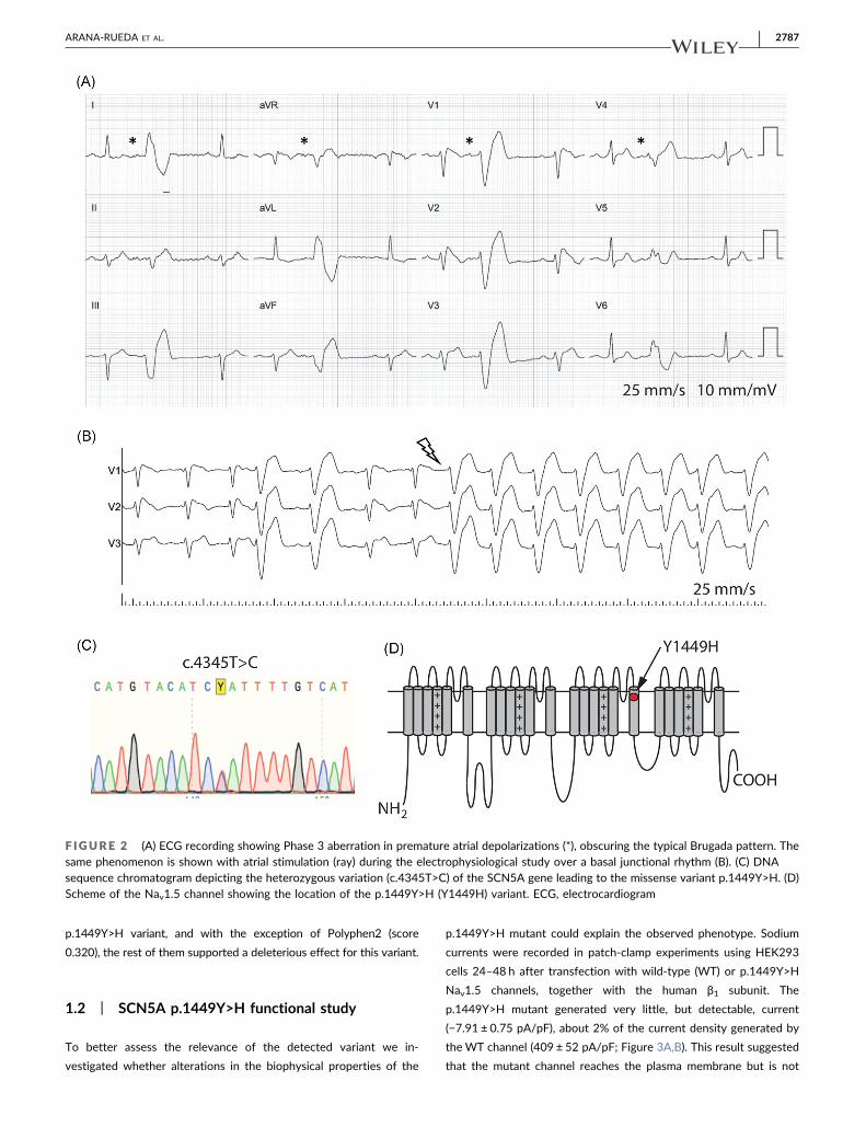
p.1449Y>H variant, and with the exception of Polyphen2 (score
0.320), the rest of them supported a deleterious effect for this variant.
1.2 | SCN5A p.1449Y>H functional study
To better assess the relevance of the detected variant we in-
vestigated whether alterations in the biophysical properties of the
p.1449Y>H mutant could explain the observed phenotype. Sodium
currents were recorded in patch‐clamp experiments using HEK293
cells 24–48 h after transfection with wild‐type (WT) or p.1449Y>H
Nav1.5 channels, together with the human β1 subunit. The
p.1449Y>H mutant generated very little, but detectable, current
(−7.91 ± 0.75 pA/pF), about 2% of the current density generated by
theWT channel (409 ± 52 pA/pF; Figure 3A,B). This result suggested
that the mutant channel reaches the plasma membrane but is not
F IGURE 2 (A) ECG recording showing Phase 3 aberration in premature atrial depolarizations (*), obscuring the typical Brugada pattern. Thesame phenomenon is shown with atrial stimulation (ray) during the electrophysiological study over a basal junctional rhythm (B). (C) DNAsequence chromatogram depicting the heterozygous variation (c.4345T>C) of the SCN5A gene leading to the missense variant p.1449Y>H. (D)Scheme of the Nav1.5 channel showing the location of the p.1449Y>H (Y1449H) variant. ECG, electrocardiogram
ARANA‐RUEDA ET AL. | 2787

fully functional. Our SCN5A constructs include an extracellular FLAG
tag, located in the domain I (DI) S1‐S2 linker of the Nav1.5 protein.4
To analyze whether p.1449Y>H mutant traffics to the plasma
membrane, anti‐FLAG immunofluorescence was performed using
non‐permeabilized cells. Confocal microscopy analysis revealed that
the mutant channel reaches the plasma membrane (Figure 3C), con-
firming that the variant does not significantly alter channel trafficking.
Several reports have described BrS variants displaying dominant‐
negative effects,5,6 as a result of the dimerization and coupled gating
of theWT and mutant Na+ channel α‐subunits.6 Thus, and mimicking
the heterozygous condition (Het), we tested whether p.1449Y>H
variant could exert any dominant‐negative effect on WT Nav1.5
channel. WhenWT and p.1449Y>H channels were co‐expressed (in a
1:1 ratio), sodium channel currents were recorded, with a peak cur-
rent density of 253 ± 20 pA/pF, approximately 60% of that observed
in the cells transfected with the WT channel (Figure 3A,B). Further
comparison of the WT and Het channels biophysical properties dis-
carded any major dominant‐negative effect of the p.1449Y>H variant
(see Figure S2 and Table S1).
2 | DISCUSSION
This article shows how patients with BrS can develop a CLBBB totally
masking the typical ECG pattern, and highlights the awareness that
must be taken for a correct diagnosis if both conditions are present.
Through a clinical and functional study, we demonstrate how the
p.1449Y>H SCN5A variant causes a significant loss of function in so-
dium channel that may be associated with this overlapping phenotype.
BrS is an inherited disorder associated with sudden death along
with the signature of a characteristic ECG pattern in precordial leads.
Thus, the current clinical diagnosis of BrS is based on the demonstra-
tion of a typical ECG pattern either spontaneous or after IC pharma-
cological challenge1 (Figure 1A). Nowadays, these criteria are not
replaceable by any other diagnostic method. However, this finding can
be clinically difficult, as the pattern is characterized by its dynamic
behavior and the association with various and often extensive con-
duction diseases. The SCN5A gene, which encodes the cardiac sodium
channel α subunit Nav1.5, is the most common BrS‐associated gene,
but is found in only 20%–25% of probands.1 This channel is responsible
for excitability and impulse conduction in the contractile myocardium
and specialized conduction system, and is also implicated in re-
fractoriness and repolarization. Thus, loss of function variants in SCN5A
have been associated not only with BrS, but also with early repolar-
ization syndrome, a variety of conduction diseases and overlapping
syndrome.1,3 Indeed, there is a significant clinical and genetic overlap
between BrS and progressive cardiac conduction disease, and both
conditions may coexist or manifest in isolated forms in carriers of the
same variant within the same family.3 Maury et al.,7 studied the pre-
valence of conduction disturbances in patients with BrS and reported a
high proportion of complete right bundle branch block (CRBBB). In such
cases, even experienced electrophysiologists may have difficulties in
F IGURE 3 Functional analysis of thep.1449Y>H mutant. (A) Representative currenttraces obtained from HEK293 cells transfectedwith Nav1.5 WT, p.1449Y>H (Y1449H), or WT+p.1449Y>H (Het) channels and the Navβ1subunit. (B) Current density‐voltage relationshipsin the same groups of cells. (C) Representativeconfocal images from HEK293 cells transfectedwith the FLAG‐tagged Nav1.5 WT andp.1449Y>H constructs. Scale bar: 20 μm
2788 | ARANA‐RUEDA ET AL.

discriminating between BrS and CRBBB by ECG alone. Nevertheless,
the prevalence of other intraventricular conduction abnormalities in BrS
patients is less known, with CLBBB estimation of less than 1%. In
CLBBB the ventricles are activated sequentially, first the right ventricle
via the right bundle and then the left ventricle, slowly activated through
the septum with forces directed towards the left, posteriorly, and in-
feriorly. As a result, the ECG changes indicative of BrS are not seen in
precordial leads, as they are buried within a wider QRS complex due to
delayed activation of the left ventricle. Our case, with a CLBBB with
Phase 3 characteristics developed during follow‐up, is a clear example
of this combination and illustrates the total concealing of the ECG
pattern, making impossible a BrS diagnosis in cases of persistent
CLBBB. In such cases, high clinical suspicion, based on age at pre-
sentation, associated disorders, symptomatology, and family history, is
necessary for ruling out this rare association. The role of specific pacing
maneuvers to unmask the BrS pattern under persistent CLBBB, as
occurs in concealed BrS due to CRBBB, remains to be explored.
At this time, there is a general concern about the pathogenicity of
many variants in SCN5A previously identified as implicated in BrS, but
of ambiguous significance following the current guidelines for classifi-
cation (up to 63% VUS).2,7 Nowadays, functional evidence is considered
the major score driver for pathogenicity assumption for missense var-
iants. According to a recent re‐evaluation of the American College of
Medical Genetics and Genomics and the Association for Molecular
Pathology (ACMG‐AMP) rules for assessment of pathogenicity,2,9 the
SCN5A variant identified in this report can be classified as pathogenic,
since it fulfills the following criteria: (i) the p.1449Y>H variant is a
missense variant that leads to the replacement of tyrosine by histidine
at position 1449, in the transmembrane S6 region of DIII, which is part
of the pore region of the Nav1.5 channel and, according to the SCN5A
variant browser, is a hotspot region for BrS1 (ACMG category PM1,
moderate evidence);8 (ii) other variants producing amino acid changes
in the same residue has been previously established as pathogenic and
associated to clinical BrS (p.1449Y>S) and also to conduction disease
with partial loss of function in in vitro studies (p.1449Y>C; PS1, strong
evidence).9–12 These results indicate that the conserved Y1449 is
crucial for the proper functioning of the Nav1.5 channel, and that its
alteration induces dramatic changes in channel activity; and (iii)
p.1449Y>H mutant encodes a not fully functional channel that gen-
erates extremely small (2% as compared to the WT) sodium currents,
and a decrease more than 50% in the peak current is significantly
associated with BrS1 penetrance (PS3, strong evidence).2,9
Recently, Ciconte et al.,13 demonstrated that BrS carriers of
SCN5A pathogenic variants exhibit a more aggressive clinical pre-
sentation and a greater epicardial substrate on electrophysiological
studies, associating genotype with phenotypic expression. Although
p.1449Y>H confers a high penetrance in the cases analyzed in this
study, no one has symptoms of severity during close follow‐up.
However, a hypothetical more serious phenotype could be developed
with aging, since some experimental studies have demonstrated the
effect of aging and myocardial fibrosis on the decline of expression
and function of sodium channels.3 Special awareness should be given
to these patients, monitoring the possible progression of the disease
(symptoms, periodical ECG, and Holter evaluation) and periodically
reassessing the risk of sudden death in a multiparametric manner.
3 | CONCLUSIONS
The combination of BrS and CLBBB as part of an overlap syndrome
totally masks the typical BrS ECG pattern needed for diagnosis.
We demonstrate how the p.1449Y>H SCN5A variant produces a
significant loss of function in sodium channel which is manifested as a
marked phenotypical expression.
ACKNOWLEDGMENTS
Wewould like to thank Drs. Sara Pagans and Ramón Brugada for the kind
gift of the pcDNA3‐SCN5A plasmid containing the hNav1.5 sequence,
and Dr. Eva Delpón for the hNavβ1 cDNA. This study was supported by
grants from the Sociedad Española de Cardiología (Ritmo 2017), and the
Consejería de Salud of the Junta de Andalucía (PI‐0365‐2017).
CONFLICT OF INTERESTS
The authors declare that there are no conflict of interests.
DATA AVAILABILITY STATEMENT
The data that support the findings of this study are available on
request from the corresponding author. The data are not publicly
available due to privacy or ethical restrictions.
ORCID
Eduardo Arana‐Rueda http://orcid.org/0000-0001-8132-7045
Alonso Pedrote http://orcid.org/0000-0003-1677-2956
Antonio Castellano http://orcid.org/0000-0003-3955-5137
REFERENCES
1. Antzelevitch C, Yan G‐X, Ackerman MJ, et al. J‐Wave syndromes
expert consensus conference report: emerging concepts and gaps inknowledge. Europace. 2017;19:665‐694.
2. Denham NC, Pearman CM, Ding WY, et al. Systematic re‐evaluationof SCN5A variants associated with Brugada syndrome. J CardiovascElectrophysiol. 2019;30:118‐127.
3. Asatryan B, Medeiros‐Domingo A. Molecular and genetic insights intoprogressive cardiac conduction disease. Europace. 2019;21:1145‐1158.
4. Wu G, Ai T, Kim JJ, et al. alpha‐1‐syntrophin mutation and the long‐QT syndrome: a disease of sodium channel disruption. Circ ArrhythmElectrophysiol. 2008;1:193‐201.
5. Keller DI, Rougier JS, Kucera JP, et al. Brugada syndrome and fever:
genetic and molecular characterization of patients carrying SCN5Amutations. Cardiovasc Res. 2005;67:510‐519.
6. Clatot J, Ziyadeh‐Isleem A, Maugenre S, et al. Dominant‐negativeeffect of SCN5A N‐terminal mutations through the interaction ofNav1.5 α‐subunits. Cardiovasc Res. 2012;96:53‐63.
7. Richards S, Aziz N, Bale S, et al. Standards and guidelines for the in-
terpretation of sequence variants: a joint consensus recommendationof the American College of Medical Genetics and Genomics and theAssociation for Molecular Pathology. Genet Med. 2015;17:405‐424.
8. Kroncke BM, Glazer AM, Smith DK, Blume JD, Roden DM. SCN5A(Nav1.5) variant functional perturbation and clinical presentation: variantsof a certain significance. Circ Genomic Precis Med. 2018;11(5):e002095.
ARANA‐RUEDA ET AL. | 2789
9. Kapplinger JD, Tester DJ, Alders M, et al. An international com-pendium of mutations in the SCN5A‐encoded cardiac sodiumchannel in patients referred for Brugada syndrome genetic testing.Heart Rhythm. 2010;7:33‐46.
10. Jiménez‐Jáimez J, Álvarez M, Algarra M, et al. Low clinical pene-
trance in causal mutation carriers for cardiac channelopathies. RevEspañola Cardiol. 2013;66:275‐281.
11. Hothi SS, Ara F, Timperley JP. Y1449C SCN5A mutation associatedwith overlap disorder comprising conduction disease, Brugada syn-drome, and atrial flutter. J Cardiovasc Electrophysiol. 2015;26:93‐97.
12. Glazer AM, Wada Y, Li B, et al. High‐throughput reclassification of
SCN5A variants. Am J Hum Genet. 2020;107:111‐123.13. Ciconte G, Monasky MM, Santinelli V, et al. Brugada syndrome ge-
netics is associated with phenotype severity. Eur Heart J. 2021;42(11):1082‐1090.
SUPPORTING INFORMATION
Additional Supporting Information may be found online in the
supporting information tab for this article.
How to cite this article: Arana‐Rueda E, Pezzotti MR,
Pedrote A, et al. Brugada syndrome masked by complete
left bundle branch block: A clinical and functional study of
its association with the p.1449Y>H SCN5A variant.
J Cardiovasc Electrophysiol. 2021;32:2785‐2790.
https://doi.org/10.1111/jce.15215
2790 | ARANA‐RUEDA ET AL.


![[MétroPole] Principe du Block Automatique Lumineux](https://static.fdocuments.fr/doc/165x107/55cf9d52550346d033ad1e23/metropole-principe-du-block-automatique-lumineux.jpg)