Biotransformation of the Insecticide Lindane by the White Rot Basidiomycete ...
9
Pestic. Sci. 1996, 41, 5 1-59 Biotransformation of the Insecticide Lindane by - the White rot basidiomycete Phanerochaete 1 chrysospor 1uw2 Christian Mougin,"" Claude Pericaud," Christian Malosse," Chantal Laugerob & Marcel Astherb INRA, Unit6 de Phytopharmacie et M6diateurs Chimiques, Route de Saint-Cyr, F-78026 VERSAILLES INRA, Laboratoire de Biotechnologie des Champignons Filamenteux, Facult6 des Sciences de Luminy, Cedex, France Parc Scientifique et Technologique, case postale 925, F-13288 MARSEILLE Cedex 09, France (Received 23 October 1995; accepted 21 December 1995) Abstract: Biotransformation of the insecticide lindane by the white rot basidio- mycete Phanerochaete chrysosporium has been investigated in liquid cultures. Some polar metabolites and carbon dioxide were produced from the pesticide. Among the metabolites identified were tetrachlorocyclohexene, tetra- chlorocyclohexene epoxide and tetrachlorocyclohexenol. When used as a sub- strate, tetrachlorocyclohexene was also converted by the fungus to tetrachlorocyclohexenol, polar metabolites and carbon dioxide. Three incubation conditions leading to low and high peroxidase production were assayed. Data from these experiments, as well as in-vitro incubations with purified enzymes, ruled out any involvement of the peroxidases in lindane biotransformation and mineralization. Moreover, 1-aminobenzotriazole (a P450 inactivator) drastically reduced pesticide metabolism. Conversely, phenobarbital (a P450 inducer) did not significantly increase lindane breakdown. Key words: basidiomycete, lindane, biotransformation, metabolites, P450 mono- oxygenase. 1 INTRODUCTION microbiological and biochemical aspects involved in pollutant transformation. For many years, the white rot The insecticide lindane (1, Fig. 1 ; la,2a,3P,4a,5a,6j- basidiomycete Phanerochaete chrysosporium Burdsall hexachlorocyclohexane, y-HCH) has been extensively has been a useful agent for liquid effluent and soil bio- used worldwide, despite its persistence in the environ- remediation. The extended biodegradative properties of ment, tendency toward bioaccumulation and toxicity to the fungus toward numerous environmentally persistent higher animals, as well as possible toxicological and chemicals'0*'' have been credited to its lignin- environmental problems of its residues.'S2 It is still used degrading system (LDS), which includes mainly extra- in France for agricultural and medical purposes, and cellular lignin peroxidases (Lips) and residual pesticide has been detected in living manganese-dependent peroxidases (MnPs)." The organism^,^ soils and surface water^.^ Thus, the micro- enzymes are produced under substrate-limiting growth bial metabolism of lindane remains a current subject of conditions, and they are not induced by pollutants. The in~estigation.~-' extracellular peroxidases can also act on compounds The use of micro-organisms for bioremediation with low water solubility, and/or adsorbed on solid par- requires an understanding of all the physiological, ticles. Nevertheless, the mechanisms by which white rot fungi degrade pollutants are known for only a few * To whom correspondence should be addressed. models. Pestic. Sci. 0031-613X/96/$09.00 51 1996 SCI. Printed in Great Britain
Transcript of Biotransformation of the Insecticide Lindane by the White Rot Basidiomycete ...

Pestic. Sci. 1996, 41, 5 1-59
Biotransformation of the Insecticide Lindane by - the White rot basidiomycete Phanerochaete
1 chrysospor 1uw2
Christian Mougin,"" Claude Pericaud," Christian Malosse," Chantal Laugerob & Marcel Astherb
INRA, Unit6 de Phytopharmacie et M6diateurs Chimiques, Route de Saint-Cyr, F-78026 VERSAILLES
INRA, Laboratoire de Biotechnologie des Champignons Filamenteux, Facult6 des Sciences de Luminy, Cedex, France
Parc Scientifique et Technologique, case postale 925, F-13288 MARSEILLE Cedex 09, France (Received 23 October 1995; accepted 21 December 1995)
Abstract: Biotransformation of the insecticide lindane by the white rot basidio- mycete Phanerochaete chrysosporium has been investigated in liquid cultures. Some polar metabolites and carbon dioxide were produced from the pesticide. Among the metabolites identified were tetrachlorocyclohexene, tetra- chlorocyclohexene epoxide and tetrachlorocyclohexenol. When used as a sub- strate, tetrachlorocyclohexene was also converted by the fungus to tetrachlorocyclohexenol, polar metabolites and carbon dioxide. Three incubation conditions leading to low and high peroxidase production were assayed. Data from these experiments, as well as in-vitro incubations with purified enzymes, ruled out any involvement of the peroxidases in lindane biotransformation and mineralization. Moreover, 1-aminobenzotriazole (a P450 inactivator) drastically reduced pesticide metabolism. Conversely, phenobarbital (a P450 inducer) did not significantly increase lindane breakdown.
Key words: basidiomycete, lindane, biotransformation, metabolites, P450 mono- oxygenase.
1 INTRODUCTION microbiological and biochemical aspects involved in pollutant transformation. For many years, the white rot
The insecticide lindane (1, Fig. 1 ; la,2a,3P,4a,5a,6j- basidiomycete Phanerochaete chrysosporium Burdsall hexachlorocyclohexane, y-HCH) has been extensively has been a useful agent for liquid effluent and soil bio- used worldwide, despite its persistence in the environ- remediation. The extended biodegradative properties of ment, tendency toward bioaccumulation and toxicity to the fungus toward numerous environmentally persistent higher animals, as well as possible toxicological and chemicals'0*'' have been credited to its lignin- environmental problems of its residues.'S2 It is still used degrading system (LDS), which includes mainly extra- in France for agricultural and medical purposes, and cellular lignin peroxidases (Lips) and residual pesticide has been detected in living manganese-dependent peroxidases (MnPs)." The organism^,^ soils and surface water^.^ Thus, the micro- enzymes are produced under substrate-limiting growth bial metabolism of lindane remains a current subject of conditions, and they are not induced by pollutants. The in~estigation.~-' extracellular peroxidases can also act on compounds
The use of micro-organisms for bioremediation with low water solubility, and/or adsorbed on solid par- requires an understanding of all the physiological, ticles. Nevertheless, the mechanisms by which white rot
fungi degrade pollutants are known for only a few * To whom correspondence should be addressed. models.
Pestic. Sci. 0031-613X/96/$09.00 51
1996 SCI. Printed in Great Britain
52 C . Mougin et al.
1
cJ? CI CI
2
CI
Cl CI
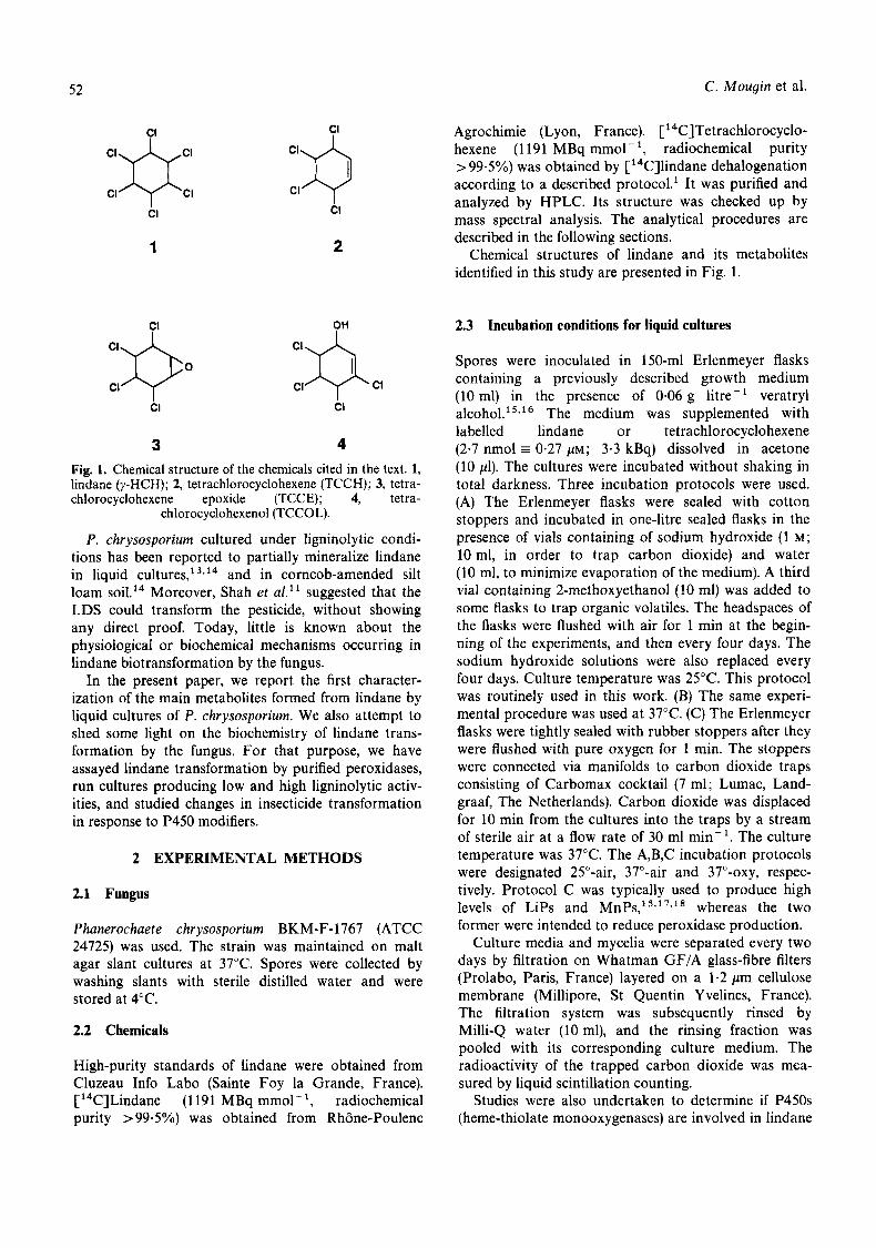
3 4 Fig. 1. Chemical structure of the chemicals cited in the text. 1, lindane (pHCH); 2, tetrachlorocyclohexene (TCCH); 3, tetra- chlorocyclohexene epoxide (TCCE); 4, tetra-
chlorocyclohexenol (TCCOL).
P. chrysosporium cultured under ligninolytic condi- tions has been reported to partially mineralize lindane in liquid cultures,' 3 9 1 4 and in corncob-amended silt loam ~ 0 i l . l ~ Moreover, Shah et a/." suggested that the LDS could transform the pesticide, without showing any direct proof. Today, little is known about the physiological or biochemical mechanisms occurring in lindane biotransformation by the fungus.
In the present paper, we report the first character- ization of the main metabolites formed from lindane by liquid cultures of P. chrysosporium. We also attempt to shed some light on the biochemistry of lindane trans- formation by the fungus. For that purpose, we have assayed lindane transformation by purified peroxidases, run cultures producing low and high ligninolytic activ- ities, and studied changes in insecticide transformation in response to P450 modifiers.
2 EXPERIMENTAL METHODS
2.1 Fungus
Phanerochaete chrysosporium BKM-F-1767 (ATCC 24725) was used. The strain was maintained on malt agar slant cultures at 37°C. Spores were collected by washing slants with sterile distilled water and were stored at 4°C.
2.2 Chemicals
High-purity standards of lindane were obtained from Cluzeau Info Lab0 (Sainte Foy la Grande, France). ['4C]Lindane (1191 MBq mmol- ', radiochemical purity > 99.5%) was obtained from Rh6ne-Poulenc
Agrochimie (Lyon, France). [ ''C]Tetrachlorocyclo- hexene (1 191 MBq mmol- ', radiochemical purity =- 99.5%) was obtained by ['4C]lindane dehalogenation according to a described protocol.' It was purified and analyzed by HPLC. Its structure was checked up by mass spectral analysis. The analytical procedures are described in the following sections.
Chemical structures of lindane and its metabolites identified in this study are presented in Fig. 1.
2.3 Incubation conditions for liquid cultures
Spores were inoculated in 150-ml Erlenmeyer flasks containing a previously described growth medium (10 ml) in the presence of 0-06 g litre-' veratryl alcohol.' 5 9 1 6 The medium was supplemented with labelled lindane or tetrachlorocyclohexene (2.7 nmol E 0.27 p ~ ; 3.3 kBq) dissolved in acetone (10 pl). The cultures were incubated without shaking in total darkness. Three incubation protocols were used. (A) The Erlenmeyer flasks were sealed with cotton stoppers and incubated in one-litre sealed flasks in the presence of vials containing of sodium hydroxide (1 M; 10 ml, in order to trap carbon dioxide) and water (10 ml, to minimize evaporation of the medium). A third vial containing 2-methoxyethanol (10 ml) was added to some flasks to trap organic volatiles. The headspaces of the flasks were flushed with air for 1 min at the begin- ning of the experiments, and then every four days. The sodium hydroxide solutions were also replaced every four days. Culture temperature was 25°C. This protocol was routinely used in this work. (B) The same experi- mental procedure was used at 37°C. (C) The Erlenmeyer flasks were tightly sealed with rubber stoppers after they were flushed with pure oxygen for 1 min. The stoppers were connected via manifolds to carbon dioxide traps consisting of Carbomax cocktail (7 ml; Lumac, Land- graaf, The Netherlands). Carbon dioxide was displaced for 10 min from the cultures into the traps by a stream of sterile air at a flow rate of 30 ml min-'. The culture temperature was 37°C. The A,B,C incubation protocols were designated 25"-air, 37"-air and 37"-oxy, respec- tively. Protocol C was typically used to produce high levels of Lips and MnPs,".' ','* whereas the two former were intended to reduce peroxidase production.
Culture media and mycelia were separated every two days by filtration on Whatman GF/A glass-fibre filters (Prolabo, Paris, France) layered on a 1.2 pm cellulose membrane (Millipore, St Quentin Y velines, France). The filtration system was subsequently rinsed by Milli-Q water (10 ml), and the rinsing fraction was pooled with its corresponding culture medium. The radioactivity of the trapped carbon dioxide was mea- sured by liquid scintillation counting.
Studies were also undertaken to determine if P450s (heme-thiolate monooxygenases) are involved in lindane

Biotransformation of lindane by Phanerochaete chrysosporium 53
biotransformation. For that purpose, we assayed the ability of P450 modifiers to alter the pesticide trans- formation rate in P. chrysosporium cultures under 25"-air conditions. The modifiers were: 1- aminobenzotriazole (ABT), a classical mechanism-based inactivator of P450s from mammals" and higher plants" and phenobarbital (PB) an inducer of P450s in higher plant tissues.21 The chemicals were added to the cultures in acetone (100 pl) without any effect of the solvent on fungal growth. Controls were supplemented with a same volume of acetone, and all samples were analysed four, eight and 12 days after treatment.
In all experiments, uninoculated sterile media were maintained as controls.
2.4 Enzymatic assays with crude extracellular fluid
Lip activity was determined from the rate of oxidation of veratryl alcohol to veratraldehyde at 30°C as described by Tien and Kirk.22 Assays were performed with culture fluid (400 pl). MnP activity was also deter- mined spectrophotometrically by the method of Paszczynski et ~ 1 . ~ ~ with vanillylacetone as a substrate. Assays were performed with extracellular fluid (100 or
Enzymatic activities were expressed in nanokatals : 200 pl).
1 nkat ml-' is equivalent to 60 U litre-'.
2.5 Analytical procedures for pesticide compounds
Filtered medium fractions (15 ml) were concentrated on a C,, guard column MCH-10 (3 cm x 4 mm ID; Varian, Les Ulis, France) at a flow rate of 1 ml min-' with an isocratic pump (Varian 9001). Elution of labelled compounds was then achieved onto the analyti- cal column ODS-80TM (25 cm x 4.6 mm ID; Varian) with a Varian 9010 pump delivering a solvent system composed of acetonitrile and water, each acidified with phosphoric acid (0.5 g litre-'). Elution began with 1% acetonitrile for 3 min, followed by a linear increase to 100% acetonitrile over 15 min, and a stationary phase of 10 min. The radioactivity of the column eluate was monitored by an HPLC LB 507 A radioactivity monitor (EG&G, Evry, France). UV absorbance was also monitored with a variable wavelength detector (Varian 9050).
To analyse the radioactivity in the mycelium, filtered fungal pellets were homogenized in acetonitrile + water (7 + 3 by volume, 5 ml) for 1 min. The homogenate was diluted with water (5 ml) and extracted with dichloro- methane (3 x 12.5 ml). After concentration of the organic phase and dissolution in methanol (500 pl), ali- quots (1OOp1) were injected in HPLC through a 7125 Rheodyne valve. The elution of labelled compounds was achieved using the conditions described above.
The total radioactivities in the cell biomass were determined by combustion in a model 307 oxidizer (Packard, Rungis, France).
2.6 Mass spectral analysis
Lindane metabolites were firstly resolved by HPLC analysis of the growth medium and then collected separately. Acetonitrile contained in the eluate was evaporated under a stream of nitrogen, and the remain- ing acidic aqueous fraction was extracted by diethyl ether. After concentration, Electron-Impact (EI, 70 eV) mass spectral analysis (GC-MS) was performed on a Nermag (Quad Service, Argenteuil, France) quadrupole R 10-1OC analyser piloted by a SIDAR acquisition system. Samples (2 pl aliquots) were introduced by G C on a Girdel serie 32 chromatograph. The gas chromato- graphic conditions were as follows: BPX-5 (SGE, Ville- neuve St-Georges, France) capillary column, 25 m x 0.32 mm ID with 0.25 pm film thickness; carrier gas, helium at 69 kPa; temperature programme 10O-24O0C, 10°C min- ; Ross Injector temperature, 240°C.
2.7 Fungal biomass determination
Growth was measured in terms of the mycelial dry weight. The filtered mycelium was dried for one day at 90°C.
2.8 Experimental error
Each experiment was done in triplicate and repeated twice. Results are expressed as means. The standard deviation was less than 10% of the mean.
3 RESULTS
3.1 formed by Phanerochaete chrysosporium
Characterization of some lindane metabolites
The amounts of radioactivity present in the culture medium, in the cell biomass and evolved as carbon dioxide were followed in liquid cultures of P. chryso- sporium maintained under 25"-air conditions, and are shown in Fig. 2A. During the mycelium growing phase (10 days), the radioactivity detected in the mycelium increased linearly with time to reach 49.6% of the initial radioactivity. Consequently, the radioactivity measured in the medium decreased. A slight but significant decrease of the radioactivity was noticed in the mycel- ium between days 10 and 14. About 3.9% of the initial

54 C. Mougin et al.
I
Y ' . - t t
t - c .- (L
O 20 1 *I 0 0 2 4 6 8 1 0 1 2 1 4
L 0
* 0 2 4 6 8 1 0 1 2 1 4
Incubation time (days)
Fig. 2. Profiles of (A) mass balance analysis and (B) metabo- lite content in liquid cultures of Phanerochaete chrysosporium in the presence of [14C]lindane. (A) (0) medium, (0) mycel- ium (A) [14C]carbon dioxide; (B) (0) unretained fraction (0) minor metabolites + b (H) metabolite a (A) metabolite c (0)
metabolite d.
radioactivity was trapped as [' 4C]carbon dioxide after 14 days of culture. At this time, the total radioactivity recovered amounted to 81.0% .of the initial radioac- tivity. This poor recovery is mainly due to lindane vola- tilization, because radioactivity was found in the traps for organic volatiles in control flasks.
Then, changes in metabolite contents were monitored in cultures of P. chrysosporium. Only lindane was extracted from the mycelium. The efficiency of the extraction protocol, evaluated by combustion of other mycelia, was up to 95%. The analysis of the radioac- tivity in the culture media evidenced a fraction not retained by the guard column. It amounted to 9.1% of the initial radioactivity after 14 days (Fig. 2B). The radioactivity in that aqueous fraction was not extracted by organic solvents, such as chloroform or diethyl ether, suggesting that it included very polar compounds. Attempts to resolve them after concentration of the aqueous phase and injection by the Rheodyne valve were also unsuccessful. By contrast, the radioactivity retained on the guard column was resolved into residual lindane and four main metabolites more polar than the pesticide parent. These metabolites, called a, b, c, d , exhibited retention times of 16.5, 17.4, 18.1 and 19.5 min, respectively. The main compounds, c and d,
showed a joint evolution with a maximal amount at day 6, followed by a decrease in the medium (Fig. 2B). Another degradation product, metabolite a, represented 6.7% of the initial radioactivity after 14 days, and accu- mulated during the experiment. A last fraction including minor metabolites and metabolite b never exceeded 1.5% of the initial radioactivity. No physicochemical degradation of the pesticide occurred in uninoculated sterile controls.
Higher amounts of metabolites were obtained from P. chrysosporium cultures treated with 5.1 PM of lindane. Veratryl alcohol (a component of the growth
was omitted since this compound is a per- sistent contaminant during metabolite purification and analysis processes. Lindane transformation was also noticed under that culture condition. The metabolites were collected and analysed separately. The structure of the most polar compound (metabolite a) cannot be proved, because of poor GC chromatography behav- iour. Attempts to analyse it by direct introduction in the mass spectrometer were also unsuccessful because of the small amount of material. A similar problem occurred for metabolite b.
GC-MS mass spectra of the less polar metabolite d showed dissociation patterns with fragment ions of m/z 183, 147, 122 and 111 (Fig. 3), containing respectively three, two, two and one chlorine atoms. Although molecular ion (m/z 218, based on 3sCl) was very low in EI mode, the fragmentation pattern was identical to that of the tetrachlorocyclohexene (TCCH),lVz4 ion at m/z 122 corresponding to an anti-Diels-Alder mecha- nism.
GC-MS analysis of metabolite c showed the presence of two different compounds with distinct retention times ( c l : 4.15 and c2 : 4.33 min, respectively) under the peak collected by HPLC. Spectra of compounds c1 and c2 (Fig. 3) showed intense cluster ions of m/z 199, 163 and 128, corresponding to the step-by-step loss of chlorine atoms from a starting molecule containing four chlorine atoms. The main difference between the two compounds consisted in an intense fragment ion at m/z 138 (certainly due to an anti-Diels-Alder reaction) present in the c2 spectrum and totally non-existent in the c 1 spec- trum. Moreover, spectra of compounds c2 and d exhibited a similar pattern, but with a difference of 16 amu due to an oxygen atom for ions at m/z 138 (122 + 16), 163 (147 + 16) and 199 (183 + 16). This allowed us to identify this compound ( c2 ) as an isomer of tetrachlorocyclohexenol (TCCOL) with an allylic hydroxyl group.6-2s Common features of c 1 and c 2 spectra suggested a tetrachlorocyclohexene epoxide (TCCE) structure for the former ( c l ) . That hypothesis was in agreement with the chromatographic properties of epoxide and hydroxylated compounds by G C analysis,2s and with the mechanism of TCCOL forma- tion from TCCH involving attack of oxygen followed by the double bond migration.892s

Biotransformation of lindane by Phanerochaete chrysosporium
- 55
TCCH I47
I l l
51 77
122 183
I ~Idl 1,l IIIII, 1 1 1 1 1 1,l ,Ill1 II A. I .I . I. 11d I
163 TCCE 99
65 I35 199 75
51
09
II .I J. (I 3 1 1
3.2 Tetrachlorocyclohexene transformation by Phanerochaete chrysosporium
128
, 111.. .. I 8 , . I , L I l 1 , l .
We studied also the transformation of TCCH by liquid cultures of P. chrysosporium to confirm the identity of the main metabolites described. Figure 4A shows that the radioactivity divided between the culture medium and the fungal biomass. It was less abundant in the latter compartment (9.4% of the initial radioactivity after 12 days) by comparison with incubations in pres- ence of lindane (49.6%, Fig. 2A). A lag phase of four days was noticed before the beginning of mineralization, and labelled carbon dioxide represented 7.1% after 12 days of culture. Total radioactivity decreased during the experiment and amounted finally to 62.0% of the initial radioactivity. These results showed an important loss of radioactivity due to volatilization of TCCH and related degradation products.
TCCH content decreased quickly in the culture medium. Conversely, some polar degradation products called a, B and x were produced. They exhibited reten- tion times of 16.2, 17.2 and 18.1 min, respectively. Their evolution is shown in Fig. 4B. Extracts from incu- bations with TCCH and lindane have been co- chromatographed. Retention times of the peaks attributed to the most apolar metabolite x corre- sponded well to those of the mixture TCCE + TCCOL. Conversely, metabolites a and did not coelute with metabolites a and b from lindane incubation extracts. No degradation occurred in uninocolutated sterile con- trols.
138
99 6s 13
50
111 P~~ I 1
3.3 Effects of incubation conditions on peroxidase production by Phanerochaete chrysosporium cultures
I67 TCCOL
I99
I , . , , I , 220 1 1
I
The involvement of the LDS in lindane degradation was firstly assayed by using purified Lips and MnPsI7 exhibiting high levels of veratryl alcohol and van- illyacetone oxidases (83.0 and 195.0 nkat ml-', respectively). Lindane was not transformed by purified enzymes alone or in mixture under oxidizing conditions (data not shown) nor reducing conditions." In both cases, the addition of culture medium to reaction mix- tures, in order to initiate possible co-reactions" was also unsuccessful.
LDS production was measured in cultures grown in the presence of lindane, and under the three culture conditions previously defined. Figure 5 shows that the production of Lips and MnPs is affected by culture conditions. Among the three incubation conditions used, only the 37O-oxy allowed the maximal production of the LDS. In that case, after a one-day lag phase, Lip production increased to its maximal value after four days of culture, and Lip activity reached 16 nkat ml-' (Fig. 5A). Then, it decreased until the end of the experi- ments. Under the 37"-air condition, the maximal pro- duction occurred at day 5 , and amounted to 50% of the previous value. The production profile obtained at 25"-air was quite similar to that obtained under 37"- oxy, with a maximum production at day 6.
A similar pattern occurred for MnP production (Fig. 5B). Nevertheless, the lack of pure oxygen was detri- mental to the substantial production of enzymes at

C . Mougin et al. 56
>, U > c V m
U
.-
.-
.- E - m E
.- c .- .- Lc 0
#
x > U
m 0 U
U .- .- .- E - m C
.- c .- .- - 0
ZR
1 0 0
80
60
40
2oLe 0 2 4 6 8 10 1 2
I I I
30;- ' 6
25
20
1 5
1 0
5
0 0 2 4 6 8 1 0 1 2
Incubation time (days)
Fig. 4. Profiles of (A) mass balance analysis and (B) metabo- lite content in liquid cultures of Phanerochaete chrysosporium in the presence of ['"CITCCH. (A) (0) medium (0) mycelium (A) ['"C] carbon dioxide; (B) (0) unretained fraction (0)
minor metabolites + B(u) metabolite a (A) metabolite x .
37"C, whilst their production was drastically reduced under 25"-air.
3.4 Effects of incubation conditions on lindane mineralization and degradation by Phunerochuete chrysosporium
Lindane mineralization was followed in the same incu- bations. The results are reported in Fig. 6A. Whatever the incubation conditions, mineralization of lindane occurred, and constituted between 3-0 and 4.5% of the initial radioactivity after 14 days of culture. It began after two days of culture at 3TC, but the lag phase was more pronounced at 25°C. No mineralization was noted in uninoculated sterile controls.
The effects of culture conditions on total metabolite formation were also followed, taking into account both the retained and unretained fractions resulting from HPLC concentration. Total metabolite formation appeared to be quite similar when the fungus was cul- tured under 37"- and 25O-air conditions (Fig. 6B). The amount of metabolites represented 18.2-18.8% of the initial radioactivity after 14 days of culture, whereas 15.1% were found under 37O-oxy. No degradation was noticed in uninoculated sterile controls. Comparable
zol- -7 1 5
U
c 0 m .-
n 5
0 0 2 4 6 8 1 0 1 2 1 4
i
6 25
\ U m A! = csoj 15 A
0 2 4 6 8 1 0 1 2 1 4 Incubation time (days)
Fig. 5. Profiles of (A) lignin and (B) manganese-dependent peroxidase production by Phanerochaete chrysosporium. (0)
25"-air (m) 37"-air (A) 37"-oxy.
I I I I I
-
-
I 7 I
0 2 4 6 8 1 0 1 2 1 4
6
1 5 -
1 0 -
S - ZR /
0 I 1 I I 1
0 2 4 6 8 1 0 1 2 1 4 Incubation time (days)
Fig. 6. (A) Mineralization and (B) total metabolite formation from ['"Cllindane by Phanerochaete chrysosporium. (0 )
25"-air (D) 37"-air (A) 37"-oxy.

Biotransformation of lindane by Phanerochaete chrysosporium 57
changes in separate metabolite amounts occurred under the three culture conditions.
3.5 Effects of P450 modifiers on lindane transformation in cultures of Phanerochaete chrysosporium
When applied to fungal cultures at to M, ABT inhibited lindane mineralization (Fig. 7A) after four, eight and 12 days of incubation. The inhibition increased with ABT concentration. In a similar way, residual lindane amounts in the medium increased in response to ABT treatment to reach 178.4% of the untreated controls (Fig. 7B). ABT M drastically reduced the formation of all metabolites in the retained and unretained fractions resulting from medium con-
I I
A 120 1
4
4 8 1 2
8 1 2 Incubation time (days)
Fig. 7. Effects of aminobenzotriazole on (A) minera1izat.m and (B) residual lindane content in medium from liquid cul-
tures of Phanerochaete chrysosporium.
centration, which was totally inhibited at the highest modifier concentration (Table 1).
PB was assayed on cultures of P. chrysosporium at and low2 M. Both concentrations induced moder-
ate inhibitory effects on lindane mineralization noticed at day 4. Pesticide mineralization was identical in both PB-treated and control cultures after eight and 12 days of incubation (data not shown). The residual lindane in the medium was slightly increased by PB treatment after eight and 12 days of culture, but was unaffected during the early days of the experiment. In general, PB treatments modified the transformation of lindane to polar metabolites a and b. They also reduced TCCH amounts after eight and 12 days of culture, TCCE and TCCOL amounts being increased (Table 1).
Both modifiers were themselves without effect on fungal growth (data not shown).
4 DISCUSSION
The results presented in this paper confirm those pre- viously r e p ~ r t e d . ' ~ , ' ~ They show that the white rot fungus P. chrysosporiurn transforms the insecticide lindane and partially mineralizes the molecule. We have characterized for the first time the main lindane metab- olites from P. chrysosporium liquid cultures. Unfor- tunately, the small amounts of material available have prevented any detailed scheme of lindane transform- ation in P. chrysosporium cultures. Nevertheless, we suggest TCCH, TCCE and TCCOL structures which agree with published studies in vertebrates or micro- o r g a n i s m ~ . ~ - ~ ~ ~ ~ ~ ~ ~
Although there is a general consensus among investi- gators that the LDS is responsible for pollutant trans- formation, direct proofs with purified enzymes are rarely presented, and indirect data are not convincing. However, new hypotheses have now been reported. For example, the ability of P. chrysosporium to mineralize DDT was shown to be independent of the formation of the LDSZ6 Then, many researchers postulate that other fungal enzymatic systems can catalyse xenobiotic degra- dation reactions as an alternative or a complement of the LDS.27s28 Numerous data also suggest that both
TABLE 1 Effects of Aminobenzotriazole (ABT) and Phenobarbital (PB) on Metabolite and Lindane Contents of Medium from Liquid Cultures of Phanerochaete chrysosporium after 12 Days
of Incubation
TCCOL + Incubation Unretained a + b TCCE (c) TCCH ( d ) conditions fraction (% of radioactivity in medium) Lindane
Untreated controls 34.3 8.5 3.1 4.3 49.8 ABT (10-3 M) 11.6 0.0 0.0 0.0 88.4 PB (lo-' M) 37.9 1.1 5.2 0.0 55.8

58 C . Mougin et al.
intra- and extracellular enzymes may be sequentially involved in the degradation process of organic com- pounds, as recently reported in the case of chlorophe-
In this paper, we show that the main first steps of lindane degradation may not involve Lips and MnPs. These results complement previous studies with purified enzymes acting under reducing conditions. l1 Increasing the oxygen level and the temperature in cultures has a strong activating effect on the rate of production of Lips and MnPs, as has also been described for the rate of lignin degradati~n.~' On the other hand, no strong differences were found in the rates of mineralization and transformation of lindane when the fungus was grown under 100% oxygen versus atmospheric conditions. Similar results have been reported previously in the case of DDT.31 A comparison between Figs 5 and 6 does not show any correlation between pesticide mineralization/ transformation and LiPs/MnPs production. Lindane transformation is maximal when peroxidase production (especially MnPs) is low. Conversely, it is lower under classical optimal ligninolytic conditions.
In living organisms, detoxication processes are mainly mediated by P450s. Such enzymatic systems are poorly known in micro-organisms, and have never been described in P . chrysosporium. Lindane undergoes a reductive dechlorination by vicinal dihaloelimination to yield TCCH. It is now established that the first step in biological breakdown of lindane is reductive in various cases. That reaction (probably catalyzed by P450s) occurred with rat liver microsomes and NADPH,9932v33 and with cell-free extracts of bacteria.34 The subsequent steps involve oxidation reactions. TCCH is oxidized to TCCOL by microsomal fractions from both rat liver and house fly abdomen.9325 It requires NADPH and is inhibited by classical P450 inhibitors (carbon monoxide, piperonyl butoxide and SKF-525A). Little is known on the enzymatic systems involved in fungi.
Although appearing contradictory, our results obtained in P . chrysosporium cultures using classical P450 modifiers are consistent with the involvement of these systems in one or more steps of lindane metabo- lism. ABT inhibits the formation of all the isolated metabolites. In that case, a slight carbon dioxide release is still observed. On the other hand, PB does not affect lindane mineralization, yet it seems to modify the profile of isolated metabolites, with an increased oxidation of TCCH to TCCE and TCCOL. The distinct effects of the modifiers on lindane transformation can be explained. ABT is a mechanism-based inactivator effi- cient on numerous P450 isoforms. In our experiments, it inhibits the enzymes involved in the first steps (dechlorination and oxidation) of lindane transform- ation. The lack of metabolites is then responsible for the inhibition of mineralization. Conversely, P450 inducers are known to be highly specific for P450 gene families. For that reason, PB may be without notable effects on
the isozymes involved in the first step of lindane break- down (dechlorination), although stimulating the follow- ing reaction (oxidation). Nevertheless, an important step for lindane mineralization (ring opening) may not involve P450 enzymes. It remains unaffected by P450 modifiers, and may not be stimulated by high amounts of metabolites. A similar lack of effect of PB on chloro- toluron oxidation has been already reported35 in wheat cell suspension cultures, although the transformation reactions were P450-mediated.36 Moreover, we cannot exclude a rapid transformation of PB by the fungus, before exertion of any strong inducing effect.
Our results, as well as those from many other labor- atory studies with fungal cultures have yielded an exten- sive list of xenobiotics susceptible to degradation by P . chrysosporium in liquid culture conditions. The results presented above clearly establish that the fungus is able to metabolize lindane without involving Lips and MnPs. For the first time, they allow us to postulate that P450s can be active systems for pesticide detoxication in P . chrysosporium.
ACKNOWLEDGEMENTS
We gratefully acknowledge the generous gift of [ 14C]- labeled lindane from RhBne-Poulenc Agrochimie (Lyon). The authors thank Prof. J.-M. Sigoillot (INRA, Luminy), Drs P. Gaillardon and V. Chaplain (INRA, Versailles) for their interest for this work, as well as Dr T. M. Vogel (RhBne Poulenc Industrialisation, Decines) for comments on the manuscript.
REFERENCES
1. Marks, T. S., Allpress, J. D. & Maule, A., Dehalogenation of lindane by a variety of porphyrins and corrins. Appl. Enuiron. Microhiol., 55 (1989) 1258-61.
2. Waliszewski, S. M., Residues of lindane, HCH isomers and HCB in the soil after lindane application. Enuiron.
3. de Cruz, I., Lacroix, G., Mougin, C . & Grolleau, G., Resi- dues of chlorinated pesticides in eggs of Grey Heron (Ardea cinerea I,.): Contribution of capillary gas chroma- tography Ion-Trap mass detection. J . High Resol. Chro- rnatogr., 19 (1996) 62-4.
4. Belamie, R., Contamination des eaux de surface par les produits phytosanitaires. In Alteration et restauration de la qualiti des eaux continentales. INRA ed., Port-Leucate,
5. Engst, R., Macholz, R. M. & Kujawa, M., Lindane metab- olism. Res. Rev., 68 (1 977) 59-90.
6. Engst, R., Macholz, R. M. & Kujawa, M., Recent state in lindane metabolism. Res. Rev., 72 (1979) 71-95.
7. Macholz, R. M. & Kujawa, M., Recent state of lindane metabolism. Part 111. Res. Rev., 94 (1985) 119-49.
8. Straube, G., Microbial transformation of hexachloro- cyclohexane. Zentralbl. Mikrohiol., 146 (1991) 327-38.
9. Deo, P. G., Karanth, N. G. & Karanth, N. G. K., Biodeg- radation of hexachlorocyclohexane isomers in soil and food environment. Crit. Rev. Microhiol., 20 (1994) 57-78.
Pollut., 82 (1993) 289-93.
1992, pp. 21-6.
Biotransfurmatiun of lindane by Phanerochaete chrysosporium 59
10. Higson, F. K., Degradation of xenobiotics by white rot fungi. Rev. Environ. Cont. Toxicol., 122 (1991) 11 1-52.
11. Shah, M. M., Barr, D. P., Chung, N. & Aust, S. D., Use of white rot fungi in the degradation of environmental chemicals. Toxicol. Lett., 64/65 (1 992) 493-501.
12. Barr, D. P. & Aust, S. D., Mechanisms white rot fungi use to degrade pollutants. Environ. Sci. Technol., 28 (1994) 78A-87A.
13. Bumpus, J. A., Tien, M., Wright, D. & Aust, S. D., Oxida- tion of persistent environmental pollutants by a white rot fungus. Science (Washington), 228 (1 985) 1434-6.
14. Kennedy, D. W., Aust, S. D. & Bumpus, J. A., Compara- tive biodegradation of alkyl halide insecticides by the white rot fungus, Phanerochaete chrysosporium (BKM-F- 1767). Appl. Enuiron. Microbiol., 56 (1990) 2347-53.
15. Capdevilla, C., Moukha, S., Ghyczy, M., Theilleux, J., Gelie, B., Delattre, M., Corrieu, G. & Asther, M., Charac- terization of peroxidase secretion and subcellular organiz- ation of Phanerochaete chrysosporium INA-12 in the presence of various soybean phospholipid fractions. Appl. Environ. Microhiol., 56 (1990) 381 1-16.
16. Mougin, C., Laugero, C., Asther, M., Dubroca, J., Frasse, P. & Asther, M., Biotransformation of the herbicide atra- zine by the white rot fungus Phanerochaete chrysosporium. Appl. Environ. Microbiol., 60 (1994) 705-8.
17. Bonnarme, P., Delattre, M., Corrieu, G. & Asther, M., Peroxidase secretion by pellets or immobilized cells of Phanerochaete chrysosporium BKM-F-1767 and INA-12 in relation to organelle content. Enzyme Microb. Technol.,
18. Asther, M., Bellon-Fontaine, M.-N., Capdevilla, C. & Corrieu, G., A thermodynamic model to predict Phanero- chaete chrysosporium INA-12 adhesion to various solid carriers in relation to lignin peroxidase production. Biotech. Bioengin., 35 (1990) 477-82.
19. Ortiz de Montellano, P. R. & Reich, N. O., Inhibition of cytochrome P-450 enzymes. In Cytochromes P-450, ed. P. R. Ortiz de Montellano. Plenum, New York, 1986, pp.
20. Reichhart, D., Simon, A., Durst, F., Mathews, J. M. & Ortiz de Montellano, P. R., Autocatalytic inactivation of plant cytochrome P450 enzymes: selective inactivation of cinnamic acid 4-hydroxylase from Helianthus tuberosus by 1-aminobenzotriazole. Arch. Biochem. Biophys., 216 (1982)
21. Reichhart, D., Salaiin, J. P. & Durst, F., Induction by manganese, ethanol, phenobarbital and herbicides of microsomal cytochrome P-450 in higher plant tissues. Arch. Biochem. Biophys., 196 (1979) 301-3.
22. Tien, M. & Kirk, T. K., Lignin-degrading enzyme from Phanerochaete chrysosporium: purification, character- ization and catalytic properties of a unique H,O,- requiring oxygenase. Proc. Natl Acad. Sci. U S A , 81 (1984)
13 (1991) 727-33.
273-3 14.
522-9.
2280-4.
23. Paszczynski, A., Huynh, V. B. & Crawford, R., Compari- son of ligninase-1 and peroxidase M, from the white rot fungus Phanerochaete chrysosporium. Arch. Biochem. Biophys., 244 (1986) 750-65.
24. Heritage, A. D. & MacRae, I. C., Identification of interme- diates formed during the degradation of hexachloro- cyclohexanes by Clostridium sphenoides. Appl. Environ. Microbiol., 33 (1977) 1295-7.
25. Tanaka, K., Kurihara, N. & Nakajima, M., Oxidative metabolism of tetrachlorocyclohexenes, pentachlorocyclo- hexenes and hexachlorocyclohexenes with microsomes from rat liver and house fly abdomen. Pestic. Biochem. Physiol., 10 (1979) 79-95.
26. Kohler, A., Jager, A., Willershausen, H. & Graf, H., Extra- cellular ligninase of Phanerochaete chrysosporium Burdsall has no role in the degradation of DDT. Appl. Microh. Bio- technol., 29 (1988) 618-20.
27. Dhawale, S. W., Dhawale, S. S. & Dean-Ross, D., Degra- dation of phenanthrene by Phanerochaete chrysosporium occurs under ligninolytic as well as nonligninolytic condi- tions. Appl. Enuiron. Microbiol., 58 (1992) 3000-6.
28. Joshi, D. K. & Gold, M. H., Degradation of 2,4,5-trichlo- rophenol by the lignin-degrading basidiomycete Phanero- chaete chrysosporium. Appl. Environ. Microbiol., 59 (1993)
29. Armenante, P. M., Pal, N. & Lewandowski, G., Role of mycelium and extracellular protein in the biodegradation of 2,4,6-trichlorophenol by Phanerochaete chrysosporium. Appl. Environ. Microbiol., 60 (1994) 1711-18.
30. Gold, M. H. & Alic, M., Molecular biology of the lignin- degrading basidiomycete Phanerochaete chrysosporium. Microb. Rev., 57 (1993) 605-22.
31. Fernando, T., Aust, S. D. & Bumpus, J. A., Effects of culture parameters on DDT [l,l,l-trichloro-2,2-bis(4- ch1orophenyl)ethanel biodegradation by Phanerochaete chrysosporium. Chemosphere, 19 (1989) 1387-98.
32. Chadwick, R. W., Chuang, L. T. & Williams, K., Dehy- drogenation: a previously unreported pathway of lindane metabolism in mammals. Pestic. Biochem. Physiol., 5
33. Tanaka, K., Kurihara, N. & Nakajima, M., Oxidative metabolism of lindane and its isomers with microsomes from rat liver and house fly abdomen. Pestic. Biochem. Physiol., 10 (1979) 96-103.
34. Ohisa, N., Yamaguchi, M. & Kurihara, N., Lindane deg- radation by cell-free extracts of Clostridium rectum. Arch. Microbiol., 125 (1980) 221-5.
35. Canivenc, M.-C., Cagnac, B., Cabanne, F. & Scalla, R., Induced changes of chlorotoluron metabolism in wheat cell suspension cultures. Plant Physiol. Biochem., 27 (1989)
36. Mougin, C., Cabanne, F., Canivenc, M.-C. & Scalla, R., Hydroxylation and N-demethylation of chlorotoluron by wheat microsomal enzymes. Plant Sci., 66 (1990) 195-203.
1779-85.
(1975) 575-86.
193-201.