A SEVERE CASE OF ACUTE NECROTIC …umbalk.org/wp-content/uploads/2018/03/26.A-SEVERE... ·...
5
Archives of the Balkan Medical Union Copyright © 2018 Balkan Medical Union vol. 53, no. 1, pp. 155-159 March 2018 RÉSUMÉ Un cas grave de pancréatite necrotique aiguë causée par une duplication des cystes duodénaux Introduction. La duplication kystique duodénale est une malformation congénitale qui survient au cours du développement embryonnaire du tube digestif. C’est une pathologie bénigne diagnostiquée le plus souvent en néonatologie ou dans l’enfance, et le dia- gnostic chez l’adulte est difficile et non spécifique. La DCD est une cause reconnue d’obstruction digestive, de pancréatite aiguë, d’ictère obstructif et, rarement, d’hémorragie digestive haute. Présentation de cas. Nous rapportons le cas d’un adulte de sexe masculin aux antécédents de douleurs abdominales et d’épisodes récurrents de pancréatite aiguë. La CT de balayage a montré une pancréatite aiguë sévère Balthazar C avec une thrombose partielle de la veine splénique et une masse kystique au niveau de la seconde partie du duodénum. La formation sous-épithéliale écho-endoscopique était la sous-mu- queuse II. On a réalisé une cystotomie endoscopique avec l’évacuation complète du liquide aux résultats cli- niques favorables. ABSTRACT Introduction. Duodenal duplication cyst (DDC) is a rare congenital malformation that appears in the em- bryonic development of the digestive tract. It is a be- nign condition usually diagnosed in infancy and early childhood, being a rare and difficult diagnosis in adult population. DDC is a recognized cause of duodenal obstruction, acute pancreatitis, obstructive jaundice and even digestive hemorrhage. Case presentation. We report the case of a young adult male with abdominal pain history, who presents with recurrent episodes of acute severe necrotic pan- creatitis. The abdominal computed tomography scan revealed a Balthazar C necrotic pancreatitis with par- tial thrombosis of the splenic vein and a cystic mass in the second part of the duodenum. The endoscopic ultrasonography (EUS) established that the duodenal cystic lesion came from the second layer, meaning the submucosa. We performed endoscopic cystotomy with complete evacuation of the fluid content into the duo- denum, with favorable clinical outcome. Conclusions. The particularity of the case is repre- sented by the low incidence of this pathology and the rare form of presentation, meaning acute pancreati- tis probably from pancreatic ductular hypertension caused by the DDC. CASE REPORT A SEVERE CASE OF ACUTE NECROTIC PANCREATITIS CAUSED BY DUODENAL CYST DUPLICATION Gabriel Constantinescu 1 , Daniela Tabacelia 2 , Mihai Ciocirlan 3 , Liliana Mirea 4 , Catalina Diaconu 1 , Vasile Sandru 1 , Madalina Ilie 1 1 Department of Gastroenterology, Emergency Clinical Hospital, Bucharest, Romania 2 Department of Gastroenterology, „Sfanta Maria Clinical Hospital“, Bucharest, Romania 3 Department of Gastroenterology, „Agrippa Ionescu Emergency Clinical Hospital“, Bucharest, Romania Corresponding author: Dr. Madalina Ilie Department of Gastroenterology, Clinical Emergency Hospital, Bucharest, Romania Phone +40723179913; email: [email protected]
Transcript of A SEVERE CASE OF ACUTE NECROTIC …umbalk.org/wp-content/uploads/2018/03/26.A-SEVERE... ·...
Archives of the Balkan Medical UnionCopyright © 2018 Balkan Medical Union
vol. 53, no. 1, pp. 155-159March 2018
RÉSUMÉ
Un cas grave de pancréatite necrotique aiguë causée par une duplication des cystes duodénaux
Introduction. La duplication kystique duodénale est une malformation congénitale qui survient au cours du développement embryonnaire du tube digestif. C’est une pathologie bénigne diagnostiquée le plus souvent en néonatologie ou dans l’enfance, et le dia-gnostic chez l’adulte est difficile et non spécifique. La DCD est une cause reconnue d’obstruction digestive, de pancréatite aiguë, d’ictère obstructif et, rarement, d’hémorragie digestive haute.Présentation de cas. Nous rapportons le cas d’un adulte de sexe masculin aux antécédents de douleurs abdominales et d’épisodes récurrents de pancréatite aiguë. La CT de balayage a montré une pancréatite aiguë sévère Balthazar C avec une thrombose partielle de la veine splénique et une masse kystique au niveau de la seconde partie du duodénum. La formation sous-épithéliale écho-endoscopique était la sous-mu-queuse II. On a réalisé une cystotomie endoscopique avec l’évacuation complète du liquide aux résultats cli-niques favorables.
ABSTRACT
Introduction. Duodenal duplication cyst (DDC) is a rare congenital malformation that appears in the em-bryonic development of the digestive tract. It is a be-nign condition usually diagnosed in infancy and early childhood, being a rare and difficult diagnosis in adult population. DDC is a recognized cause of duodenal obstruction, acute pancreatitis, obstructive jaundice and even digestive hemorrhage.Case presentation. We report the case of a young adult male with abdominal pain history, who presents with recurrent episodes of acute severe necrotic pan-creatitis. The abdominal computed tomography scan revealed a Balthazar C necrotic pancreatitis with par-tial thrombosis of the splenic vein and a cystic mass in the second part of the duodenum. The endoscopic ultrasonography (EUS) established that the duodenal cystic lesion came from the second layer, meaning the submucosa. We performed endoscopic cystotomy with complete evacuation of the fluid content into the duo-denum, with favorable clinical outcome.Conclusions. The particularity of the case is repre-sented by the low incidence of this pathology and the rare form of presentation, meaning acute pancreati-tis probably from pancreatic ductular hypertension caused by the DDC.
CASE REPORT
A SEVERE CASE OF ACUTE NECROTIC PANCREATITIS CAUSED BY DUODENAL CYST DUPLICATION
Gabriel Constantinescu1, Daniela Tabacelia2, Mihai Ciocirlan3, Liliana Mirea4, Catalina Diaconu1, Vasile Sandru1, Madalina Ilie1
1 Department of Gastroenterology, Emergency Clinical Hospital, Bucharest, Romania2 Department of Gastroenterology, „Sfanta Maria Clinical Hospital“, Bucharest, Romania3 Department of Gastroenterology, „Agrippa Ionescu Emergency Clinical Hospital“, Bucharest, Romania
Corresponding author: Dr. Madalina Ilie
Department of Gastroenterology, Clinical Emergency Hospital, Bucharest, Romania
Phone +40723179913; email: [email protected]
A severe case of acute necrotic pancreatitis caused by duodenal cyst duplication – Constantinescu et al
156 / vol. 53, n. 1
INTRODUCTION
Duodenal duplication cyst (DDC) is a rare con-genital malformation that appears in the embryonic development of the digestive tract. It is a benign con-dition usually diagnosed in infancy and early child-hood, being a rare and difficult diagnosis in adult population, because of the nonspecific symptoms re-lated to the size and location of the lesion1. Duodenal cysts are very rare, the most common site of gastro-intestinal duplications is in the ileum, the esopha-gus, and the colon. Duplication cysts are surrounded
with a gastrointestinal epithelial lining2. They are commonly tubular or cystic and they may commu-nicate with the pancreatic and biliary system; in rare cases, the communication with the digestive lumen is present3. DDC is a recognized cause of duodenal ob-struction, acute pancreatitis, obstructive jaundice and even digestive hemorrhage. The treatment of symp-tomatic DDC was traditionally surgical but now, due to the new endoscopic techniques, this can be done without considerable morbidity4.
CASE PRESENTATION
A 43-year-old male patient presented at our hos-pital with intense epigastric and back pain, associated with nausea and vomiting. His medical history was pos-itive for recent recurrent necrotic pancreatitis. At clini-cal examination, a raised heart rate, cold sweat hands and diffuse abdominal tenderness were observed. The blood tests revealed inflammatory syndrome with leukocytosis (14 630/mm3), hyperfibrinogenemia and pancreatic reaction, with elevated amylase and lipase (1132 U/L, 2112 U/L, respectively); renal and hepatic function tests were within normal ranges.
The abdominal ultrasonography showed a fat liver, with normal gallbladder and biliary tree, and
Conclusions. La particularité du cas est représentée par la faible incidence de la pathologie et la forme rare de la présentation, c’est-à-dire une pancréatite aiguë due à l’hypertension canalaire pancréatique provoquée par la DDC.
Mots-clés: duplication kystique duodénale, pancréa-tite nécrotique aiguë sévère, cystotomie endoscopique.
Key words: duodenal cyst duplication, severe acute necrotic pancreatitis, endoscopic cystotomy.
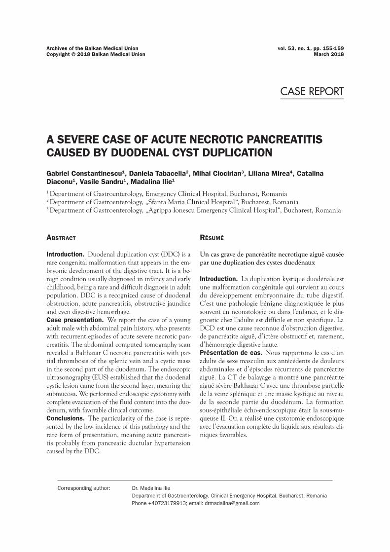
Figure 1. Abdominal ultrasound image showing pancreatic pseudocyst.
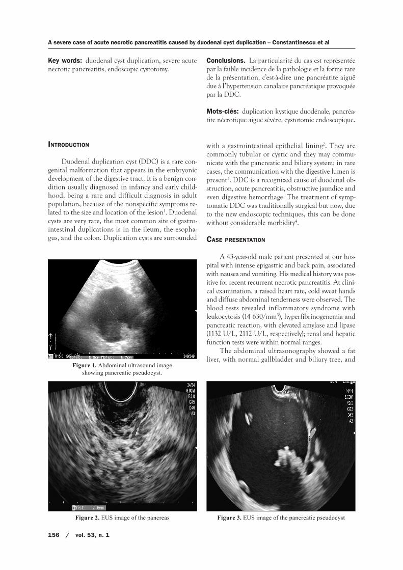
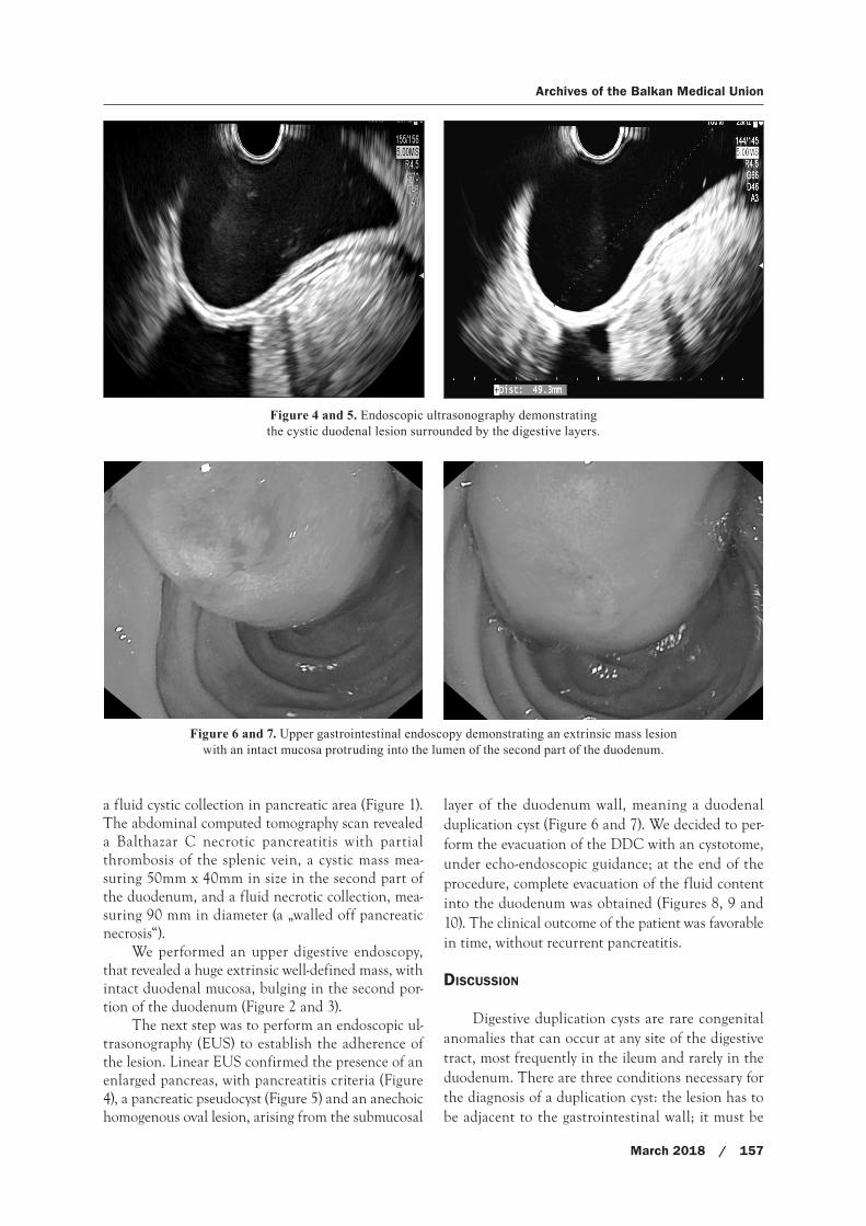
Figure 2. EUS image of the pancreas Figure 3. EUS image of the pancreatic pseudocyst
Archives of the Balkan Medical Union
March 2018 / 157
a fluid cystic collection in pancreatic area (Figure 1). The abdominal computed tomography scan revealed a Balthazar C necrotic pancreatitis with partial thrombosis of the splenic vein, a cystic mass mea-suring 50mm x 40mm in size in the second part of the duodenum, and a fluid necrotic collection, mea-suring 90 mm in diameter (a „walled off pancreatic necrosis“).
We performed an upper digestive endoscopy, that revealed a huge extrinsic well-defined mass, with intact duodenal mucosa, bulging in the second por-tion of the duodenum (Figure 2 and 3).
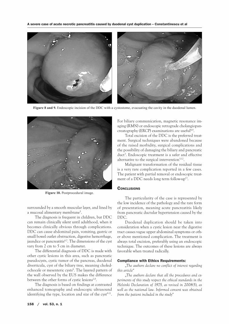
The next step was to perform an endoscopic ul-trasonography (EUS) to establish the adherence of the lesion. Linear EUS confirmed the presence of an enlarged pancreas, with pancreatitis criteria (Figure 4), a pancreatic pseudocyst (Figure 5) and an anechoic homogenous oval lesion, arising from the submucosal
layer of the duodenum wall, meaning a duodenal duplication cyst (Figure 6 and 7). We decided to per-form the evacuation of the DDC with an cystotome, under echo-endoscopic guidance; at the end of the procedure, complete evacuation of the fluid content into the duodenum was obtained (Figures 8, 9 and 10). The clinical outcome of the patient was favorable in time, without recurrent pancreatitis.
DISCUSSION
Digestive duplication cysts are rare congenital anomalies that can occur at any site of the digestive tract, most frequently in the ileum and rarely in the duodenum. There are three conditions necessary for the diagnosis of a duplication cyst: the lesion has to be adjacent to the gastrointestinal wall; it must be
Figure 4 and 5. Endoscopic ultrasonography demonstrating the cystic duodenal lesion surrounded by the digestive layers.
Figure 6 and 7. Upper gastrointestinal endoscopy demonstrating an extrinsic mass lesion with an intact mucosa protruding into the lumen of the second part of the duodenum.
A severe case of acute necrotic pancreatitis caused by duodenal cyst duplication – Constantinescu et al
158 / vol. 53, n. 1
surrounded by a smooth muscular layer, and lined by a mucosal alimentary membrane5.
The diagnosis is frequent in children, but DDC can remain clinically silent until adulthood, when it becomes clinically obvious through complications. DDC can cause abdominal pain, vomiting, gastric or small bowel outlet obstruction, digestive hemorrhage, jaundice or pancreatitis6,7. The dimensions of the cyst vary from 2 cm to 5 cm in diameter.
The differential diagnosis of DDC is made with other cystic lesions in this area, such as pancreatic pseudocysts, cystic tumor of the pancreas, duodenal diverticula, cyst of the biliary tree, meaning choled-ochocele or mesenteric cysts8. The layered pattern of the wall observed by the EUS makes the difference between the other forms of cystic lesions2,8.
The diagnosis is based on findings at contrasted enhanced tomography and endoscopic ultrasound identifying the type, location and size of the cyst9-11.
For biliary communication, magnetic resonance im-aging (RMN) or endoscopic retrograde cholangiopan-creatography (ERCP) examinations are useful4,8.
Total excision of the DDC is the preferred treat-ment. Surgical techniques were abandoned because of the raised morbidity, surgical complications and the possibility of damaging the biliary and pancreatic duct3. Endoscopic treatment is a safer and effective alternative to the surgical intervention5,12.
Malignant transformation of the residual tissue is a very rare complication reported in a few cases. The patient with partial removal or endoscopic treat-ment of a DDC needs long term follow-up13.
CONCLUSIONS
The particularity of the case is represented by the low incidence of the pathology and the rare form of presentation, meaning acute pancreatitis likely from pancreatic ductular hypertension caused by the DDC.
Duodenal duplication should be taken into consideration when a cystic lesion near the digestive tract causes vague upper abdominal symptoms or oth-er above mentioned complication. The treatment is always total excision, preferably using an endoscopic technique. The outcomes of these lesions are always favorable when treated radically.
Compliance with Ethics Requirements:„The authors declare no conflict of interest regarding
this article“„The authors declare that all the procedures and ex-
periments of this study respect the ethical standards in the Helsinki Declaration of 1975, as revised in 2008(5), as well as the national law. Informed consent was obtained from the patient included in the study“
Figure 8 and 9. Endoscopic incision of the DDC with a cystostome, evacuating the cavity in the duodenal lumen.
Figure 10. Postprocedural image.
Archives of the Balkan Medical Union
March 2018 / 159
REFERENCES
1. Lewin KJ, Riddel RH, Weinstein WM. Small and large bowel structure, developmental and mechanical disor-ders. In: Lewin KJ, Riddel RH, Weinstein WM (eds.). Gastrointestinal Pathology and Its Clinical implications. New York: Igaku Shoin, 1992: 734-736.
2. Gress FG, Savides TJ. „Endoscopic ultrasongraphy“ third edition, chapter 17, Gastrointestinal subepithelial masses, 2016;17:141-142.
3. Wieczorek RL, Seidman I, Ranson JH, Ruoff M. Congenital duplication of the stomach: case report and review of the English literature. Am J Gastroenterol 1984;79(8):597-602.
4. Al Traif I, Khan MH. Endoscopic drainage of a duodenal duplication cyst. Gastrointest Endosc 1992;38:64-65.
5. Faigel DO, Burke A, Ginsberg GG, et al. The role of endo-scopic ultrasound in the evaluation and management of foregut duplications. Gastrointest Endosc 1997;45(1):99-103.
6. Yamauchi Y, Hoshino S, Yamashita Y, Funamoto S, Ishida K, Shirakusa T. Successful resection of an infected duodenal duplication cyst after percutaneous cyst drainage: Report of a case. Surg Today. 2005;35:586–9.
7. Maksymyuk VV, Ivanchuk MA, Sheremet MI, et al. Prognostic markers in acute pancreatitis. The Archives of the Balkan Medical Union 2017; 52(3): 263-271.
8. Chen JJ, Lee HC, Yeung CY, Chan WT, Jiang CB, Sheu JC. Meta-analysis: the clinical features of the duodenal duplica-tion cyst. Journal of Pediatric Surgery. 2010;45(8):1598–1606.
9. Redondo-Cerezo E, Pleguezuelo-Diaz J, de Hierro ML, et al. Duodenal duplication cyst and pancreas divisum caus-ing acute pancreatitis in an adult male. World Journal of Gastrointestinal Endoscopy. 2010;2(9):318–320.
10. Diaconu C. „Ecografia abdominală“. În „Explorări funcțio-nale în medicina internă“, coordonator Camelia Diaconu. Editura ALL, București, 2016.
11. Diaconu C. „Tomografia computerizată abdominală“. În „Explorări funcționale în medicina internă“, coordonator Camelia Diaconu, Editura ALL, București, 2016.
12. Sezgin O, Altiparmak E, Yilmaz U, Saritas U, Sahin B. Endoscopic management of a duodenal duplication cyst associated with biliary obstruction in an adult. J Clin Gastroenterol. 2001;32:353–5.
13. Kawahara H, Takahashi T, Okada A. Characteristics of duo-denal duplications causing pancreatitis in children and ado-lescents: A case report and review of the literature. J Pediatr Gastroenterol Nutr. 2002;35:372–6.