
450 - Department of Computer Sciencerht/RHT Papers/1992... · ' - 450 - AUGMENTATION DE PRECISION...
20
' - 450 - AUGMENTATION DE PRECISION DU GESTE OPERATOIRE A L'AIDE D'UN SYSTEME INFORMATIQUE INTEGRE POUR LA CHIRURGIE. Russell H. Taylor, Howard A. Paul, Court B. Cutting, Brent Mittelstadt, William Hanson, Peter Kazanzides, Bela Musits, Yong-Yil Kim, Alan Kalvin, Betsy Haddad, Deljou Khoramabadi, David Larose. RESUME: Ce papier decrit un travail recent sur deux systemes informatiques integres visant a aider un chirurgien a planifier et realiser des procedures chirurgicales precises. Le premier systeme utilise une interface utilisateur interactive et un traitement simple d'images de· tomodensitometrie (TDM) pour permettre a un chirurgien de definir le placement d'une prothese de hanche implantee sans ciment dans le femur du patient. Ce systeme utilise en suite un robot actif pour usiner avec precision la forme prealablement definie dans I' os. Des experiences in vitro ont demontre une tres nette augmentation en precision de la co'incidence de l'implant avec l'os, par rapport a la technique manuelle. Une validation clinique sur des chiens necessitant I' implantation d'une prothese est en cours. Le second systeme repose sur I'utilisation de modeles geometriques 3D du crane du patient obtenus a partir d'images TDM afin d'aider un chirurgien a planifier des osteotomies optimales. Ce systeme utilise des capteurs temps reel et un systeme de guidage passif pour aider le chirurgien a realiser I 'intervention pre-definie avec precision. Des experiences preliminaires in-vitro sur des modeles de cranes plastiques ont permis de repositionner des fragments osseux avec une precision sub-millimetrique. Cet article presente une breve discussion de !'application pour chaque systeme, suivie d'une description generale du systeme, de la procedure chirurgicale et un resume des experiences conduites jusqu'ici. En conclusion, nous discutons de !'architecture du systeme theorique informatique inregre qui pourrait etre a partir des systemes presentes ici. Mots cles : Robotique chirurgicale, chirurgie assistee par ordinateur, planning chirurgical. ABSTRACT: AUGMENTATION OF HUMAN PRECISION in COMPUTER-INTEGRATED SURGERY This paper describes recent work on two computer integrated systems to assist surgeons in the planning and execution of precise surgical procedures. The first system uses interactive graphics and simple image processing of Computed Tomography (CT) to allow a surgeon to specify where a cementless hip implant is to be placed in the patient's femur and uses an active robot to accurately machine the required shape in the patient's bone. In vitro experiments have demonstrated an order-of-magnitude improvement in the precision of fit and placement compared to standard manual techniques and a clinical trial in dogs needing hip replacement surgery is underway. The second system uses CT- derived 3D geometric models of the patient's skull to assist the surgeon to plan optimal craniofacial osteotomies and relies on realtime sensing, online "advice", and passive manipulation aids to assist the surgeon to execute the plans accurately. Preliminary in- vitro experiments on plastic skull models with the surgical component of this system have demonstrated sub-millimeter alignment accuracies for bone fragment relocation. We provide· a brief discussion of the application problem to be solved by each system, followed by a system overview, a description of the surgical procedure, and a summary of experience to date. We conclude by discussing a hypothetical "computer integrated surgery" architecture which might evlove from systems like those described here. Key Words : Robotic surgery, computer-assisted surgery, presurgical planning. Innov. Tech. Biol. Med., Vol. 13, n° 4, 1992.
Transcript of 450 - Department of Computer Sciencerht/RHT Papers/1992... · ' - 450 - AUGMENTATION DE PRECISION...
' - 450 -
AUGMENTATION DE PRECISION DU GESTE OPERATOIRE A L'AIDE D'UN SYSTEME INFORMATIQUE INTEGRE POUR LA CHIRURGIE.
Russell H. Taylor, Howard A. Paul, Court B. Cutting, Brent Mittelstadt, William Hanson, Peter Kazanzides, Bela Musits, Yong-Yil Kim, Alan Kalvin,
Betsy Haddad, Deljou Khoramabadi, David Larose.
RESUME: Ce papier decrit un travail recent sur deux systemes informatiques integres visant a aider un chirurgien a planifier et realiser des procedures chirurgicales precises. Le premier systeme utilise une interface utilisateur interactive et un traitement simple d'images de· tomodensitometrie (TDM) pour permettre a un chirurgien de definir le placement d'une prothese de hanche implantee sans ciment dans le femur du patient. Ce systeme utilise en suite un robot actif pour usiner avec precision la forme prealablement definie dans I' os. Des experiences in vitro ont demontre une tres nette augmentation en precision de la co'incidence de l'implant avec l'os, par rapport a la technique manuelle. Une validation clinique sur des chiens necessitant I' implantation d'une prothese est en cours. Le second systeme repose sur I 'utilisation de modeles geometriques 3D du crane du patient obtenus a partir d'images TDM afin d'aider un chirurgien a planifier des osteotomies optimales. Ce systeme utilise des capteurs temps reel et un systeme de guidage passif pour aider le chirurgien a realiser I 'intervention pre-definie avec precision. Des experiences preliminaires in-vitro sur des modeles de cranes plastiques ont permis de repositionner des fragments osseux avec une precision sub-millimetrique. Cet article presente une breve discussion de !'application pour chaque systeme, suivie d'une description generale du systeme, de la procedure chirurgicale et un resume des experiences conduites jusqu'ici. En conclusion, nous discutons de !'architecture du systeme theorique informatique inregre qui pourrait etre con~u a partir des systemes presentes ici.
Mots cles : Robotique chirurgicale, chirurgie assistee par ordinateur, planning chirurgical.
ABSTRACT:
AUGMENTATION OF HUMAN PRECISION in COMPUTER-INTEGRATED SURGERY
This paper describes recent work on two computer integrated systems to assist surgeons in the planning and execution of precise surgical procedures. The first system uses interactive graphics and simple image processing of Computed Tomography (CT) to allow a surgeon to specify where a cementless hip implant is to be placed in the patient's femur and uses an active robot to accurately machine the required shape in the patient's bone. In vitro experiments have demonstrated an order-of-magnitude improvement in the precision of fit and placement compared to standard manual techniques and a clinical trial in dogs needing hip replacement surgery is underway. The second system uses CTderived 3D geometric models of the patient's skull to assist the surgeon to plan optimal craniofacial osteotomies and relies on realtime sensing, online "advice", and passive manipulation aids to assist the surgeon to execute the plans accurately. Preliminary invitro experiments on plastic skull models with the surgical component of this system have demonstrated sub-millimeter alignment accuracies for bone fragment relocation. We provide· a brief discussion of the application problem to be solved by each system, followed by a system overview, a description of the surgical procedure, and a summary of experience to date. We conclude by discussing a hypothetical "computer integrated surgery" architecture which might evlove from systems like those described here.
Key Words : Robotic surgery, computer-assisted surgery, presurgical planning.
Innov. Tech. Biol. Med., Vol. 13, n° 4, 1992.

- 451 -
Augmentation of Human Precision in Computer-Integrated Surgery
Russell H. Taylor(l), Howard A. Paul(2), Court B. Cutting(3) Brent Mittelstadt (2), William Hanson (4), Peter Kazanzides (2), Bela Musits (2)
Yong· Yil Kim ( 1,5), Alan Kalvin(l), Betsy Haddad(2), Deljou Khoramahadi(2), David Larose(l)
(I) IBM T. J. Watson Research Center; Yorktown Heights, New York 10598
telephone: (914)784-7796; fax: (914)784-6282; email: [email protected]
(2) Integrated Surgical Systems;829 W. Stadium Ln.;Sacramento Ca 95834
(.\) Institute of Reconstructive Plastic Surgery; NYU Medical Center; NY, NY 10016
(4) IBM Palo Alto Science Center; Page Mill Rd; Palo Alto, Ca. 94305
(5) Present address: Korea Inst. of Science & Technology; Seoul, Korea
Introduction: An Emerging Partnership
Recent advances in medical imaging technology (CT, MRI, PET, etc.), coupled with advances in
computer-based image processing and modelling capabilities have given physicians an unprecedented
ability to visualize anatomical structures in live patients, and to use this information in diagnosis and
treatment planning. Furthermore, the potential synergy between precise computer based planning and
precise surgical execution is very great, especially in areas such as neurosurgery and orthopaedics
where CT, MRI, and other 3D medical imaging modalities are already finding routine clinical use. If it is possible to execute a procedure accurately, then it makes sense to develop methods to extract
accurate geometric information from the images and to use computer optimization methods to develop
the best possible surgical plan. Similarly, the design of custom implants or other surgical aids
becomes more sensible if there is some assurance that they will be placed where they are designed to
go. Finally, consistent execution makes experimental or clinical studies more meaningful.
L'.nt'ortunately, the precision of surgical planning often greatly exceeds that of surgical execution.
Typically, precise surgical execution has been limited to procedures (such as brain biopsies) for which
a suitable stereotactic frame is available. The inconvenience and restricted applicability of these
devices has led many researchers to explore the use of robotic devices to augment a surgeon's ability
lo pcrt'orm geometrically precise tasks planned from computed tomography (CT) or other image data
le.g .. 111. 12]. [3]. (41, [5], [6], [7][8] ).
The ultimate goal of this research is a partnership between men (surgeons) and machines (com
puters and robots) that seeks to exploit the capabilities of both to do a task better than either can do
alone. Of course surgeons are already pretty impressive. They are very dexterous and are relatively
'lrnng. They are highly trained to exploit a variety of tactile, visual, and other cues. "Judgementally"
rnntrolled. they understand what is going on in surgery and use their dexterity, senses, and experience 10 cxcL·utc the procedure. However, they also get tired. They cannot see (or tolerate) ionizing radi
ation. Tht'y are most efficient when working at a certain geometric scale and lose dexterity if forced 10 11 Drk in very small spaces or with clumsy instruments. Unassisted, they cannot reliably move
instruments exact distances, exert precomputed forces, or achieve precise angular alignments.
On the other hand, machines are very precise and untiring and can be equipped with any number of
scn.,nJ) feedback devices. Numerically controlled robots can move a surgical instrument through an
exactly defined trajectory with precisely controlled forces. They can react very quickly to external
'1gnals and have relatively little difficulty in communicating with other computer-based systems. Of
Innov. Tech. Biol. Med., Vol. 13, n° 4, 1992.
- 452 -
course, they lack judgement and are in many ways quite clumsy. They cannot easily locate the patient. Their potential speed and strength may make surgeons nervous, since it might be hard to stop them in time if they start to do something wrong. By nature, the surgeons want to control everything that goes on in the operating room. However, they are being asked to rely on a complex (and perhaps a bit mysterious) machine to help carry out their job. Can they trust it?
The most obvious way to prevent a robotic device from making an undesired motion is to make it incapable of moving of its own accord. Motorless manipulators have been implemented, in which joint encoders are used to provide feedback to the surgeon on where his instruments are relative to his image-based surgical plan (e.g., [4], [5], [9], [8]). This approach has many advantages, since the surgeon remains very much in control of what he is doing. The system functions primarily as an additional information source helping the surgeon relate the position of his instruments to medical images used to plan the procedure. One drawback is that the mechanical structure of the arm can make the actual manipulation of the instruments somewhat clumsy. This has led a number of researchers to investigate non-mechanical means of tracking instruments. For example, Meyer-Ebrecht et. al. [10] use three CCD cameras to track LED beacons affixed to the surgeon's instruments. We also use an optical tracking device, which is discussed below and in [ 11]
Another important limitation of passive instrument tracking is that it is often very difficult for a person to align a tool accurately in six degrees of freedom with only positional feedback. Passive manipulators, permitting free motion until locked, have however been implemented for limb positioning, tissue retraction, instrument holding, and other applications in which accuracy is not important [12] [13]. In another case, Davies [14] implemented a three degree-of-freedom passive manipulation aid for prostate surgery, which was used clinically, after prototyping the necessary motions on an active robot.
In cases where only a single motion axis is required during the "in contact" phase of the surgery, the robot may be used essentially as a motorized stereotactic frame ([2], [15], [16], et al). A passive tool guide is placed at the desired position and orientation relative to the patient; brakes are applied; and robot power is turned off before any instrument touches the patient. The surgeon provides whatever motive force is needed for the surgical instruments themselves and relies on his own tactile senses for further feedback in performing the operation. This approach ameliorates, but does not entirely eliminate, the safety issues raised by the presence of an actively powered robot in close proximity to the patient and operating room personnel. Furthermore, maintaining accurate positioning is not always easy, since many robots tend to "sag" a bit when they are turned off or to "jump" when brakes are applied. Leaving power turned on and relying on the robot's servocontroller to maintain position introduces further safety exposures. Finally, the approach is limited to cases where a passive guide suffices. The surgeon cannot execute a complex pre-computed trajectory.
Active devices that move while touching the patient are less common. A voice-commanded limb positioning robot has been developed for orthopaedic applications where the surgeon's strength, rather than his precision, must be augmented [ 12]. Kelly et al have developed a motorized stereotactic frame for use with laser neurosurgery [I] [17]. Drake, et. al. used a general-purpose industrial manipulator to do much the same thing on an exceptional basis for 6 patients [ 6] .1 Davies has developed a motorized version of his device for prostetectomies [ 18).
This paper will discuss experimental work with two systems, one active and the other passive, aimed at significantly improving the precision of surgical procedures inherently requiring high degreeof-freedom motions while "in contact" with the patient. The active system uses a general-purpose
Drake's paper includes a perceptive discussion of the safety tradeoffs involved in using an essentially unmodified industrial robot for neurosurgery. In this case, there was little choice. Standard methods could not be used and, indeed, had been tried unsuccessfully on 4 of the patients.
- 453 -
manipulator to provide an order-of-magnitude improvement (compared to standard manual techniques)
in implant placement and fit for orthopaedic hip replacement surgery, and explicitly addresses many of
the safety concerns associated with the use of such devices. A clinical trial on dogs needing hip
replacements is underway, as are efforts to qualify and replicate the system for human clinical trials.
The passive system combines 30 modelling, interactive graphics, realtime sensing and a remote center
of motion passive manipulator to assist a surgeon in the planning and execution of precise craniofacial
surgery. This relatively new work is presently targeted at an in-vitro demonstration on plastic skull
models and does not specifically address clinical qualification. However, early experiments with the
system are quite promising and the technology demonstrated is in many ways complementary to that
developed for the orthopaedic surgery robot.
For each system, we will provide a brief discussion of the application problem to be solved. This
will be followed by a system overview, a description of the surgical procedure, and a summary of
experience to date. Finally, we will conclude with a more general discussion of a hypothetical ulti
mate "computer integrated surgery" system.
Augmentation with an Active Robot: Hip Replacement Surgery
Over the past several years, researchers at IBM, the University of California at Davis and Integrated
Surgical Systems, Inc., have developed an image-directed robotic system to augment the performance
of human surgeons in precise bone machining procedures in orthopaedic surgery, with cementless total
hip replacement surgery as an initial application.2
About half of the 300,000 total hip replacement operations performed in the US and Europe each
year use cementless implants, where stability of the implant, uniform stress transfer from the implant
to the bone, and restoration of the proper biomechanics critically affect the outcome of the surgery. In
tum, these factors are significantly affected by the proper placement of the implant relative to the bone
and by the accuracy with which the femoral cavity can be prepared to match the implant shape.
Recently reported research confirms that gaps between implant and bone significantly reduce bone
ingrowth [25). A recent study ([19], [24]) of the standard manual broaching method for preparing the
femoral cavity found that the gaps between the implant and the bone were commonly 1-4 mm and that
the overall hole size was 36% larger than the broach. Only 21 percent of an implant inserted into the
hole would actually touch bone. Furthermore, the surgeon had only limited control over where the
broach actually placed the hole.
Thus, our goals were, first, to provide the surgeon with a suitable means to specify where the
implant should go relative to the patient's anatomy and then to exploit the robot's precision to machine
out a cavity of exactly the right shape and size in the place specified. This application inherently
requires computer controlled motion of the robot's end-effector while it is in contact with the patient.
Thus, we have had to pay considerable attention to safety checking mechanisms, as well as to pro
viding means for planning the procedure, registering the patient, robot, and plan coordinate systems,
and accurately executing the plan.
Much of the material in this section has also appeared as a brief article in the J. of the Robotic Society of Japan[7J . Additional material may be found in a number of conference papers [19], [20], [21], [22J, [23], [24]. A full-length journal article is being prepared.

- 454 -
System Overview
The overall application architecture is illustrated in Figure I and may be broken down, roughly,
into (offline) planning and (online) surgical components. The system has gone through considerable
evolution over the years (and continues to evolve). The description here focuses on the version now
being used in a veterinary clinical trial.
Presurgical Planning
The CT-based presurgical planning system [23] is implemented on an IBM RS/6000 workstation,
and incorporates CT data set handling, simple image processing, and interactive graphics functions.
The CT coordinates of marker objects used for intraoperative registration of the plan to the patient are
determined automatically by image processing. The selection of an appropriate orthopaedic implant
and determination of where it is to be placed relative to the patient's femur is now done interactively.
The surgeon selects orthographic cross sectional slices through the CT image of the bone, which are
resampled and displayed. He then selects the implant shape from a computer-aided-design (CAD)
library of possible designs, and uses a mouse to position and orient the implant model within the
volumetric CT data set. As he does this, the workstation generates the implant cross sections corre
sponding to the selected slices through the CT data and displays them. When the surgeon is satisfied,
the coordinates of each locating marker, together with the implant identification and desired location
are written to a file. In the future, we expect that the computer will assist the surgeon by computing
and displaying appropriate "goodness-of-fit" measures, and, eventually, proposing optimized positions
and custom implant designs.
IMPLNTEO BONE
~ PRESURGICAL IMPLANT DESIGNS
PLANNING '/
~ rmIJ ____ J~":~' ---L.~-~ "'\
CALIBRATION PIN POS. IMPLANT SHAPE DATA IMPLANT PLACEMENT
( MOTION
MONITOR
\
IMPLANTED BONE
3D IMAGES MODELS
ONLINE DISPLAY
HANDHELD TERMINAL
Figure 1. Architecture of Hip Replacement Surgery System. The system consists of a presurglcal planning component and a surgical component. In the system used for the veterinary clinical trial, the motion monitoring and robot control functions are subsumed within the robot controller.

(a ) ( b) ( c )
l d ) ( e ) ( f )
,~~
( g ) ( h ) ( i )
Figure 2. Surgical Procedure for Hip Surgery. (a) presurgical planning display; (b) fixated cadaver bone; (c), (d) manual guiding to approximate pin position; (e) tactile search for a pin; (f) cutting the shape; (g) online display (h) final result; (i) operating room scene for first clinical trial.

- 456 -
Operating Room System
The operating room system consists of several components. A .fixation system is used to hold the patient's femur firmly to the robot base during the procedure. The five-axis robot is a modified SCARA manipulator with an added pitch axis, six degree-of-freedom force sensor, and a standard high-speed surgical cutting tool. During surgery, all but the robot's end-effector is covered by a sterile sleeve; the end-effector is separately sterilized. The robot controller provides servocontrol, low-level monitoring, sensor interfaces, and higher-level application functions. During surgery, the force sensor is used to support redundant safety checking, tactile search to locate the aligning pins, and compliant motion guiding by the surgeon.
The man-machine interface includes an online display system which combines data generated by the planning system with data transmitted from the robot controller to show progress of the cutting procedure superimposed on the CT-derived image views used in planning. A gas-sterilized hand-held terminal allows the surgeon to interact with the system during the course of the operation. This terminal supports manual guiding, motion enable, emergency power on/off, and similar functions. It is also used to select appropriate pre-programmed error recovery procedures should the need arise.
The robot control subsystem performs extensive safety checking (21] and monitors cutter force to make sure that the robot does not exert excessive force on the patient. Either the surgeon or the controller is able to freeze (inhibit) all robot motion or to tum off manipulator and cutter power in response to recognized exception conditions. If this happens, the surgeon must explicitly re-enable motion from the hand-held terminal.
Summary of Surgical Procedure
Titanium locating pins are used to register the CT images used to plan the surgery to the patient in the operating room. Before surgery, locating pins are inserted into the patient's greater trocanter and femoral condyles, and a computed tomography (CT) scan is made of the leg. Image processing techniques are used to locate the top center of each pin relative to CT coordinates.
The actual surgical procedure is illustrated in Figure 2. Briefly, the patient is prepared and draped in as usual. Surgery continues normally until the femoral head has been removed. At this point the femur is clamped in a fixation device that rigidly attaches it to the base of the robot. A combination of manual guiding and tactile search is used to locate the top center of each pin in robot coordinates. The robot controller then computes the appropriate transformation between CT and robot coordinates and uses this information to machine out the implant cavity. At this point, the pins can be removed and the surgery procedes in a normal manner.
Experience
Extensive rehearsals were conducted on plastic and cadaver bones and on foam test blocks, in order to verify basic system accuracy and to gain confidence in overall system behavior (26]. In the key "bottom line" experiment, test shapes were machined in foam blocks held in a test fixture into which three locating pins had been implanted and located on CT images of the fixture. The dimensional error of the test shapes (as measured on a coordinate measuring machine accurate to = 0.01 mm) was Jess than 0.08 mm and the total placement error was less than 0.2 mm. Similar test shapes machined in cadaver bone gave dimensional errors less than 0.05 mm and placement errors of about 0.4 mm. Thus, it seems fair to conclude that at least an order-of-magnitude improvement in overall accuracy has been achieved.
A clinical trial, conducted by one of the authors (Paul), was begun in May I 990 on family pets needing hip replacement surgery. By October 1991, 26 dogs had been operated on, all successfully.

- 457 -
Since this is a clinical trial, no bone histology data will be available until after the dogs live out their natural life-span. However, they have all recovered nicely and are doing well. The time required for the robotic portion of the procedure is about 30 minutes, including placing the femur into the fixator (5-7 minutes), locating the pins (5-10 minutes), and cutting (about 15 minutes). As experience is gained, the procedure is being refined, and these times may be expected to be reduced.
Work on a next generation system suitable for use in human surgery is well advanced, and incorporates both enhancements to the presurgical planning system and a fully integrated five-axis robot with additional mechanical safety features, redundant encoders, and additional internal and external safety checking.
Augmentation with Passive Aids: Cranio-Facial Surgery
Researchers at IBM and NYU Medical Center have recently begun research on computer-integrated methods for optimal planning and augmented execution of precise osteotomies to correct cranio-facial malformations. In these procedures, the facial bones are cut into multiple fragments, relocated and fixed together to give the patient a more normal facial appearance.
A simple system, in which bone fragment motions are planned based on analysis of morphometric landmarks obtained from radiographs and custom inter-occlusal splints are used to help with surgical execution, was earlier developed at NYU and is in routine clinical use there [27]. However, we expect that better surgical plans could be obtained from analysis of 30-CT models of bone surfaces. The present surgical procedure is very time consuming and (even with the splints) is still of limited accuracy. Furthermore, the splints force the surgeon to make compromises in his surgical plan, since the soft palette and other tissue still remain attached to the maxilla and can only be stretched so far. There is thus significant synergy between better planning methods and better means of executing the plans that are developed.
Our present goal is to develop an integrated planning and execution system based on 30 models derived from CT data. These models will be used both for optimal planning and interactive presurgical simulation of the planned procedure and for realtime online "advice" to the surgeon during execution. The requirements and (consequently) the present emphasis of this work are somewhat different than those for orthopaedic bone machining. The surgeon's primary requirement is accurate feedback about where the bone fragments are relative to their planned positions. The actual manipulations performed are rather more dexterous than those required during the broaching phase of hip replacement surgery, and the surgeon relies rather more on tactile feedback to assure him that all is well. On the other hand, significantly fewer individual geometrically precise motions are required for bone fragment relocation than for machining a complex implant shape. These considerations have led us to concentrate, at least initially, on sensing the relative location of the bone fragments, providing interactive feedback to the surgeon, and using relatively simple passive manipulation aids to help him achieve the desired alignment. As experience and confidence in the sensing, feedback, and monitoring systems are gained, the apparatus may possibly be automated to provide greater convenience to the surgeon.3
System Overview
The system structure is illustrated in Figure 3, and once again may be broken down into presurgical
If is interesting to note that a somewhat similar evolution is reported by Davies, et at, who have developed a system for assisting with prostetectomies [ 18]. The motions required were first demonstrated in-vitro using a general purpose robot. A completely passive special purpose device was then developed and used clinically. Once confidence was gained, the device was instrumented and, finally, motorized.
'
' ' ' ' : --+-!
: Images
:
- 458 -
Presurgical Planning
@ r------1
i .--------!
' ' ' : "Normal" Data
i ij
..._ __ __, Operating Room System
* Realtime Sensing
* Plan Following
* Model-to-Reality Registration
* Graphics, Speech, and other
Man-Madline Interfaces
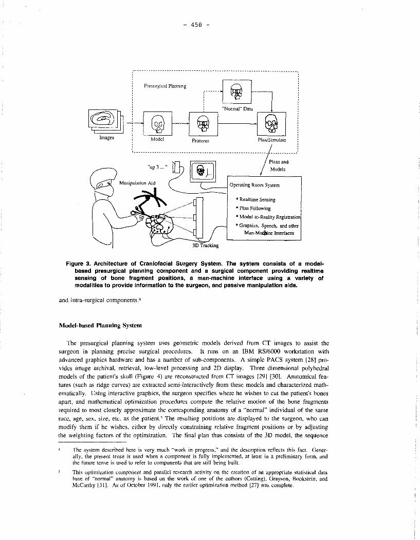
Figure 3. Architecture of Cranlofacial Surgery System. The system consists of a modelbased presurgical planning component and a surgical component providing realtlme sensing of bone fragment positions, a man-machine interface using a variety of modalities to provide information to the surgeon, and passive manipulation aids.
and intra-surgical components.4
Model-based Planning System
The presurgical planning system uses geometric models derived from CT images to assist the surgeon in planning precise surgical procedures. It runs on an IBM RS/6000 workstation with advanced graphics hardware and has a number of sub-components. A simple PACS system [28] provides image archival, retrieval, low-level processing and 20 display. Three dimensional polyhedral models of the patient's skull (Figure 4) are reconstructed from CT images [29] [30]. Anatomical features (such as ridge curves) are extracted semi-interactively from these models and characterized mathematically. Using interactive graphics, the surgeon specifies where he wishes to cut the patient's bones apart, and mathematical optimization procedures compute the relative motion of the bone fragments required to most closely approximate the corresponding anatomy of a "normal" individual of the same race, age, sex, size, etc. as the patient." The resulting positions are displayed to the surgeon, who can modify them if he wishes, either by directly constraining relative fragment positions or by adjusting the weighting factors of the optimization. The final plan thus consists of the 30 model, the sequence
The system described here is very much "work in progress," and the description reflects this fact. Generally, the present tense is used when a component is fully implemented, at least in a preliminary form, and the future tense is used to refer to components that are still being built.
This optimization component and parallel research activity on the creation of an appropriate statistical data base of "normal" anatomy is based on the work of one of the authors (Cutting), Grayson, Bookstein, and McCarthy [31 ]. As of October 1991, only the earlier optimization method [27 J was complete.

- 459 -
Figure 4. Skull Model Reconstructed from CT Images
and approximate location of the cuts, the location of key anatomical features used to register the patient to the model, and the planned relative motion of the bone fragments.
Surgical System
The surgical system assists the surgeon in carrying out his surgical plan, and incorporates both a "real time" (i.e., predictable latency) PC/AT, which supports all sensor and manipulator interfaces and provides a limited backup graphics capability, and an RS/6000 which supports 3D graphics and higher level functions.
The Sensing Subsystem provides information needed to register the reality on the operating table with the models from the surgical plan, for tracking motion of bone fragments, surgical instruments, etc. The principal geometric sensor is a Northern Digital Optotrak (tm) 3D digitizer, which tracks multiple light emitting diode (LED) beacons to an accuracy of about ± 0.1 mm over a I m volume, and automatically combines subsets of beacon positions to produce 6D rigid body coordinates. In our usual configuration, the unit returns 32 3D positions and up to eight 6D positions every 50 ms. In addition to its speed and accuracy, this system has several advantages for operating room use. Unlike electromagnetic field sensors, it is unaffected by metal and is relatively hard to confuse with stray reflections. The beacons are small and relatively easy to affix to the patient, the manipulator hardware, surgical instruments, and other objects whose postions must be tracked. One such instrument is a pointer, which will be used to locate anatomical landmarks on the patient, to allow the surgeon to verify his proposed placement of cuts relative to the surgical plan and other similar uses. A force sensor mounted on the passive manipulation aid (below) will be used to provide additional safety monitoring of manipulation forces. In the future, we anticipate incorporating a a number of additional sensing modalities, including normal computer vision, realtime radiography, and (possibly) redundant kinesthetic sensors in the manipulation aids to provide continuity when visual endpoint sensing is temporarily blocked.
- 460 -
The Surgeon Interface uses a variety of modalities (graphics, synthetized voice, tonal cues,
programmable impedence of manipulator joints, etc.) to provide online, realtime "advice" to the surgeon,
based on the sensed relationship between the surgical plan and surgical execution. Although, we
eventually expect to provide quite a sophisticated interface that draws heavily on "virtual reality"
technologies and techniques, our initial plans are much more modest Very simple realtime graphics and
auditory cues are provided for alignment. An online 3D model display will provide somewhat more
detailed "snapshots" of bone fragment positions relative to the surgical plan.
Passive Manipulation Aids assist the surgeon in precisely aligning bone fragments or in
aligning his instruments relative to the patient. The defining characteristic of such aids is that the surgeon
provides all the motive force. Generally, the manipulation aid should interfere as little as possible with
the surgeon's tactile "feel" for what is happening to the patient, while preserving the desired alignment
once it is achieved. At the same time, it is extremely difficult for most people to achieve accurate six
degree of freedom alignments without the use of external aids to constrain unwanted motions. Our
approach is to develop manipulation aids with manually actuated (or computer controlled) brakes to
provide selective Jocking of orthogonally decoupled degrees-of-freedom resolved in a tool frame centered at
a work point reasonably far removed from the mechanism. In such a mechanism, the revolute axes are all
mutually perpendicular and intersect at the work point. Each motion axis of the mechanism only affects
one rotational or translational degree of freedom of a bone fragment or other object rigidly held at the
work point. This permits the surgeon to work on only one or two degrees of freedom at a time without
disturbing those which have already been aligned. Our present implementation (Figure 5) of a three axis
coarse positioning system, a 6 DOF fine positioing system, and a standard 6 DOF adjustable tooling
clamp [10). The fine positioning system consists of three counterbalanced linear stages carrying a
conventional 0, axis and crossed goniometer cradle 0, and 0, axes with a rotation center about 150 mm
from the mechanism. One advantage of the coarse-fine structure is that it permits relatively large work
volumes while limiting the inertia that the surgeon must cope with. The modularity is similarly very
useful for experimentation.
Surgical Procedure It is anticipated that surgery will procede normally up to the point where the surgeon is ready to
perform the first planned osteotomy. At this point, the surgeon will affix three or more LED beacons to
each (future) bone fragment with standard 1.5 mm percutaneous "K wires". Next, one or more "views" of
the patient will be obtained to measure the relative position of the beacons, which are all rigidly affixed to
the skull. The position of the beacons relative to the skull (and, hence, the coordinate transformations of
the bone fragments relative to the beacons) will then be determined by pointing to known landmarks
(which may be artificially implanted or anatomical) while simultaneously observing the beacon positions.
Once the skull is located, the pointer and feedback system may be used to assist the surgeon in locating
and verifying the exact placement of his planned cuts relative to his surgical plan. The cuts may then be made in whatever order is most convenient for the surgeon and may be done all at once or interspersed
with realignment motions, as appropriate6.
Once a bone fragment B . has been cut free, the surgeon must manipulate it so that it is in l
the pre-planned position and orientation relative to another bone fragment B .. To do this, the surgeon
will place the center-of-rotation of the passive manipulator's goniometer cradd stages over the planned
6 In contrast, Cutting and Grason's present manual technique of interocclusal splints [24] requires that the cuts and realignment motions be sequenced so that the portion of the face being realigned always remains rigidly affixed to the teeth. This technique is moderately accurate but tedious and also can force compromises in the surgical plan. The more common "freehand" techniques for multisegment craniofacial osteotomies do not constrain the order the cuts are made, but also do not provide any help to the surgeon in positioning the fragments accurately.

- 461 -
Coarse
Coarse x
...._~Axe y ~Axex
t Axe z R z R
x
Figure 5. Structure of Passive Manipulation Aid. Consists of 3 coarse positioning axes, a 6 axis remote-center-of-motion fine posltloner, and an adtustable Instrument holder. The coarse positioning axes are used to position the fine posltloner center-of-motion at the desired center-of-motion of a bone fragment. Since the fine posltloner motions are orthogonally decoupled at the remote center, each rototatlonal and translational degree of freedom may be realigned Independently and locked by the surgeon.
center-of-rotation of fragment B;. This fragment will be grasped firmly with standard bone forceps, which will be rigidly affixed to the manipulation aid by use of the adjustable end-of-arm tooling. The manipulation aid may now be used to realign fragment B; relative to B1 In a typical alignment strategy, this will be done by first unlocking all "fine motion" degrees of freedom and manipulating the fragment into its approximate desired position, with the surgeon relying on his own tactile feedback and the force sensor information to verify that there is no undesired obstruction. Then, each degree-offreedom will be successively brought into alignment and locked. As discussed earlier, the kinematic design of manipulation aid assures that adjustment of one degree-of-freedom will not undo the alignment of other degrees of freedom.7
Once B; has been aligned, standard surgical screw and plate methods will then be used to affix it to Bi. This process will be repeated until all bone fragments have been repositioned. Surgery will then
procede normally.
Experiments and Status
As of October 1991, many components of the total system architecture have been implemented, but
system integration is far from complete. Within the planning system, initial programs for image processing, model construction, and feature extractions have been completed. Substantial progress has been made on the interactive graphics and surgical simulation components. Independent work is underway in to construct the anatomical data base. Work on a full-blown surgical plan optimizer
There may be a small amount of "cross talk" for large angular reorientations, necessitating an additional iteration through successive degrees of freedom. In practice, this is not a significant problem, since the alignment steps are quite quick and at most one additional iteration (if any) is usually all that is required to achieve the desired accuracy.
- 462 --
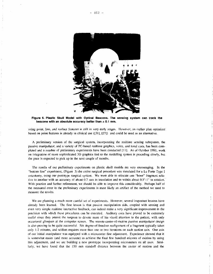
Figure 6. Plastic Skull Model with Optical Beacons. The sensing system can track the beacons with an absolute accuracy better than± 0.1 mm.
using point, line, and surface features is still in very early stages. However, an earlier plan optimizer based on point features is already in clinical use ([31 ], [27]) and could be used as an alternative.
A preliminary version of the surgical system, incorporating the realtime sensing subsystem, the passive manipulator, and a variety of PC-based realtime graphics, voice, and tonal cues, has been completed and a number of preliminary experiments have been conducted [11]. As of October 1991, work on integration of more sophisticated 3D graphics tied to the modelling system is proceding slowly, but the pace is expected to pick up in the next couple of months.
The results of our preliminary experiments on plastic skull models are very encouraging. In the "bottom line" experiment, (Figure 3) the entire surgical procedure was simulated for a Le Forte Type I osteotomy, using our prototype surgical system. We were able to relocate one "bone" fragment relative to another with an accuracy of about 0.7 mm in translation and to within about 0.5°-1° in rotation. With practice and further refinement, we should be able to improve this considerably. Perhaps half of the measured error in the preliminary experiments is most likely an artifact of the method we used to measure the results.
We are planning a much more careful set of experiments. However, several important lessons have already been learned. The first lesson is that passive manipulation aids, coupled with sensing and even very simple realtime interactive feedback, can indeed make a very significant improvement in the precision with which these procedures can be executed. Auditory cues have proved to be extremely useful since they permit the surgeon to devote more of his visual attention to the patient, with only occasional glimpses at the computer screen. The remote-center-of-motion passive manipulator design is also proving to be quite successful. Six degree-of-freedom realignment of a fragment typically takes only 1-2 minutes, and seldom requires more than one or two iterations on each motion axis. One axis of our initial manipulator was equipped with a micrometer fine adjustment. Experience showed that it is somewhat easier (and more accurate) to achieve the final few hundred microns of motion by using this adjustment, and we are building a new prototype im:orporating micrometers on all axes. Similarly, we have found that the 150 mm standoff distance between the center of motion and the
'" (a)
( c)
- 463 -
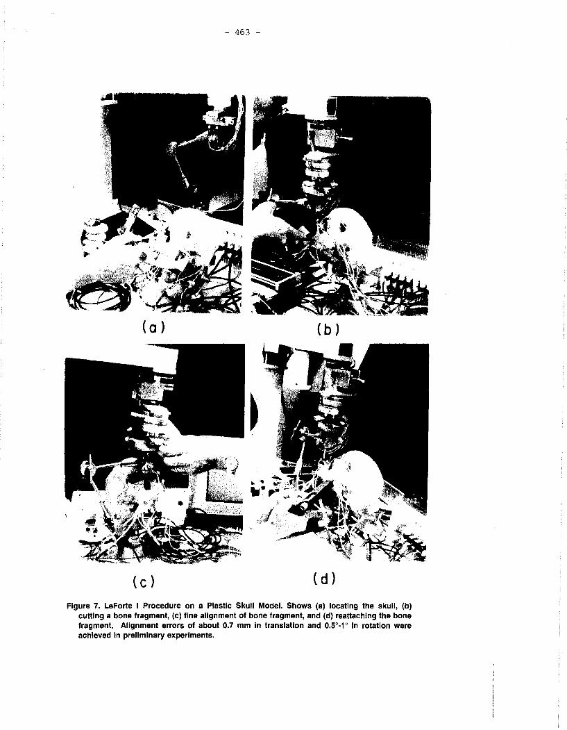
(b)
( d)
Figure 7. LeForte I Procedure on a Plastic Skull Model. Shows (a) locating the skull, (b) cutting a bone fragment, (c) fine alignment of bone fragment, and (d) reattaching the bone fragment. Alignment errors of about 0.7 mm in translation and 0.5°-1° in rotation were achieved in preliminary experiments.

- 464 -
goniometer cradles is a hit too cramped, and this distance is being increased substantially in the new
model.
Although our present work explicitly does not address clinical qualification issues such as sterility, patient compatibility, etc., in principle these issues should he relatively simpler to address than those
associated with an active robot, since it is easy to guarantee that no unwanted motions will occur. The biggest issue would likely he he qualification of the realtime sensing system for use in an operating room and verification that the actual bone fragment positions correspond to the positions being reported hy the system.
Discussion
Although the two example systems described above differ considerably in individual technological components and overall emphasis of effort, their architectures have many similarities. This is not acci
dental; their components can be viewed as complementary building blocks in the man-machine partnership alluded to in the introduction.
The outline of the ultimate "Computer Integrated Surgery" architecture are beginning to become clear, and many of the essential elements are indeed avaliable in nacent form today. These include:
• A 1111/fied data hase, which will eventually contain essentially all information about a patient. Much of this data will be in the form of medical images gathered from a variety of sensors and modalities, together with anatomical models derived from the images. Such a data base would be be a logical extension of the Picture Archival and Communications Systems (PACS) that are now
being introduced.
• Powerful presurgical planning and postsurgical analysis systems, typically implemented on computer workstations, but often backed-up by supercomputers and special purpose processors, will be used to process the patient data. A key element of this processing will be the derivation of 3D engineering models of the patient's anatomy and the use of these models to assist the physician in analyzing the patient's condition. This processing will include both precise quantitative computations (e.g., numerical optimizations, finite elements, etc.) and qualitative analysis augmented by powerful graphics and visualization hardware.
The surgeon will use this analysis to plan an optimal surgical procedure, to simulate its execution, and (possibly) to simulate future healing by the patient. If necessary, he will design appropriate custom implants or instruments and will send the design files off to an automated manufacturing facility to have them fabricated.
The planning workstation would also interact with other computers to order any necessary consumable items, schedule the operating room and surgical personnel, and instruct the hospital staff on any preoperative procedures needed for the patient. It would also communicate with device and implant manufacturer's computers to order necessary consumables, custom instrumentation, and custom (or off-the-shelf) implantable devices.
for post-operative followup, the workstation would assist the surgeon in analysis of intraoperative and postoperative medical images, to assess how well the planned surgical procedure
was carried out and of how well the patient's recovery is proceding. It would be used to assist in
planning additional rehabilitation procedures or any follow-up surgery that might be required. In
- 465 -
addition to these per-patient uses, it would perform efficacy analyses that could help improve the planning and execution of future surgical interventions.
• Online surgical systems will assist the surgeon in carrying out the planned procedure. These systems will vary in specific embodiment depending on the application, but will typically include several key elements:
- A surgical workstation will have access to all data from the preoperative planning system. Indeed, it most likely will be essentially the same workstation, with additional interfaces for interoperative sensors, man-machine interfaces, and device control. It will be responsible for tracking progress of the surgical plan, managing realtime sensors and manipulation devices, and communicating with the surgeon/
Sensing Subsystems provide information needed to register the actuality on the operating table with the models from the surgical plan, and for tracking the progress of the surgical procedure. tracking motion of bone fragments, surgical instrument<;, etc. These might include conventional computer vision, realtime radiography and ultrasound devices, special purpose geometric sensors, force sensors, voice recognition systems, intraoperative MRI or CT, etc.
- Surgeon Interface Subsystems will use a variety of modalities (graphics, synthesized voice, tonal cues, programmable impedence of manipulator joints, etc.) to provide online, realtime "advice" to the surgeon, based on the sensed relationship between the surgical plan and surgical execution. In cases where a robotic device is making preprogrammed motions, they will permit the surgeon to monitor the progress of the surgery and intervene in appropriate ways. In cases where the surgeon is providing the motive forcem they will provide feedback to assist him in accurately executing the planned procedure.
- Manipulation aids are provided to assist the surgeon in precisely aligning his instruments relative to the patient or in executing the mechanical parts of the planned procedure. Such aids will run the gamut from semi-autonomous, continuously controlled robots, to teleoperated devices, to point-to-point robots and motorized stereotactic frames, to passive devices of various degrees of sophistication.
Different surgical procedures will require different specific devices, and different surgeons will have different needs and preferences. 8 It is unlikely that any one single system will meet all needs, and so it seems reasonable to speculate that over time a "library" of modular building blocks or standardized subsystems with differing capabilities might evolve, together with appropriate interface standards allowing them to be put together in various ways, as appropriate. Although some of these subsystems (such as the prostetectomy "robot" described in [ 18]) may be rather specialized, others will be adaptable to many different procedures. Such generality is important since it permits the cost of qualifying a device for clinical use (and the replication and marketing cost of supplying it to hospitals) to be spread over many different procedures.
Conclusion
This paper has discussed two systems for augmentation of human prec1s10n in surgery. One of these stresses the use of an active robot to execute relatively complex trajectories required to machine
Indeed, one of the co-authors (Paul) has pointed out that he would take a somewhat different approach for orthopaedic osteotomies than that proposed here for craniofacial osteotomies.
- 466 -
bone in orthopaedic surgery. The other stresses realtime sensing, online "advice," and passive manipulation to help a surgeon precisely relocate bone fragments in craniofacial osteotomies. One essential aspect of both applications is the use of medical imaging and modelling to help the surgeon develop an optimal surgical plan and the concommitent synergy between computer-based presurgical planning and precise surgical execution.
Furthermore, although each application is interesting and important in iself, the underlying technology is clearly more broadly applicable. These systems may be viewed as steps in the evolution of a partnership between people and machines that exploits the capabilities of both to do precise surgical tasks better than either could do alone.
Acknowledgements
A great many people have contributed to one aspect or another of the experimental systems discussed in this paper. Although many of their names appear in the references and (indeed) on papers providing fuller descriptions of these systems, we would like to take this opportunity to explicitly acknowledge their critical role. Joel F. Zuhars (ISS), Edward Glassman (IBM), William L. Bargar, MD (Sutter Hospital), and Bill Williamson (!SS) have all contributed substantially to the development of the hip replacement system, as have Steve Lamb (OSI, Inc), Bob Olyha (IBM), Ken Honeycutt (IBM), Kip Harris (IBM), and many others. Marilyn Noz (NYU), Robert Olyha (IBM), Nils Bruun (IBM), and Dieter Grimm (IBM) all contributed substantially to the craniofacial surgery system, as did Rudi Schmidt (IBM). Leon Kehl, Chris Hockey, Walter Carpini, and Michelle Melluish of Northern Digital, Inc., provided invaluable consultation and support for the optical tracking system that is the principal positional sensor used in the craniofacial surgery system and that was also used as a prototype redundant position sensor for the hip surgery system.
References
[11 Bruce A. Kall, Patrick J. Kelly, and Stephan J. Goerss, "Interactive Stereotactic Surgical System for the Removal of lntracranial Tumors Utilizing eh C02 Laser and CT-Derived Database," IEEE Transactions on Biomedical Engineering, pp. 112-116, February 1985.
[2] Y. S. Kwoh, J. Hou, E. Jonckheere, and S. Hayati, "A Robot with Improved Absolute Positioning Accuracy for CT Guided Stereotactic Surgery," IEEE Transactions on Biomedical Engineering, pp. 153-161, February 1988.
[3] S. Lavallee, "A New System for Computer Assisted Neurosurgery," Proc. 1 /'th IEEE Engineering in Medicine and Biology Conf, pp. 926-927, Seattle, Nov 1989.
[4] Y. Kosugi, E. Watanabe, J. Goto, T. Watanabe, S. Yoshimoto, K. Takakura, and J. Ikebe, "An Articulated Neurosurgical Navigation System Using MRI and CT Images," IEEE Transactions on Biomedical Engineering, pp. 147-152, February 1988.
[5] R. L. Galloway, C. A. Edwards, J. G. Thomas, S. Scheiner, and R. J. Maniunas, "A new device for Interactive, Image Guided Surgery," Proc. SPIE Medical Imaging V, 1991.
[61 James M. Drake, Michael Joy, Andrew Goldenberg, and David Kreindler, "Robotic and Computer Assisted Resection of Brain Tumors." Proc. Fifth Int. Conj. 011 Advanced Robotics, pp. 888-892, Pisa, June 199 l.
[7] Russell H. Taylor, Howard. A. Paul, Brent D. Mittelstadt, William Hanson, Peter Kazanzides, Joel F. Zuhars, Edward Glassman, Bela L. Musits, William L. Bargar, and William Williamson, "An Imagebased Robotic System for Hip Replacement Surgery," Journal of the Robotics Society of Japan, pp. 111-116, October 1990.

- 467 -
[81 Ludwig Adams, Joachim M. Gilsbach, Werner Krybus, Dietrich Meyer-Ebrecht, Ralph Mosges, and Georg Schlondorff, "CAS - A Navigation Support for Surgery," 3d Imaging in Medicine, pp. 411-423, Springer-Verlag Berlin Heidelberg, 1990.
[9] S. Lavallee, "Computer Assisted Interventionist Imaging: Application to the Vertebral Column Surgery," Proc. 12'th IEEE Engineering in Medicine and Biology Conf, pp. 430-431, Phila., Nov. 1990.
[ 191 Ludwig Adams, A. Knepper, W. Krybus, D. Meyer-Ebrecht, G. Pfeiffer, R. Rueger, and M. Witte, "Navigation Support for Surgery by means of Optical Position Detection and Real-Time 3D Display," Proc Computer Aided Radiology 91, 1991.
[11] Russell H. Taylor, Court B. Cutting, Yong-yil Kim, Alan D. Kalvin, David Larose, Betsy Haddad, Deljou Khoramabadi, Marilyn Noz, Robert Olyha, Nils Bruun, and Dieter Grimm, "A Model-Based Optimal Planning and Execution System with Active Sensing and Passive Manipulation for Augmentation of Human Precision in Computer-Integrated Surgery," Proc. 1991 Int Symposium on Experimental Robotics, Toulouse, France, June 25-27 1991.
[121 J. A. McEwen, C.R. Bussani, G. F. Auchinleck, and M. J. Breault, "Development and Initial Clinical Evaluation of Pre-Robotic and Robotic Retraction Systems for Surgery," Proc. Second Work~hop on Medical and Health Care Robots, pp. 91-101, Newcastle-on-Tyne, Sept. 1989.
[13] Elmed Incorporated., Retract-Robot (tm), 1990.
[141 B. L. Davies, R. D. Hibberd, A. Timoney, and J. Wickham, "A Surgeon Robot for Prostatectomies," Proc. Second Workshop on Medical and Health Care Robots, pp. 91-101, Newcastle-on-Tyne, Sept. 1989.
[151 P. Cinquin, S. Lavallee, and J. Demongeot, "Computer Assisted Medical Interventions," Proc. Second Workshop on Medical and Health Care Robots, pp. 91-101, Newcastle-on-Tyne, Sept. 1989.
[16] J. L. Garbini, R. G. Kaiura, J. A. Sidles, R. V. Larson, and F. A. Matson, "Robotic Instrumentation in Total Knee Arthroplasty," Proc. 33rd Annual Meeting, Orthopaedic Research Society, p. 413, San Francisco, January 1987.
[ 171 Patrick J. Kelly, Bruce A. Kall, Stephan Goerss, and Franklin Earnest, "Computer-assisted Stereotaxic laser Resection of Intra-Axial Brain Neoplasms," J. Neurosurg, pp. 427-439, March 1986.
LI 8] B. L. Davies, R. D. Hibberd, A. Timoney, and J. Wickham, "A Surgeon Robot for Prostatectomies," Proc. Fifth lmemational Con.fereczce on Advanced Robotics, pp. 871-875, Pisa, June 1991.
[19] H. A. Paul, D. E. Hayes, W. L. Bargar, and B. D. Mittelstadt, "Accuracy of Canal Preparation in Total Hip Replacement Surgery Using Custom Broaches," Proc. First International Symposium 011 Custom Made Prostheses, Dusselforf, October 1988.
(201 Howard. A. Paul, Brent D. Mittelstadt, Joel Zuhars, William L. Bargar, Peter Kazanzides, B. Williamson, and T. C. Hsia, "Surgical Procedure for Robotic Hip Replacement," Proc. /2'th IEEE Medi
cine & Biology Cmif.. Phila .. 1990.
[21] Russell H. Taylor, Howard. A. Paul, Peter Kazanzides, Brent D. Mittelstadt, William Hanson, Joel F. Zuhars, Bill Williamson, Bela L. Musits, Edward Glassman, and William L. Bargar, "Taming the Bull: Safety in a Precise Surgical Robot," Proc. 1991 Cm!ference on Advanced Robotics, Pisa, Italy, June
1991.
[22] Russell H. Taylor, Howard. A. Paul, Brent D. Mittelstadt, William Hanson, Peter Kazanzides, Joel F. Zuhars, Edward Glassman, Bela L. Musits, William L. Bargar, and William Williamson, "An Imagebased Robotic System for Precise Orthopaedic Surgery," Proc. 12'th IEEE Medicine & Biology Conf,
Phila., November 1990.
- 468 -
123] W. H. Hanson, H. A. Paul, Bill Williamson, and Brent Mittelstadt, "Orthodock: A Computer System for Presurgical Planning," Proc. /2'th IEEE Medicine & Biolof!y Con{. Phila., 1990.
124] H. Paul. B. Mittelstadt. W. Bargar, P. Kazanzides, B. Williamson, J. Zuhars, R. Taylor, and W. Hanson, "Accuracy of Implant Interface Preparation: Hand-held Broach vs. Robot Machine Tool," Proc. Ortlwpaedic Research Society, Washington, D. C., (to appear) A 1992.
125J P. M. Sandborn, S. D. Cook. W. Spies, and M. Koster, "Tissue Response in Porous Coated Implants Locking Initial Bone Apposition," J. of Arthro11lasty, December 1988.
[26] B. D. Mittelstadt, **** In Preparation *'"**, PhD thesis, University of California at Davis, 1992.
[27 \ Court Cutting, MD, Barry Grayson, DDS, and Hie Chun Kim, "Precision Multi-Segment Bone Positioning Using Computer Aided Methods in Craniofacial Surgical Applications," Proc. 12'th IEEE Medicine & Biologv Con{, Phi la., November 1990.
128] M. E. Noz and G. Q. Maguire, "QSH: A Minimal but Highly Portable Image Display and Processing Toolkit," Computer Methods and Programs in Biomedicine, pp. 229-240, Nov. 1988.
[291 A. D. Kalvin, C. B. Cutting, B. Haddad, and M. Noz, "Constructing topologically connected surfaces for the comprehensive analysis of 3-D medical structures," Proc. SP/E Medical Imaging Conference V, San Jose, February 1991.
[30) A. D. Kalvin, Segmentation and Surface-based Modelling of Objects in JD Biomedical Images, PhD thesis. New York University, New York, 1991.
[31] Court Cutting. MD and Barry Grayson, DDS, "Three Dimensional Computer Aided Planning of Craniofacial Surgical Procedures," Plastic and Reconstructive Surgery, 1986.

\\ INNOVATION ET TECHNOLOGIE
EN BIOLOGIE ET MEDECINE UN JOURNAL EUROPEEN DE GENIE BIOLOGIOUE ET MEDICAL POUR LA PROMOTION DES APPLICATIONS DE LA RECHERCHE.
SOMMAIRE VOL. 13, N° 4, 1992
NUMERO THEMATIQUE
ROBOTIQUE CHIRURGICALE
- GMCAO : GESTES MEDICO-CHIRURGICAUX ASSISTES PAR ORDINATEUR. METHODOLOGIE, APPLICATIONS MEDICALES, RESUL TATS. CINQUIN P., DEMONGEOT J., TROCCAZ J., LAVALLEE S., CHAMPLEBOUX G., BRUNIE L., LEITNER F., SAUTOT P., MAZIER B., PEREZ A., DJ AID M., FORTIN T., CHENIN M., CHAPEL A., ................................................................................................. 373
- PREMIERS PAS VERS LA DISSECTOMIE ET LA REALISATION DE PROTHESES DU GENOU A L'AIDE DE ROBOTS. FADDA M., MARTELLI S., DARIO P., MARCACCI M., ZAFFAGNINI S., VISANI A., .......... 394
- AIDE AU REPERAGE TRIDIMENSIONNEL POUR LA CHIRURGIE DE LA BASE DU CRANE. ADAMS L., KNEPPER A., KRYBUS W., MEYER-EBRECHT D., PFEIFER G., RUGER R., WITTE M., ....................................................................................................................... 409
- CONTRAINTES MECANIQUES : SOLUTION POUR LE PROBLEME DE SECURITE EN ROBOTIQUE CHIRURGICALE? DAVIES B.L., HIBBERD R.D., NG W.S., TIMONEY A.G., WICKHAM J.E.A., ....................... .425
- STEREOT AXIE MAG NE TI QUE : DEPLACEMENT D'IMPLANTS DANS LE CERVEAU, ASSISTES PAR ORDINATEUR ET GUIDES PAR IMAGERIE. RITTER R.C., GRADY M.S., HOWARD III M.A., GILLIES G.T., .......................................... 437
- AUGMENTATION DE PRECISION DU GESTE OPERATOIRE A L'AIDE D'UN SYSTEME INFORMATIQUE INTEGRE POUR LA CHIRURGIE. TAYLOR R.H., PAUL H.A., CUTTING C.B., MITTELSTADT B., HANSON W., KAZANZIDES P., MUSITS B., KIM Y.Y., KALVIN A., HADDAD B., KHORAMABADI D., LAROSE D., .................................................................................................................... .450